


ISSN: 0973-7510
E-ISSN: 2581-690X
Indian lilac or neem (Azadirachta indica) is found in tropical and subtropical regions of the Indian subcontinent. Each part of the tree is a source of various phytochemicals. Neem gum is an exudate from mature parts of the plant stem. Biochemically, it has an acidic pH range (5–6) and is composed of monosaccharides, saponins, phenols, and tannins. This study aimed to elucidate the diversity of neem gum-associated microflora through high throughput metagenomics approach using 16S rRNA variable region sequencing. The bacterial community of neem gum was dominated by Firmicutes (~82%), Proteobacteria (~18%), and Actinobacteria (~0.02%). Among the genera, Lactococcus was found to be the most dominant bacterium. The predominance of Lactococcus in neem gum is probably due to its acidic nature, which provides a suitable microenvironment for its proliferation. In addition, Lactococcus and beneficial microorganisms such as Pseudomonas, Burkholderia, Pantoea, Klebsiella, and Methylobacterium were also present in the gum. This study highlights the fact that neem gum can be exploited as a unique source of microorganisms for biotechnological and agricultural applications.
Azadirachta indica, Neem Gum, Metagenomics, 16S rDNA (V3-V4 region), MiSeq
Neem (Azadirachta indica) is a tree native to India that has been used as a traditional medicine since ancient times.1 Neem gum is a natural resin extracted from neem trees, which is obtained by tapping the tree trunk and solidifying it upon exposure to air. Neem gum is used in various applications, viz: food additives,2 cosmetic products,3 and as a traditional remedy for various ailments. Studies have shown that neem gum has antifungal, antibacterial, and anti-inflammatory properties4-6 and is a good source of carbohydrates and amino acids.7 Neem gum has been used in traditional medicine for its health-promoting effects and recent research has confirmed its benefits, including its ability to improve gut health and boost the immune system.2 Despite its potential health benefits, more research is needed to fully understand the composition and properties of neem gum as well as its potential risks and side effects. However, its natural origin and low toxicity make it an attractive alternative to synthetic additives and remedies. Tannins, also present in neem gum, are known for their astringent and antimicrobial effects.8 In addition, neem gum is rich in polysaccharides, which are complex sugars that have been shown to have immune-boosting and anti-inflammatory effects.
Various endophytic microflora has been isolated from various parts of neem plants.9-12 These microbes have also been characterized for their secondary metabolite-production capabilities.
Plant-associated microbes play an important role in plant growth and physiology, and thus, directly and indirectly influence humans.13 These microbes produce many useful compounds that are important for agriculture and industry.14 Neems have been widely used to investigate endophytes and their secondary metabolites.15 The microbiota present in each part of neem plants, such as the bark, root, stem, and leaves, are well known, but with respect to the neem gum microbiome, no study has been conducted yet.16-18 For in-depth analysis of plant-related microbiomes, the metagenomic approach offers the necessary platform for a robust high-throughput technique.19 The metagenomics approach has greatly helped explore the actual microbial biodiversity found in the atmosphere. Besides being a remarkable reservoir of novel genes and enzymes, herbal compounds, bioactive substances, and bioprocesses, the microbiota can contribute appreciably to the direction of a sustainable environment.
Realizing the potential of neem gum as a source of unique microflora and genes, we explored its microbiome using a metagenomic approach to decipher the microbial community structure.
Sample collection and genomic DNA isolation
Neem gum was collected under sterile conditions from a neem plant located in Maunath Bhajan (25.90°N, 83.49°E), Uttar Pradesh, India, and processed for DNA isolation. The collected samples were then dissolved in sterile saline. The dissolved sample was filtered on 0.2-micron nylon filter paper using a vacuum filter chamber, and after filtration, the filter paper was placed in Lysis buffer (10 mM Tris-HCl, 2 mM EDTA, 1% SDS) at 4°C and sonicated at 4°C by pulse on for 5 s and pulse off for 30 s for 9 min. Sonicated samples were centrifuged at 10,000 rpm for 15 min at 4°C, and the collected pellet was dissolved in 2 ml of TE buffer (Himedia, India), and DNA was extracted with the commercially available PureLink™ Microbiome DNA Purification (Invitrogen by Thermo Fisher Scientific) Kit as per the manufacturer protocol. DNA purity was checked by determining the A260/280 ratio using a Nanodrop.
Preparation of 2x 300 MiSeq library Cluster generation and sequencing
Bacterial 16S rRNA gene hypervariable regions V3-V4 were amplified using the V3V4F (CCTACGGGNGGCWGCAG) and V3V4R (GACTACHVGGGTATCTAATCC) primers.20 The primers were modified to include Illumina sequencing adapters as overhangs. 25 ng of DNA was used by PCR using the KAPA HiFi Hot Start Ready Mix. The PCR involved an initial denaturation of 95°C for 5 min followed by 25 cycles of 95°C for 30 s, 55°C for 45 s, 72°C for 30 s, and a final extension at 72°C for 7 min. The amplicons were purified using amplification beads to remove unused primers. Libraries were quantified using the Qubit dsDNA High-Sensitivity Assay Kit. Sequencing was performed using an Illumina MiSeq with a 2 × 300 PE V3 sequencing kit.
Data analysis
Quality control checked reads were imported into Mothur,21 pairs were aligned, and contigs were obtained. The contigs were screened for errors; the most effective ones between 300 bp and 550 bp were retained and contigs with ambiguous base calls were discarded. High-quality contigs were checked for identical sequences and redundant entries were merged. The filtered contigs were processed and classified into taxonomic outlines using the Silva v.128 database.22
Taxonomic distribution of bacterial flora in neem gum
Taxonomic annotation of neem gum was performed to determine the composition of bacteria inherent to the niche. A rarefaction curve (Figure 1) shows the degree of diversity of a given variety of reads in a sample. Surprisingly, no significant compositional diversity of bacterial taxa was observed in neem gum. Alpha diversity (Figure 2) is an accurate measure of the relative abundance and richness of bacteria in a given sample.

Figure 1. Rarefaction curve

Figure 2. Alpha diversity measurements. It represents different Alpha diversity indices of which Chao1 and ACE represent the richness of the sample and Shannon, Simpson, Inv Simpson and Fisher represent both richness and relative abundance
Distribution of bacterial phyla in neem gum
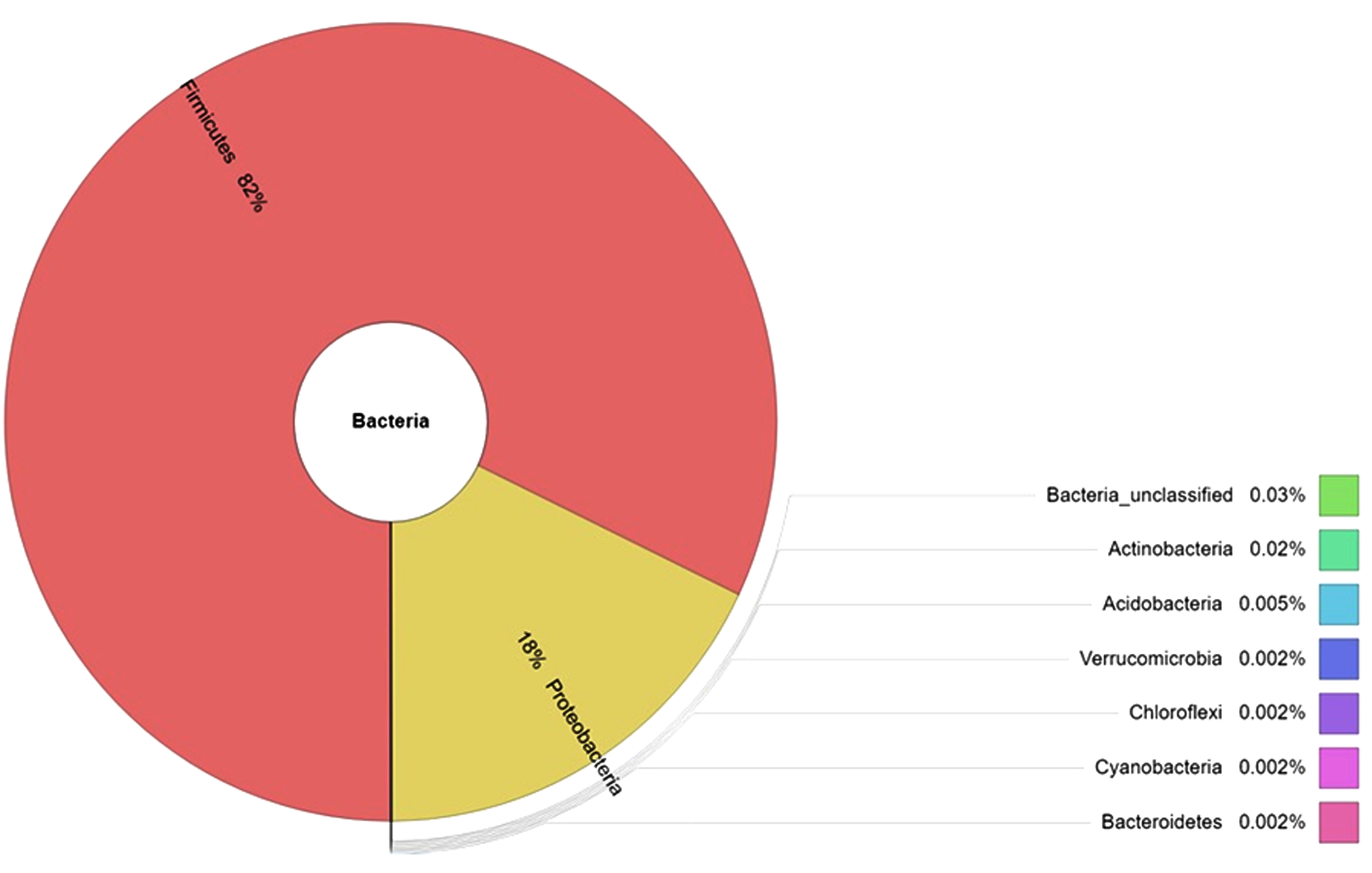
The sample analysis revealed the presence of nine phyla. Firmicutes (~82%) were found to be the most dominant, followed by Proteobacteria (~18%), Actinobacteria (~0.02%), and other phyla such as Acidobacteria, Bacteroidetes, Verrucomicrobia, Chloroflexi, Cynobacteria, and unclassified bacteria were (< 0.01%) (Figure 3, Figure 4a).

Figure 3. Top 10 Phylum abundance distribution
 4(a)
4(a)
 4(b)
4(b)
 4(b)
4(b)

4(d)
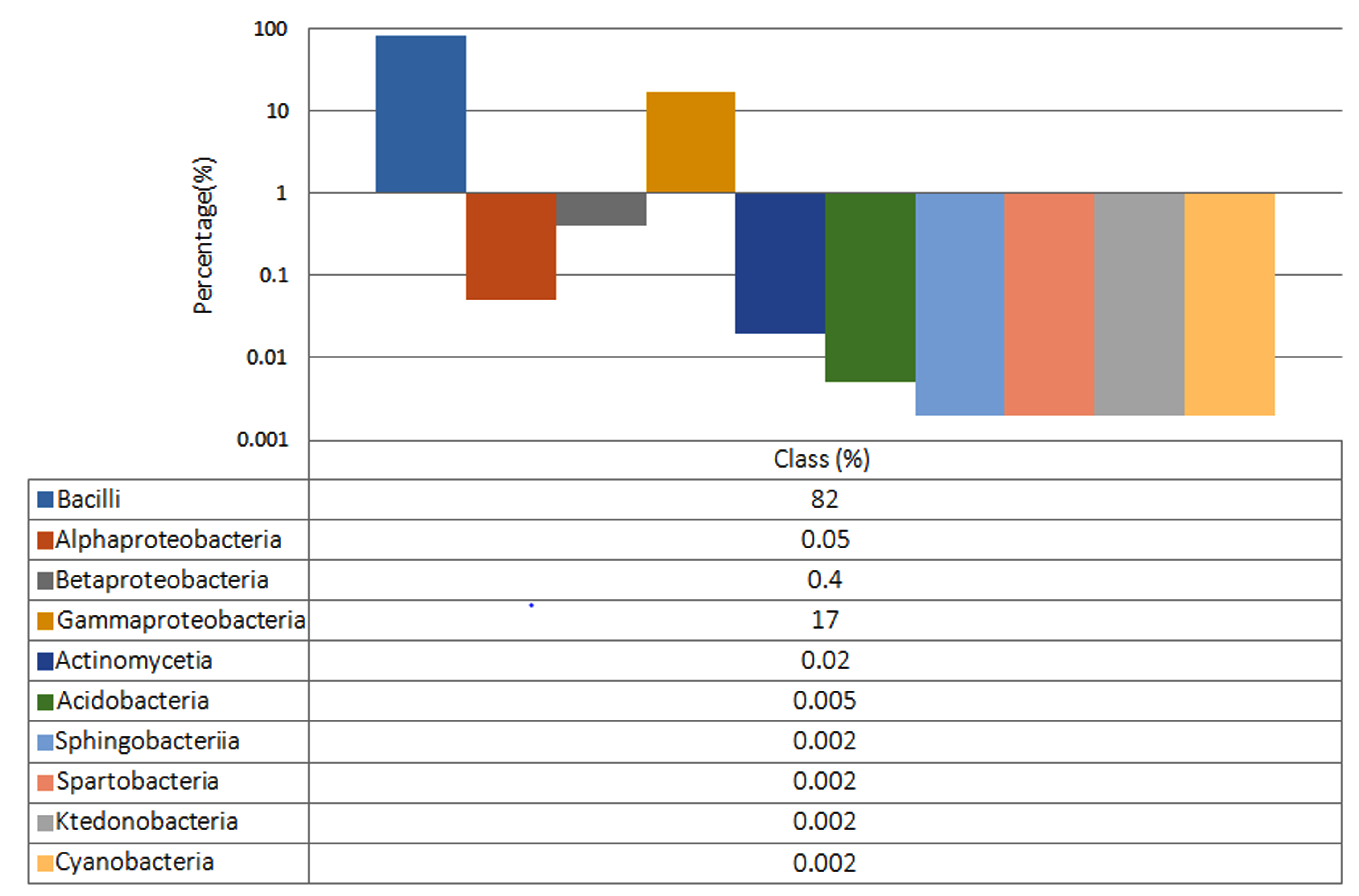
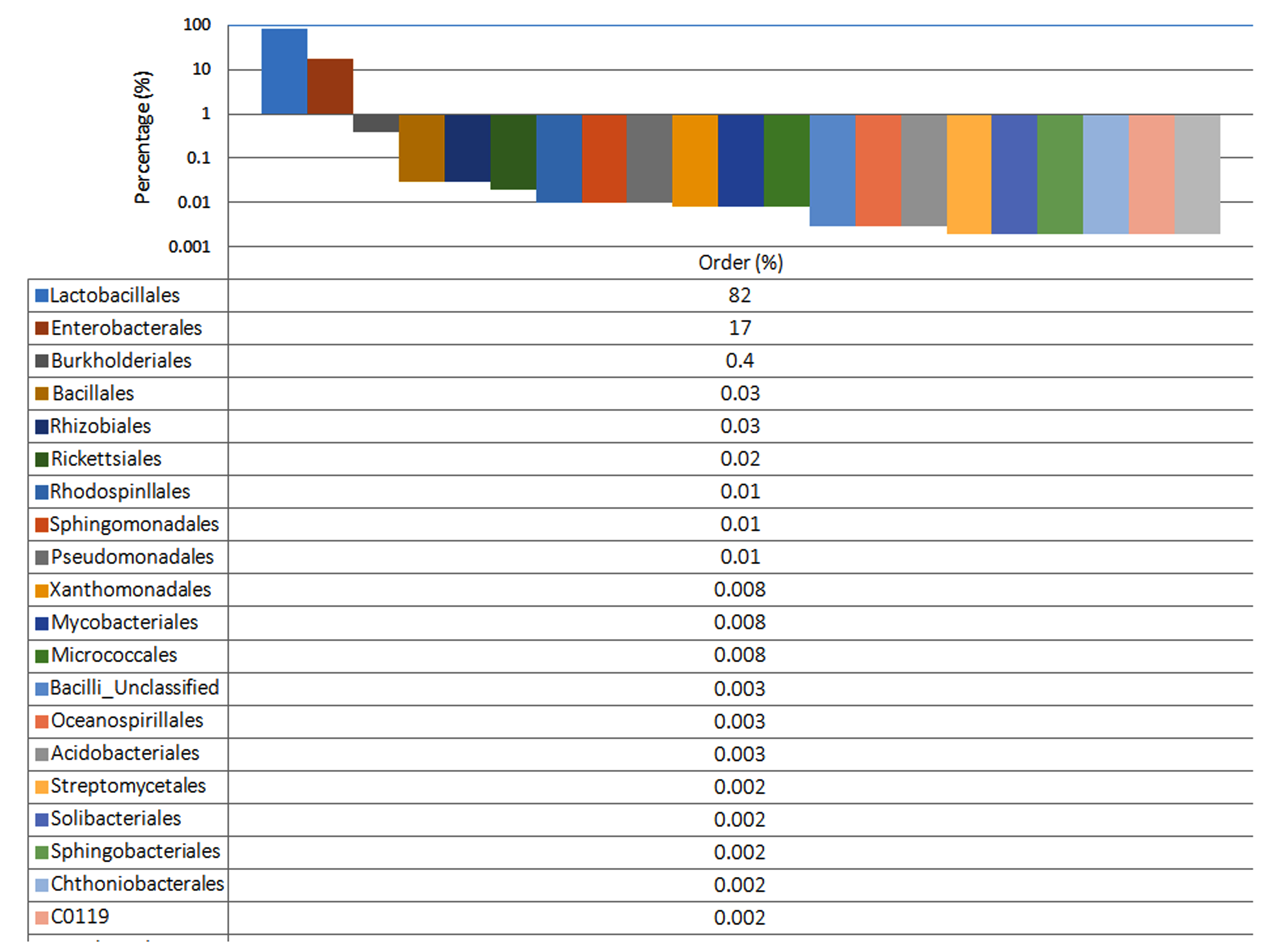
Figure 4. Distribution of bacteria at (a) Phylum level (b) class level (c) order level (d) Genus level
Distribution of bacterial class in neem gum
Distribution at the class level revealed the predominance of Bacilli (82%), followed by Gamma-proteobacteria (17%), Betaproteobacteria (0.4%), Alpha-proteobacteria (0.05%), Actinomycetes (0.02%), Acidobacteria (0.005%), and other classes such as Sphingobacteria, Spartobacteria, Ktedonobacteria, and Cyanobacteria were equally distributed (0.002%) in the sample (Figure 4b).
Distribution of order of bacterial flora in neem gum
While investigating the order richness, nine bacterial orders viz. Lactobacillales (82%), Enterobacteriales (17%), Burkholderiales (0.4%), Bacillales (0.03%), Rhizobiales (0.03%), Rickettsiales (0.02%), Rhodospinllales (0.01%), Sphingomonadales (0.01%), Pseudomonadales (0.01%) were found to be dominant in the in sample.(Figure 4c).
Distribution of bacterial genera in neem gum
The top 20 genera were Lactococus (82%), followed by Unclassified Enterobacteriaceae (10%), Pantoea (4%), and Kosakonia (3%) were most abundant. (Figure 4d). Neem gum samples showed a high abundance of Lactococcus.
This study aimed to acquire a clear idea to define the presence of a diverse microbial population in neem gum. Neem gum can be used as a source of nutrients for microbial growth; microbes can digest the soluble polysaccharide content in neem gum.23 It was evident from the results that at the phylum level, members of Firmicutes were more abundant in neem gum than other phyla. Besides Firmicutes, the phyla Proteobacteria, Actinobacteria, Acidobacteria, Bacteroidetes, and Verrucomicrobia were identified. However, class-level diversity analysis suggested that members of Bacilli were predominant in neem gum. In neem gum samples, all taxonomic strata (phylum, class, order, family, and genus) indicated the presence of diverse genera. At the genus level, Lactococcus was predominantly present in the sample. Along with Lactococcus, other genera, such as Pantoea, Kosakonia, Burkholderia-Paraburkholderia, Massiia, Shimwella, streptococcaceae, Klebsiella, Cronobacter, Weissella, Methylobacterium, Sodalis, Paenibacterium, Sporolactobacillus, Sphingomonas, Citrobacter, and Corynebacterium were also detected in the samples. The predominance of Lactococcus in neem gum is probably due to its acidic nature, which provides a suitable microenvironment for its proliferation. Earlier studies have shown that Lactococcus exerts antagonistic activity against many pathogens by producing secondary metabolites.24-26 Lactococcus has a high tolerance to low pH and produces different exopolysaccharides with viscous physical properties. Exopolysaccharides are stabilizing, emulsifying, and antioxidant agents that enhance the antibacterial, anticoagulant, and antioxidant activities of microbes. Lactococcus has antagonistic activity against many pathogens through the production of secondary metabolites such as nisin peptide, lacticine, and bacteriocin. Some of the antimicrobial properties of neem gum may be due to the presence of bacteria, such as Lactococcus, and the low pH of the gum. The phytochemical composition of neem gum has been reported to comprise prebiotic oligosaccharides, which are highly beneficial for increasing the population of Lactococcus, which is well known as a good bacterium and gut commensal in humans,2,27 Some studies have shown that neem gum can potentially enhance the growth of healthy microbes and can be used as a prebiotic drink.28
The present metagenomic analysis showed a diverse bacterial population present in the sample and some of which were reported as plant growth promoters and some are for antimicrobial and secondary metabolite producers.29,30 Other than Lactococcus, Streptococcus, Pseudomonas, and Burkholderia showed antagonistic activity against pathogens. Pantoea, Klebsiella, and Methylobacterium have been reported as potential plant growth promoters and have shown beneficial roles in agriculture and industry. Pseudomonas and Pentoea have been reported to be biological control agents against various pathogens. They produce various antibiotics such as pantocins, herbicolins, microcins, and phenazines. Pseudomonas spp. are metabolically versatile, producing various secondary metabolites. Pseudomonas produces several surface-active lipopeptides (LPs). Because they produce a range of LPs, different Pseudomonas spp. have been studied in recent years for their potential as biocontrol agents and for enhancing industrial enzyme production in extreme environments.31-35 Metagenomic analysis indicated that the colonization of the microbial community in neem gum is a complex consequence and requires thorough investigation to understand the mechanism and guiding forces of colonization. In summary, it can be concluded that neem gum is a good source of diverse microorganisms which can have multiple agricultural and biotechnological applications.
ACKNOWLEDGMENTS
The authors would like to thank Director, ICAR-NBAIM, Maunath Bhanjan (Uttar Pradesh) for providing necessary support for conducting the research work and acknowledge AMITY Institute of Biotechnology, Amity University, Noida (Uttar Pradesh), India.
CONFLICT OF INTEREST
The authors declare that there is no conflict of interest.
AUTHORS’ CONTRIBUTION
AKS, HC, AS and SS designed and supervised the study. AKS designed and supervised individual experiments. PS performed the experiments. PS, HC and AS evaluated and analyzed the experiments. PS and AKS wrote the manuscript with the contributions from all the authors. All authors read and approved the final manuscript for publication.
FUNDING
None.
DATA AVAILABILITY
All datasets generated or analyzed during this study are included in the manuscript.
ETHICS STATEMENT
Not applicable.
- Kumar VS, Navaratnam V. Neem (Azadirachta indica): Prehistory to contemporary medicinal uses to humankind, Asian Pac J Trop Biomed. 2013;3(7):505-514.
Crossref - Suntari D, Siswadi DD, Hamzah Z, Handayani AT, Setyowati DI . Effectiveness test of neem gum (Azadirachta indica) solution against the viability of Escherichia coli and Salmonella typhimurium (in vitro). Jurnal Pangan dan Agroindustri. 2022;10(1).
Crossref - Tomar A, Verma G, Phogat S, Singh M. Neem in Health and Cosmetics, Neem: A Treatise, IK International Publishing House Pvt Ltd., New Delhi. 2009:461-85.
- Vijayakumar S, Divya M, Vaseeharan B, et al. Biogenic preparation and characterization of ZnO nanoparticles from natural polysaccharide Azadirachta indica. L.(neem gum) and its clinical implications. Journal of Cluster Science. 2021;32:983-93.
Crossref - Neihaya HZ, Zahraa SH and Kadhem H. Antibacterial and anti-biofilm activities of Neem gum (Azadirchta indica) and Arabic gum (Acacia senegal) extracts on human pathogenic bacteria. Al-KufaUniv J Biol. 2020;12(2):80-90. https://www.researchgate.net/publication/346448381_Antibacterial_and_anti-biofilm_activities_of_Neem_gum_Azadirchta_indica_and_Arabic_ gum_Acacia_senegal_extracts_on_human_pathogenic_bacteria.Accessed February 2021.
- Nagpal M, Raj N, Thakur GS and Aggarwal G. Improved solubility of itraconazole binary dispersions using neem gum: Development and characterization of topical gel. Curr Bioact Compd. 2019;15(4):399-407.
Crossref - Mukherjee S, and Srivastava HC. The structure of neem gum. J Am Chem Soc 1955;77(2):422-423.
Crossref - Kopparam M, Reddy MR. Neem Gum as an Alternative to Synthetic Pharmaceutical Excipients. RGUHS Journal of Pharmaceutical Sciences. 2021;11(3):22-31.
Crossref - Chutulo EC, Chalannavar RK. Endophytic mycoflora and their bioactive compounds from Azadirachta indica: A comprehensive review. J Fungi. 2018;4(2):42.
Crossref - Kharwar RN, Sharma VK, Mishra A, et al. Harnessing the phytotherapeutic treasure troves of the ancient medicinal plant Azadirachta indica (Neem) and associated endophytic microorganisms. Planta Medica. 2020; 86(13/14):906-940.
Crossref - Sarah R, Idrees N, Tabassum B. A Review of Endophytic Microbiota of Medicinal Plants and Their Antimicrobial Properties. Core Microbiome: Improving Crop Quality and Productivity. 2022:1-9.
Crossref - Saxena P, Chakdar H, Singh A, Shirodkar S, Srivastava AK. Microbial diversity of Azadirachta indica (Neem) gum: An unexplored niche. J Appl Biol Biotechnol. 2023;11(2):209-219.
Crossref - Castronovo LM, Vassallo A, Mengoni A, M et al. Medicinal plants and their bacterial microbiota: A review on antimicrobial compounds production for plant and human health. Pathogens. 2021;10(2):106.
Crossref - Senn S, Pangell K, Bowerman AL. Metagenomic Insights into the Composition and Function of Microbes Associated with the Rootzone of Datura inoxia. BioTech. 2022;11(1):1.
Crossref - Agasimundin VB, Rangiah K, Sheetal A, Gowda M. Neem Microbiome. Neem Genome. 2019:111-123.
Crossref - Rajagopal R, Suryanarayanan TS. Isolation of endophytic fungi from leaves of neem (Azadirachta indica). Curr Sci. 2000;78:1375-137. https://www.jstor.org/stable/24104047
- Verma VC, Gond SK, Kumar A, et al. The endophytic mycoflora of bark, leaf, and stem tissues of Azadirachta indica A. Juss (Neem) from Varanasi (India). Microbial Ecology. 2007;54:119-125.
Crossref - Verma VC, Gond SK, Kumar A, et al. Endophytic Actinomycetes from Azadirachta indica A. Juss.: Isolation, Diversity, and Antimicrobial Activity. Microb Ecol. 2008;57:749-756.
Crossref - Oliveira C, Shakiba E, North D, et al. 16S rRNA Gene-Based Metagenomic Analysis of Rhizosphere Soil Bacteria in Arkansas Rice Crop Fields. Agronomy. 2022;12(1):222.
Crossref - Klindworth A, Pruesse E, Schweer T, et al. Evaluation of general 16S ribosomal RNA gene PCR primers for classical and next-generation sequencing-based diversity studies. Nucleic Acids Res. 2013; 41(1): e1-e1.
Crossref - Schloss PD, Westcott SL, Ryabin T, et al. Introducing mothur: Open-Source, Platform-Independent, Community-Supported Software for Describing and Comparing Microbial Communities. Appl Environ Microbiol. 2009;75(23):7537-7541.
Crossref - Quast C, Pruesse E, Yilmaz P, et al. The SILVA ribosomal RNA gene database project: improved data processing and web-based tools. Nucleic Acids Res. 2013;41(D1):D590-D596.
Crossref - Hamzah Z, Sulistyani E, Hafizh C, et al. Neem Gum (Azadirachta Indica) Solution Poten-tial for Improving Viability of Bifidobacterium longum and Lactobacillus acidohilus Bacteria. Indonesian Journal of Life Sciences. 2022;4(1):120-128.
Crossref - Soundharrajan I, Yoon YH , Muthusamy K, et al. Iso-lation of Lactococcus lactis from whole crop rice and determining its probiotic and anti-microbial properties towards gastrointestinal associated bacteria. Microorganisms. 2021;9(12):2513.
Crossref - Nehal F, Sahnoun M, Smaoui S, Jaouadi B, Bejar S, Mohammed S. Characterization, high production and antimicrobial activity of exopolysaccharides from Lactococcus lactis F-mou. Microb Pathog. 2019;132:10-19.
Crossref - Jawan R, Abbasiliasi S, Mustafa S, Kapri MR, Halim M, Ariff AB. In vitro evaluation of potential probiotic strain Lactococcus lactis Gh1 and its bacteriocin-like inhibitory substances for potential use in the food industry. Probiotics and Antimicrobial Proteins. 2021;13:422-440.
Crossref - Von Wright A, Axelsson L. Lactic acid bacteria: an introduction. In Lactic acid bacteria, CRC Press. 2019:1-16.
Crossref - Hamzah Z, Handayani AT, Setyowati DI, Sulistyani E, Wiyono HT, Indahyani DE. Baluran neem gum drinking water as a prebiotic candidate, Acta Sci Pol Technol Aliment. 2022;21(4):449-458.
Crossref - Qi X, Wang E, Xing M, Zhao W, Chen X . Rhizosphere and non-rhizosphere bacterial community composition of the wild medicinal plant Rumex patientia. World J Microbiol Biotechnol. 2012;28(5):2257-2265.
Crossref - Singh G, Mukerji KG. Root exudates as determinant of rhizospheric microbial biodiversity. Microbial Activity in the Rhizoshere. 2006:39-53.
Crossref - Suman A, Shukla L, Marag PS, Verma P, Gond S, Prasad JS . Potential use of plant colonizing Pantoea as generic plant growth promoting bacteria for cereal crops. J Environ Biol. 2020;41(5):987-994.
Crossref - Choudhary P, Waseem M, Kumar S, Subbarao N, Srivastava S, Chakdar H. Y12F mu-tation in Pseudomonas plecoglossicida S7 lipase enhances its thermal and pH stability for industrial applications: a combination of in silico and in vitro study. World J Microbiol Biotechnol. 2023;39(3):75.
Crossref - Tariq M, Hasnain N, Rasul I, et al. Reconnoitering the ca-pabilities of nodule endophytic Pantoea dispersa for improved nodulation and grain yield of chickpea (Cicer arietinum L.), World J Microbiol Biotechnol. 2023;39(3):85.
Crossref - Gamit HA, Naik H, Chandarana KA, Chandwani S, Amaresan N. Secondary metabo-lites from methylotrophic bacteria: their role in improving plant growth under a stressed environment. Environ Sci Pollut Res. 2023;30(11):28563-28574.
Crossref - Gohil RB, Raval VH, Panchal RR, Rajput KN. Plant growth promoting activities and effect of fermented panchagavya isolate Klebsiella sp. PG-64 on Vigna radiate, World J Microbiol Biotechnol. 2023;39(2):41.
Crossref
© The Author(s) 2023. Open Access. This article is distributed under the terms of the Creative Commons Attribution 4.0 International License which permits unrestricted use, sharing, distribution, and reproduction in any medium, provided you give appropriate credit to the original author(s) and the source, provide a link to the Creative Commons license, and indicate if changes were made.