


ISSN: 0973-7510
E-ISSN: 2581-690X
Acute and chronic wounds are the major cause of death according to World Health Organization (WHO), in which, antimicrobial resistance is considered to be a major plight. In this regard, our study is aimed at developing an antimicrobial agent using the mucus of Pangasianodon hypophthalmus against the clinically resistant microbial pathogens and to evaluate the cell cytotoxicity and cell viability followed by an in vitro wound healing analysis. The evaluation of antimicrobial activity was performed through well diffusion method and micro dilution method. The cell cytotoxicity and cell viability were assessed using MTT assay. The cell migration and in vitro wound healing was performed using scratch assay. The acidic extracts of mucus showed antimicrobial activity against the eight different selected bacterial strains while the organic extract showed against seven bacterial strains. L929 showed a cell viability of 102.96% at a concentration of 75 µg/mL and did not show cell toxicity effect up to the concentration of 300 µg/mL. In the in vitro wound healing analysis, the cell migration rate was 99.27% in the treated cells while, the untreated showed only 94.68%. The current research work clearly shows that the mucus of P. hypophthalmus possesses antimicrobial activity and wound healing potency. Furthermore, gene expression analysis and in vivo trials have to be performed for a thorough understanding of the actual cellular mechanism of wound healing.
Pangasianodon hypophthalmus, Cell Cytotoxicity, Cell Migration, Mucus Extracts, Wound Healing
Acute and chronic wounds have been a major cause of death throughout the world. It is estimated that 1-2% of the population in the developed countries suffer from chronic wounds.1 The commonly found wounds are due to diabetes, accidental injuries and burns. Given the situation of the increased type II diabetes, comorbid issues like diabetic foot ulcer is increasing steadily out of which at least 5% of diabetic patients are affected and 1% of them go for amputations.2 As per the report provided by WHO in the year 2021, 3.16 million lives are lost due to unintentional injuries per year. One of the noninfectious physical agents of wounds is the burn.3 According to WHO, it is also estimated that 1,80,000 deaths are caused due to burns in the middle and low income countries. Microbial Infections and host microbiota-associated pathogens are some of the reasons for improper wound care. Based on the above mentioned facts and analyses, there is definitely a need to develop a novel approach towards wound healing and its processes.
Fish skin mucus is the primary barrier between the environment and fish thus, performing a major role in protecting the fish from major pathogens.4 Skin mucus plays a vital role in fish health, as it is crucial to the innate immune system that serves as the first physical and chemical barrier against various infections.5 The mucosal layer also helps in regulating the movement of solute and water molecules across the skin surface for major functions like respiration, osmotic regulations, communication, abrasions and entanglement of particulate materials.6 The goblet cells, club cells and sacciform cells are the three different types of cells involved in the production of mucus in fish epidermal region. In the fish epidermal surfaces and gill surfaces, are many goblet cells that secrete mucus. These cells create granules of mucus that rupture to discharge their contents.7 The sacciform cells mix their contents along with contents of goblet cells.8 Fish mucus majorly contains proteins and fatty acid which are proven to play significant role in defense against the invading pathogens. Proteases, lectins, lysosomes, antimicrobial peptides, immunoglobulin, transferrins, complement proteins, monosaturated fatty acids like oleic acid, polyunsaturated fatty acids like linoleic acids are also present in the mucus secreted by fishes.9
Depending on the species variety, breed, developmental stages, sex and environmental conditions, the viscoelasticity and rheological properties of the mucus varies. The secretion of mucus in fresh water fish when compared to marine fishes is high due to water exchange mechanisms.10 The ability of fish skin mucus to fight off bacteria that can change the mucosal microbiome and enhance the fish’s susceptibility to various diseases has attracted more attention in recent year.11 The ability of mucus to kill microbes makes it seem like a viable option for the creation of new commercial and therapeutic drugs to treat human infections.12 Bhatnagar et al., demonstrated the bactericidal role of Heteropneustes fossilis and Clarias batrachus against pathogens. Also various research work have been carried out till date with respect to the antimicrobial activity of fish mucus.5
Fishes are generally oviparous, except few fishes like sharks that undergoes a viviparous condition13. The skin of shark is comparatively thicker, elastic and the wound healing recovery rate along with the stress level tolerance is higher compared to other fishes. The adult male shark bites the female shark during copulation, causing wounds that are very likely to get infected by pathogens in the aquatic environment or from the oral cavity of male shark. A study also documented that the incision formed after birth of young sharks started to heal from 29th day and by 179 days, there was no visible scar found.14
The importance of bioprospecting investigations in search for novel therapeutic agents, such as antibacterial and wound healing, is due to the fact that the biochemical resources from the marine environment represent an important role in medical and industrial goods.15 In addition, anti-microbial resistance is one of the biggest risks to global health and a major driving factor to the healthcare expenditures globally.16 In this regard, the current study is mainly focused on to evaluate the antimicrobial property and wound healing potency of the mucus from Pangasianodon hypophthalmus, i.e. a fresh water viviparous, edible shark.
Collection and maintenance of specimen
Five live adult freshwater shark species P. hypophthalmus with an average length of 30 cm and an average weight of 700 g were obtained from Fisheries Research and Information Centre, Hesaraghatta, Bengaluru, Karnataka. The obtained fishes were transported to the laboratory and acclimatized to the laboratory set up for 10 days in a glass tank (1000 L) prior to sample collection. The tank was provided with high pressure aerators, heater and a pump for filtration. Almost half of the water was refilled on every alternate day to maintain the quality of water. A normal aquarium feed was used to feed the fishes twice a day.
Collection of Fish mucus
After 10 days of acclimatization, the experimental fishes were starved for 24 hours before sample collection to avoid fecal contamination. The test organism was rinsed with 10% saline to avoid contamination and for removal of debris on the surface. The mucus was sampled from five different fishes aseptically using a spatula from the anterior to the posterior end dorso-laterally, while, collection of mucus was avoided on the ventral side to eliminate anal contamination if any. The collected mucus was stored in a sterile condition at 4°C until further use.17 After collection, the fishes were released back into the tank. The above stated procedure was repeated until the required amount of sample was collected.
Microbiological characterization of crude mucus
Contamination of bacteria if any was assessed by spreading 100 µl of crude mucus on Nutrient Agar media (Himedia, Mumbai). These culture plates were incubated in a bacterial incubator for 24-36 hours at a temperature of 37°C and checked for the colony forming units. The fungal contamination was also tested by spreading 100 µl of crude sample with Potato dextrose agar media (Himedia, Mumbai).
Microbial strains
Both Gram-negative and positive bacterial strains i.e., Bacillus cereus (MTCC 430), Klebsiella pneumonia (MTCC 109), Staphylococcus aureus (MTCC3160), Pseudomonas aeruginosa (MTCC 424), Escherichia coli (MTCC443) and Enterococcus faecalis (MTCC 439) were obtained from Department of CAS (Centre for Advanced Studies) in Botany, University of Madras, Chennai. Methicillin Resistant Staphylococcus aureus, Salmonella typhi were obtained from Chettinadu Hospital, Chennai. The obtained strains were subcultured and maintained in nutrient broth media (Himedia, Mumbai, India) until further use.
Mucus extract preparation
The collected sample was filtered using Whatman No. 1 filter paper and was centrifuged at a temperature of 4°C with 10,000 rpm for 20 minutes. From the centrifuged tube, the supernatant was collected in two different falcon tubes. There were two different extracts prepared, i.e. acidic and organic extracts. The acidic extract was prepared by mixing the collected mucus and 10% acetic acid in the ratio of 1:1 as described by Diamond et al. with slight modifications,18 to prepare the acidic extract, equal volume of filtered mucus was mixed with an equal volume of 10% acetic acid and placed in the hot water bath for 5 min. The resulted solution was again centrifuged at 4°C with 10,000 rpm for 20 min. Further, the supernatant was separated out and lyophilized and the lyophilized sample was re-suspended in water and labeled as the acidic extract. The crude mucus was also collected and lyophilized separately. The organic extract was prepared by suspending the lyophilized crude mucus in Dimethylsulfoxide (DMSO).19 The organic, acidic extracts were prepared in different concentrations and tested for the antimicrobial activity.
Estimation of carbohydrates and proteins
The lyophilized fish mucus sample was re-suspended in water and dissolved completely at a concentration of 10 mg/mL and labeled as the aqueous extract. Carbohydrate was estimated using Anthrone.20 The Anthrone method, which hydrolyzes carbohydrate into glucose after acidic treatment and then dehydrates them to generate hydroxymethyl furfural, which then gives the anthrone reagent its distinctive green color, was used to calculate the amount of carbohydrates.21 The protein content was determined using the Lowry’s method. In an alkaline environment, protein peptide bonds react with Cu2+ to reduce it to Cu+, which then reacts with the Folin-Ciocalteau reagent to generate the phosphomolybdate complex, which is blue in colour.22
Antimicrobial activity assay
The Antimicrobial activity was performed using Agar well diffusion method as stated by Atef et al.23 The nutrient agar plate was spread with 100 µl of bacterial strains individually. A sterile cork borer of 5mm in diameter was used for creating the wells. The organic and acidic extracts with different concentrations i.e., 1 mg, 2.5 mg, 5 mg, 7.5 mg, 10 mg were prepared respectively and 50 µl of these extracts were loaded onto each well. DMSO was employed as the organic extract’s negative control and water as a negative control for acidic extract. An antibiotic, i.e. ampicillin was used as positive control for both the extracts. All the plates were incubated at 37°C for 18 hours and the zone of inhibition was observed and measured in diameter. Triplicates were performed for all the aforementioned tests.
Determination of minimum inhibitory concentration (MIC)
The Minimum Inhibitory concentration was performed using a serial dilution method onto a 96 well plate reader.24 The assay was performed as stated by Barnes et al. with slight modifications as mentioned below.25 The MIC was performed using the Nutrient Broth media [Himedia, Mumbai], a freshly prepared bacterial broth culture was used for the test. DMSO and water were used as negative control. Each well was loaded with 100 µl of nutrient broth and 100 µl of organic as well acidic extract samples of different concentrations i.e., 0.5 mg, 1.75 mg, 2.5 mg, 3.75 mg, 5 mg into individual wells. The initial reading was recorded separately, followed by 20 µl of the respective bacterial broth cultures were loaded onto each well and checked for the bacterial growth.
Cell cytotoxicity and viability using MTT assay
The cell cytotoxicity was evaluated using MTT assay (3-(4,5-dimethylthiazolyl-2)-2,5-diphenyltetrazolium bromide).26,27 The cryopreserved L929 mouse fibroblast cells were thawed, trypsinized and incubated for 48-92 hours at 37°C and transferred into a 5 mL Eppendorf tube. The L929 cell suspension was centrifuged at 2000 rpm to obtain the pellets. The obtained cell pellet was suspended in DMEM-HG (Dulbecco’s Modified Eagle Medium with high Glucose) medium and the cell count was adjusted such that 1 mL of suspension contains 50000 L929 Cells approximately. The L929 cells were further loaded onto a 96 well plate reader such that 200 µl of cell suspension contains 1000 cells and was incubated for 24 h at 37°C with 5% CO2. After incubation, 200 µl of test concentrations of 18.75 µg/mL, 37.5 µg/mL, 75 µg/mL, 150 µg/mL, 300 µg/mL, 600 µg/mL were loaded into respective wells for further incubation. Followed by, the aspiration of media and addition of 200 µl of 10% MTT reagent, so as to make the concentration to 0.5 mg/mL and the plate was further incubated for 3 hours with the above mentioned specifications. The crystals were separated and dissolved in 100 µl of solubilisation solution (DMSO)28 5-dimethylthiazol-2-yl. The absorbance was recorded at a wavelength of 570 nm and at 630 nm, respectively.29
Cell migration and wound healing using scratch assay
In vitro wound healing and cell migration was assessed using scratch assay.30 The L929 cells was cultured on a 12-well plate so as to create a monolayer. A thin linear scratch was performed in the middle of the wells using sterile tip. Monolayer cells was washed with 1mL of Dulbecco’s Phosphate Buffer saline twice followed by an addition of 1 mL of DMEM high glucose medium supplemented with 10% Foetal Bovine Serum (MP Biomedicals, Germany) and test samples at a concentration of 75 µg/mL and 150 µg/mL (these concentrations were chosen since they showed good results in cell viability assay) respectively and was incubated. The cell migration was assessed at an interval of 0h, 12 h, 24 h, 48 h using inverted phase contrast microscope (XDFL, Sunny Instruments, China). The percentage of cell migration was calculated by comparing the area of scratch at 0th hour and 24th hour, respectively.
Statistical analysis
For statistical analysis, two way analysis of variance (ANOVA) was used to determine the variation in the antimicrobial activity of different type of mucus extracts prepared. The probability of the statistical analysis was found to be P < 0.05.
Carbohydrate and protein estimation
The standard graph was plotted for the estimation of Carbohydrate and the R2 value of the standard graph was found to be 0.99. The estimated amount of carbohydrate present in 100 µl of the aqueous extract (10 mg/mL) was found to be 50.99 µg as shown below (Figure 1). Similarly, the standard graph for protein estimation was also plotted and the R2 value was found to be 0.99. From the standard graph, the amount of protein present in 100 µl of aqueous extract (10 mg/mL) was found to be 341.22 µg and shown below (Figure 2). Hence, the amount of carbohydrate present in 100 µl of aqueous extract (1 mg/mL) was found to be 5.99 µg/mL and the amount of protein present was 34.1 µg/mL respectively.
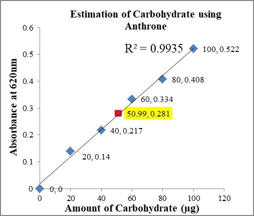
The highlighted concentration i.e., 50.99 µg was the amount of carbohydrate present in 100 µl of 10 mg/mL sample concentration
Figure 1. Standard graph for Carbohydrate estimation (R2 = 0.9935) with the aqueous extract
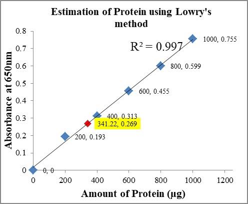
The highlighted concentration i.e., 341.22 µg was the amount of protein present in 100 µl of 10 mg/mL sample concentration
Figure 2. Standard graph for protein estimation (R2 = 0.997) with the aqueous extract
Antibacterial activity assay
The acidic extract of mucus showed highest zone of inhibition against all the eight selected bacterial strains while, the organic extract showed resistance against 7 selected bacterial strains. There was clear variation in the antibacterial activity depending on the bacterial strain type and concentration of the sample as well. Both the extracts showed significant zone of inhibition difference as shown below (Table 1 and Table 2). The selected bacterial strains for example K. pneumonia, E. coli did not show any resistance towards the commercially available ampicillin, on the other hand, the acidic and organic extracts of the fish mucus showed resistance towards the bacterial growth. From the data (Table 1) given below, P. aeruginosa at a concentration of 5 mg/mL for the organic extract showed highest zone of inhibition. It is worth noting that the higher the concentration, the lower was the antimicrobial activity. On the other hand, E. coli alone showed growth inhibition at a higher concentration as it is a high resistant bacterial strain.
Table (1):
Inhibition zones (mm) shown by organic extract against different selected pathogenic microbial strains
| Bacterial Strain | Image no. | Zone of Inhibition (mm) | MIC mg/mL | |||||
|---|---|---|---|---|---|---|---|---|
| 1 mg/mL | 2.5 mg/mL | 5 mg/mL | 7.5 mg/mL | 10 mg/mL | Ampicillin | |||
| Methicillin Resistant S. aureus | 1 | 7.3 ± 0.5 | 10.3 ± 0.5 | 0.0 ± 0.0 | 8.3 ± 0.5 | 8.3 ± 0.5 | 8.2 ± 0.5 | 1 |
| B. cereus | 3 | 10.6 ± 0.5 | 9.3 ± 0.5 | 0.0 ± 0.0 | 8.3 ± 0.5 | 0.0 ± 0.0 | 7.5 ± 0.5 | 2.5 |
| S. typhi | 5 | 10.3 ± 0.5 | 0.0 ± 0.0 | 0.0 ± 0.0 | 7.3 ± 0.5 | 7.0 ± 0.5 | 10.2 ± 0.5 | 1 |
| K. pneumonia | ND | 0.0 ± 0.0 | 0.0 ± 0.0 | 0.0 ± 0.0 | 0.0 ± 0.0 | 0.0 ± 0.0 | 0.0 ± 0.0 | ND |
| E. faecalis | 8 | 9.3 ± 0.5 | 9.6 ± 0.5 | 10.3 ± 0.5 | 11.3 ± 0.5 | 8.6 ± 0.5 | 12.0 ± 0.0 | 2.5 |
| S. aureus | 10 | 0.0 ± 0.0 | 8.6 ± 0.5 | 0.0 ± 0.0 | 8.0 ± 0.5 | 0.0 ± 0.0 | 9.3 ± 0.5 | 2.5 |
| E. coli | 12 | 0.0 ± 0.0 | 8.3 ± 0.5 | 0.0 ± 0.0 | 0.0 ± 0.0 | 9.3 ± 0.5 | 0.0 ± 0.0 | 2.5 |
| P. aeruginosa | 14 | 10.0 ± 0.5 | 9.6 ± 0.5 | 11.6 ± 0.5 | 9.3 ± 0.5 | 9.3 ± 0.5 | 12.6 ± 0.5 | 1 |
Image number should be correlated with the numbers on Figure 3. Zone of inhibition including the well diameter, values are expressed as mean ± SD. Data was analyzed using two way ANOVA and the value of p < 0.05. Highlighted values are the highest inhibition exhibited by that particular bacterial strain
Table (2):
Zone of inhibition (mm) shown by acidic extract against different selected pathogenic microbial strains
| Bacterial Strain | Image No. | Zone of Inhibition (mm) | MIC | ||||
|---|---|---|---|---|---|---|---|
| 1 mg/mL | 2.5 mg/mL | 5 mg/mL | 7.5 mg/mL | 10 mg/mL | mg/mL | ||
| Methicillin resistant S. aureus | 2 | 18.3 ± 0.5 | 13.3 ± 0.5 | 12.0 ± 0.0 | 9.6 ± 0.5 | 9.6 ± 0.5 | 2.5 |
| B. cereus | 4 | 18.3 ± 0.5 | 13.0 ± 0.5 | 14.3 ± 0.0 | 9.3 ± 0.5 | 0.0 ± 0.0 | 3.75 |
| S. typhi | 6 | 11.3 ± 0.5 | 13.0 ± 1.0 | 13.3 ± 0.5 | 7.0 ± 1.0 | 7.0 ± 1.0 | 2.5 |
| K. pneumonia | 7 | 17.0 ± 0.0 | 15.3 ± 0.5 | 13.0 ± 1.0 | 13.0 ± 0.0 | 0.0 ± 0.0 | 2.5 |
| E. faecalis | 9 | 14.0 ± 0.0 | 17.3 ± 0.5 | 19.6 ± 0.5 | 11.3 ± 0.5 | 8.0 ± 0.5 | 1.25 |
| S. aureus | 11 | 9.3 ± 0.5 | 11.3 ± 0.5 | 15.0 ± 0.0 | 8.6 ± 0.5 | 0.0 ± 0.0 | 2.5 |
| E. coli | 13 | 19.3 ± 0.5 | 14.3 ± 0.5 | 13.3 ± 0.5 | 11.0 ± 1.0 | 11.0 ± 1.0 | 1.25 |
| P. aeruginosa | 15 | 21.3 ± 0.5 | 22.6 ± 0.5 | 15.6 ± 0.5 | 9.0 ± 1.0 | 9.0 ± 1.0 | 0.5 |
Image number should be correlated with the numbers on Figure 3. Positive control remains same as shown in Table 1. Zone of inhibition including the well diameter, values are expressed as mean ± SD. Data was analyzed using two way ANOVA and the value of p < 0.05. Highlighted values are the highest inhibition exhibited by that particular bacterial strain
Methicillin resistant S. aureus showed highest zone of Inhibition for the organic extract mucus at 2.5 mg/mL concentration and for the acidic extract of mucus at 1 mg/mL concentration. B. cereus exhibited highest growth inhibition for organic and acidic extracts at 1 mg/mL concentration. S. typhi exhibited highest zone of inhibition for organic extract of mucus at 1 mg/mL and for acidic extract at 5 mg/mL. K. pneumonia did not show any inhibition for organic extract while, it showed highest zone of inhibition for the acidic extract at 1 mg/mL concentration. E. faecalis showed highest zone of inhibition for organic extract at 7.5 mg/mL and 5 mg/mL for acidic extract of mucus. S. aureus exhibited highest zone of inhibition at 2.5 mg/mL of organic extract and for acidic extract at 5 mg/mL. E. coli showed highest zone of inhibition for organic extract at a concentration of 10 mg/mL and for acidic extract at 1 mg/mL. P. aeruginosa showed highest zone of inhibition for organic extract at 5 mg/mL and for acidic extract at 2.5 mg/mL concentration. For MIC, the different bacterial strains showed different results. Both the organic and acidic extracts showed zone of inhibition at a concentration of less than 2.5 mg/mL for all the bacterial strains except the acidic extract treated against B. cereus at 3.75 mg/mL concentration. Data given below (Table 1 and Table 2) shows the zone of inhibition at different concentrations of both the extracts and image (Figure 3) shows the highest inhibition zone by the selected bacterial strains at different concentrations.
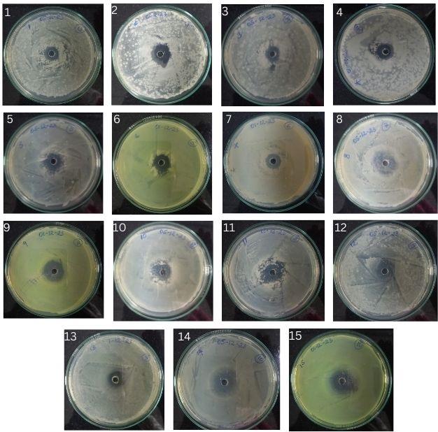
1. Organic extract Methicillin resistant S. aureus, 2. Acidic extract Methicillin resistant S. aureus, 3. Organic extract B. cereus, 4. Acidic extract B. cereus, 5. Acidic extract S. typhi, 6. Organic extract S. typhi, 7. Acidic extract K. pneumonia, 8. Organic extract E. faecalis, 9. Acidic extract E. faecalis, 10. Organic extract S. aureus, 11. Acidic extract S. aureus,
12. Organic extract E. coli, 13. Acidic extract E. coli, 14. Organic extract P. aeruginosa, 15. Acidic extract P. aeruginosa
Images represent the highest zone of inhibition by each bacteria i.e highlighted in the table
Figure 3. Zone of inhibition using Agar well diffusion method against selected bacterial strains
Cytotoxicity effects of mucus
For the evaluation on the biological effects of mucus on normal cell viability and morphology, L929 cells were treated with different concentration (18.75-600 µg/mL). As shown in below (Figure 4A and 4B), the aqueous extract of mucus exhibited no or lack of cytotoxicity as the viability was not less than 50%. At the above stated concentrations, the percentage viability of L929 cells was 102% and 99.78% at a concentration of 75 µg/mL and 150 µg/mL, respectively compared to the untreated cells. The concentration of mucus and their cell viabilities respectively did not show IC50 until the concentration of 600 µg/mL which is a proven fact that there is an absence of toxicity effect for the selected concentration.

Images represented were captured using florescent microscope (XDFL series) at a magnification of 100 µm
Figure 4A. Representative images of L929 cells i.e untreated, 75 µg/mL, 150 µg/mL for cytotoxicity evaluation
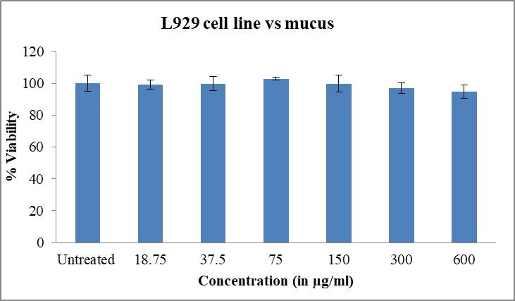
Results show the average of triplicates performed ±SD. (p < 0.05)
Figure 4B. Cell viability calculated for different concentrations along with the untreated cells. Results show the average of triplicates performed ±SD. (p < 0.05)
In-vitro cell migration and wound repair
Apart from the cell cytotoxicity and cell viability assays, the cell proliferation and migration property was also assessed using in vitro scratch assay. The L929 cells (Figure 5A) shows the wound healing property of mucus compared to the untreated cells as the aqueous extract of mucus showed complete closure of the wound area in a time duration of 24 hours by inducing the migration of L929 cells towards the scratch area(Figure 5B). As shown below (Figure 5A), mucus at a concentration of 75 µg/mL assisted in the increased cell migration to 81.60% at 12th hour, 99.26% at 24th hour and at 150 µg/mL to 79.32% at 12th hour and 99.27% after 24 hours. On the other hand (figure 5A), the capacity of the cell migration was found to be 99.27% at 24th hour while the untreated cells showed only 65.44% at 12th hour and 94.68% at 24th hour. Thus remarking the wound healing property of mucus evidently.
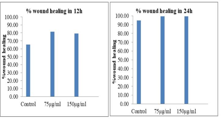
Wound healing percentage was calculated by comparing the final gap area to initial gap area (0th hour)
Figure 5A. Represents the percentage of cell migration at the interval of 12 hours and 24 hours
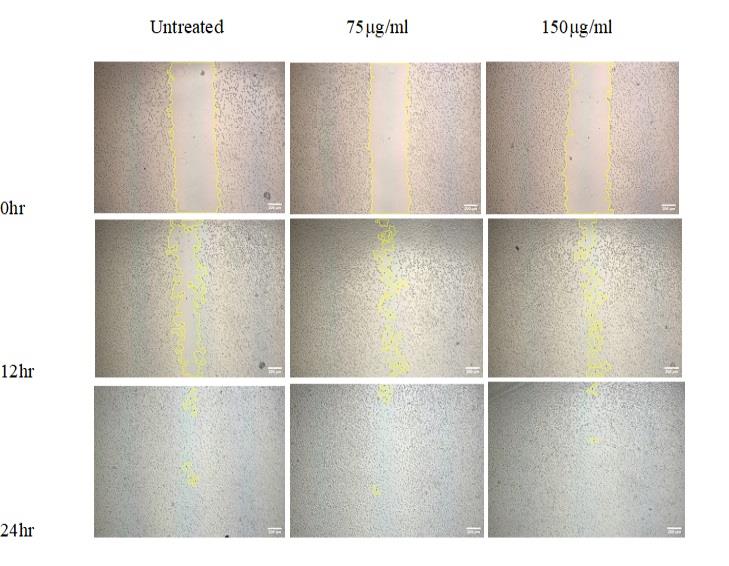
Wound healing area was analyzed using ImageJ (Fiji) software. Images represented were captured using florescent microscope (XDFL series) at a magnification of 100 µm
Figure 5B. Represents the rate of cell migration and the area of the migrated cells when compared to the initial scratch
Based on the existing literature survey and research, the antimicrobial activities of mucus along with the different mucus extracts from different species of fishes have been extensively studied.5,31 From the available literatures, it is clearly evident that the antimicrobial activity is totally dependent only on different fish species including the extraction protocol and strain of the selected clinically resistant pathogens.32,33 From the current studies, it is seen that the acidic extract of P. hypophthalmus showed highest antibacterial activity towards the selected clinically resistant strains which is similar to that of kumara et al.,34 who evaluated the crude extracts of Hypophthalmichthys nobilis, Cyprinus carpio and Ctenopharyngodon idella. In spite of the existing reports on the antimicrobial activity of mucus extracts from different fishes, our study is the first one to assess the antimicrobial activity of mucus from a viviparous fish against the clinically resistance pathogenic bacteria.
The acidic extract of mucus from
P. hypophthalmus showed highest antimicrobial activity through the well diffusion method along with the MIC assay when compared to the commercially available drug ampicillin. Therefore, we suggest that this mucus can be used as an alternative source of an antimicrobial agent for treatment of wounds. The current study was evaluating the actual healing mechanism/potency of the mucus on wounds and the healing processes via MTT assay and Scratch assay.
Furthermore, the mucus extracts of P. hypophthalmus accelerated cell viability by showing an enhanced growth of L929 cells in the MTT assay performed. This is in alignment with the work carried out by Nessma et al.,19 where the organic extract of snail mucus of E. desertorum showed higher viability on human fibroblast cells. However, the existing research is more focused on to the cell viability while in vitro analysis of wound healing is not explored to a maximum extent. In the scratch assay performed in our current study, at the 12th hour itself, the percentage of wound closure showed around 15% increase which was found to be significant. It is to be critically noted that the initial phase of wound healing processes, i.e. hemostasis, inflammation, proliferation is highly impacted by the mucus, which is a clear evidence provided on to the potency of the mucus to be used as a therapeutic agent.
On the other hand, it is not shocking that there are differences found within the different species of fishes taken for different studies that the effect could be pertaining to their composition and mechanical properties. A biological matrix i.e., the mucosal layer acts as a barrier between the fish and its surroundings where, the molecular constituents of it are crucial when performing multiple functions, such as mechanical support, physiological functions and immunological functions.35,36 The fish mucus is known to contain multiple antimicrobial peptides. The antimicrobial peptides (AMP) that also called Host Defense Peptides, are the peptides with up to 80 amino acid residues encoded by different genes.37 They are primarily distinguished by their antibacterial, cationic and amphipathic chemical nature. Almost all classes of fish species contain these old innate immune components. In eukaryotic cells, their mature forms are frequently cysteine-rich molecules with several intra-molecular disulfide bonds.38 Certain AMP families are able to control the type and degree of activity of their members by preserving or breaking these links.39 For example, certain research on a specific set of human beta-defensins suggest that the reduction of such bonds alter the function of defensins (one of the most studied families of AMPs) by inhibiting their chemotactic activities and inducing their direct antibacterial genes.38
Therefore, we conclude in our present study that the mucus of P. hypophthalmus could be a possible major resource of a biomaterial that can be used for the antimicrobial activity and wound healing processes. Furthermore, the molecular composition of this fish mucus is to be explored to confirm the actual potency along with the in vivo, gene expression studies to elucidate the definite mechanism of action.
The present research work has demonstrated the potency of mucus of P. hypophthalmus fish as an efficient antimicrobial agent, showing highly significant application in wound healing and therapeutic purposes. Furthermore, the human trials along with the other in vivo studies have to be performed to ascertain the same.
ACKNOWLEDGMENTS
The authors would like to thank CHRIST (Deemed to be University) for providing the necessary support and guidance to carry out the present research work, and Dr. A. John Paul, Assistant Professor, St. Joseph’s University for his guidance and support.
CONFLICT OF INTEREST
The authors declare there is no conflict of interest.
AUTHORS’ CONTRIBUTION
MP and GM conceptualized the study and applied methodology. MP collected resources and performed supervision. GM performed data curation, funding acquisition and investigation. MP performed validation. GM wrote, reviewed and edited the manuscript. Both authors read and approved the final manuscript for publication.
FUNDING
This work was supported by the fellowship provided by ‘KSTEPS, DST, Govt. of Karnataka, India, with fellowship award no.: LIF-05:2023-24.
AVAILABILITY OF DATA
All datasets generated or analyzed during this study are included in the manuscript.
ETHICS STATEMENT
Not applicable.
- Gottrup F. A specialized wound-healing center concept: importance of a multidisciplinary department structure and surgical treatment facilities in the treatment of chronic wounds. Am J Sur. 2004;187(5):S38-S43.
Crossref - Margolis DJ. Epidemiology of Wounds. Measurements in Wound Healing. 2012:145-153.
Crossref - Bhate-Deosthali P, Lingam L. Gendered pattern of burn injuries in India: a neglected health issue. Reprod Health Matters. 2016;24(47):96-103.
Crossref - Caballero S, Galeano AM, Lozano JD, Vives M. Description of the microbiota in epidermal mucus and skin of sharks (Ginglymostoma cirratum and Negaprion brevirostris) and one stingray (Hypanus americanus). Peer J. 2020;8:e10240.
Crossref - Tiralongo F, Messina G, Lombardo BM, Longhitano L, Volti GL, Tibullo D. Skin Mucus of Marine Fish as a Source for the Development of Antimicrobial Agents. Front Mar Sci. 2020;7:541853.
Crossref - Magnadottir B. Innate immunity of fish (overview). Fish Shellfish Immunol. 2006;20(2):137-151.
Crossref - Dash S, Das SK, Samal J, Thatoi HN. Epidermal mucus, a major determinant in fish health: a review. Iran J Vet Res. 2018;19(2):72-81.
- Fasulo S, Tagliafierro G, Contini A, et al. Ectopic expression of bioactive peptides and serotonin in the sacciform gland cells of teleost skin. Arch Histol Cytol. 1993;56(2):117-125.
Crossref - Swain P, Dash S, Sahoo P, et al. Non-specific immune parameters of brood Indian major carp Labeo rohita and their seasonal variations. Fish Shellfish Immunol. 2007;22(1-2):38-43.
Crossref - Shephard KL. Functions for fish mucus. Rev Fish Biol Fisheries. 1994;4(4):401-429.
Crossref - Kumari U, Nigam AK, Mitial S, Mitial AK. Antibacterial properties of the skin mucus of the freshwater fishes, Rita rita and Channa punctatus. Eur Rev Med Pharmacol Sci. 2011;15(7):781-786.
- Gomathy M, John PA, Velayuthannair K. A systematic review of fish based biomaterials on wound healing and antiinflammatory processes. Adv Wound Care. 2023;13(2):83-96.
Crossref - Tomita T, Toda M, Murakumo K, et al. Five-Month Incubation of Viviparous Deep-Water Shark Embryos in Artificial Uterine Fluid. Front Mar Sci. 2022;9:825354.
Crossref - Chin A, Mourier J, Rummer JL. Blacktip reef sharks (Carcharhinus melanopterus) show high capacity for wound healing and recovery following injury. Conserv Physiol. 2015;3(1):cov062.
Crossref - da Cunha MG, Franchin M, Galvao L, et al. Antimicrobial and antiproliferative activities of stingless bee Melipona scutellaris geopropolis. BMC Complement Altern Med. 2013;13(1):23.
Crossref - Marston HD, Dixon DM, Knisely JM, Palmore TN, Fauci AS. Antimicrobial Resistance. JAMA. 2016;316(11):1193.
Crossref - Shabir U, Dar JS, Bhat AH, Ganai BA, Khan IA. Isolation and characterization of β-defensin-like protein 1 from epidermal mucus of fungal infected fish (Cyprinus carpio) and assessment of its antimicrobial potencies. Aquac Rep. 2022;23:101056.
Crossref - Diamond G, Zasloff M, Eck H, Brasseur M, Maloy WL, Bevins CL. Tracheal antimicrobial peptide, a cysteine-rich peptide from mammalian tracheal mucosa: peptide isolation and cloning of a cDNA. Proc Natl Acad Sci USA. 1991;88(9):3952-3956.
Crossref - EL-Zawawy NA, Mona MM. Antimicrobial efficacy of Egyptian Eremina desertorum and Helix aspersa snail mucus with a novel approach to their anti-inflammatory and wound healing potencies. Sci Rep. 2021;11(1):24317.
Crossref - Husemann E. Methods in Carbohydrate Chemistry. Herausgeg. v. R. L. Whistler, R. J. Smith, J. N. BeMiller und M. L. Wolfrom . Vol. IV: Starch. Academic Press, New York-London 1964. 1. Aufl., XVI, 335 S., zahlr. Abb. u. Tab., geb. $ 13.50. Angewandte Chemie. 1965;77(5):226-227.
Crossref - Anitha S, Manivannan A. Nutritional evaluation of potato milk produced by ultrasonication–A functional alternative for bovine milk. Food and Humanity. 2023;1:684-688.
Crossref - Yadav DK, Yadav M, Rani P, et al. Screening of best growth media for Chlorella vulgaris cultivation and biodiesel production. Biofuels. 2023;15(3):271-277.
Crossref - Atef NM, Shanab SM, Negm SI, Abbas YA. Evaluation of antimicrobial activity of some plant extracts against antibiotic susceptible and resistant bacterial strains causing wound infection. Bull Natl Res Cent. 2019;43(1):144.
Crossref - Shiromi PSAI, Hewawasam RP, Jayalal RGU, Rathnayake H, Wijayaratne WMDGB, Wanniarachchi D. Chemical Composition and Antimicrobial Activity of Two Sri Lankan Lichens, Parmotrema rampoddense, and Parmotrema tinctorum against Methicillin-Sensitive and Methicillin-Resistant Staphylococcus aureus. Evid Based Complement Alternat Med. 2021;2021:1-18.
Crossref - Barnes VL, Heithoff DM, Mahan SP, House JK, Mahan MJ. Antimicrobial susceptibility testing to evaluate minimum inhibitory concentration values of clinically relevant antibiotics. STAR Protocols. 2023;4(3):102512.
Crossref - Alley MC, Scudiero DA, Monks A, et al. Feasibility of drug screening with panels of human tumor cell lines using a microculture tetrazolium assay. Cancer Res. 1988;48(3):589-601.
- Najm AAK, Azfaralariff A, Dyari HRE, et al. Anti-breast cancer synthetic peptides derived from the Anabas testudineus skin mucus fractions. Sci Rep. 2021;11(1):23182.
Crossref - Scudiero DA, Shoemaker RH, Paull KD, et al. Evaluation of a soluble tetrazolium/formazan assay for cell growth and drug sensitivity in culture using human and other tumor cell lines. Cancer Res. 1988;48(17):4827-4833.
- Gerlier D, Thomasset N. Use of MTT colorimetric assay to measure cell activation. J Immunol Methods. 1986;94(1-2):57-63.
Crossref - Hu Z, Yang P, Zhou C, Li S, Hong P. Marine Collagen Peptides from the Skin of Nile Tilapia (Oreochromis niloticus): Characterization and Wound Healing Evaluation. Marine Drugs. 2017;15(4):102.
Crossref - Diaz-Puertas R, Adamek M, Mallavia R, Falco A. Fish Skin Mucus Extracts: An Underexplored Source of Antimicrobial Agents. Marine Drugs. 2023;21(6):350.
Crossref - Li J, Xie S, Ahmed S, et al. Antimicrobial Activity and Resistance: Influencing Factors. Front Pharmacol. 2017;8:364.
Crossref - Leng W, Wu X, Xiong Z, et al. Study on antibacterial properties of mucus extract of snakehead (Channa argus) against Escherichia coli and its application in chilled fish fillets preservation. LWT. 2022;167:113840.
Crossref - Kumari S, Tyor AK, Bhatnagar A. Evaluation of the antibacterial activity of skin mucus of three carp species. Int Aquat Res. 2019;11(3):225-239.
Crossref - Ángeles EM. An Overview of the Immunological Defenses in Fish Skin. ISRN Immunol. 2012;2012:1-29. doi
Crossref - Hancock REW, Haney EF, Gill EE. The immunology of host defence peptides: beyond antimicrobial activity. Nat Rev Immunol. 2016;16(5):321-334.
Crossref - Munk JK, Ritz C, Fliedner FP, Frimodt-Møller N, Hansen PR. Novel Method To Identify the Optimal Antimicrobial Peptide in a Combination Matrix, Using Anoplin as an Example. Antimicrob Agents Chemother. 2014;58(2):1063-1070.
Crossref - Schroeder BO, Wu Z, Nuding S, et al. Reduction of disulphide bonds unmasks potent antimicrobial activity of human β-defensin 1. Nature. 2011;469(7330):419-423.
Crossref - Mahlapuu M, Hakansson J, Ringstad L, Bjorn C. Antimicrobial Peptides: An Emerging Category of Therapeutic Agents. Front Cell Infect Microbiol. 2016;6.
Crossref
© The Author(s) 2024. Open Access. This article is distributed under the terms of the Creative Commons Attribution 4.0 International License which permits unrestricted use, sharing, distribution, and reproduction in any medium, provided you give appropriate credit to the original author(s) and the source, provide a link to the Creative Commons license, and indicate if changes were made.