


ISSN: 0973-7510
E-ISSN: 2581-690X
Bovine brucellosis is an infectious pathology that compromises animal well-being and causes significant economic losses for livestock farmers. This disease leads to abortions and is caused by the bacterium Brucella abortus. In this research, the prevalence of bovine brucellosis (Brucella abortus) was estimated in General Proaño Parish using the Rose Bengal test and confirmation through competitive ELISA. Methodologically, The research was carried out between February and August 2023 on 100 animals from 25 locations in the Proaño parish, in the Morona canton, Ecuador. Blood serum was obtained from five breeds of cattle: Charolais, Holstein, Brown Swiss, Mestiza and Jersey. The collected samples were transported to the laboratory for their respective analysis. Five milliliters of venous blood were collected from the coccygeal vein, from which 1 mL of blood serum was obtained. The serum was used for disease diagnosis through the Rose Bengal test, and those that tested seropositive were reconfirmed using the competitive ELISA immunoassay method. The clinical analyses determined that there is a 0% prevalence of the pathology; based on these results, we can state that the null prevalence of Brucella may be due to environmental and geographical factors that influence its presence and transmission. Furthermore, the precision and sensitivity of the diagnostic methods used are crucial; however, the ELISA and Rose Bengal methods may have been insensitive and did not detect mild infections. Finally, Brucella infection may not be present during sampling due to temporal fluctuations in its prevalence. In conclusion, there is no scientific evidence of bovine brucellosis as the primary cause of abortions and gestational losses in the study area.
Brucellosis, Abortions, Rose Bengal, Competitive ELISA Test
Brucellosis, a zoonotic pathology caused by the genus Brucella, represents a significant concern in global bovine livestock. Known as infectious abortion or “Bangs,” this disease has been identified in various regions, including Latin America, Mediterranean countries, and Saudi Arabia.1 The positive relationship between Brucella abortus colonization and gestational losses in cattle is undeniable, although other pathogens such as Campylobacter fetus, Leptospira spp., Trichomonas fetus, among others, are also common causes of abortions.2
In the bovine context, brucellosis exhibits a global distribution, being endemic in some countries, including ours, where a low national seroprevalence is recorded, although in certain provinces, its rate may be moderate. The movement of animals between different regions can contribute to the proliferation of this pathology.3 The transmission routes are diverse and include animal body secretions such as semen, aborted fetuses, contaminated fetal membranes, as well as direct inhalation and oral ingestion of contaminated food, forages, or water.4
Brucellosis not only impacts animal health but also poses a threat to public health as it is a potential zoonosis transmissible to humans.4 In the province of Morona Santiago, where livestock farming is a crucial economic activity, the presence of brucellosis could have economic and social health consequences for this sector. Although control measures have been implemented, the lack of up-to-date information on the prevalence of the disease is a cause for concern, highlighting the need for timely detection3
The standard diagnostic methods for bovine brucellosis are serology and microbiology. Early detection through these tools is essential for disease control and eradication. Currently, the diagnosis in live dairy cattle involves detecting anti-Brucella antibodies in serum or milk, as well as isolating Brucella from milk samples.5 The history of the identification of Brucella abortus in the Crimean War in 1887 by Dr. David Bruce and the discovery of the disease by Dr. Bernhard Bang in 1987 have been fundamental milestones in understanding and controlling this disease.6
Brucellosis represents one of the greatest challenges in livestock production, particularly due to gestational losses in the late stage of pregnancy, leading to significant economic consequences. An abortion can result in losses ranging from $90 to $1900, depending on the production system and the genetic value of the animals.7 The causes of abortions in cattle are diverse and include infectious agents, toxic agents, heat stress, genetic abnormalities, twin pregnancies, and mastitis, with infections being the most common.8
The economic impacts of brucellosis are notable, affecting reproductive performance, milk and meat production, and generating other associated costs. Originating from Brucella abortus, this zoonotic disease affects humans through contact with secretions from aborted animals or the consumption of contaminated products.9 Globally, 924,121 cases have been recorded in bovine livestock, with the Americas having the highest prevalence.10 In General
Proaño Parish, crucial for agricultural and livestock production, the lack of research on the prevalence of bovine brucellosis underscores the urgent need for sanitary measures and local control programs.9
The functional anatomy of the bovine reproductive system, comprising ovaries, oviduct, uterus, cervix, vagina, and external genitals, plays a crucial role in reproduction. Progesterone, in particular, regulates the endometrial glands during gestation, providing an optimal environment for embryonic development. The pathophysiology of bovine abortion, defined as the interruption of gestation and loss of the conceived product, can occur at different stages, with embryonic losses before 42 days and abortions after 260 days being less common.11
Brucella abortus, one of the infectious agents causing abortion, induces placentitis and endometritis, resulting in abortions, vaginal discharges, and stillbirths. Additionally, it affects calves with weakness and higher mortality, leading to retained placenta, metritis, and a reduced lactation period, impacting calf nutrition.12 Pregnant females exhibit symptoms, while non-pregnant ones are asymptomatic. Necropsy reveals granulomatous inflammatory lesions in the reproductive tract and other tissues.13 The diagnosis of bovine brucellosis aims to understand the distribution and prevalence through direct or serological tests, with the isolation of Brucella spp. being a slow process.14,15
Diagnostic methods for brucellosis involve microbiological samples, such as milk or vaginal swabs from live animals, tissues obtained in necropsy, or fluid from testicular hygroma bursitis.15 Standard serological tests, such as agglutination, complement fixation, fluorescence polarization, and ELISA, use the O-side chain of Brucella abortus polysaccharide as an antigen. Originally developed to detect infections, these tests share structural similarities with the LPS of other bacteria.16,17
The Rose Bengal test, used in the diagnosis of bovine brucellosis, employs whole cells of Brucella abortus stained with the reagent. This method, quick and simple, facilitates screening and the assessment of disease prevalence in animals, being especially useful for veterinarians processing numerous samples daily.18 However, it has limitations, such as potential false positives due to antibodies generated by vaccination, and it is recommended to confirm results in suspicious cases.19 Positivity is determined by observing agglutination under indirect light.20 Despite its limitations, the Rose Bengal test remains valuable for early detection and monitoring of bovine brucellosis.
The competitive ELISA test quantifies antibodies binding to the antigen, using spectrophotometry to measure the conversion of a transparent substrate into a colored product. With high sensitivity and specificity, this immunoassay technique is essential for routine diagnostics, allowing differentiation between vaccinated and infected animals. Its specificity comes from plates coated with bacterial lipopolysaccharides and a specific antibody for an epitope of the O-polysaccharide of the S-lipopolysaccharide antigen.21 A monoclonal antibody is used, competing with serum antibodies; those with over 28% competition are considered positive, as evidenced by the color of the serum.22 Negative samples lack antibodies, while positive ones inhibit the binding of the antigen to the O-polysaccharide epitope, manifested in the color of the positive serum.23
Finally, bovine brucellosis is prevented and eradicated through vaccination programs and diagnostic tests with the culling of infected animals. Livestock control, annual check-ups, serological tests, and milk analysis are key measures. Effective vaccines, based on modified live bacteria, should be selected according to local government recommendations. Consuming pasteurized dairy products and practicing sanitary measures are essential to prevent brucellosis in humans.24,25
With all this background, the objective of this study was focused on: Determining the Prevalence of Bovine Brucellosis (Brucella abortus) through the Rose Bengal test and confirmation with competitive ELISA.
The present study was conducted in rural areas of the General Proaño parish, belonging to the Morona canton in the Morona Santiago province, Ecuador. Between February and August 2023, blood serum samples were taken from 100 grazing cows (88% males and 12% females) from 25 different localities. The age distribution of the cows was as follows: 35% were between 12 and 36 months, 29% between 36 and 60 months, 23% over 60 months, and 13% 12 months or younger. The cattle breeds analyzed were Charolais, Holstein, Brown Swiss, Mestiza, and Jersey. Most of the animals appeared healthy, with the exception of two that had a history of abortions in the last third of gestation.
Experiment management
Sampling and labeling
The sampling process involved extracting 3-5 mL of venous blood from the coccygeal vein and placing it in yellow Vacutainer tubes with a separator gel. This facilitated clot formation and the extraction of blood serum for subsequent analysis. The sample was labeled with a unique code, including the owner’s initials, the animal’s number, the initial letter of its gender, and the breed. This code allowed for the identification and traceability of the samples, with the results recorded in the Vetelab laboratory.
Sample transport
The blood sample, stored in a cooler at 4°C after being labeled, was transported for analysis in the laboratory. Upon receiving the sample, the cooler and test tubes were disinfected. The log sheet was completed, quantifying the samples and determining the amount of necessary reagents.
In the Rose Bengal test, a microplate was used. Drops of negative control and positive control serum were added to the first wells. Then, a drop of the Brucella abortus strain S99 suspension was applied alongside these drops. In the remaining well, 500 µL of the test sample was added and homogenized with the control bacteria suspension. After 2 minutes, antibody precipitations were observed in positive cases and no changes in negative cases.
For the competitive ELISA test, a microplate with wells was used. The test sample and brucellosis standards were placed in the wells. One hundred microliters (100 µL) of anti-Brucella serum were added, homogenizing to form antigen-antibody complexes. After one hour of incubation at room temperature, the supernatant was removed through repeated washes. A colorimetric substrate was applied, sealing the plate until a color change occurred. Antibody measurement was performed using a spectrophotometer, evaluating absorbance and optical density to adjust the calibration curve and calculate the percentage of anti-Brucella antibodies.
Among the response variables, the race, gender, age, and sensitivity of diagnostic techniques (Rose Bengal and ELISA) were analyzed, for which the following formula was applied:
Sensibility = [A / A + C] × 100
Formula 1: Formula for calculating the sensitivity of the diagnostic tests under study.
Where; A: True positives; B: True positives + False negatives.
Specificity = [Negative / Negative + false positive] × 100
The specificity of the diagnostic techniques was also analyzed by applying the following formula:
Formula 2: Formula for calculating the specificity of the diagnostic tests under study. Where: AN: True negatives; FP: False positives.
The prevalence was also analyzed, which was assessed using the results obtained in the laboratory, with the following formula:
Prevalence= [ Number of positive animals
Number of sampled animals ] × 100
Formula 3: Formula for estimating the prevalence of bovine brucellosis in
General Proaño Parish.
Serodiagnosis using the Rose Bengal test
In the analysis of the 25 farms sampled and a total of 100 animals studied, subjected to serodiagnosis with the Rose Bengal test, 100% were seronegative (Table 1).
Table (1):
Serodiagnosis by Rose Bengal test
| #Farm | Negatives | Positives | Total |
|---|---|---|---|
| Frequency | Frequency | ||
| 1 | 4 | – | 4 |
| 2 | 4 | – | 4 |
| 3 | 4 | – | 4 |
| 4 | 4 | – | 4 |
| 5 | 4 | – | 4 |
| 6 | 4 | – | 4 |
| 7 | 4 | – | 4 |
| 8 | 2 | – | 2 |
| 9 | 4 | – | 4 |
| 10 | 4 | – | 4 |
| 11 | 4 | – | 4 |
| 12 | 4 | – | 4 |
| 13 | 3 | – | 3 |
| 14 | 4 | – | 4 |
| 15 | 4 | – | 4 |
| 16 | 4 | – | 4 |
| 17 | 4 | – | 4 |
| 18 | 4 | – | 4 |
| 19 | 4 | – | 4 |
| 20 | 3 | – | 3 |
| 21 | 4 | – | 4 |
| 22 | 4 | – | 4 |
| 23 | 5 | – | 5 |
| 24 | 4 | – | 4 |
| 25 | 7 | – | 7 |
| Total | 100 | 0 | 100 |
In the research of Cruz26 in Zamora Chinchipe, a seroprevalence of 5.42% was found. Mainato & Vallecillo27 in Cañar reported 29.3% positivity. In contrast, in General Proaño, no positive cases were found. Calderon et al.28 in El Carmen, Manabi, documented 6.55% seropositivity. These results contrast with the null prevalence in General Proaño, indicating geographic variability and highlighting coastal areas with higher rates of bovine brucellosis.
Diagnosis using the competitive ELISA test
The competitive ELISA test was performed on two animals from locality 6, both with a history of abortions in the last third of gestation. The test focused on these specific animals to determine if the abortions were related to a Brucella infection. The results were negative for the pathogen, considering percentage inhibition (PI) values greater than 50 as positive and ≤ 50 as negative. The values obtained as shown in Table 2 were 48.94 and 45.92 PI, indicating that the abortions were not due to Brucella, but to another cause.
Table (2):
Diagnosis by competitive ELISA test
| #Farm | Negatives | Positives | Total |
|---|---|---|---|
| Frequency | Frequency | ||
| 6 | 2 | – | 2 |
| Case 1 | Case 2 | ||
| PI: | 48.94 | 45.92 |
According to Tovar & Yepes,29 competitive ELISA is more specific and sensitive, being accessible and reliable in livestock diagnostics. Martinez,30 highlights the variability of reliability (90.53-99.9%) depending on the exposure of the animal. In this investigation, competitive ELISA confirmed the seronegativity of suspected cases, supporting the usefulness and accuracy of the method.
Prevalence
According to the results of the Rose Bengal and competitive ELISA tests, none of the animals in the General Proaño parish were positive for brucellosis, resulting in a prevalence of 0%. Moyano et al.31 found in Zamora and Morona Santiago, areas of pastoral rope systems, that 100% of 48 animals were negative for brucellosis. It coincides with this study, where no animal was diagnosed as seropositive. In Azuay, Mainato,32 determined a prevalence of 7.8% in milk from 153 farms. However, in General Proaño, a prevalence of 0% was evident. Escobar et al.33 carried out geo-referencing of bovine brucellosis in Santo Domingo de los Tsachilas from 2012 to 2016, observing a significant decrease in prevalence, indicating the effectiveness of the national bovine brucellosis eradication program in the province.
Relationship between the variable race
According to the results presented in the Figure, in the General Proaño parish, the predominant breed is Charolais with 41%, followed by Brown Swiss with 24%, Mestiza with 23%, while the Holstein dairy breeds and Jersey represent 12% each. In an investigation in the Sinai Parish, Sagbay34 found that 64.14% of the adult bovine units were Charolais, followed by Charolais x Mestiza F1 at 26.07%. Yari35 evaluated the prevalence of infectious bovine rhinotracheitis in General Proaño, observing 51.3% of Charolais, 25.9% of Brown Swiss, 14.6% of Holstein, 3.8% of Mestiza, 0.6% of Jersey, and 3.8% of crosses. These findings coincide with the preeminence of Charolais in the present investigation and support the presence of the other races mentioned.
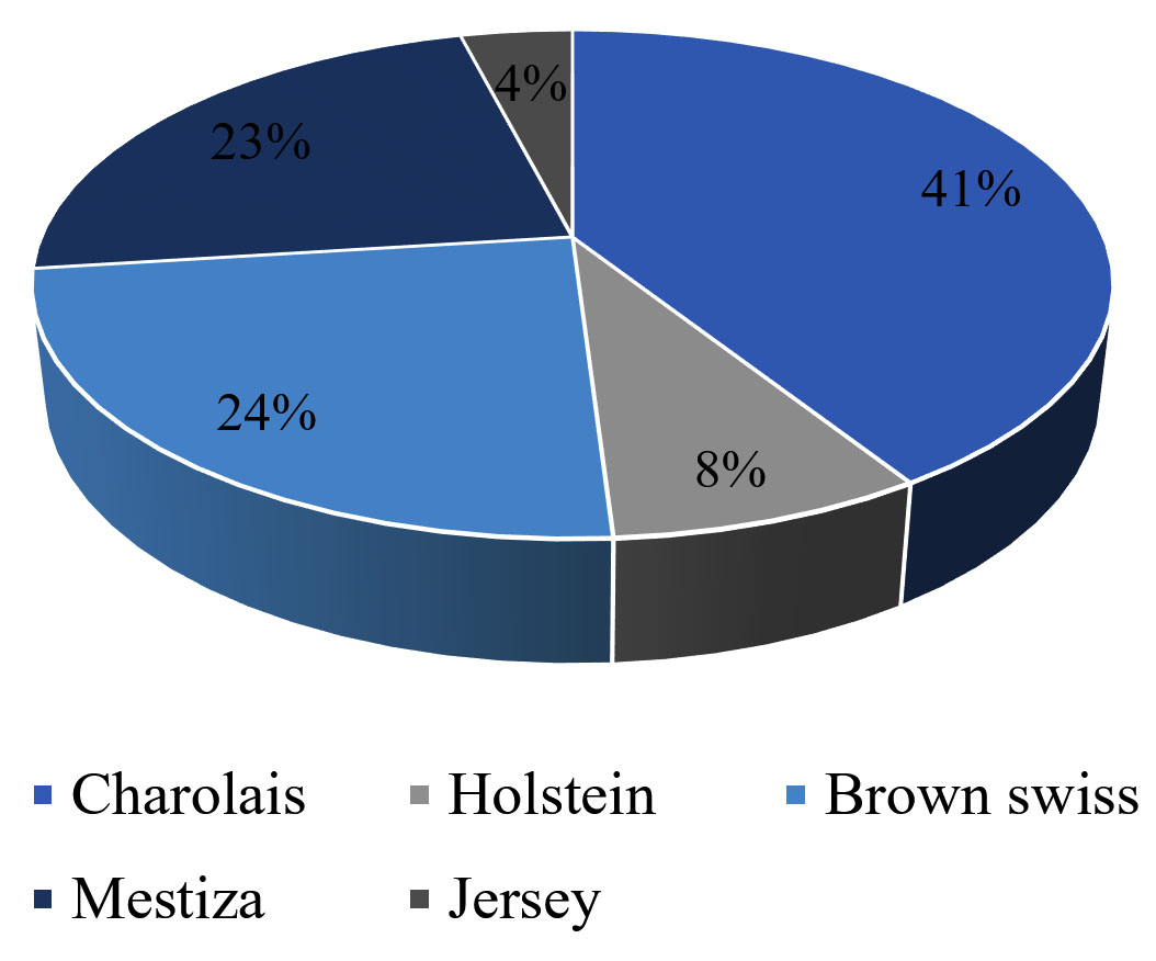
Figure. Bovine breeds analyzed
By applying clinical diagnostics, using the Rose Bengal test in randomly selected animals and competitive ELISA in cattle with clinical signs, the prevalence of bovine brucellosis in General Proaño Parish was 0% in February and March. Tests were conducted on 100 animals, with zero positivity in antibody agglutination using the Rose Bengal method. Although clinical signs were observed in two animals from Lot 6, reconfirmation with competitive ELISA showed PI of 48.94 and 45.92, both below 50, confirming their seronegativity. These randomly applied methods indicate reliable results without biases.
ACKNOWLEDGMENTS
The authors would like to thank the Universidad Estatal de Bolívar, especially the technical staff of the Laboratorio General de la Facultad de Ciencias Agropecuarias, for allowing and supporting the development of the research.
CONFLICT OF INTEREST
The authors declare that there is no conflict of interest.
AUTHORS’ CONTRIBUTION
KVE conceptualized, designed the study and performed sample collection. JMM executed the laboratory tests. RR developed the testing protocol and performed data analysis. JMM collaborated in the interpretation of results. JMM and FBM cordinated and supervised the study. KVE and JMM wrote the manuscript. RR and FBM reviewed the manuscript. All authors read and approved the final manuscript for publication.
FUNDING
This research was funded by the General Proaño Parish GAD (Morona Santiago).
DATA AVAILABILITY
All datasets generated or analyzed during this study are included in the manuscript.
ETHICS STATEMENT
This research was approved by the Ethical Committee of the State University of Bolivar, Ecuador.
- Carrillo PC, Gotuzzo E. Brucellosis. Revista Peruana de Medicina Experimental y Salud Publica. 2014;14(1):63-66
- Galvao KN, Bicalho RC, Jeon SJ. Symposium review: The uterine microbiome associated with development of uterine disease in dairy cows. J Dairy Sci. 2019;102(12):11786-11797.
Crossref - Cabrera V. Prevalence of bovine brucellosis in the Limon Indanza Canton, Morona Santiago Province. Cuenca: State University of Cuenca. 2018.
- Owens CE, Daniels KM, Ealy AD, Knowlton KF, Cockrum RR. Potential mechanisms of interaction between bacteria and the reproductive tract of dairy cattle. J Dairy Sci. 2020;103(11):10951-10960.
Crossref - Marianelli C, Martucciello A, Tarantino M, Vecchio R, Iovane G, Galiero G. Evaluation of Molecular Methods for the detection of Brucella species in water buffalo milk. J Dairy Sci. 2008;91(10):3779-3786.
Crossref - Mode S, Katterer M, Quebatte M, Dehio C. Antibiotic presistence of intracellular Brucella abortus. Plos Negl Trop Dis. 2022;16(7):e0010634.
Crossref - De Vries A. Economic Value of pregnancy in Dairy Cattle. J Dairy Sci. 2006 89(10):3876-3885.
Crossref - Norman HD, Miller RH, Wright JR, Hutchinson JL, Olson KM. Factors associated with frequency of abortions recorded through dairy herd improvement test plans. J Dairy Sci. 2012;95(7):4074-4084.
Crossref - Mag, Agrocalidad. Manual of Procedures for the Care and Control of Bovine Brucellosis in Ecuador. Quito, Pichincha, Ecuador. 2016. https://www.agrocalidad.gob.ec/wp-content/uploads/2020/05/resolucion-0131.pdf
- OPS. https://www.paho.org/es/temas/zoonosis
- Szelenyi Z, Gyori D, Boldizsar S, et al. Pregnancy and stillbirth losses in dairy cows with singleton and twin pregnancies. Acta Vet Hung. 2019;67(1):115-126.
Crossref - Bonillaa L, Salamancab RAA, Fajardoc CFC. Brucella abortus: brucellosis and new diagnostic tests in cattle. Veterinary Medicine and Animal Husbandry. 2021:1-16: https://repository.ucc.edu.co/server/api/core/bitstreams/0821f568-7da8-4881-b297-fe1c77b0d66e/content
- Silva M. Bovine Brucellosis. Symposium Review: Uruguayan Buiatric Conference. 2001. https://bibliotecadigital.fvet.edu.uy/handle/123456789/1/browse?type=subject&value=BRUCELOSIS
- Kaden R, Ferrari S, Jinnerot T, Lindberg M, Wahab T, Lavander M. Brucella abortus: determination of survival times and evaluation of methos for detection in several matrices. BMC Infect Dis. 2018;18(1):259.
Crossref - Di Bonaventura G, Angeletti S, Lanni A, Petitti T, Gherardi G. Microbiolical laboratory diagnosis of human brucellosis: An Overview. Pathogens. 2021;10(12):1623.
Crossref - Bulashev AK, Ingirbay BK, Mukantayev KN, Syzdykova AS. Evaluation of chimeric proteins for serological diagnosis of brucellosis in cattle. Vet World. 2021;14(8):2187-2196.
Crossref - Ducrotoy MJ, Munoz PM, Conde-Alvarez R, Blasco JM, Moriyon I. A systematic review of current immunological tests for the diagnosis of cattle brucellosis. Prev Vet Med. 2018;151:57-52.
Crossref - Ezama A, Gonzalez JP, Majalija S, Bajunirwe F. Assessing short evolution brucellosis in a highly Brucella spp. endemic cattle keeping population of western Uganda: a complementary use of Rose Bengal test and IgM rapid diagnostic test. BMC Public Health. 2018;18(1):315.
Crossref - Chacon-Diaz C, Zabalza-Barangua A, San Roman B, et al. Brucella abortus S19 GFP-tagged vaccine allows the serological identification of vaccinated cattle. Plos One. 2021;16(11):e0260288.
Crossref - Keshavarzi F, Abdolmohammadi Z. Detection of Brucella abortus using nested-PCR molecular method. Clin Lab. 2021;67(11).
Crossref - Kalleshamurthy T, Skariah S, Rathore Y, et al. Comparative evaluation of fluorescence polarization assay and competitive ELISA for the diagnosis of bovine brucellosis vis-a-vis sero-monitoring. J Microbiol Methods. 2020;170:105858.
Crossref - Gall D, Nielsen K. Improvements to the competitive ELISA for detection of antibodies to Brucella abortus in cattle sera. Journal of Immunoassay. 1994;15(3):277-291.
Crossref - Gaviria Obregon O. Risk factors associated with Brucella abortus seropositivity in livestock farms in the department of Putumayo, Colombiaabortus in livestock farms in the department of Putumayo, Colombia. Universidad La Salle.. 2020:144. https://ciencia.lasalle.edu.co/cgi/viewcontent.cgi?article=1013&context=maest_agrociencias
- Quinteros A. Vaccination Coverage of Calves with the S19 Strain Against Bovine Brucellosis in the Municipality of Capinota. Universidad Mayor de San Simon, Cochabamba. 2021. http://ddigital.umss.edu.bo/handle/123456789/27791
- Martinez P. Brucelosis bovina-Sintomas y tratamiento. 2019. https://www.expertoanimal.com/brucelosis-bovina-sintomas-y-tratamiento-24264.html
- Herrera L, Castillo JC, Manuel J. Determination of the seroprevalence of bovine brucellosis and associated factors in the canton El Pangui, province of Zamora Chinchipe. National University of Loja. 2022. https://dspace.unl.edu.ec/jspui/handle/123456789/24754
- Mainato S, Vallecillo A. Seroprevalence of bovine brucellosis in the province of Canar, Ecuador. Maskana, Animal production. 2017;8:25-28. https://publicaciones.ucuenca.edu.ec/ojs/index.php/maskana/article/view/1480
- Menendez JCC, Goicochea CAB, Aguayo MDZ, De la Cruz Veliz LM, Zambrano PFR. Seroprevalence of bovine brucellosis and its relationship with abortion in reproductive age in the canton of El Carmen, province of Manabi, Ecuador. The technique of Review of Agrosciences. 2019;(21):87-96.
Crossref - Karina TC, Maria YE. Bovine brucellosis and the effectiveness of diagnostic tests. Ibague, Tolima, Colombia. 2022. https://repository.ucc.edu.co/entities/publication/df6da037-d35c-4b70-aff6-a308124d16cd
- Martinez LT. Prevalence of Bovine Brucellosis (Brucella abortus) using detection methods such as the Milk Ring Test (MRT) and the Competitive Elisa Test (Elisa-C). A descriptive review. Thesis Antonio Narino University. Popayan. Colombia. 2021:73. https://repositorio.uan.edu.co/items/1761fc5b-0654-4d64-ba0f-c1fa15b573cc
- Moyano J, Lopez J, Vargas J, Quinteros O. Prevalence of infectious diseases in the Ecuadorian Amazon. Proceedings of the XXXIX National and International Congress of Buiatrics 2015 “Lic. Luis Bravo Tornel”. 2015;37-39. https://fmvz.unam.mx/zootecnia/Prueba/ammveb/congreso.html
- Alfonso MAE. Frequency and geographic referencing of milk samples positive for brucellosis in farms in Azuay. Cuenca, Ecuador. University of Cuenca. 2021. http://dspace.ucuenca.edu.ec/handle/123456789/36317
- Escobar S, Romero E, Gualpa F. GEO-Referencing Of the Prevalence of Bovine Brucellosis (Brucella abortus) in Santo Domingo de los Tsachilas. Spamciencia Journal. 2017;(1):59-66. https://revistasespam.espam.edu.ec/index.php/Revista_ESPAMCIENCIA/article/view/136
- Sagbay K. Characterization of the Production Systems of Beef Cattle in the Sinai Parish, Canton Morona, Morona Santiago Province. Macas, Morona Santiago, Ecuador. ESPOCH. 2022. http://dspace.espoch.edu.ec/handle/123456789/17094
- Yari BM. Prevalence of Infectious Bovine Rhinotracheitis (IBR) in Cattle Herds of the General Proaño Parish, Morona Canton in the Province of Morona Santiago. Macas, Morona Santiago, Ecuador. ESPOCH. 2022. http://dspace.espoch.edu.ec/handle/123456789/18419
© The Author(s) 2024. Open Access. This article is distributed under the terms of the Creative Commons Attribution 4.0 International License which permits unrestricted use, sharing, distribution, and reproduction in any medium, provided you give appropriate credit to the original author(s) and the source, provide a link to the Creative Commons license, and indicate if changes were made.