


ISSN: 0973-7510
E-ISSN: 2581-690X
 , Shaziya Sulthana1, Vijay Ramesh1, E. Sumitha2 and Sunita C. Mesta1
 Actinomycetes are filamentous, Gram-positive bacteria of major ecological and biomedical importance due to their exceptional ability to produce diverse bioactive secondary metabolites encoded by numerous biosynthetic gene clusters. In the present study, actinomycetes were isolated from rhizospheric and non-rhizospheric soil samples and systematically characterized using morphological, biochemical, and molecular approaches. The isolates were screened for antimicrobial, antioxidant, and L-glutaminase-producing potential, followed by 16S rRNA gene sequencing for taxonomic identification. Several isolates exhibited strong antibacterial activity against clinically significant pathogens, including Staphylococcus aureus (NCIM 2073) and Escherichia coli (NCTC 9001). Antioxidant potential evaluated using the DPPH radical scavenging assay confirmed the presence of redox-active metabolites. Efficient production of L-glutaminase, an amidohydrolase enzyme was observed. To complement the experimental findings, in silico molecular docking studies were performed to investigate the interaction of selected bioactive compounds with relevant target proteins, providing mechanistic insights into binding affinity and molecular stability. The docking results revealed favourable binding energies and key hydrogen-bond and hydrophobic interactions, supporting the observed biological activities. Overall, the integrated experimental and in silico approach highlights soil-derived actinomycetes as versatile and sustainable bioresources with significant pharmaceutical and biotechnological potential, emphasizing their role in combating antimicrobial resistance and enabling the rational development of novel therapeutic and industrial products.
Actinomycetes, L-glutaminase, Antimicrobial Activity, Antioxidant Activity, Molecular Docking, Secondary Metabolites
Actinomycetes are a group of unicellular filamentous, Gram-positive bacteria that play a crucial role in microbial biotechnology and soil ecosystems. Their principal ecological niche is soil, where they actively assist in the decomposition of organic materials and the recycling of nutrients. They are widely distributed in both terrestrial and aquatic environments. Actinomycetes are morphologically characterized by mycelial growth, aerial and substrate hyphae, and spore production. Until molecular and phylogenetic analysis, these traits were considered as fungus.1 Their genomes are generally high in guanine and cytosine (G+C content), which is linked to genetic stability and adaptability to challenging environmental circumstances. Numerous bioactive metabolites are produced by these microbes. More than two-thirds of all naturally derived antibiotics currently used in clinical settings come from the genus Streptomyces, which is the most studied and commercially exploited. Actinomycetes are the source of traditional antibacterial drugs including streptomycin, tetracycline, erythromycin, rifampicin, and chloramphenicol, which have transformed contemporary medicine. Actinomycete-derived metabolites have enormous therapeutic potential due to their antifungal, antiviral, antiparasitic, anticancer, and immunosuppressive characteristics in addition to their antibacterial action.2 Actinomycetes are becoming more widely acknowledged as important sources of industrial enzymes and antioxidant metabolites in addition to antibacterial substances. Numerous species generate redox-active molecules, flavonoids, and phenolic compounds that can scavenge reactive oxygen species, reducing oxidative stress linked to inflammatory illnesses, cancer, ageing, and neurological conditions. Assays like DPPH, ABTS, and FRAP have been frequently used to assess the antioxidant potential of actinomycetes.3 Because of these characteristics, actinomycetes are now useful in nutraceutical and functional food applications in addition to pharmaceutical. In addition to its usage in flavor enhancement, biosensors, and biocatalysis, L-glutaminase has gained special attention due to its therapeutic value in the treatment of cancer. Recent years have seen a resurgence of scientific interest in the study of actinomycetes from various sources due to the rapid growth of multidrug-resistant diseases. In this regard, finding potential candidates with antibacterial, antioxidant, and enzyme-producing ability still requires the systematic isolation, characterization, and functional assessment of actinomycetes. Thus, the goal of this study is to investigate soil actinomycetes as multipurpose bioresources with important uses in the biotechnological, industrial, and medicinal fields.4
Collection and isolation of samples
Actinomycetes were isolated from six soil samples using the viable plate count method and serial dilution up to 10-9 The samples were plated on Actinomycetes Isolation Agar (Himedia) supplemented with griseofulvin and fluconazole, and were subsequently incubated at 35 °C. Pure cultures were obtained by streak plating.5
Morphological and biochemical characterization
The morphological characterization was done, which included Gram staining, aerial and substrate mycelium, and spore morphological studies by the cover slip method. The biochemical tests included Catalase, indole, methyl red, Voges-Proskauer, citrate utilization, gelatin hydrolysis, and carbohydrate fermentation tests that provide details on metabolic profiles.6
Genomic DNA isolation (CTAB method)
Bacterial isolates were cultivated in LB broth, then centrifugation was used to extract the cells. After suspending the pellet in CTAB extraction buffer, it was incubated for 30 minutes at 60 °C. Chloroform:isoamyl alcohol (24:1) was used to extract the supernatant following centrifugation. After precipitating DNA with cold isopropanol, it was cleaned with 70% ethanol, allowed to air dry, and then dissolved in TE buffer. DNA concentration was measured after RNase A treatment.
DNA purification
A commercial spin-column kit was used to further purify the isolated DNA in accordance with the protocol. Before PCR amplification, the purified DNA was quantified and eluted in elution buffer.7
PCR amplification of 16S rRNA gene
The universal primers 27F (5′-AGAGTTTGATCCTGGCTCAG-3′) and 1492R (5′-GGTTACCTTGTTACGACTT-3′) were used to amplify the 16S rRNA gene. Template DNA (25-40 ng), PCR buffer, MgCl2, dNTPs, forward and reverse primers, Taq DNA polymerase, and nuclease-free water were all included in each 25 µL PCR reaction. Initial denaturation at 95 °C for two minutes, thirty cycles of denaturation at 95 °C for thirty seconds, annealing at 50 °C for thirty seconds, extension at 72 °C for one minute, and a final extension at 72 °C for ten minutes comprised the PCR cycling conditions.8
Gel extraction and purification
The appropriate bands were removed and purified using a gel extraction kit in accordance with the manufacturer’s instructions after PCR products were resolved on an agarose gel.9
Sanger sequencing
The same primers were used to perform Sanger sequencing on purified PCR results. Following conventional cycling conditions for sequencing reactions, ethanol-EDTA precipitation was used for post-reaction purification. After being denatured in Hi-Di formamide, the refined products were fed into an automated DNA Sequencer. Sequence analysis software, such as FinchTV, BioEdit, and ChromasLite, was used to examine chromatogram files (ab1). For identifying bacteria, high-quality sequences were put together and compared with reference sequences in the NCBI BLAST database.10
Ligand preparation
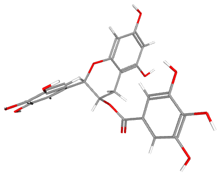
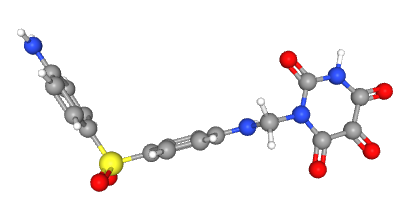
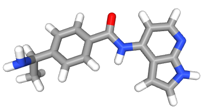
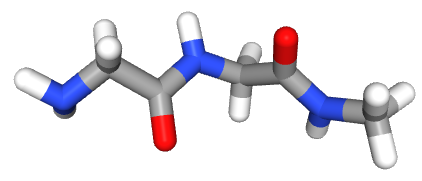
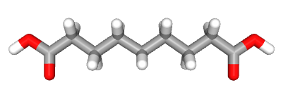
Bioactive ligand constituents associated with actinomycetes were retrieved from the PubChem database based on an extensive review of the literature. Five selected ligands-Epigallocatechin gallate (CID 65064), Allantodapsone (CID 234402), CID 9810884, N-amino-N-[2-(methylamino)-2-oxoethyl]acetamide (CID 439199), and Azelaic acid (CID 2266)-were downloaded in SMILES and SDF formats. These ligands were prepared by adding hydrogen atoms, assigning appropriate charges, and performing energy minimization before further in silico molecular docking analysis (Table 1).11
Table 1. List of selected bioactive ligands associated with actinomycetes, including their PubChem compound IDs and corresponding chemical structures, used for ligand preparation and subsequent in silico molecular docking studies
No. |
Ligands |
Molecule names |
Structure |
|---|---|---|---|
1. |
65064 |
Epigallocatechin gallate |
 |
2. |
234402 |
Allantodapsone |
 |
3. |
9810884 |
9810884 |
 |
4. |
439199 |
Amino-N-[2-(methylamino)- 2-oxoethyl]acetamide |
 |
5. |
2266 |
Azelaic acid |
 |
Protein selection and preparation
The three-dimensional crystal structures of Fibronectin-binding protein (PDB ID: 1KZN) and Glycoprotein adhesin SasA (PDB ID: 4F20) were retrieved from the RCSB Protein Data Bank (PDB). These structures were prepared using the protein preparation wizard of Maestro v13.3 to fix all the problems in the existing structures. The bond orders were assigned, hydrogen bonds optimized, and missing loops and side chains were filled using Prime. Additionally, water molecules were Removed12 (Figure 1).
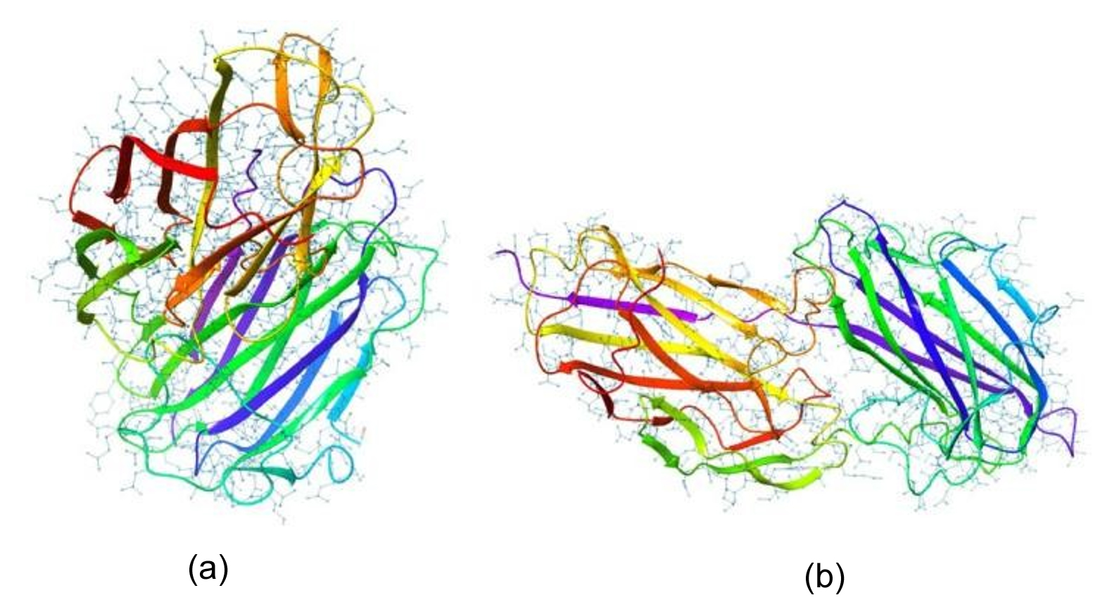
Figure 1. 3D structures of proteins: (a) Fibronectin-binding protein (PDB ID: 1KZN); (b) Glycoprotein adhesin SasA (PDB ID: 4F20)
Binding site prediction
The binding sites of proteins were retrieved using the Sitemap tool of the Schrodinger Suite. The top-ranked potential binding site was selected in the receptor grid generation tool of the suite to generate a grid surrounding the binding site amino acid residues for both proteins, which generated grid zip files as the output.13
Molecular docking
Molecular docking studies were performed using Schrodinger Glide software to evaluate the interactions between selected ligands and key biofilm- and resistance-associated protein targets. The crystal structures of Glycoprotein adhesin SasA from Staphylococcus aureus (PDB ID: 4F20), Fibronectin-binding protein from Staphylococcus aureus (PDB ID: 1KZN), and an extended-spectrum β-lactamase (ESBL) protein from Escherichia coli were retrieved from the Protein Data Bank. These proteins were selected due to their critical roles in biofilm formation, host adhesion, and antibiotic resistance. Docking analyses were carried out to determine binding affinities and key molecular interactions, thereby identifying potential inhibitory ligands against the selected protein targets.14
Antioxidant activity (DPPH Assay)
The DPPH (2,2-diphenyl-1-picrylhydrazyl) assay measures antioxidant activity by measuring a compound’s capacity to quench the stable DPPH free radical. Ascorbic acid was maintained as standard. Spectrophotometric measurements reveal a colour shift from deep purple to yellow when DPPH is reduced by electron or hydrogen donation.15 The absorbance was measured at 517 nm and the percentage of scavenging activity was determined using the formula
% Radical scavenging activity= [1-(Absorbance of control/Absorbance of sample)] ×100
Screening for L-glutaminase production
Primary screening
Agar plates enriched with L-glutamine and phenol red as a pH indicator were used for primary screening. Around colonies, a shift in color from yellow to pink or red suggested the formation of L-glutaminase as a result of ammonia release and the ensuing rise in pH.16,17
Semi-qualitative
Actinomycetes were grown in a production medium that included glucose, mineral salts (NaCl, KH2PO4, MgSO4), and L-glutamine as the nitrogen source. The cultures were shaken and incubated for 48-72 hours at 30-37 °C. Whatman No. 1 filter paper was used to filter the broth after it had been incubated, and the filtrate was gathered for Semi-Qualitative analysis.16,17
Antibacterial activity of Actinomycetes
Primary screening
The actinomycete culture was streaked as a single streak on the media and then incubated for four to five days. After growing, the bacterial culture was incubated at the proper temperature and streaked perpendicularly. The decreased growth near the streaked line was indicative of the antibacterial activity.18
Secondary
The actinomycetes cultures were grown in Starch Casein Nitrate Broth at 35 °C for 7 days. Filtration was done to obtain the culture extract. The 24 hours old test organisms were Escherichia coli and Staphylococcus aureus cultures that were 24 hours old were utilized as test organisms on Mueller-Hinton agar. The perpendicular streak method was used to assess antibacterial activity; antibacterial potential was shown by the inhibition of bacterial growth close to the actinomycete streak.18
Isolation and characterization
A total of 58 actinomycete isolates were obtained from six soil samples. Among these, 20 isolates were selected for further study based on distinct morphological characteristics. The distribution of isolates across the six soil samples is presented in Table 2.
Table 2. Distribution of actinomycete isolates obtained from six soil samples, indicating the number of isolates recovered and selected for further characterization
Soil Samples |
Soil Colour |
Total Isolates Obtained |
Selected Isolates for study |
|---|---|---|---|
S1 |
Black soil |
11 |
4 |
S2 |
Black soil |
09 |
3 |
S3 |
Red soil |
07 |
4 |
S4 |
Lateritic |
08 |
3 |
S5 |
Lateritic |
13 |
3 |
S6 |
Red soil |
10 |
3 |
Total |
58 |
20 |
The isolates were powdery, velvety, slimy, and cottony, and their pigmentation ranged from chalky white to greyish white. All isolates were Gram-positive. The coverslip study of isolates showed a variety of spore chain configurations, such as spiral, hook, and rectus (straight). All isolates had positive catalase activity and negative gelatin hydrolysis and carbohydrate fermentation of all tested sugars, according to biochemical characterization, which revealed variable reactions. Five isolates were positive for Indole, seven isolates were positive for Methyl red, five isolates were positive for Voges-Proskauer, six isolates were positive for citrate and one negative for catalase. The morphological and microscopic characteristics of representative isolates are shown in Figure 2, while the biochemical characteristics are summarized in Table 3.
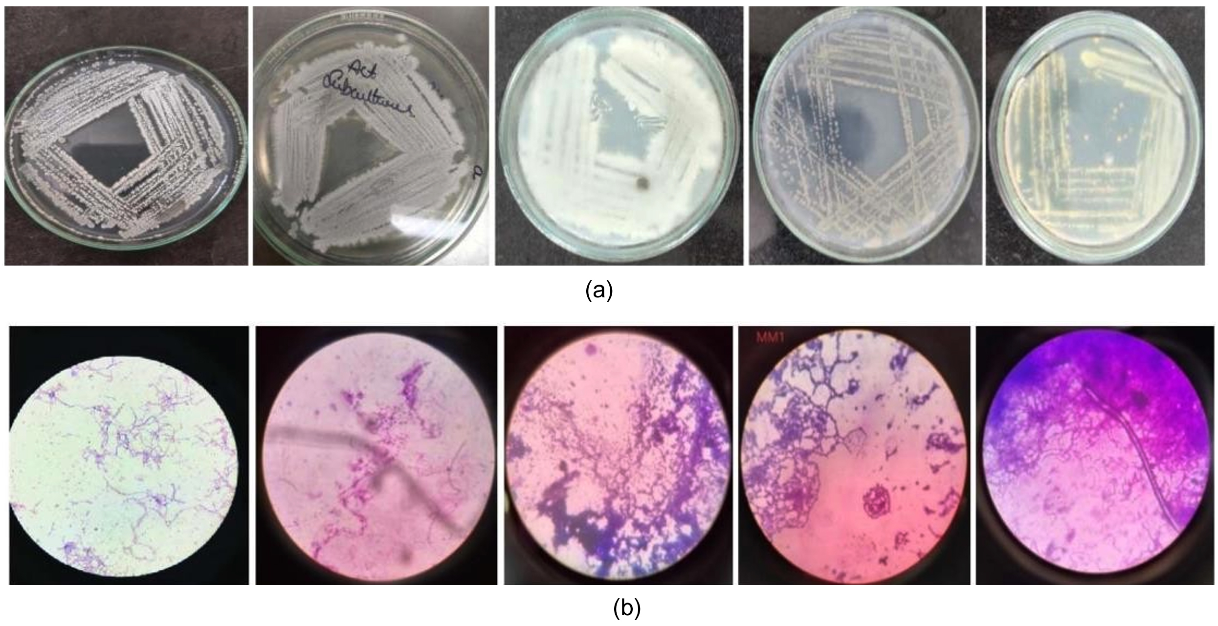
Figure 2. (a) Representative colony morphology of actinomycete isolates observed on agar plates, showing characteristic growth patterns; (b) Representative Gram-stained and spore morphology of actinomycete isolates revealing rectus, flexibilis and open hook arrangements under light microscopy
Table 3. Biochemical characterisation of actinomycete isolates (AM1-AM20), including Gram staining and enzymatic and metabolic test results
Isolate Code |
Grams Staining |
Catalase |
Indole |
Methyl Red |
Voges-Proskauer |
Citrate Utilization |
Gelatine Hydrolysis |
Carbohydrate Fermentation |
|---|---|---|---|---|---|---|---|---|
AM1 |
+ |
+ |
− |
+ |
− |
+ |
− |
− |
AM3 |
+ |
+ |
− |
− |
+ |
− |
− |
− |
AM6 |
+ |
+ |
+ |
− |
− |
+ |
− |
− |
AM7 |
+ |
+ |
+ |
+ |
+ |
+ |
− |
− |
AM9 |
+ |
+ |
− |
+ |
− |
− |
− |
− |
AM11 |
+ |
+ |
+ |
− |
+ |
+ |
− |
− |
AM13 |
+ |
+ |
− |
+ |
− |
+ |
− |
− |
AM15 |
+ |
+ |
− |
− |
+ |
− |
− |
− |
AM17 |
+ |
+ |
+ |
+ |
− |
+ |
− |
− |
AM18 |
+ |
+ |
− |
− |
− |
− |
− |
− |
AM19 |
+ |
+ |
− |
+ |
+ |
+ |
− |
− |
AM20 |
+ |
− |
− |
− |
− |
− |
− |
− |
Where: (+) Positive reaction; (−) Negative reaction
Bioinformatics analysis
The potent isolate AM7 was further confirmed through 16S rRNA gene sequencing and the 16S rRNA gene sequence (799 bp) showed a very strong match, according to the NCBI database. With an E-value of 0.0, high query coverage (~93%), and ~99%-100% sequence identity, Streptomyces indiaensis had the highest BLAST similarity (Figure 3). These results confirm that the isolate belongs to the genus Streptomyces and is closely related to Streptomyces indiaensis. This molecular identification supports its taxonomic position based on 16S rRNA analysis. The GenBank accession number is JQ065726.1.
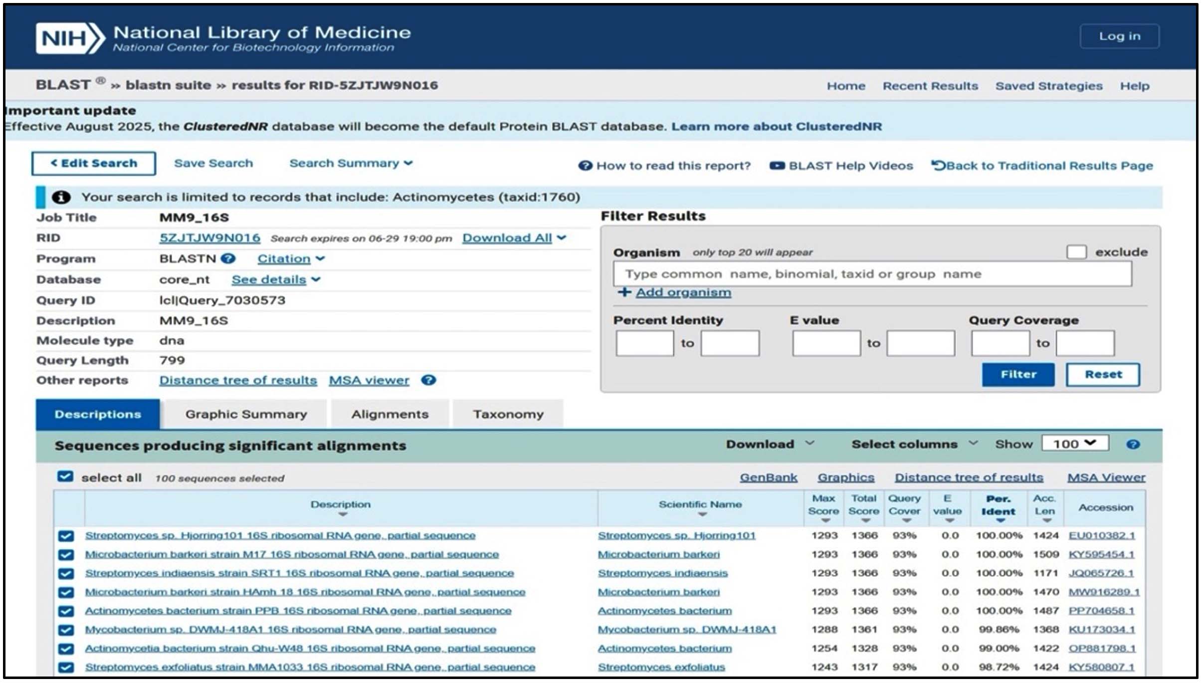
Figure 3. BLASTN analysis of the 16S rRNA gene sequence showing significant similarity with actinomycete taxa, confirming the phylogenetic affiliation of the isolate based on high sequence identity and query coverage
Protein-ligand interaction analysis
The docking study of Glycoprotein adhesin SasA (PDB ID: 4F20) from Staphylococcus aureus demonstrated significant interactions with the tested phytocompounds. Among the evaluated molecules, ligand Epigallocatechin gallate showed the highest binding affinity with a docking score of -8.878 kcal/mol, forming six hydrogen bonds, indicating a strong and stable interaction. The detailed docking interactions and binding scores of protein 4F20 are summarized in Table 4. Allantodapsone exhibited a binding energy of -6.577 kcal/mol with three hydrogen bonds, suggesting favorable binding within the active site of the protein. Ligand ID 9810884 displayed a docking score of -6.281 kcal/mol, forming two hydrogen bonds, whereas ligand Amino-N-[2-(methylamino)-2-oxoethyl]acetamide showed moderate binding affinity (-3.599 kcal/mol) with three hydrogen bonds. The weakest interaction was observed with ligand ID 2266, which exhibited a low binding energy of -0.560 kcal/mol despite forming three hydrogen bonds. Taken together, the docking analysis indicates that ligand 65064 and ligand Allantodapsone possess the greatest potential to inhibit fibronectin-mediated biofilm formation in S. aureus, warranting further experimental validation.
Table 4. Docking results of Protein 4F20 Staphylococcus aureus
| No. | Protein Name and ID | Molecule Name | H-bond | D-Scores | Ligand interactions |
|---|---|---|---|---|---|
| 1. | 4F20 Crystal structures Staphylococcus aureus | Epigallocatechin gallate | 6 | -8.878 | 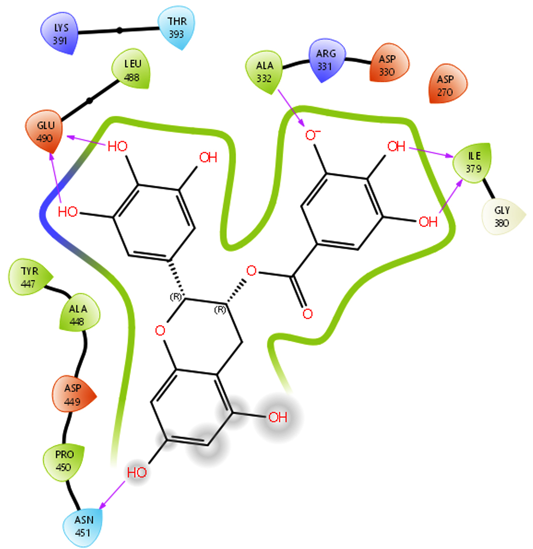 |
| 2. | Allantodapsone | 3 | -6.577 | 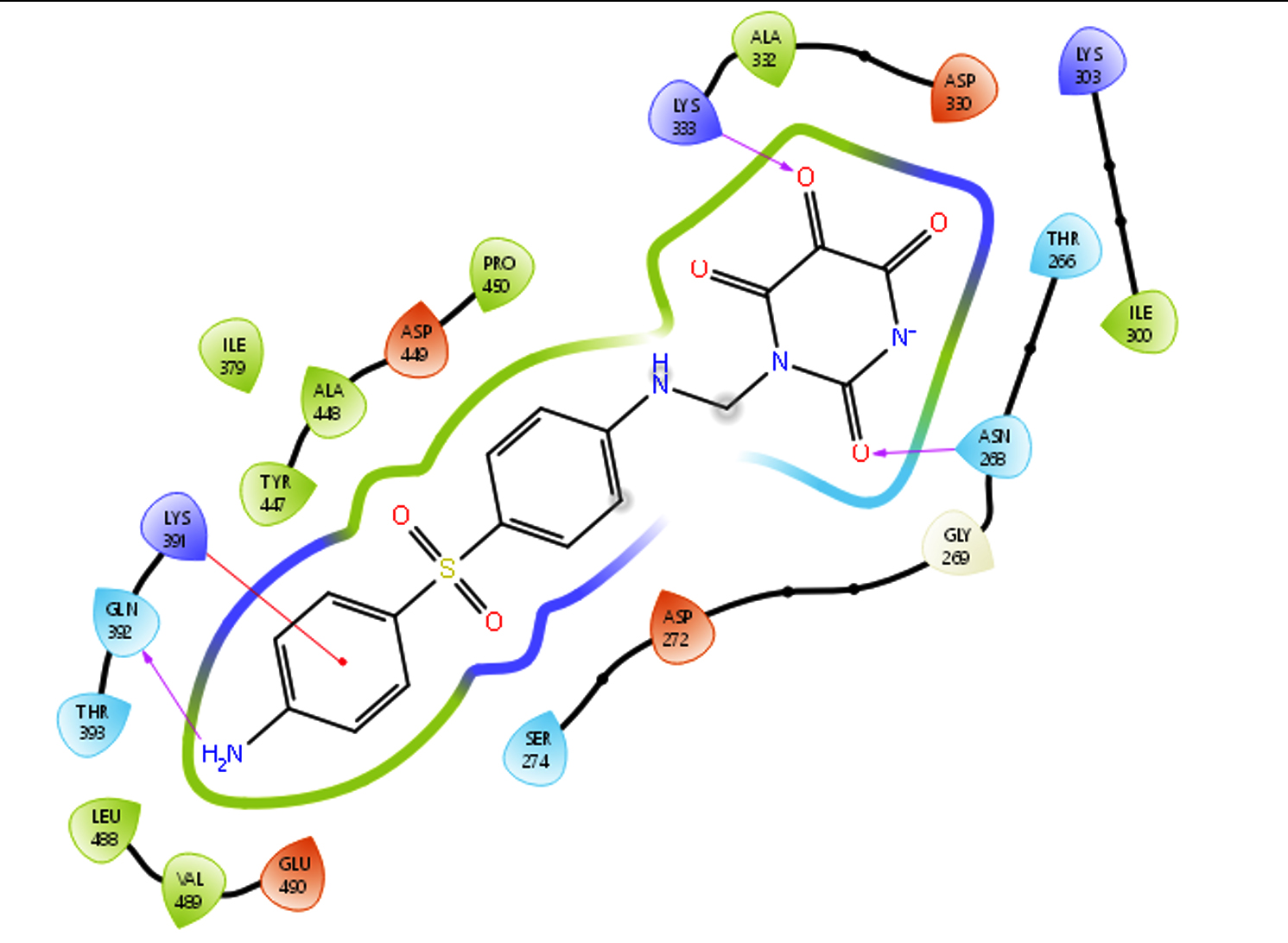 |
|
| 3. | 9810884 | 2 | -6.281 | 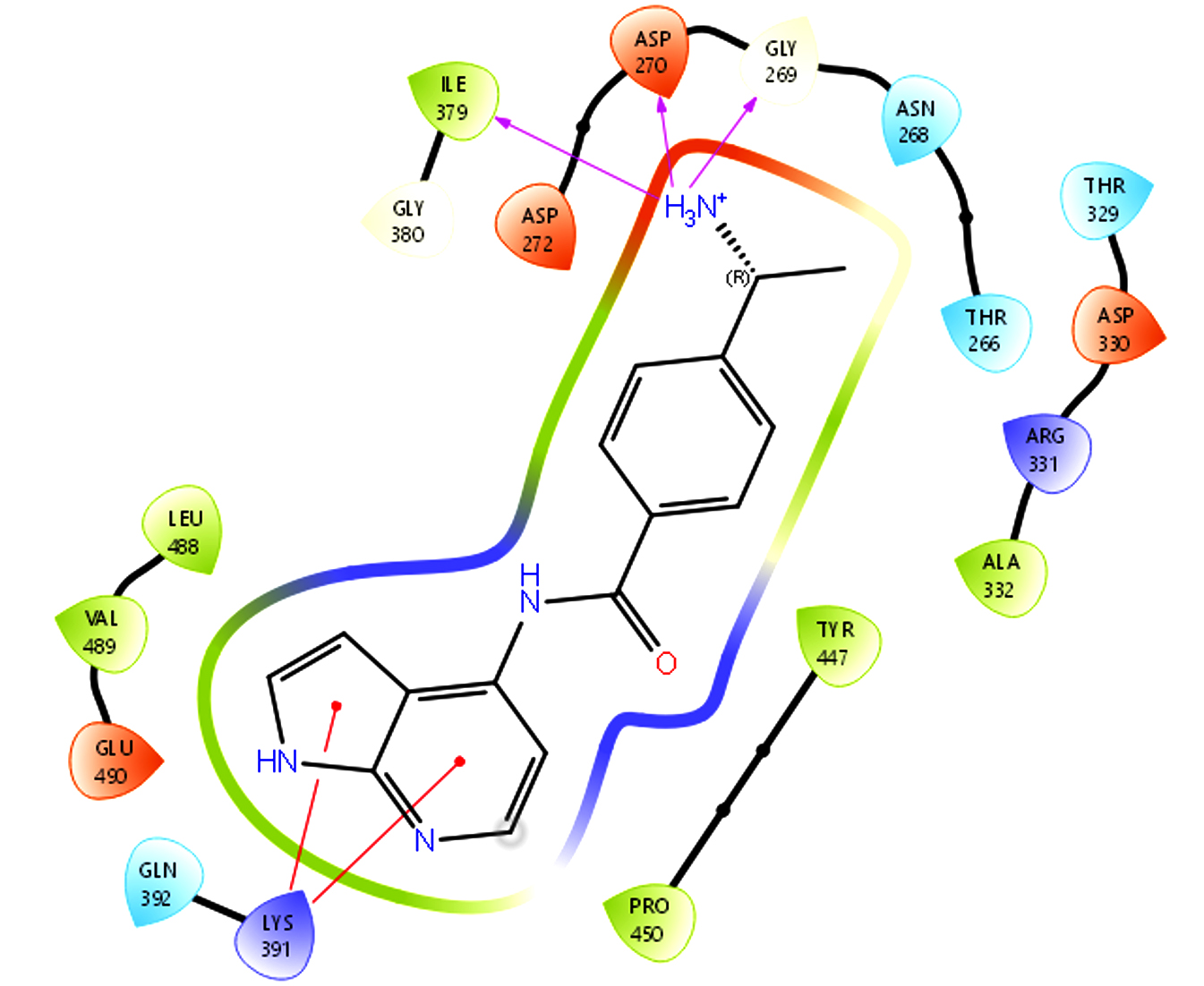 |
|
| 4. | Amino-N-[2-(methylamino)- 2-oxoethyl]acetamide | 3 | -3.599 | 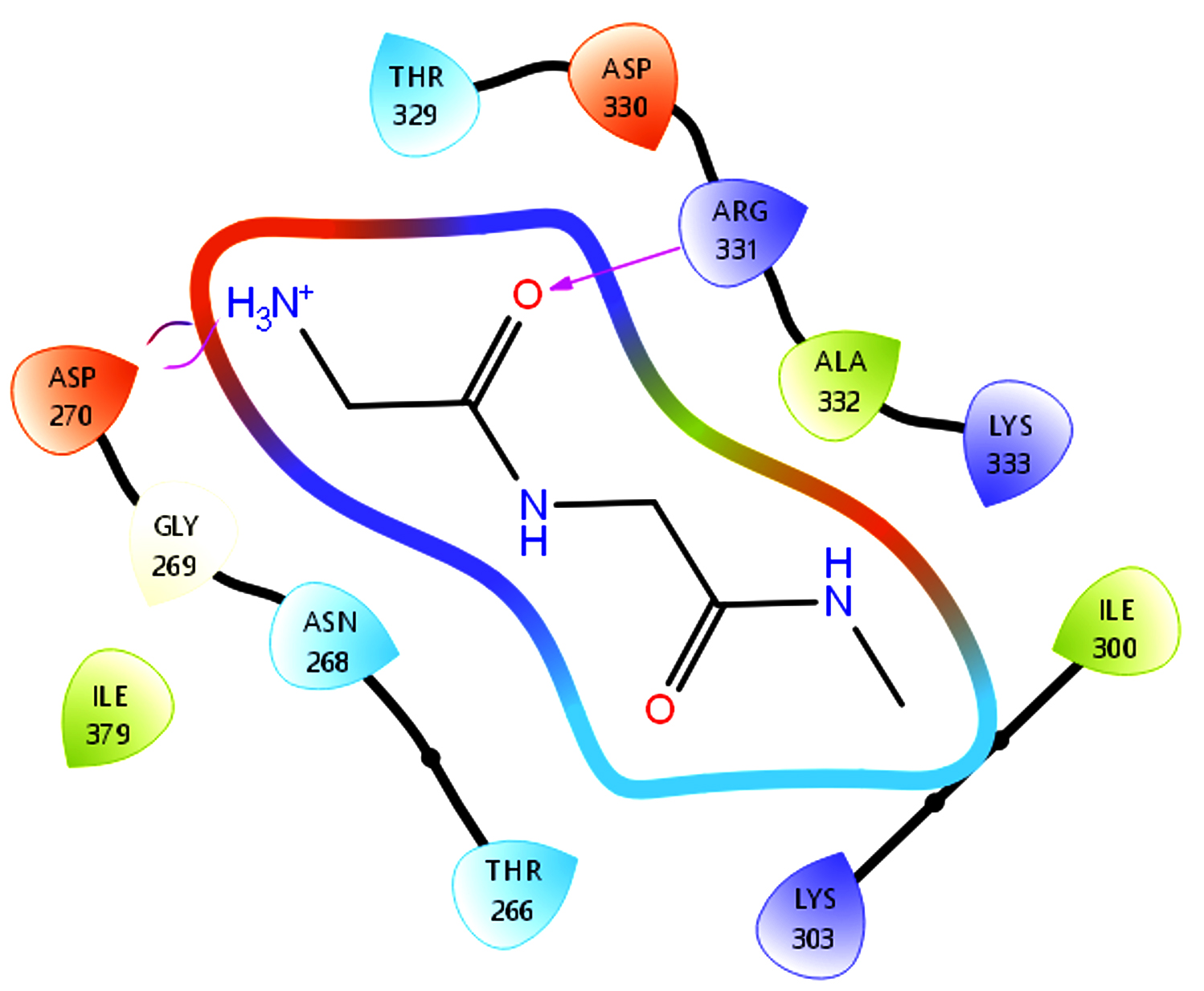 |
|
| 5. | Azelaic acid | 3 | -0.560 | 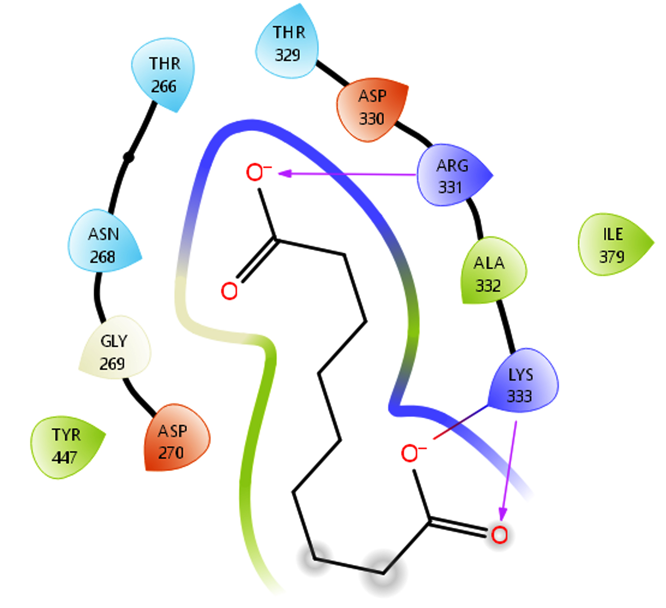 |
The molecular docking analysis of the target protein 1KZN revealed strong and variable binding interactions with the selected phytocompounds. Among the tested molecules, ligand Allantodapsone exhibited the highest binding affinity with a docking score of -11.349 kcal/mol, forming three hydrogen bonds, indicating a highly stable protein-ligand complex. Ligand Epigallocatechin gallate also showed excellent binding, with docking scores of -10.440 kcal/mol (four hydrogen bonds) and -9.610 kcal/mol (five hydrogen bonds) in different binding conformations, suggesting consistent interaction stability. Ligand ID 9810884 demonstrated strong binding affinities of -10.366 kcal/mol with one hydrogen bond and -7.104 kcal/mol with two hydrogen bonds, reflecting orientation-dependent interactions within the binding pocket. Ligand Amino-N-[2-(methylamino)-2-oxoethyl]acetamide showed moderate binding affinity with a docking score of -7.950 kcal/mol and three hydrogen bonds, whereas the weakest interaction was observed for ligand, which exhibited a docking score of -3.121 kcal/mol with two hydrogen bonds. Overall, the docking results indicate that ligand Allantodapsone and Epigallocatechin gallate possess the strongest interaction potential with the 1KZN protein, highlighting them as promising candidates for further experimental validation. The docking interactions and binding affinities of ligands with protein 1KZN are presented in Table 5.
Table 5. Docking results of protein 1KZN Escherichia coli
| No. | Protein Name and ID | Molecule Name | H-bond | D-Scores | Ligand interactions |
|---|---|---|---|---|---|
| 1. | 1KZN Escherichia coli | Allantodapsone | 3 | -11.349 | 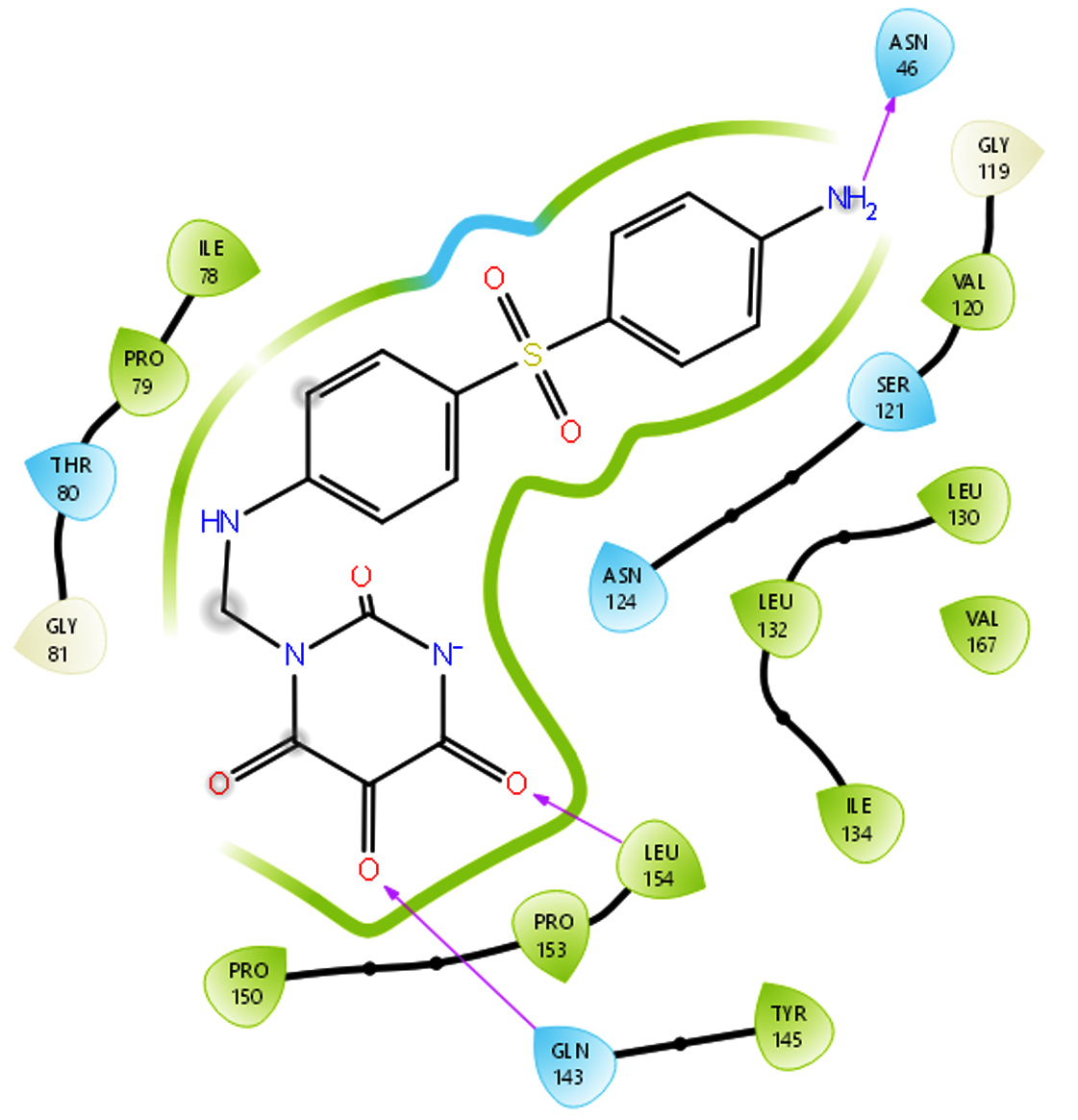 |
| 2. | Epigallocatechin gallate | 4 | -10.440 | 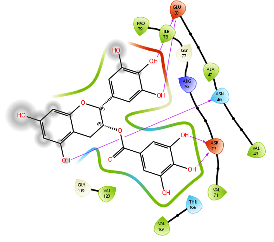 |
|
| 3. | 9810884 | 1 | -10.366 | 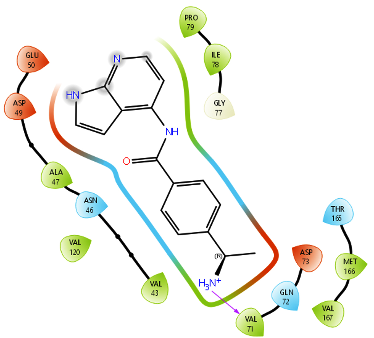 |
|
| 4. | Amino-N-[2-(methylamino)- 2-oxoethyl]acetamide | 3 | -7.950 | 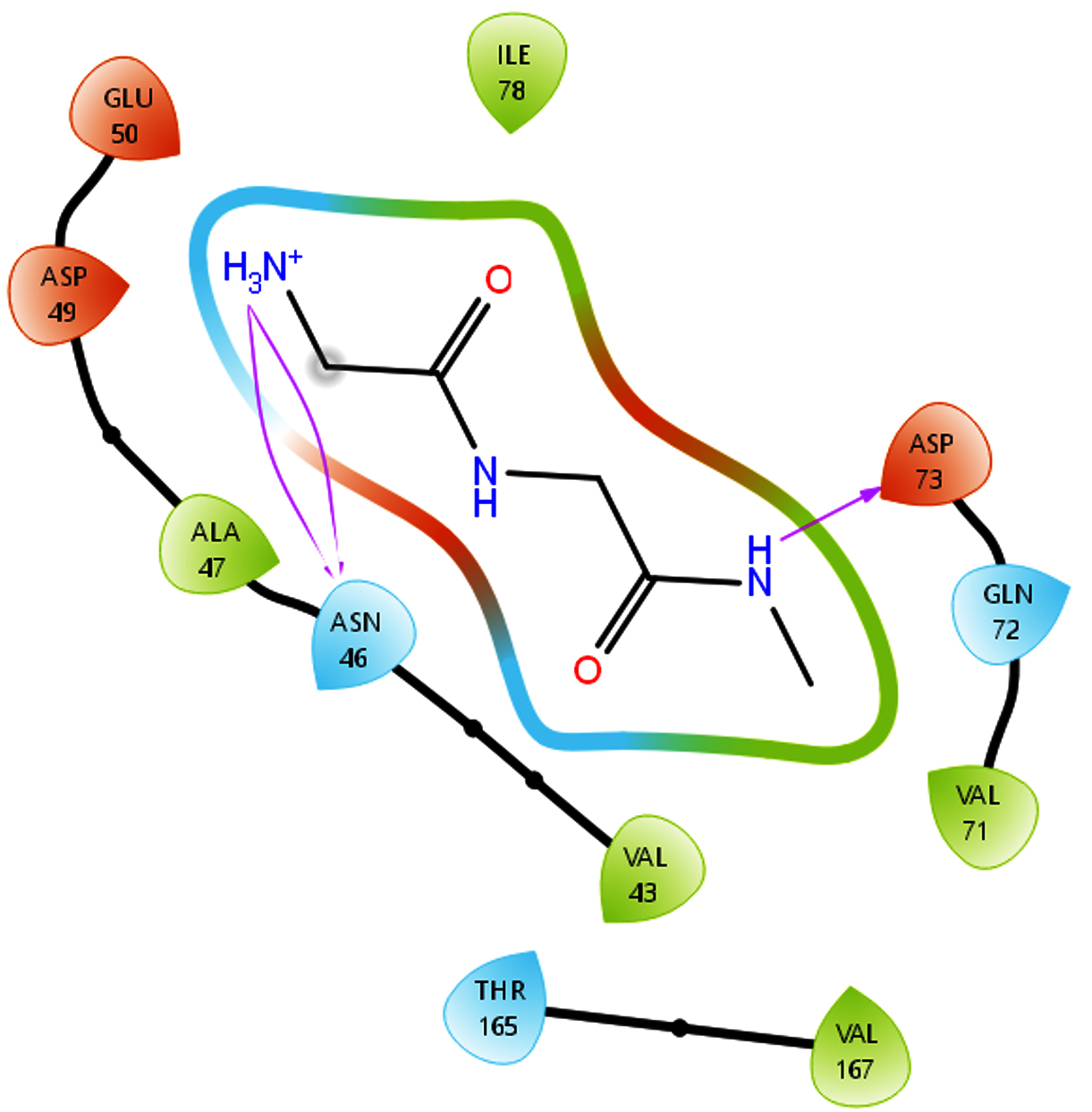 |
|
| 5. | Azelaic acid | 2 | -3.121 | 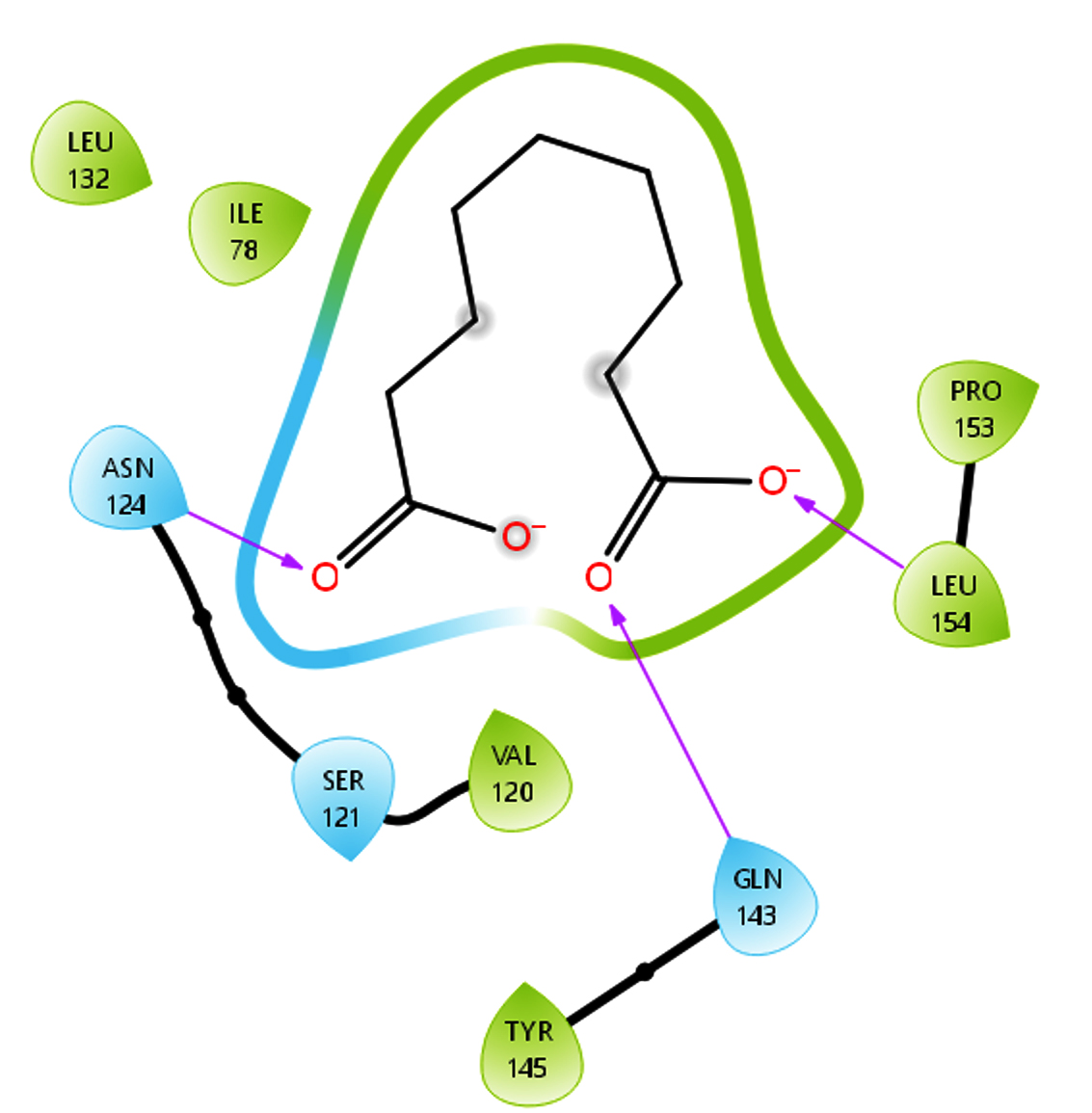 |
After the systematic isolation, morphological and biochemical characterization of actinomycetes, followed by genomic DNA isolation, 16S rRNA gene amplification, gel purification, and Sanger sequencing, the biological potential of the identified isolates was further validated through in silico molecular docking studies. The quality and integrity of the isolated genomic DNA were confirmed by agarose gel electrophoresis as shown in Figure 4. The docking analysis provided a scientific link between the experimentally observed antimicrobial and enzyme-producing activities and their possible molecular mechanisms. Selected Ligands were docked against key target proteins associated with pathogenicity and biofilm formation in Staphylococcus aureus and Escherichia coli, revealing strong and stable protein-ligand interactions characterized by favorable binding energies and multiple hydrogen bonds. Notably, ligands such as Epigallocatechin gallate and Allantodapsone showed high binding affinity toward fibronectin and the 1KZN protein, supporting their potential role in disrupting microbial adhesion, biofilm formation, and survival pathways. Thus, the in silico docking results scientifically validate and complement the experimental findings, strengthening the conclusion that actinomycete-derived bioactive compounds represent promising candidates for the development of novel antimicrobial and biotechnological applications.
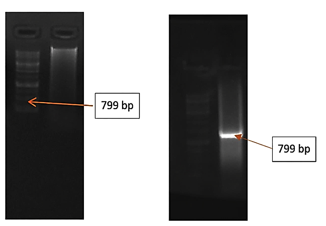
Figure 4. Agarose gel electrophoresis showing intact genomic DNA isolated from the actinomycete isolate, with a clear high-molecular-weight DNA band indicating good quality extraction
Antioxidant activity (DPPH)
The actinomycete extracts exhibited significant free radical scavenging activity in the DPPH assay, indicating the presence of redox-active secondary metabolites. The standard calibration curve and DPPH radical scavenging activity are illustrated in Figure 5. Among the isolates, AM7 showed the highest percentage inhibition, demonstrating strong antioxidant potential compared to other isolates.
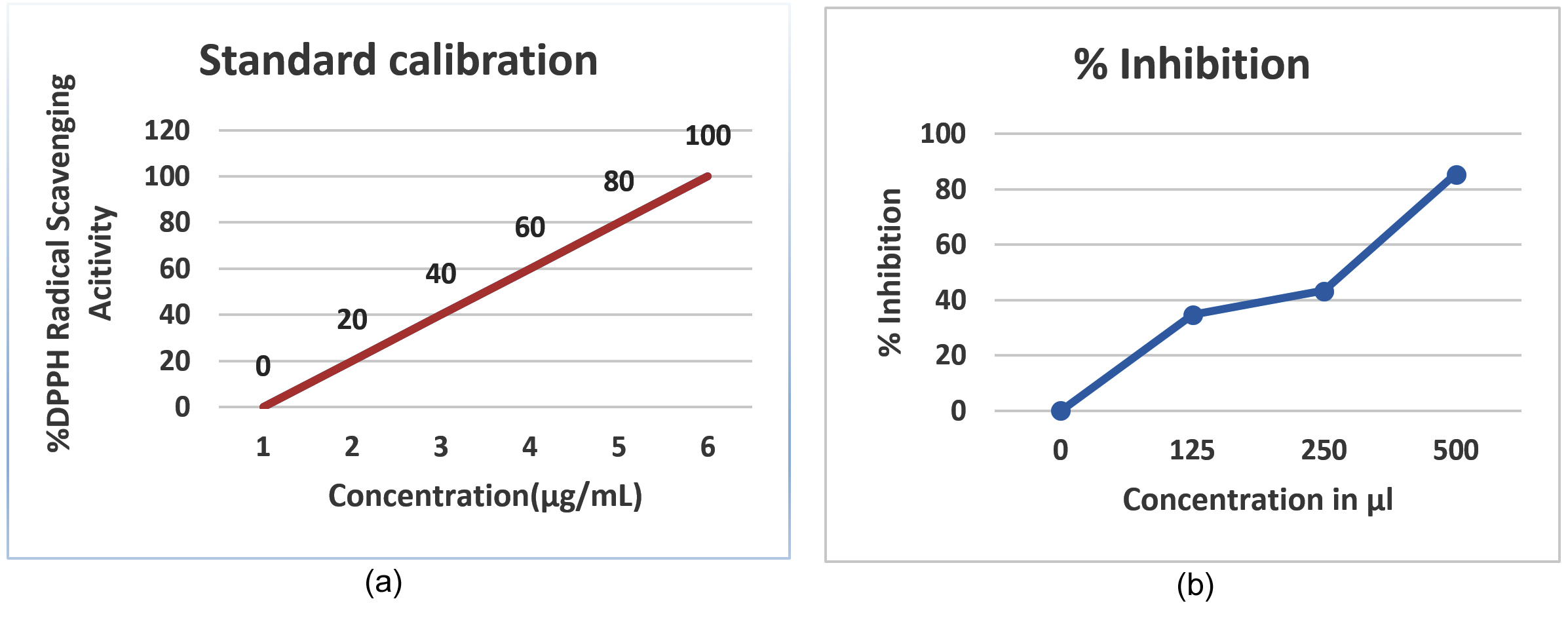
Figure 5. (a) Standard calibration curve of ascorbic acid showing the linear relationship between absorbance and concentration (µg); (b) DPPH radical scavenging assay depicting percentage inhibition as a function of sample concentration (µl)
Screening for L-glutaminase
Primary screening using phenol red–L-glutamine agar revealed distinct pink zones around isolate AM7, indicating efficient L-glutaminase production due to ammonia release and an alkaline shift in the medium. The results are depicted in Figure 6.
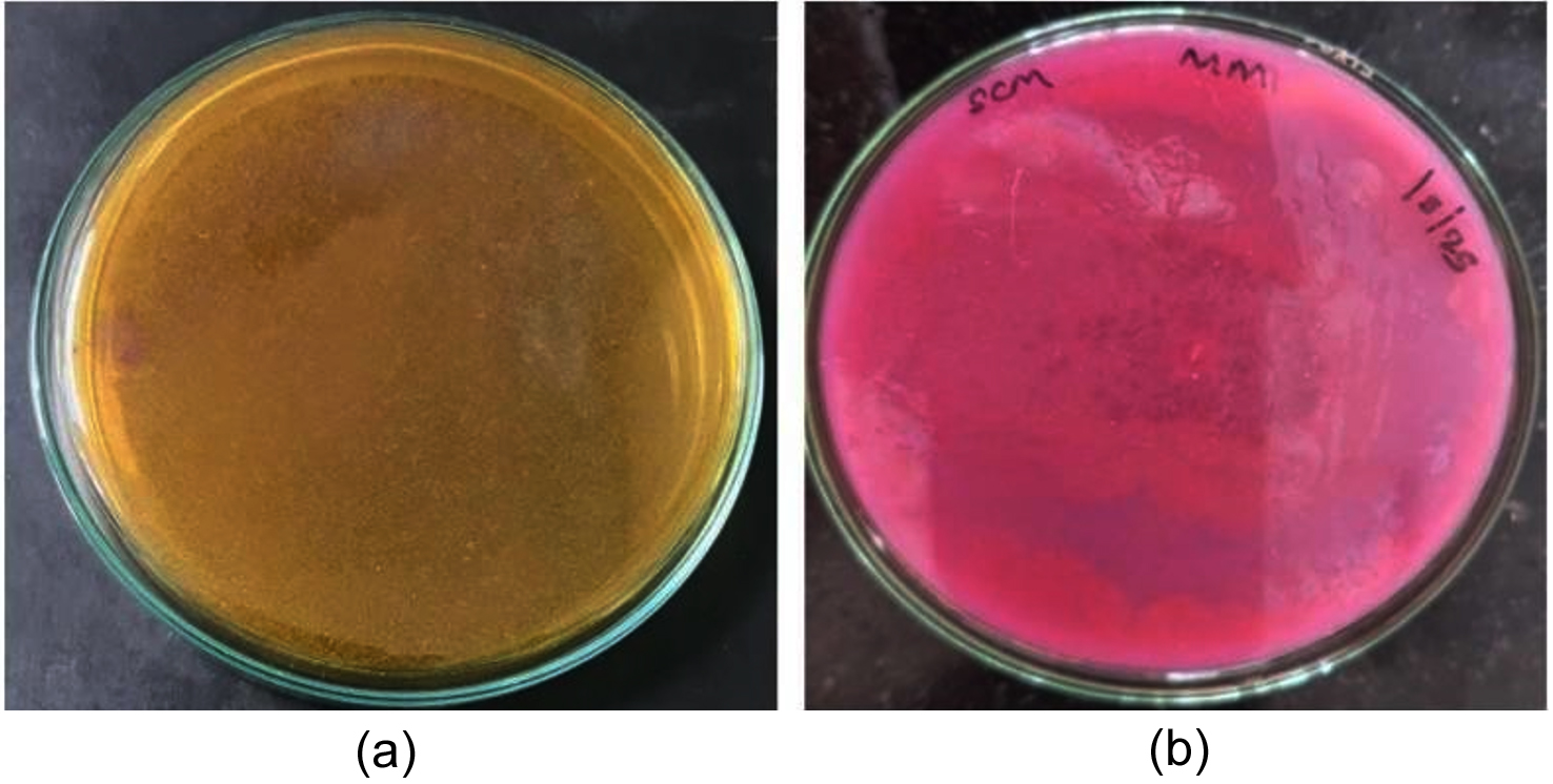
Figure 6. A) Control B) Positive result for Primary screening of L-glutaminase on phenol red-L-glutamine agar showing a distinct pink halo around isolate AM7 due to alkaline shift from L-glutaminase activity, while other isolates exhibit weak or no color change
Antibacterial activity
Primary screening
Twelve isolates (AM1, AM3, AM6, AM7, AM9, AM11, AM13, AM15, AM17, AM18, AM19, AM20) inhibited both the test organisms. Isolate AM7 showed distinct inhibitory activity against both Escherichia coli and Staphylococcus aureus during primary screening using the perpendicular streak method, whereas other isolates showed little to no inhibition. The primary antibacterial screening results are shown in Figure 7.
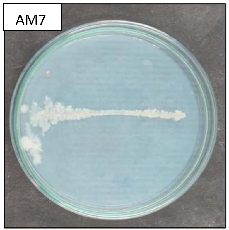
Figure 7. Primary antibacterial screening of actinomycete isolates by the perpendicular streak method showed distinct inhibitory activity of isolate AM7 against Escherichia coli and Staphylococcus aureus, while other isolates exhibited weak or no inhibition
Secondary screening
Around six isolates (AM1, AM7, AM9, AM11, AM13, AM17) showed inhibition. Further study revealed that AM7 exhibited potent and repeatable antibacterial action, indicating the generation of broad-spectrum antimicrobial secondary metabolites, while the other isolates showed no discernible inhibition, according to antibacterial screening using the perpendicular streak method. The secondary antibacterial activity results are summarized in Table 6 and illustrated in Figure 8.
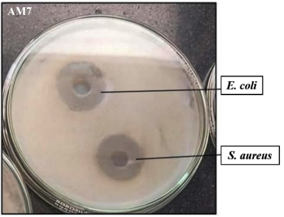
Figure 8. Secondary antibacterial screening of actinomycete isolates by the perpendicular streak method showing clear zones of inhibition, with isolate AM7 exhibiting strong and reproducible broad-spectrum antibacterial activity compared to other isolates displaying weak or no inhibition
Table 6. Antibacterial activity of actinomycete isolates against Escherichia coli and Staphylococcus aureus expressed as zones of inhibition (mm). Streptomycin was used as the positive control
Isolate Code |
Escherichia coli |
Staphylococcus aureus |
|---|---|---|
Streptomycin |
38 mm |
37 mm |
AM1 |
23 mm |
24 mm |
AM7 |
28 mm |
33 mm |
AM9 |
19 mm |
21 mm |
AM11 |
27 mm |
31 mm |
AM13 |
23 mm |
26 mm |
AM17 |
17 mm |
21 mm |
*Values represent the diameter of inhibition zones measured in millimetres (mm), indicating the relative antibacterial efficacy of each isolate
The present study demonstrates that soil-derived actinomycetes constitute a robust and versatile source of bioactive metabolites and industrially important enzymes.19 By isolating actinomycetes from both rhizospheric and non-rhizospheric environments, this work captured ecological diversity that directly translated into functional and metabolic variability. The consistent expression of antimicrobial, antioxidant, and enzymatic activities underscores the metabolic plasticity driven by complex biosynthetic gene clusters characteristic of this group. Several isolates displayed strong antibacterial activity against Staphylococcus aureus and Escherichia coli, indicating the production of metabolites capable of targeting both Gram-positive and Gram-negative bacterial systems.20 Such broad-spectrum inhibition strongly suggests interference with conserved cellular pathways, including cell wall synthesis, membrane integrity, or protein function. In the context of escalating antimicrobial resistance, these findings reinforce the relevance of actinomycetes as a critical reservoir for the discovery of next-generation antimicrobial agents.21
The significant antioxidant activity observed through DPPH radical scavenging assays confirms the synthesis of redox-active secondary metabolites. These compounds actively neutralize free radicals and may contribute to cellular protection against oxidative damage, positioning actinomycete-derived antioxidants as promising candidates for therapeutic and nutraceutical development. The coexistence of antimicrobial and antioxidant properties within the same isolates further highlights their multifunctional metabolic potential. Moreover, the efficient production of L-glutaminase by selected isolates establishes their importance in biotechnological and biomedical applications. By catalyzing glutamine hydrolysis, L-glutaminase plays a crucial role in food biotechnology, biosensor design, and anticancer strategies that exploit glutamine dependency in malignant cells. The concurrent synthesis of L-glutaminase and secondary metabolites suggests coordinated metabolic regulation that enhances industrial feasibility.22
Importantly, in silico molecular docking analyses complemented the experimental data by revealing strong binding affinities and stable molecular interactions between selected bioactive compounds and target proteins. These computational insights elucidate key hydrogen bonding and hydrophobic interactions responsible for ligand stabilization and biological activity. The integration of in vitro screening with in silico validation strengthens the mechanistic interpretation of the results and provides a rational framework for lead optimization. Collectively, this study positions actinomycetes as strategically valuable microorganisms for pharmaceutical innovation and industrial biotechnology.14
The present investigation confirms that soil-derived actinomycetes are a prolific and sustainable source of biologically active secondary metabolites and industrially important enzymes. Isolates obtained from rhizospheric and non-rhizospheric soils demonstrated marked antimicrobial activity against Staphylococcus aureus and Escherichia coli, along with significant antioxidant potential as evidenced by effective DPPH radical scavenging. The efficient production of L-glutaminase further emphasizes the biotechnological relevance of these strains for applications in food biotechnology, biosensor development, and anticancer strategies.
In the current study, in silico molecular docking analyses substantiated the experimental findings by revealing strong and stable interactions between selected bioactive compounds and target proteins. The docked ligands exhibited favorable binding energies ranging from approximately -3.88 to -8.36 kcal/mol, indicating moderate to high binding affinity. Among the tested compounds, ligand CID 9810884 demonstrated the highest affinity with a docking score of -8.358 kcal/mol and formed two stabilizing hydrogen bonds, while ligand CID 65064 showed consistent binding across multiple poses with up to six hydrogen bonds, reflecting enhanced interaction stability. Other ligands displayed moderate docking scores (-5.30 to -5.44 kcal/mol) supported by 3-5 hydrogen bonds, further validating their binding potential. Collectively, the correlation between strong docking scores, hydrogen bond interactions, and experimental bioactivity highlights the reliability of the integrated in vitro-in silico approach. This study establishes actinomycetes as promising candidates for the rational discovery and development of novel antimicrobial agents, antioxidant compounds, and enzyme-based therapeutics, providing a solid scientific foundation for future metabolite purification, structural elucidation, and translational research.
ACKNOWLEDGMENTS
The authors thank JSS Academy of Higher Education & Research, Mysuru, for providing infrastructure support for the current research.
CONFLICT OF INTEREST
The authors declare that there is no conflict of interest.
AUTHORS’ CONTRIBUTION
MA and SCM conceptualized the study. MA and ES contributed to methodology. MA contributed to data curation and formulation of the study. SS contributed to investigation, isolation and screening of Actinomycetes, formal analysis, and data interpretation. ES and SCM performed validation. ES and VR contributed to bioinformatics and statistical analysis. MA and SS wrote the manuscript. ES and VR reviewed the manuscript. SCM supervised the study and contributed to manuscript editing and overall project administration. VR revised the manuscript. All authors read and approved the final manuscript for publication.
FUNDING
None.
DATA AVAILABILITY
All datasets generated or analyzed during this study are included in the manuscript.
ETHICS STATEMENT
This article does not contain any studies on human participants or animals performed by any of the authors.
- Obbels D, Verleyen E, Mano MJ, et al. Bacterial and eukaryotic biodiversity patterns in terrestrial and aquatic habitats in the Sør Rondane Mountains, Dronning Maud Land, East Antarctica. FEMS Microbiol Ecol. 2016;92(6):fiw041.
Crossref - Cao WR, Yu XR, Sun YY, Jiang MY, Xu XD, Li YJ. Ornithinimicrobium laminariaesp. nov., isolated from the kelp Laminaria japonica. Int J Syst Evol Microbiol. 2022;72(1):10.1099/ijsem.0.005187.
Crossref - Giacalone D, Schutt E, McRose DL. The phospho-ferrozine assay: a tool to study bacterial redox-active metabolites produced at the plant root. Appl Environ Microbiol. 2025;91(1):e0219424.
Crossref - Tork SE, Aly MM, Elsemin O. A new l-glutaminase from Streptomyces pratensis NRC 10: Gene identification, enzyme purification, and characterization. Int J Biol Macromol. 2018;113:550-557.
Crossref - Smith AJ, Hall V, Thakker B, Gemmell CG. Antimicrobial susceptibility testing of Actinomyces species with 12 antimicrobial agents. J Antimicrob Chemother. 2005;56(2):407-409.
Crossref - Hegazy GE, Olama ZA, Abou-Elela GM, Ramadan HS, Ibrahim WM, El Badan DES. Biodiversity and biological applications of marine actinomycetes-Abu-Qir Bay, Mediterranean Sea, Egypt. J Genet Eng Biotechnol. 2023;21(1):150.
Crossref - Panatula N, Guntuku G, Palla MS, Muthyala MKK, Meka M, Jagadeeswara Reddy D. Isolation, Screening and Identification of Biosurfactant Producing Strain Nocardiopsis dassonvillei var B2 From Oil Contaminated Soil. J Basic Microbiol. 2024;64(12):e2400504.
Crossref - Ghashghaei S, Etemadifar Z, Tavassoli M, Mofid MR. Optimization of Degenerate PCR Conditions for Reducing Error Rates in Detection of PKS and NRPS Gene groups in Actinomycetes. Avicenna J Med Biotechnol. 2023;15(1):28-37.
Crossref - Hazim Abdul Hameed D, Hussein Ali E. Extraction and Purification of Extracellular L-Glutamate Oxidase from Streptomyces. Arch Razi Inst. 2021;76(4):769-779.
Crossref - Diongue K, Dione JN, Diop A, et al. Direct 16S/ITS rRNA Gene PCR Followed by Sanger Sequencing for Detection of Mycetoma Causative Agents in Dakar, Senegal: A Pilot Study Among Patients with Mycetoma Attending Aristide Le Dantec University Hospital. Mycopathologia. 2024;189(5):80.
Crossref - Souza RAC, Costa WRP, de F Faria E, et al. Copper (II) complexes based on thiosemicarbazone ligand: Preparation, crystal structure, Hirshfeld surface, energy framework, anti-Mycobacterium activity, in silico and molecular docking studies. J Inorg Biochem. 2021;223:111543.
Crossref - Garg A, Singhal N, Kumar M. Discerning novel drug targets for treating Mycobacterium avium ss. paratuberculosis-associated autoimmune disorders: an in silico approach. Brief Bioinform. 2021;22(3):bbaa195.
Crossref - Badineni M, Malkhed V, Rumandla L, Malikanti R, Vadija R, Mustyala KK. Structure Elucidation and Identification of Novel Lead Molecules against Sulfur Import Protein cysA of Mycobacterium tuberculosis. Curr Protein Pept Sci. 2023;24(7):589-609.
Crossref - Nyambo K, Tapfuma KI, Adu-Amankwaah F, et al. Molecular docking, molecular dynamics simulations and binding free energy studies of interactions between Mycobacterium tuberculosis Pks13, PknG and bioactive constituents of extremophilic bacteria. Sci Rep. 2024;14(1):6794.
Crossref - Shatri AMN. Biochemical characterization of actinomycete from Namibia rocky crest mountainous soil and analyzing their bioactive metabolites for antagonistic effect against human respiratory pathogens. Pan Afr Med J. 2024;48:12.
Crossref - Afonso Kessler de Andrade G, de Vargas MVM, Goulart SNB, et al. Screening of endophytic fungi from Antarctic mosses: Potential production for L-asparaginase free of glutaminase and urease activity. J Biotechnol. 2023;377:1-12.
Crossref - Wardah ZH, Chaudhari HG, Prajapati V, Raol GG. Application of statistical methodology for the optimization of L-glutaminase enzyme production from Streptomyces pseudogriseolus ZHG20 under solid-state fermentation. J Genet Eng Biotechnol. 2023;21(1):138.
Crossref - Takatsuki A, Hosoda J, Tamura G, Arima K. Screening of antiviral antibiotics from Actinomycetes: correlation between antiviral activity, cytotoxicity and antibacterial activity. (Studies on antiviral and antitumor antibiotics. XI). J Antibiot. 1969;22(4):171-173.
Crossref - Silveira MAV, Santos SMBD, Okamoto DN, et al. Atlantic Forest’s and Caatinga’s semiarid soils and their potential as a source for halothermotolerant actinomycetes and proteolytic enzymes. Environ Technol. 2023;44(11):1566-1578.
Crossref - Aryal S, Adhikari R, Regmi B, Joshi DR. Antibacterial Compounds of Actinomycetes Isolated From Altitude Soils. J Nepal Health Res Counc. 2025;22(4):784-791.
Crossref - Chen M, Xia L, Wu C, et al. Microbe-material hybrids for therapeutic applications. Chem Soc Rev. 2024;53(16):8306-8378.
Crossref - Hassan MG, El-Sayyad GS, Abdel-Monem MO, et al. Unravelling the outcome of L-glutaminase produced by Streptomyces sp. strain 5 M as an anti-neoplasm activity. Microb Cell Fact. 2025;24(1):4.
Crossref
© The Author(s) 2026. Open Access. This article is distributed under the terms of the Creative Commons Attribution 4.0 International License which permits unrestricted use, sharing, distribution, and reproduction in any medium, provided you give appropriate credit to the original author(s) and the source, provide a link to the Creative Commons license, and indicate if changes were made.