


ISSN: 0973-7510
E-ISSN: 2581-690X
Pyocyanin is a blue-green phenazine pigment synthesized by Pseudomonas aeruginosa that has significant biotechnological applications. The present study aims to investigate pyocyanin extracts of multi-drug resistant P. aeruginosa isolates sourced from hospital wastewater and evaluate their antimicrobial activity against a panel of clinically relevant pathogens such as Staphylococcus aureus, Salmonella typhi, Achromobacter xylosoxidans and Candida albicans. Nutrient broth and King’s A broth supplemented with 1% nutrient supplements such as rice water and groundnut cake powder were used as a production medium. Extracted pyocyanin was confirmed by FTIR analysis. The isolates P8 and P9 demonstrated of varying concentrations of pyocyanin in different media. Isolate P8 showed maximum pyocyanin production in King’s A broth compared to nutrient broth with pyocyanin yields 14.34 (µg/mL) and 5.63 (µg/mL), respectively, without the nutrient supplements. Preliminary antimicrobial activity of the pyocyanin extracts exhibited substantial inhibition of tested bacterial culture at a concentration of 25 mg/µl; however, did not show any antifungal activity against tested fungi.
Pseudomonas aeruginosa, Pyocyanin, Pigment Production, Bioactive Compounds, Antimicrobial Agents
Pseudomonas aeruginosa is a gram-negative, aerobic, rod-shaped uniflagellate bacterium. It is remarkably adaptable and metabolically versatile microbe that demonstrates broad habitat tolerance, exhibiting ubiquitous distribution in various ecological niches such as soil, water, animals, sewage, hospitals, oil-contaminated areas, and sinks.1 It is an opportunistic pathogen that poses a serious threat particularly in healthcare settings and in individuals with compromised immune systems. P. aeruginosa generates a substantial diversity of extracellular pigments such as pyocyanin, pyomelanin, pyorubrin, and fluorescein. One of the significant features is the production of soluble blue-green pigment pyocyanin.2,3 Bacteria depend on secondary metabolites in order to survive in the environment and pyocyanin is majorly secreted by P. aeruginosa. Pyocyanin is important as a quorum sensing (QS) signaling molecule as well as a virulence factor for P. aeruginosa.4 Pyocyanin pigment is a redox-active nitrogen-containing heterocyclic secondary metabolite with a molecular structure of 5-methyl-1-hydroxyphenazine.5 The two enzymes PhzM and PhzS, are involved in the synthesis of pyocyanin from the precursor phenazine. The synthesis of this pigment is influenced by iron concentration, as adding iron to a low-phosphate medium enhances the production of pyocyanin and related phenazine pigments by bacterium species.6 Carbon and nitrogen sources in growth media also affect pyocyanin synthesis. Pyocyanin is a zwitterion, and has a low molecular weight and enables permeation through biological membranes. At neutral pH, it is characterized by a blue-greenish color, and at alkaline pH, it changes to pink-red. The antagonistic activity of pyocyanin is due to its unique redox potential fiercely converting oxygen to the toxic superoxide radical.7 Its electron transferrable nature allows it to be used in a variety of industries, including medical, agriculture, and industry, while also having a good impact on the environment.8 Pyocyanin holds potential for use as a biocontrol agent. Studies show that the secondary metabolite, pyocyanin exhibits antibacterial, antiviral, antifungal, and anticoagulant properties.4 Studies also reported anticancer properties of pyocyanin agent due to its cytotoxic effect on tumor cells.9 Pyocyanin serves as a redox mediator in biosensors and microbial fuel cells, a food colorant, a preservative, and a textile colorant in the industry due to its antibacterial and antibiofilm activity.10 Bacterial infections have become difficult to treat due to increasing drug resistance and necessitate alternate methods to combat drug resistant pathogens. Pyocyanin has potent antibacterial activity against both Gram-positive and Gram-negative bacteria and also have antifungal properties.11 Several microbial species including bacteria and fungi have the ability to flourish in a range of conditions, causing infections that are highly common in humans due to many types of exposure. In the present study, selected human pathogens were chosen to represent those that are associated with infections that transmit through various ways including contaminated food and water, direct contact and airborne transmission through the inhalation of respiratory droplets and dust particles. Because of their close association with the Pseudomonas in Cystic Fibrosis infections A. xylosoxidans was also chosen. Therefore, the aim of the present study was extraction of pyocyanin pigment from P. aeruginosa, and evaluate its microbicide effect against significant human pathogens such as Gram-positive S. aureus, Gram-negative S. typhi, A. xylosoxidans and fungi C.albicans.
Collection and characterization of Pseudomonas aeruginosa
The current study utilized P. aeruginosa (n = 10) sourced from hospital wastewater, obtained from the institutional repository.
Molecular screening of Pseudomonas spp.
Molecular assay was done by using a P. aeruginosa species-specific primer for the outer membrane lipoprotein OpRL gene. (OprL1 F: ATGGAAATGCTGAAATTCGGCCT; OprL2 R: TCTTCAGCTCGACGCGACG) of P. aeruginosa (amplicon size 151 bp). A volume of 30 µl was used for Polymerase Chain Reaction (PCR) using Bio-Rad S1000, Thermocycler with reaction conditions, initial denaturation at 94°C for 5 min, denaturation at 95°C 30 sec, annealing at 55°C for 30 sec, extension at 72°C 30 sec and a final extension 72°C for 7 min. The amplified products were visualized by electrophoresis in 1.5% agarose gel, stained with ethidium bromide and visualized in a gel documentation system (Bio-Rad, USA).12,13
Preliminary screening and media optimization for pyocyanin pigment production
All confirmed isolates were checked for the production of pyocyanin by streaking onto the cetrimide agar. Only the colonies that showed vigorous blue-green pigmentation were selected for further analysis. Two growth media, Nutrient Broth (NB) and King’s A medium amended with 1% of nutrient sources were used for the production of pyocyanin. Briefly, 100 µL of P. aeruginosa liquid culture consisting of cell density 3×10⁶ CFU/mL was inoculated into 100 mL NB and King’s A medium supplemented with 1% of nutrient supplements (NS) rice water and/or groundnut cake powder and incubated at 37°C in a shaker incubator (160 rpm) up to log phase. After 72 hours of incubation, the culture medium was subjected to pyocyanin extraction. Bacteria grown without NS was considered as control and the growth of the strain was monitored during the study.14
Extraction and purification of pyocyanin pigment
The pigment was extracted using the chloroform method.15 Isolates were grown in nutrient broth and King’s A Media with or without NS for 72 hours and centrifuged at 10000 rpm for 10 minutes and the supernatant was collected. To the supernatant, chloroform was added at a ratio of 2:1, and mixed thoroughly to get a bluish-green pigment. To this pigment, 0.1 N of HCl was added and mixed well to obtain an acidified pink color layer which was then neutralized with 0.2 NaOH, the neutralized layer was treated with chloroform and kept in a vacuum evaporator for drying. The chloroform fraction, blue in color due to the presence of blue-colored pyocyanin was concentrated and purified by a silica gel column having a diameter of 3 cm and 60 cm length. The column was packed with silica having a mesh size of 200 and equilibrated using a chloroform-methanol solvent system in the ratio 1:1 and the concentrated pyocyanin fraction was loaded into the column. Chloroform-methanol solvent system was used as the mobile phase to separate pyocyanin. The blue-colored pyocyanin fraction was collected and concentrated in a vacuum rotary evaporator at 40°C. Dried pyocyanin was stored at 4°C until further use.16
Characterization of extracted pyocyanin pigment
The purified pyocyanin was analyzed by spectroscopic method. The UV-visible spectrophotometer was used to measure the maximum absorption of the solution at 520 nm. Concentrations were expressed as micrograms of pyocyanin produced per milliliter of culture supernatant.15
Concentration of pyocyanin (µg/mL) = OD520nm × 17.072
Fourier Transform Infrared (FTIR) analysis
Specific functional groups of extracted pyocyanin were observed using FTIR analysis and confirmed.16 Characterization was done at Poornayu Research Labs (Bangalore).
Antimicrobial efficacy of extracted pyocyanin pigment
Antibacterial assay of extracted pyocyanin pigment was carried out by well diffusion technique with DMSO served as control according to the protocol described by Saha et al.17 Wells with 0.5 mm diameter was made on sterile Muller Hinton agar plates. Three of the test bacteria namely S. aureus, A. xylosoxidans and S. typhi (previously confirmed and obtained from institutional repository) were inoculated into Muller Hinton broth (MHB) and incubated at shaker incubator at 37°C with 120 rpm until the turbidity reached 0.5 MacFarland. From this 0. 1 mL of the culture was swabbed homogenously onto Muller Hinton agar. 40 µL of extracted pyocyanin was added to the well and the plates were incubated at 37°C for 24 hours. The inhibition zone around the wells was measured in millimeters. The antifungal activity of the pyocyanin was determined by agar well diffusion technique, according to method described in CLSI guidelines 2020 at a concentration of purified pyocyanin 40 mg/L obtained by adding DMSO. Briefly C. albicans were grown in MHB upto 0.5 MacFarland and swabbed of MHA with 2% glucose and 0.5 µg/mL methylene blue. An aliquot of 50 µL of these concentrations was tested against Candida albicans MTCC 227 and were performed in triplicates.
P. aeruginosa isolates were obtained from the institutional repository and revived. All ten isolates were subjected to various biochemical tests and confirmed. All 10 isolates were found to be Gram-negative, rod-shaped bacilli that were positive for oxidase, catalase, and citrate utilization tests and negative for Indole, Methyl red, and Vogues Proskauer tests.
P. aeruginosa isolates were further screened by molecular method, PCR using a species-specific primer specific for the outer membrane lipoprotein. Figure 1 presents the DNA amplification at 151 bp confirming the presence of P. aeruginosa.
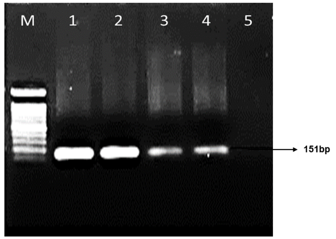
Figure 1. Gel image showing the band for P. aeruginosa. Lane M: Marker 100 bp; Lane 1-4: Positive isolates, Lane 5: Negative control
Preliminary screening and culture conditions for pyocyanin production
All the confirmed isolates were streaked onto the cetrimide agar for preliminary screening for pyocyanin production. The colonies with strong blue-green pigment production, P8 and P9 isolates were selected for further optimization studies. The effect of incubation time for pyocyanin production for the two isolates P8 and P9 were tested at 72 and 96 hours. Two flasks were inoculated with the culture, and incubation was carried out for 4 days, i.e. 96 hours to observe the production of pyocyanin. Production of pyocyanin gradually decreased by increasing the incubation time from 72 hours to 96 hours which was clearly shown by the change in the color from green to yellow as seen in Figure 2. Hence, the incubation time of 72 hours was considered optimum for pyocyanin production.
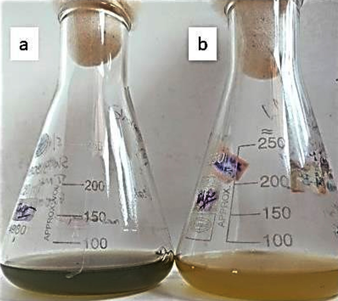
Figure 2. Effect of incubation time on pyocyanin production in Nutrient broth medium (a) 72 hr (b) 96 hr
Pigment production by P8 and P9 isolates was carried out in Nutrient broth and King’s A broth medium with and without supplements such as 1% rice water and ground nut cake powder. Nutrient broth and King’s A broth without supplement which served as control and showed better concentration when compared with medium supplemented with carbon sources. The highest production of pyocyanin was observed with P8 isolate about 5.63 (µg/mL) without any supplements in Nutrient broth. In King’s A medium, also maximum pigment production was observed in P8 isolate 14.34 (µg/mL) without any supplements. Overall, isolate P8 exhibited better yield when compared to P9 isolate in both the medium. It was observed that the production of pyocyanin amended with inexpensive carbon sources like rice water and ground nut cake did not show any observable effect on the pyocyanin yield.
Purification and characterization of pyocyanin pigment
UV-Spectrophotometer analysis confirmed the extracted pyocyanin with a maximum absorbance of 0.97 at 365 nm wavelength.
FTIR data revealed the presence of carbonyl group (C=O) observed at 1666 cm-1, 1688 cm-1, C=N at 1572 cm-1, 1463 cm-1, respectively for P8 and P9 isolates, and the aromatic stretching of C-H was observed at 2956 cm-1 for both P8 and P9. Figure 3 shows the FTIR spectra of the extracted pyocyanin pigment of P8 isolate.
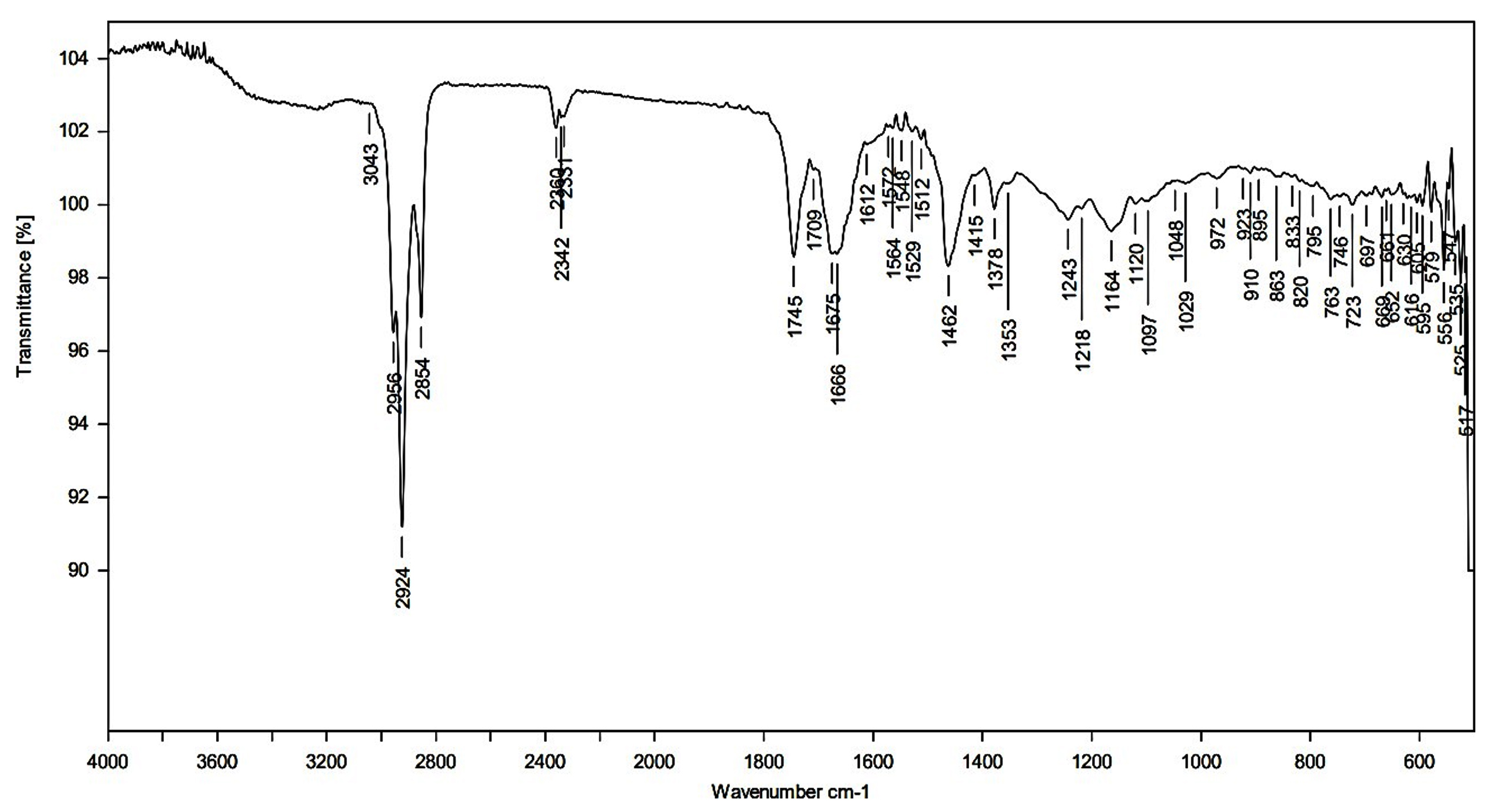
Figure 3. FTIR spectroscopic analysis of the pyocyanin pigment extracted from P8 isolate
Antimicrobial efficacy of extracted pyocyanin pigment
The antimicrobial susceptibility tests of pyocyanin were performed against pathogenic strains of S. aureus, S. typhi, and A. xylosoxidans as shown in Figure 4. The inhibition activity was observed by the zone of clearance around the pigment. S. aureus, A. xylosoxidans, and S. typhi showed a significant zone of inhibition. Pyocyanin extracted from P8 and P9 isolates did not show any inhibition zone with tested fungi.
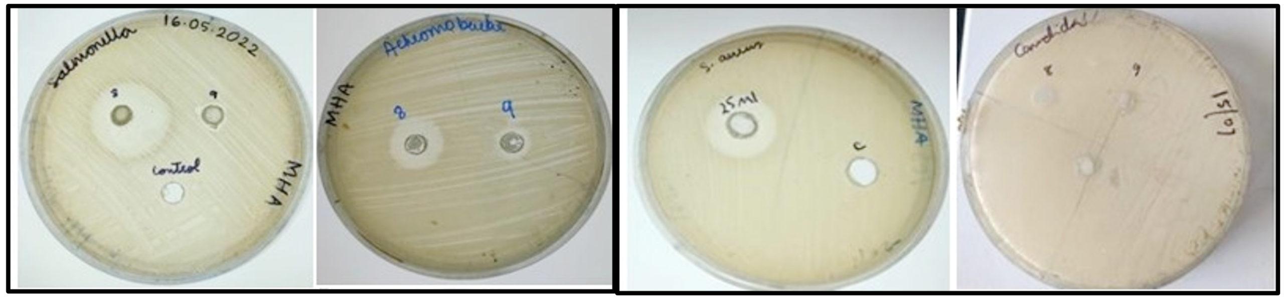
Figure 4. Representative images of antagonistic activity of extracted pyocyanin showing zone of inhibition against tested bacteria and fungi
Hospital wastewater are a major reservoir of antibiotic-resistant bacteria which might harbor bacteria with potential industrial applications.18 P. aeruginosa being opportunistic bacterium with economic significance is widely used for various applications due its ability to secrete phenazine.4 In order to increase the yield of pyocyanin pigment produced by Pseudomonas strains, use of an inexpensive medium was investigated. Different culture conditions and nutrient supplements affect the overall production of pyocyanin pigment. On observing the pigment production at two different temperatures of incubation, the production of pyocyanin gradually decreased by increasing the incubation time from 72 hours to 96 hours which was clearly shown by the change in the color from green to yellow as seen in Figure 2. Hence, the incubation time of 72 hours can be considered optimum for pyocyanin production. Pyocyanin production depends on the incubation period. A study by Rani et al. reported the same incubation time, i.e. 72 hours as the optimum for pyocyanin production. There were remarkable changes in the color of the media after 72 hours of incubation.19 Similar results were obtained in a study by Elbargisy, where it was observed that the optimum conditions for pyocyanin production are to use King’s A fluid medium with inoculated medium incubated at 37°C with shaking at 200 rpm for a period of three to four days.20 Both shaking and longer incubation periods (3-4 days) improved pyocyanin production. Longer incubations start to create additional pigments including pyoverdine, pyomelanin, and pyorubin, which slow down the production of pyocyanin and make it more difficult to extract the pigment.21
Overall, isolate P8 exhibited better yield when compared to P9 isolate in both Nutrient Broth and King’s A broth medium. Carbon and nitrogen sources highly affect the production of pyocyanin.22 However, in this study it was observed that the media amended with carbon sources such as rice water and ground nut cake did not show any effect on the pyocyanin yield. Pyocyanin production was maximum in King’s A broth upto 14.34 µg/mL compared to nutrient broth which yielded 5.63 µg/mL. Production of pyocyanin is nutrient-dependent, the natural nutrients such as fat, minerals carbohydrates, and proteins in King’s A growth medium carrying peptone, magnesium chloride, and glycerol as major chemical nutrients influence the production of pyocyanin. A detailed study of the medium composition and nutrient supplement composition was not carried out due to time constraints. The combination of nutrients both natural and chemical nutrients in King’s A medium might have increased the bacterial density and maximum pyocyanin pigment. The yield obtained in the present study without nutrient supplements may be due to the contents of the King’s A broth medium which is rich in both organic and inorganic nutrients which enable an increase in pigment production. Peptone, glycerol, and magnesium chloride supplemented in King’s A broth, whereas nutrient broth has few energy sources due to which pyocyanin yield was low.
A study for P. aeruginosa proliferation and pyocyanin production was done using media; King’s A medium, glycerol-supplemented nutrient broth (GSNB), and mineral medium. King’s A medium supported the highest increase in the growth yield for P. aeruginosa strains used. Under shaking conditions, King’s A medium resulted in the highest growth yield for both P. aeruginosa strains R1 and U3. The study demonstrated similar results that King’s A media supports good growth of Pseudomonas for pyocyanin production either in shaking or static conditions.23 For P8 and P9 isolates, FTIR measurements showed the presence of carbonyl groups (C=O) at 1666 cm-1, 1688 cm-1, and C=N at 1572 cm-1 and 1463 cm-1, respectively. For both P8 and P9 isolates, the aromatic stretching of C-H was seen at 2956 cm-1. These bands match the several functional groups that are present in pyocyanin and all result is in accordance with previous studies.24 The intensity of the pyocyanin concentration exhibited maximum absorbance at 365 nm wavelength, which is typical of the pyocyanin molecule. Studies have reported pyocyanin exhibits an absorbance band at 230-380 nm.25
Antimicrobial activity screening of the extracted pyocyanin showed inhibition of pathogenic strains of S. aureus, S. typhi, and A. xylosoxidans as shown in Figure 4. The inhibition zone of P8 and P9 against S. aurues were 24 mm and 21 mm. The highest zone of inhibition was observed for isolate P8 against S. typhi with 26 mm. Based on the results of an experimental study, it was observed that for S. aureus, a concentration of 23.78 µM is necessary for antibacterial activity.26 According to Devnath et al., Gram-negative bacteria are reportedly less susceptible to pyocyanin as compared to Gram-positive bacteria.27 Recently, Kamer et al. further demonstrated antimicrobial activity of pyocyanin against methicillin resistant S. aureus (MRSA) which highlights the potential prospective application of pyocyanin as biocontrol agent.28
The possible mechanisms of resistance and sensitive patterns of isolates for the pyocyanin depends on the level of catalase and superoxide dismutase production by the Gram-positive and Gram-negative bacteria. The difference in the cell wall content of these bacteria might be associated with the sensitivity and resistance of bacteria to pyocyanin pigment.29 A. xylosoxidans is a genus of non-fermenting Gram-negative bacteria primarily isolated from the respiratory tract of people with cystic fibrosis and often co-exists with Pseudomonas species, it can cause a broad range of infections in hosts with other underlying conditions.30 Remarkably in this study, pyocyanin pigment of P8 and P9 showed considerable microbicidal activity against A. xylosoxidans with inhibition zone size of 19 mm and 12 mm, respectively. To the best of our knowledge, this is the first study to test the efficacy of pyocyanin against A. xylosoxidans The specificity of the targeted pathogen needs to be studied further. Fungi that commonly infect humans and cause detrimental infections include Candida spp. and Cryptococcus spp. Pyocyanin has been shown to impede the growth of several fungal species to a significant extent. Pyocyanin acts against fungi in a concentration-dependent manner depending on the fungal species. A study by Abdul-Hussien and Atia reported pyocyanin was able to inhibit the growth of Aspergillus niger, Aspergillus fumigatus, Cryptococcus neoformans, Candida tropicalis, and Candida albicans in the concentration range of 47.56 µM to 475.65 µM while Candida krusei was not as significantly inhibited.31 In the present study no antifungal effect of pyocyanin by P8 and P9 isolates were observed. Robust fungal cell wall structure and detoxifying mechanism against ROS could be the reason for the resistant towards pyocyanin.
Pyocyanin exhibits versatility in applications, serving as a food colorant, fabric dye, and therapeutic antimicrobial agent as studied by various researchers. Recognizing its multifaceted utility, the present study investigated synthesis of pyocyanin from P. aeruginosa through cost-effective fermentation techniques, in contrast to expensive synthetic methods. A comprehensive investigation into various low-cost nutrient sources can be conducted to maximize pyocyanin production by Pseudomonas spp. at different growth conditions, offering a deeper understanding of optimal conditions for enhanced synthesis. The pyocyanin extracts of P8 and P9 isolates showed substantial antibacterial activities against human pathogens which could be used in future as potential biocontrol against infections cause by these microorganisms.
The present study demonstrated that P8 and P9 isolates of hospital wastewater origin are able to produce pyocyanin at varying concentrations in different media. King’s A broth could be used for pyocyanin production at optimum culture conditions. The optimum conditions for the production of pyocyanin was found to be 37°C at 160 rpm for 72 hours in King’s A medium. The study demonstrated no significant increase in pyocyanin production using rice water and groundnut cake powder which are inexpensive energy sources. Although, potent antibacterial activity of extracted pyocyanin against selected human pathogen A. xylosoxidans may have promising applications, especially in an era where antibiotic resistance is a growing concern.
ACKNOWLEDGMENTS
The authors are grateful to Nitte University Center for Science Education and Research for the necessary facilities provided.
CONFLICT OF INTEREST
The authors declare that there is no conflict of interest.
AUTHORS’ CONTRIBUTION
MD designed the study. DR and FS performed data collection. DR, MD and FS performed data analysis. DR and MD performed data interpretation. DR prepared the first draft of the manuscript. N wrote the manuscript. MD reviewed the manuscript. All authors read and approved the final manuscript for publication.
FUNDING
None.
DATA AVAILABILITY
All datasets generated or analyzed during this study are included in the manuscript.
ETHICS STATEMENT
Not applicable.
- Crone S, Vives-Florez M, Kvich L, et al. The environmental occurrence of Pseudomonas aeruginosa. Apmis. 2020;128(3): 220-231.
Crossref - DeBritto S, Gajbar TD, Satapute P, et al. Isolation and characterization of nutrient dependent pyocyanin from Pseudomonas aeruginosa and its dye and agrochemical properties. Sci Rep. 2020;10(1):1542.
Crossref - El-Fouly MZ, Sharaf AM, Shahin AAM, El-Bialy HA, Omara AMA. Biosynthesis of pyocyanin pigment by Pseudomonas aeruginosa. J Radiat Res Appl Sci. 2015;8(1):36-48.
Crossref - Goncalves T, Vasconcelos U. Colour me blue: the history and the biotechnological potential of pyocyanin. Molecules. 2021; 26(4): 927.
Crossref - Jablonska J, Augustyniak A, Dubrowska K, Rakoczy R. The two faces of pyocyanin – why and how to steer its production? World J Microbiol Biotechnol. 2023;39(4):103.
Crossref - Baron SS, Rowe JJ. Antibiotic action of pyocyanin. Antimicrob Agents Chemother. 1981;20(6):814-820.
Crossref - Abdelaziz AA, Kamer AMA, Al-Monofy KB, Al-Madboly LA. Pseudomonas aeruginosa’s greenish-blue pigment pyocyanin: its production and biological activities. Microb Cell Fact. 2023;22(1):110.
Crossref - Diggle, Stephen P and Marvin Whiteley. Microbe Profile: Pseudomonas Aeruginosa: Opportunistic Pathogen and Lab Rat. Microbiology. 2019:166(1):30-33.
Crossref - Qin S, Xiao W, Zhou C, et al. Pseudomonas aeruginosa: pathogenesis, virulence factors, antibiotic resistance, interaction with host, technology advances and emerging therapeutics. Signal Transduct Target Ther. 2022;7(1):199.
Crossref - Alatraktchi FA, Svendsen WE, Molin S. Electrochemical Detection of Pyocyanin as a Biomarker for Pseudomonas aeruginosa: A Focused Review. Sensors. 2020;20(18):5218.
Crossref - Mudaliar SB, Bharath Prasad AS. A biomedical perspective of pyocyanin from Pseudomonas aeruginosa: its applications and challenges. World J Microbiol Biotechnol. 2024;40(3):90.
Crossref - Suresh S, Prithvisagar KS, Kumar BK, Premanath R. Unravelling the Distinctive Virulence Traits and Clonal Relationship among the Pseudomonas aeruginosa Isolates from Diabetic Patients. J Pure Appl Microbiol. 2022;16(3):1893-1908.
Crossref - Divyashree M, Mani MK, Karunasagar I. Association of exopolysaccharide genes in biofilm developing antibiotic-resistant Pseudomonas aeruginosa from hospital wastewater. J Water Health. 2022;20(1):176-184.
Crossref - Bacame-Valenzuela FJ, Perez-Garcia JA, Figueroa-Magallon ML, Espejel-Ayala F, Ortiz-Frade LA, Reyes-Vidal Y. Optimized Production of a Redox Metabolite (pyocyanin) by Pseudomonas aeruginosa NEJ01R Using a Maize By-Product. Microorganisms. 2020;8(10):1559.
Crossref - Wang Y, Newman DK. Redox reactions of phenazine antibiotics with ferric (hydr)oxides and molecular oxygen. Environ Sci Technol. 2008;42(7):2380-2386.
Crossref - Bosch A, Miann A, Vescina C, et al. Fourier transform infrared spectroscopy for rapid identification of nonfermenting gram-negative bacteria isolated from sputum samples from cystic fibrosis patients. J Clin Microbiol. 2008;46(8):2535-2546.
Crossref - Saha S, Thavasi R, Jayalakshmi S. Phenazine Pigments from Pseudomonas aeruginosa and Their Application as Antibacterial Agent and Food Colourants. Res J Microbiol. 2008;3(3):122-128.
- Divyashree M, Mani MK, Shama Prakash K, et al. Hospital wastewater treatment reduces NDM-positive bacteria being discharged into water bodies. Water Environ Res. 2020;92(4):562-568.
Crossref - Rani A, Chauhan S, Azmi W. Production and antimicrobial, antioxidant and anticancer applications of pyocyanin from isolated Pseudomonas aeruginosa. SciFed J. Ferment. Microbial Technol. 2018;1.
Crossref - Elbargisy RM. Optimization of nutritional and environmental conditions for pyocyanin production by urine isolates of Pseudomonas aeruginosa. Saudi J Biol Sci. 2021;28(1):993-1000.
Crossref - Kurachi M. Studies on the Biosynthesis of Pyocyanine. (I): On the Cultural Condition for Pyocyanine Formation. 1958.
- El Feghali PAR, Nawas T. Pyocyanin: A Powerful Inhibitor of Bacterial Growth and Biofilm Formation. Madridge J Case Rep Stud. 2018;3(1):101-107.
Crossref - Rahman PK, Pasirayi G, Auger V, Ali Z. Development of a simple and low cost microbioreactor for high-throughput bioprocessing. Biotechnol Lett. 2009;31(2):209-214.
Crossref - Matilla MA, Udaondo Z, MaaB S, Becher D, Krell T. Virulence Induction in Pseudomonas aeruginosa under Inorganic Phosphate Limitation: a Proteomics Perspective. Microbiol Spectr. 2022;10(6):e0259022.
Crossref - Jennings KR. Spectrometric identification of organic compounds (Fifth Edition) Silverstein RM, Bassler GC, Morrill TC. Wiley, New York. Organic Mass Spectrometry. 1991;26(9):813.
Crossref - Hamad MN, Marrez DA, El-Sheribieny SM. Toxicity evaluation and antimicrobial activity of purified pyocyanin from Pseudomonas aeruginosa. Biointerface Res Appl Chem. 2020;10:6974-90
- Devnath P, Uddin MK, Ahamed F, Hossain MT, Manchur MA. Extraction, Purification and Characterization of pyocyanin produced by Pseudomonas aeruginosa and evaluation for its antimicrobial activity. Int Res J Biological Sci. 2017;6(5):1-9.
- Kamer AMA, Abdelaziz AA, Al-Monofy KB, et al. Antibacterial, antibiofilm, and anti-quorum sensing activities of pyocyanin against methicillin-resistant Staphylococcus aureus: in vitro and in vivo study. BMC Microbiol. 2023;23(116).
Crossref - Moayedi A, Nowroozi J, Sepahy AA. Effect of fetal and adult bovine serum on pyocyanin production in Pseudomonas aeruginosa isolated from clinical and soil samples. Iran J Basic Med Sci. 2017;20(12):1331-1338.
Crossref - Isler B, Kidd TJ, Stewart AG, Harris P, Paterson DL. Achromobacter Infections and Treatment Options. Antimicrob Agents Chemother. 2020;64(11):e01025-20.
Crossref - Abdul-Hussein ZR, Atia SS. Antimicrobial Effect of Pyocyanin Extracted from Pseudomonas aeroginosa. Eur J Exp Bio. 2016;6(6):1-3.
© The Author(s) 2024. Open Access. This article is distributed under the terms of the Creative Commons Attribution 4.0 International License which permits unrestricted use, sharing, distribution, and reproduction in any medium, provided you give appropriate credit to the original author(s) and the source, provide a link to the Creative Commons license, and indicate if changes were made.