

ISSN: 0973-7510
E-ISSN: 2581-690X
Xylanases shows a large variety of function and have unique importance in the biotechnology and industries due to their potential applications. The major applications of Xylanases are in Textile industry, Biofuel production, Bread making, Pulp bleaching and Food industries. This study is focused on identification and screening of fungi, which can produce Xylanase. Xylanase producing fungi were obtained by congo red method of screening from soil collected from different area. The selection was done on the basis of colony size maximum dimeter 2.5. Four isolates showed clear zone around them that proving to be xylanolytic in nature. The maximum clear zone diameter was measured to be around 7.50 cm in Apergillus versicolor.
Xylanase, Prelimnary Screening, molds.
Xylanase [E.C.3.2.1.8] (Shallom and Shoham 2003) are a class of hydrolytic enzymes which can hydrolyze the straight polysaccharide â-1, 4-xylan which is present in the secondary cell wall and forms an interface between lignin and other polysaccharides (Dhiman et al. 2008). The term covers a range of non cellulose polysaccharides composed, in various proportions, of monosaccharide units such as D-xylose, D-mannose, D-glucose, L-arabinose, D-galactose, D-glucuronic acid and D-galacturonic acid. Classes of hemicellulose are named according to the main sugar unit. Thus, when a polymer is hydrolyzed and yields xylose, it is a xylan; in the same way, hemicelluloses include mannans, glucans, arabinans and galactans (Whistler and Richards 1970; Viikari et al.1994; Uffen 1997; Ebringerova and Heinze 2000).
Collection of soil
The soil samples were collected from top soil (7.5 cm deep) in sterilized polythene bags which were cold-sterilized by UV-radiation for at least 12 h. The glassware was sterilized in the hot-air oven at 160oC for 2 h. Growth media and distilled water blanks were autoclaved at 121oC for 15 min. The collected soil samples were inoculated within 24 h. If delayed, samples were stored at 4oC in refrigerater. The pH of soil was determined using pH meter.
Culture media
For selective isolation of xylanase producing fungi Czapeck’s Dox Agar with Birch wood xylan 5.0g, Pepton 5.0g , Yeast extract 5.0g, MgSO4 0.2g, KH2PO4 1.0g , Agar-Agar 20.0g, Distilled water 1000 ml. pH of the medium s adjusted to 5.50 sterilized at 121°C and 15 lb of pressure for 20 min and poured into sterile petri dishes.
Isolation of fungal colonies
Soil sample equivalent to 1 g dry weight of soil sample was added to 10 ml of sterilized distilled water to make soil suspension. The suspension was vigorously shaken on a magnetic shaker for 30 min to obtain uniform suspension of microorganism. Transferred 1 ml of suspension to series of tubes containing 9 ml of sterilized distilled water with a sterile pipette under aseptic condition to make 10-1 dilution up to 10-5 dilution and plated on Czapeck’s Dox Agar media over spreading of 100 µl of sample from 10-3 to 10-5 for selective isolation of xylanase producing fungi.
Triplicates were maintained for each dilution. Single isolated colonies on plates were picked and further purified on fresh plates for axenic cultures and maintained on Czapeck’s Dox Agar medium.
Screening of isolates for Xylanase production
Xylanase producing fungal strain from different soil sample were isolated using dilution plate technique containing Czapek’s agar medium containing xylan as the sole carbon source. The medium was poured in sterile petriplates and allowed to solidify. After solidifying the medium fungal strain were inoculated in plate by the sterilized inoculating loop. The inoculated plates were incubated for 7 days at 30R”C. After incubation the plate were observe for the formation of clear zone around the fungal growth. The zone were visible when 0.1% (w/v) Congo red was flooded on the plates for 30 min. After 30 min of incubation, plates were washed with 1M NaCl to remove excess stain.
Quantitative screening of microorganisms
The isolates exhibiting positive zone of hydrolysis were selected for secondary screening The isolates were screened by batch fermentation for investigation of its potential to synthesize Xylanase. The screening was performed by inoculating the spores of isolate in modified Czapak Dox liquid medium. All the selected isolates were inoculated in the sterilized medium taken in 150ml Erlenmeyer flasks. The inoculum was added to the sterilized medium under aseptic conditions. The flasks were incubated at 28±2°C in an incubator with static condition for 10 days. After fermentation was over the cotton plugs were removed and the fermentation broth was filtered to separate the biomass. The filtered was use as crude enzyme source after every 24 hours and used as enzyme source for the assay of xylanase activity.
Assay of xylanase
The Xylanase activity was determined by estimating the total amount of reducing sugar formed after xylanase action with Birch wood xylan as a substrate using di-nitrosalicylic acid method (Miller, 1959). 0.5 ml of crude enzyme and 1.0 ml of Sodium acetate buffer was added to 1.0 ml of 1% Xylan solution and incubated for 1 h at room temperature. 2ml of DNSA reagent was added to the reaction mixture. The enzymatic reaction was terminated by keeping the test tube at 100°C in a water bath for 10 min. After cooling, the color developed was read colorimetrically at 540 nm.
The enzyme activity was calculated by using the following formula; One unit of xylanase was defined as the amount of enzyme required to release 1µmol of xylose from birch wood xylan in one minute under standard assay conditions.
Preliminary screening
The preliminary screening was performed for selected isolates the selection of isolates was done on the basis of colony size. The fast growing isolates were selected having maximum 2.5 diameter.
Plate assay with congo red is one of the important methods to assess the xylanolytic activity of the microflora. A clear hallow zone will appear after addition of dye and destaining with NaCl on plates due to the disappearance of polysaccharide around growing colony because of utilization of polysaccharide by microorganisms with secretion of xylanase. The activity of xylanase on plate method showed the clear zone after destaining with in a range of 0.5 to 8 cm (Figure 1). The hallow zones around the fungal colonies signified the solubilisation of xylan due to the hydrolytic action of xylanases. Fungal cultures isolated through xylan containing medium and their zone of clearance on xylan congo red plate are presented in (Table 1). The highest zone was recorded in 7 number of isolate 7.50±0.05 cm and lowest zone was recorded in 6 number of isolate 0.03±0.001. (Figure II)
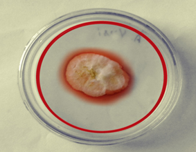
Fig. 1. The zone of hydrolysis produced by fungal strains on Czpaks dox agar medium containing xylan
Table (1):
Zone of clearance by fungal cultures isolated through xylan containing medium.
Isolation number |
Zone of Hydrolysis(cm) |
|---|---|
1 |
0.05 ± 0.001 |
2 |
0.04 ± 0.001 |
3 |
0.06 ± 0.001 |
4 |
1.42 ± 0.001 |
5 |
0.08 ± 0.001 |
6 |
0.03 ± 0.001 |
7 |
7.50 ± 0.05 |
8 |
0.04 ± 0.001 |
9 |
0.07 ± 0.001 |
10 |
0.08 ± 0.002 |
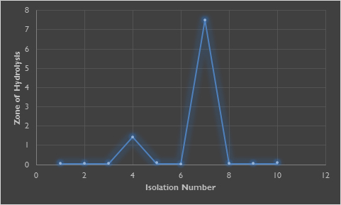
Fig. 2. Isolate no. 7 shows highest zone
- Shallom, D. and Shoham, Y. Microbial hemicellulases. Curr. Opin.Microbiol. 2003; 6: 219-228.
- Miller, G. L. Use of dinitrosalicylic acid reagent for determination of reducing sugar. Anal. chem., 1959; 31: 426-428.
- Viikari L, Kantelinen A, Sundqvist J and Linko M. Xylanases in bleaching: from an idea to the industry. FEMS Microbiol Rev. 2001; 13: 335-350.
- Bailey M J , Biely, P and Poutanen K. Interlaboratory testing of methods for assay of xylanase activity, Journal of Biotechnology. 1992; 23: 257–27
- Lowry OH, Rosebrough NJ, Farr AL, Randall RJ. Protein measurement with the Folin phenol reagent. J. Biol. chem. 1951; 193: 265-275.
- Uffen, R. L. Xylan degradation: a glimpse at microbial diversity. J Ind Microbiol Biotechnol. 1997; 19: 1–6
- Whistler,R.L. and Richards,E.L. Hemicelluloses. In: Pigman W, Horton D (eds) The carbohydrates. Academic Press, New York. 1970; 447–469
- Beg, Q.K., Kapoor, M., Mahajan, L.,Hoondal G.S. Microbial xylanases and their Industrial applications : a review, App. Microbio. Biotechnoal. 2001; 56: 326-338.
- Collins. T., C.Gerday and Feller, G. Xylanases, families and extremophilic xylanases. FEMS Microbiol. Rev. 2005; 29: 3-23
- Ball A S and McCarthy A J. Saccharification of Straw by Actinomycetes enzyme. Journal of Applied Bacteriology 1989; 66: 439-444
- Dhiman S S, Sharma J and Battan B. Industrial Applications and future prospects of Microbial Xylanases: A Review. BioResources, 2008; 3(4): 1377-1402.
- Nakamura S, Wakabayashi K, Nakai R, Aono R and Horikoshi K. Purification and some properties of an alkaline xylanase from alkaliphilic Bacillus sp. Strain 41M-1. Applied and Environmental Microbiology 1993; 59: 2311-2316.
- Srinivasan M. C. and Rele M. V. Microbial xylanases for paper industry. Current Science 1999; 77: 137-142.
- Sudan R and Bajaj B K. Productionand Biochemical characterization of xylanases from an alkali tolerant novel sp Aspergillus niveus RS2. World Journal of Microbiology and Biotechnology 2007; 23(4): 491-500.
- Taneja K, Saurabh G and Kuhad R.C. Properties and Application of a partially purified alkaline Xylanase from an Alkalophilic fungus Aspergillus nidulans 2002; KK-99.
- Azeri C, Tamer A U and Oskay M. Thermoactive cellulase-free xylanase production from alkaliphilic Bacillus strains using various agro-residues and their potential in biobleaching of kraft pulp. African Journal of Biotechnology 2010; 9(1): 63-72.
- Kulkarni N, Shendye A, Rao M. Molecular and biotechnological aspects of xylanases. FEMS Microbiol Rev 1999; 23: 411–456
- Garg G, Dhiman SS, Mahajan R, Kaur A and Sharma J. Bleach-boosting effect of crude xylanase from Bacillus stearothermophilus SDX on wheat straw pulp. New Biotechnol., 2011; 28(1):58-64
- Goldschmidt F. From Cellulose to Ethanol: Engineering Microorganisms to Produce Biofuel. Institute of Biogeochemistry and Pollutant Dynamics 2008; 1-17
- Csiszár E, Urbánszki K and Szakás G. Biotreatment of desized cotton fabric by commercial cellulase and xylanase enzymes. J MolCatal B Enzym. 2001; 11: 1065-1072
- Haltrich D, Nidetzky B, Kulbe KD, Steiner W and Zupancic. Production of fungal xylanases, Bioresource Technology 58, 1996; 137-161
- Sunna A., Antranikian G. Xylanolytic enzymes from fungi and bacteria. Crit. Rev. Biotechnol. 1997; 17: 39-67
- Nakamura S., Wakabayashi K., Nakai R., Aono R., Horikoshi K. Purification and some properties of an alkaline xylanase from alkaliphilic Bacillus sp. strain 41 M-1. App. Environ. Microbiol. 59 1993; 7: 2311-2316
- Subramaniyan S, Prema P. Biotechnology of microbial xylanases, enzymology, molecular biology and application. Crit. Rev. Biotechnol. 2002; 22: 33-64.
- Kulkarni N, Shendye A, Rao M. Molecular and biotechnological aspects of xylanases. FEMS Microbiol. Rev. 1999; 23 :411-56
- Gupta S, Bhushan B, Hoondal G S and Kuhad R C. Improved xylanase production from a haloalkalophilic Staphylococcus sp SG-13 using inexpensive agricultural residues. World Journal of Microbilogy and Biotechnology, 2001; 17: 5-8.
- Krengel, U., Dijkstra, W. Three-dimensional structure of endo-1,4—xylanase I from Aspergillus niger: molecular basis for its low pH optimum. J. Mol. Biol., 1996; 263: 70–78.
- Torronen,A.,Rouvinen, J. Structural comparison of two major endo-1,4-xylanases from Trichoderma reesei. Biochemistry, 1995; 34: 847–856.
© The Author(s) 2018. Open Access. This article is distributed under the terms of the Creative Commons Attribution 4.0 International License which permits unrestricted use, sharing, distribution, and reproduction in any medium, provided you give appropriate credit to the original author(s) and the source, provide a link to the Creative Commons license, and indicate if changes were made.