


ISSN: 0973-7510
E-ISSN: 2581-690X
In the present investigation, the phytogenic synthesis of silver nanoparticles (AgNPs) was carried out with Callicarpa macrophylla which possess multiple functional properties. The absorbance peak of the resultant AgNPs was found to be between 300 and 800 nm, with the highest absorbance at 436 nm. The phytocomponents accountable for facilitating the synthesis were determined through the utilization of Fourier-transform infrared (FTIR) analysis, which unveiled the presence of hydroxyl, amide, aldehyde, and alkene moieties. The crystal structure of the AgNPs was investigated via X-ray diffraction (XRD) analysis, which exhibited diffraction intensities at the 2-theta angle, signifying the presence of a well-defined crystalline structure of AgNPs. The polydispersity of the AgNPs was observed under a transmission electron microscope (TEM), with an average particle size of 10 to 60 nm. The AgNPs exhibited moderate antibacterial activity against Pseudomonas aeruginosa, Escherichia coli, Staphylococcus aureus. The sprouting percentage of Pisum sativum, Vigna radiata, and Macrotyloma uniflorum seeds was assessed with notable observation of increase in length of root size was maximum at AgNPs concentrations of 5 mg/mL and 2 mg/mL for Pisum sativum and Vigna radiata, respectively. However, in the case of Macrotyloma uniflorum, the highest germination rate was observed at a concentration of 10 mg/mL. Similarly, shoot length was highest in all seeds at 10 mg/mL. Furthermore, the AgNPs showed significant dye degradation capabilities, with the highest degradation rate for safranin (58%), followed by methylene blue (35%), and the least degradation observed with crystal violet (21%). Overall, the studies confirm the multi applicative properties of AgNPs synthesized from Callicarpa macrophylla.
Silver Nanoparticles, Callicarpa macrophylla, Antibacterial Activity, Seed Germination, Dye Degradation
Nanotechnology is a rapidly growing field of science that involves the development of nanoparticles for diverse applications.1 Nanoparticles are particles with a size ranging from 1 to 100 nanometers and are widely studied due to their unique properties.2 Metal nanoparticles are extensively investigated due to their ease of synthesis.3 AgNPs have captured considerable interest owing to their extraordinary characteristics, including catalytic capabilities, magnetic and optical polarization, electrical conductance, and antimicrobial efficacy.4
The conventional methods of nanoparticle synthesis involve the use of chemical and physical methods, which are bound with limitations in terms of cost, safety, and environmental impact.5 To overcome these limitations, alternative methods such as biological methods have been proposed.6 These methods involve the use of microorganisms, enzymes, fungi, and plants or their extracts to synthesize nanoparticles. Among these methods, the use of plants and microbes is more superior and advantageous due to the presence of phytocomponents and secondary metabolites that mediate the synthesis process and act as capping agents, making the nanoparticles more stable and effective.7,8
The agricultural sector has recently shown interest in the use of nanoparticles to improve crop productivity while minimizing the use of hazardous pesticides.9 The application of nanoparticles in combating microbial pathogens and promoting plant growth has shown promising results in enhancing crop productivity, which is a critical research priority worldwide for food safety and security.10 Additionally, the use of nanoparticles for degrading industrial waste, such as textile dye byproducts, has been demonstrated to be a cost-effective and environmentally friendly approach.
Based on these considerations, AgNPs were synthesized using the plant Callicarpa macrophylla, which is an ayurveda coolant herb.11 We aimed to investigate the antibacterial activity of the synthesized AgNPs against phytopathogens, their potential for plant growth promotion, and their ability to degrade dyes such as Safranin, Crystal violet, and Methylene blue.
Plant extract preparation
The plant specimen was collected from an abundant area in the Western Ghats of Karnataka. The specimen underwent thorough washing to eliminate any traces of soil particles, after which it was sectioned into small pieces. Subsequently, 20 g of the plant material was pulverized using a pestle and mortar, followed by boiling in a 250 mL Erlenmeyer flask containing 200 mL of water for a duration of 30 minutes. The resultant aqueous extract was employed for the synthesis of AgNPs.12
AgNPs: Synthesis and characterization
1 mM silver nitrate solution was added to the aqueous extract and incubated under varying conditions such as temperature, pH, and concentration. The advancement of the synthesis was initially monitored through variations in the colour of the solution, which was subsequently verified using UV-visible spectrophotometry. The progression of the reaction was monitored through periodic sampling, capturing spectra ranging from 200 to 700 nm using a Shimadzu double-beam spectrophotometer. To examine the crystalline properties of the AgNPs, XRD investigations were conducted utilizing the Rigaku Miniflex-II Desktop X-ray diffractometer instrument operated at 30 kV. The average size was determined using the Scherrer equation derived from recorded spectra, where N = K / cos, with K representing the Scherrer constant (ranging from 0.9 to 1, accounting for the shape factor), being the X-ray wavelength (1.5418), 1/2 denoting the width of the XRD peak at half height, and the Bragg angle. TEM analysis at 200 kV was carried out using the TECHNAI-T12 JEOL JEM model 2100. FTIR analysis was performed at room temperature using a JASCO FT-IR 4100 instrument, offering a resolution of 4 cm-1.13
Biological activities of AgNPs
Antibacterial assays
Well Diffusion assay
The effectiveness of the AgNPs in combating bacteria was assessed through well diffusion assay and broth dilution assay, following the procedure outlined in reference.13 Initially, nutrient agar plates were warmed and then inoculated with 106 colony-forming unit (CFU) suspensions of the test pathogens such as pseudomonas aeruginosa, Escherichia coli and Staphylococcus aureus using a sterile cork borer in a uniform manner. Subsequently, a 10 mM diameter agar well was extracted, and each well was filled with 50 μl of 10 mg/mL AgNPs solution. The plates were incubated at 37°C for 24 hours, and the extent of inhibition zones was measured and compared to a standard concentration of 1 mg/mL amoxicillin.
Broth dilution Assay
To perform the experiment, small tubes were filled with 1 mL of nutrient broth, 10 µl of the AgNPs sample, and 10 µl of bacterial pathogens, including Pseudomonas aeruginosa, Escherichia coli, and Staphylococcus aureus, for evaluation. Subsequently, the tubes were placed in an incubator and subjected to a temperature range of 37°C for a duration of 24 hours. After the incubation period, the tubes were checked for the bacterial growth indicated by the turbidity of the media. The optical density at 600 nm was measured, and the findings from the broth dilution method were recorded. Throughout the experiment, both positive and negative controls were maintained for comparison.
Plant growth promotion assay
The seed germination was performed as per the protocol described by Shinde et al.14 with slight modifications. In brief, under the sterile condition, different seeds, viz., Pisum sativum, Vigna radiata, and Macrotyloma uniflorum, were selected and washed with sterile water, followed by surface sterilisation where the samples were treated with a solution containing 3.5% sodium hypochlorite for a duration of 1 minute, followed by thorough rinsing with sterile distilled water. Different concentrations of AgNPs were treated with the selected seeds, and growth was evaluated. In order to compare the growth, a control plate was maintained without the treatment of AgNPs. Different parameters of germination were studied, such as seed germination rate (SGR), relative root growth (RRG), and germination index (GI), which were calculated.
By employing the following equations: Relative seed germination rate = (Sc/Ss) x 100, Relative root growth = (Rs/Rc) x 100, and Germination index = (SGR/RRG) x 100, where Ss represents the number of germinated seeds in the sample, Sc denotes the number of germinated seeds in the control, Rs signifies the average root length in the sample, and Rc represents the average root length in the control.15
Dye degradation assay
The photolytic degradation of dyes such as methylene blue, crystal violet, and safranin were carried out according to the protocol described by Baker et al.16 In brief, a 10 mg/mL stock solution of the respective dyes was prepared, and a 1 mg/mL AgNPs solution was added. The initial degradation progress was observed through a noticeable alteration in the color of the reaction mixture, while additional verification was obtained by utilizing UV-visible spectroscopy. The formula below was utilized to measure the percentage (%) of color removal:
D (%) = (Dye(i)-Dye(ii))/ Dye(i) *100
where D (%) is the decolorization percentage.
Dye (i) is the initial absorbance.
Dye (ii) is the final absorbance
Plant processing of Callicarpa macrophylla was successful, and the aqueous extract was centrifuged to separate the larger plant debris followed by filtration. The plant extract, after undergoing filtration, was evaluated for its potential in synthesizing AgNPs. The reaction mixture exhibited an observable color transformation, indicating the successful synthesis of AgNPs (Figure 1).
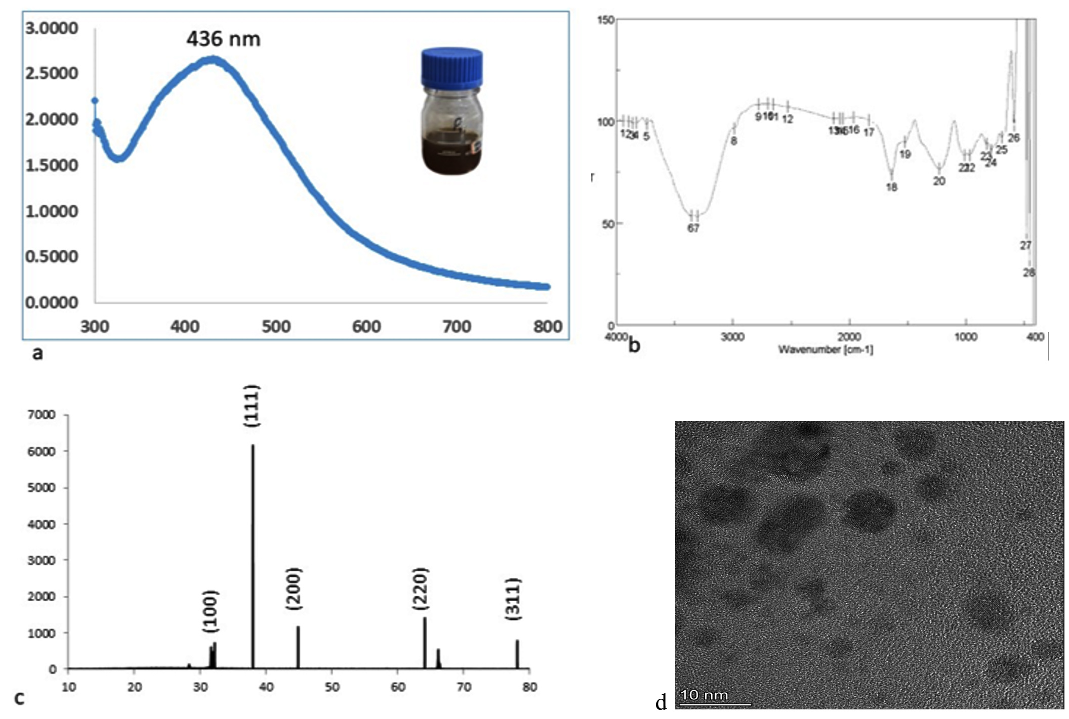
Figure 1. Characterisation of silver nanoparticles
Characteristics analysis of AgNPs
The visual changes were further confirmed with the UV-spectroscopy which displayed prominent peak between 300 to 800 nm with highest absorbance conferring at 436 nm (Figure 1a). The reaction and completion of synthesis was achieved within 15 minutes of incubation under optimised condition of variables such as alkaline pH 9, higher temperature above 80°C, and 1 mM concentration of metal salts. In the present investigation, the involvement of phytocomponents in facilitating the synthesis was investigated using FTIR analysis, which revealed the prominent presence of the hydroxyl group through a significant peak observed at 3359 cm-1, followed by the amide group corresponding to the peak at 1639 cm-1, the aldehyde group at 1226 cm-1, and the alkene group at 968 cm-1, as shown in Figure 1b. The examination of the nanoparticles’ crystal structure was conducted through XRD analysis, which revealed diffraction intensities at the 2-theta angle, confirming their crystalline nature (Figure 1c). The morphological attributes of the AgNPs were investigated using TEM, which exhibited a diverse range of sizes and shapes, indicating polydispersity among the nanoparticles. Size analysis was performed by counting 300 AgNPs, and the corresponding graph is presented in the Figure 1d, illustrating that the majority of AgNPs measured 10 to 60 nm.
Biological activities of the nanoparticles
Antibacterial Activity
The synthesised AgNPs were further evaluated for their biological activities. The results indicated a moderate level of activity against all the tested pathogens, which was confirmed through the well diffusion method (Table 1 and Figure 2a), which showed a zone of inhibition across the well. The broth dilution assay was measured via the optical density of the broth seeded with AgNPs, which was measured at 600 nm (Table 2 and Figure 2b). The test pathogens showed moderate activity for AgNPs.
Table (1):
Antibacterial measured with zone of inhibition in mm
Pathogen Used |
Antibiotic (Amoxycillin) |
Zone of inhibition |
|---|---|---|
Pseudomonas aeruginosa |
10 |
7 |
E. coli |
11 |
6 |
Staphylococcus aureus |
12 |
7 |
Table (2):
Antibacterial activity measured with optical density at 600 nm
Pathogen Used |
Control |
Plant extract |
|---|---|---|
Pseudomonas aeruginosa |
0.063 |
0.088 |
E. coli |
0.080 |
0.037 |
Staphylococcus aureus |
0.152 |
0.051 |
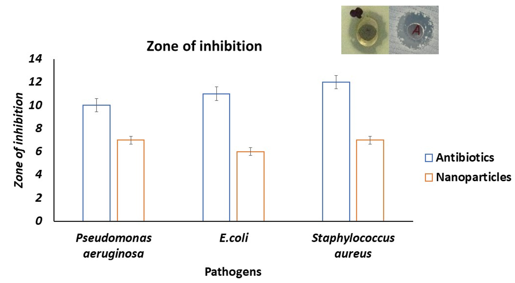
Figure 2a. Antibacterial activity of silver nanoparticles via well diffusion
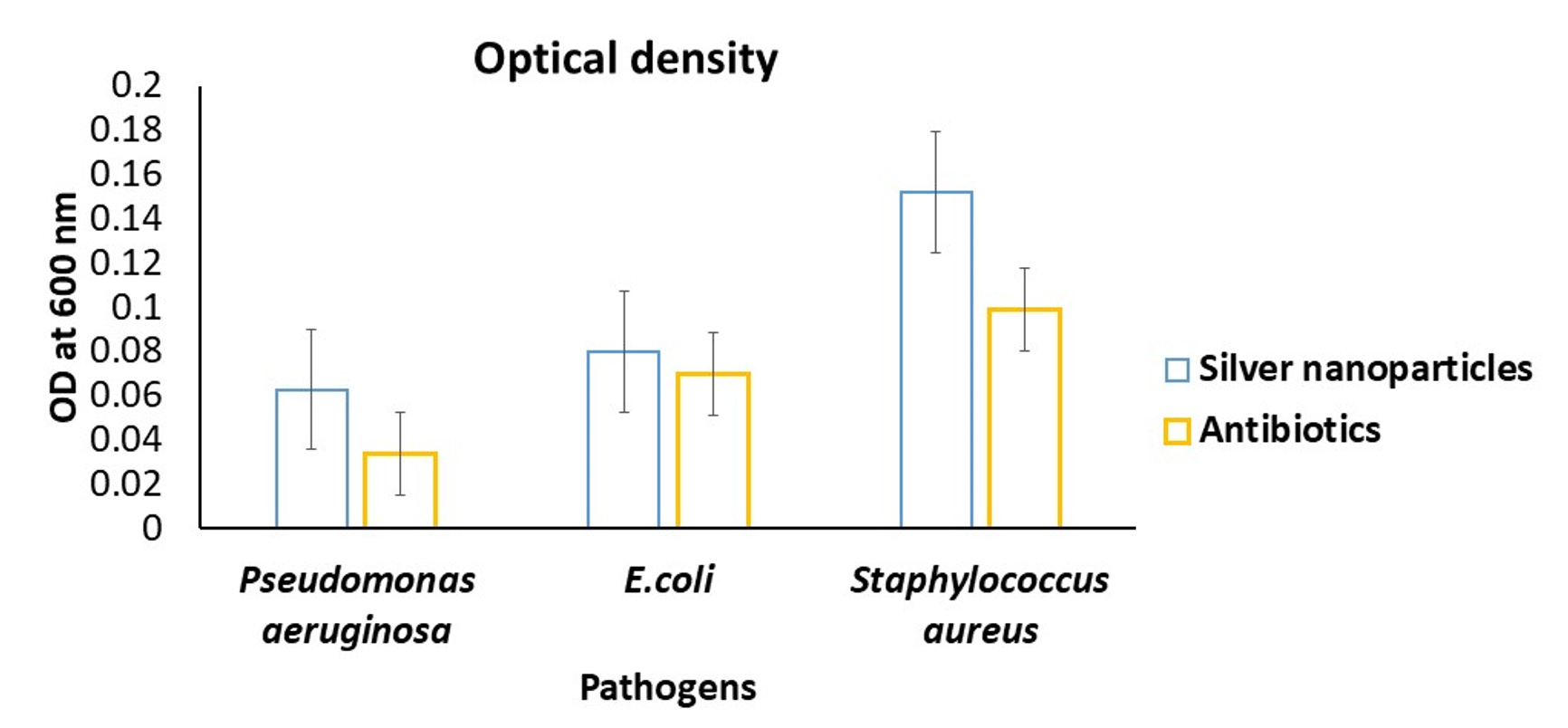
Figure 2b. Antibacterial activity of silver nanoparticles via well diffusion
Plant growth Promotion
The impact of AgNPs on the seed germination and seedling growth of the test seeds was significant. All the seeds with different concentrations of AgNPs showed germination. The measurement of the root and shoot was done. The length of the root was measured from the hypocotyls to the tip of the root. Similarly, for the shoot, length was measured from the root-hypocotyl zone to the cotyledons. The AgNPs treatment seeds were compared with the control, and different parameters such as relative root growth and germination index were calculated and tabulated in Table 3.
Table (3):
Seed germination parameters of different seeds against different concentrations of Ag-NPs
| Seeds | Concentration of AgNps | SGR | RRG | GI |
|---|---|---|---|---|
| Pisum sativum | 2 mg | 80 | 400 | 20 |
| 5 mg | 60 | 300 | 20 | |
| 10 mg | 70 | 300 | 23 | |
| Vigna radiata | 2 mg | 60 | 114 | 52 |
| 5 mg | 70 | 85 | 82 | |
| 10 mg | 90 | 171 | 52 | |
| Macrotyloma
uniflorum |
2 mg | 80 | 200 | 40 |
| 5 mg | 75 | 133 | 56 | |
| 10 mg | 60 | 133 | 45 |
Seed Germination Rate (SGR) Relative Root Growth (RRG) and Germination Index (GI)
The root length and shoot length of all the seeds are presented in Figure 3a and Figure 3b. The results obtained in the seed germination assay were conducted in triplicate and validated with a control without the treatment of AgNPs. The root length was highest with a 5 mg/mL and 2 mg/mL concentration of AgNPs for Pisum sativum and Vigna radiata, except for Macrotyloma uniflorum, which showed the highest germination at 10 mg/mL. Similarly, the shoot length was highest in all the seeds at 10 mg/mL.
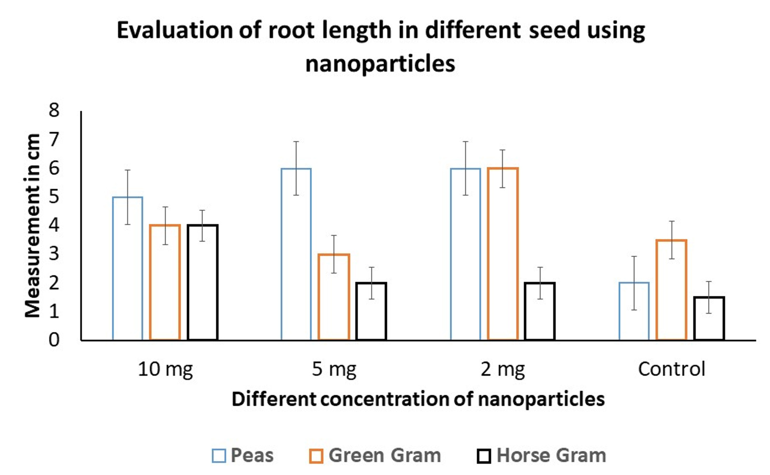
Figure 3a. Measurement of root length of seed germination
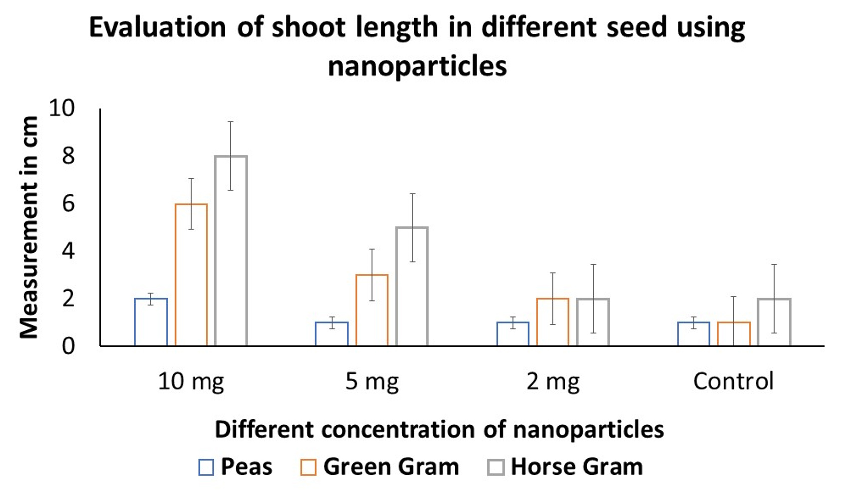
Figure 3b. Measurement of root length of seed germination
Dye Degradation
The catalytic efficiency of AgNPs in the degradation of dyes were rapid, reaching completion within a 12 hour incubation period when exposed to visible light. The confirmation of dye degradation was visually observed and further assessed through UV-visible spectroscopy, with initial and final measurements taken at specific wavelengths corresponding to different dyes. In the case of methylene blue, the degradation percentage was determined to be 35%. The degradation process of dyes exhibited rapid progress and concluded within 12 hours of the incubation period under the influence of visible light (Figure 4). For methylene blue, the degradation percentage was 35%; similarly, for safranin, the degradation percentage was 58%; and finally, for crystal violet, the degradation was 21%. Water and dye solution were used as blanks. Different time intervals (0.5, 1, 1.5, and 2 h) were taken, and optical density (OD) was measured using a spectrophotometer. Experiments were repeated three times, and the mean percentage value was recorded. Among all the evaluated dyes, the degradation of safranin was the highest.
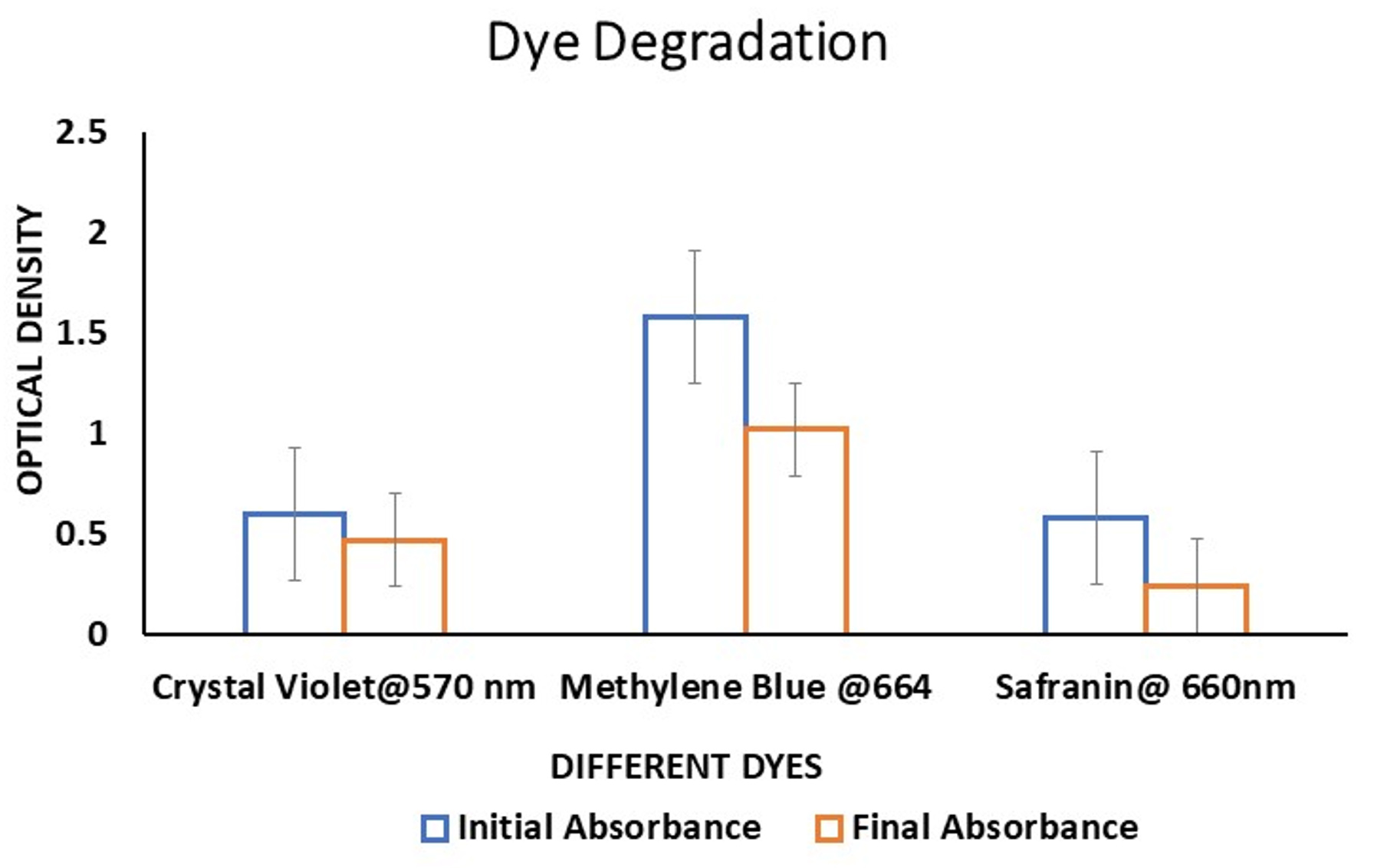
Figure 4. Dye degradation of silver nanoparticles
In this present study, the use of Callicarpa macrophylla showed synthesis of AgNPs within 30 minutes of incubation period. This rapid process can be accounted for the presence of phytocomponents of the plants.17 The studies on the plant Callicarpa macrophylla L. has shown biological activities.18 The synthesis was confirmed with difference in the color of the reaction solution to brown. This is due to the surface plasmon resonance of the AgNPs which is well documented with scientific studies.19 The impact of various factors and variables playing an important role in the synthesis of AgNPs is described with previous findings that favour the synthesis.12 The plant extract comprises myriad phytocomponents that act as reducing agents and stabilising agents. These findings align with previous research that suggests the existence of functional groups.20 Research has shown that the properties of AgNPs are dependent on their size,21,22 with the properties becoming more significant as the size decreases. In this study, the average particle size was found to be 10 to 60 nm, which confirms the aforementioned research. Which was densely coupled with the neighbouring AgNPs, forming a group or cluster zone. This can be attributed to the phytocomponents that are on the surface of the AgNPs, thus forming a group of AgNPs that can be seen in many scientific publications.22
The results were in accordance with the previous reports in the scientific literature that demonstrated the potential of AgNPs acting as antibacterial agents showing activity against Escherichia coli, Pseudomonas aeruginosa and Staphylococcus aureus.12,16,23 The role of phytocomponents to mediate and attenuate the desired activity is also considered to be of prime importance in selecting plant species.24 On a similar front, the role of AgNPs in plant growth promotion was carried out via seed germination assay. The AgNPs significantly influence seed germination in all varieties.25 Similarly, for the shoot, length was measured from the root-hypocotyl zone to the cotyledons.15 The obtained results are in accordance with the previous finding, which reports the role of AgNPs as plant growth promoters and influences seed germination.26 The seeds are protected by an outer layer that has sieving properties that sometimes prevent the entry of macromolecules. Whereas AgNPs can easily penetrate inside the seeds and enhance the uptake of water and protect them from microbial infections, which will increase germination compared to normal plant growth promoters and fungicides.27 The outcomes of dye degradation observed in this present study align with previously documented research.28,29 Although the precise mechanisms behind the degradation process have not been completely elucidated, studies suggest that the significant surface area of the AgNPs plays a crucial role. It is proposed that the excited electrons generate photogenerated excitons, which are effectively captured by the AgNPs owing to their expansive surface area.16,30
In summary, the findings of this study are significant and highlight the potential of using Callicarpa macrophylla synthesis of AgNPs for various applications, such as combating bacterial pathogens and degrading environmental contaminants like dyes. Additionally, the study also indicates that AgNPs have the potential to promote seed germination. Due to the brief nature of this article, additional studies are needed to fully understand the mechanisms underlying the antibacterial activity of AgNPs, as well as the potential toxicity of the by-products of dye degradation and AgNPs used in foliar applications for pot and greenhouse experiments.
ACKNOWLEDGMENTS
The authors would like to thank JSS Academy of Higher Education and Research for Providing the infrastructure to carry out the present study.
CONFLICT OF INTEREST
The authors declare that there is no conflict of interest.
AUTHORS’ CONTRIBUTION
SB and AP conceptualized the study. HKR performed data generation. KM, SB and HS wrote the manuscript. AP reviewed the manuscript. SNR and SB edited the manuscript. All authors read and approved the final manuscript for publication.
FUNDING
None.
DATA AVAILABILITY
All datasets generated or analyzed during this study are included in the manuscript.
ETHICS STATEMENT
Not applicable.
- Bayda S, Adeel M, Tuccinardi T, Cordani M, Rizzolio F. The history of nanoscience and nanotechnology: From chemical-physical applications to nanomedicine. Molecules. 2019;25(1):112.
Crossref - Khan I, Saeed K, Khan I. Nanoparticles: Properties, applications and toxicities. Arab J Chem. 2019;12(7):908-931.
Crossref - Chandrakala V, Aruna V, Angajala G. Review on metal nanoparticles as Nanocarriers: Current challenges and perspectives in Drug Delivery Systems. Emergent Mater. 2022;5(6):1593-1615.
Crossref - Shenashen MA, El-Safty SA, Elshehy EA. Synthesis, morphological control, and properties of silver nanoparticles in potential applications. Part Part Syst Charact. 2013;31(3):293-316.
Crossref - Lee SH, Jun BH. Silver nanoparticles: Synthesis and application for nanomedicine. Int J Mol Sci. 2019;20(4):865.
Crossref - Bahrulolum H, Nooraei S, Javanshir N, et al. Green synthesis of metal nanoparticles using microorganisms and their application in the agrifood sector. J Nanobiotechnol. 2021;19(1):86.
Crossref - Singh J, Dutta T, Kim K-H, Rawat M, Samddar P, Kumar P. ‘green’ synthesis of metals and their oxide nanoparticles: Applications for environmental remediation. J Nanobiotechnol. 2018;16(1:84).
Crossref - Ivanova N, Gugleva V, Dobreva M, Pehlivanov I, Stefanov S, Andonova V. Silver nanoparticles as multi-functional drug delivery systems. Nanomedicines. 2019.
Crossref - Alengebawy A, Abdelkhalek ST, Qureshi SR, Wang MQ. Heavy metals and pesticides toxicity in agricultural soil and plants: Ecological risks and human health implications. Toxics. 2021;9(3):42.
Crossref - Mittal D, Kaur G, Singh P, Yadav K, Ali SA. Nanoparticle-based Sustainable Agriculture and Food Science: Recent Advances and Future Outlook. Front Nanotechnol. 2020;2.
Crossref - MD(Ayu) DJVH. Priyangu Callicarpa macrophylla: Uses, research, remedies. Easy Ayurveda. 2022. https://www.easyayurveda.com/2017/03/14/priyangu-callicarpa-macrophylla/. Accessed May 11, 2023.
- Baker S, Prudnikova SV, Shumilova AA, Perianova OV, Zharkov SM, Kuzmin A. Bio-functionalization of phytogenic AG and zno nanobactericides onto cellulose films for bactericidal activity against multiple drug resistant pathogens. J Microbiol Methods. 2019;159:42-50.
Crossref - Baker S, Mohan KK, Santosh P, Rakshith D, Satish S. Extracellular synthesis of silver nanoparticles by novel Pseudomonas veronii AS41G inhabiting Annona squamosa L. and their bactericidal activity. Spectrochim Acta Part A Mol Biomol Spectrosc. 2015;136:1434-1440.
Crossref - Shinde S, Paralikar P, Ingle AP, Rai M. Promotion of seed germination and seedling growth of Zea mays by magnesium hydroxide nanoparticles synthesized by the filtrate from Aspergillus niger. Arab J Chem. 2020;13(1):3172-3182.
Crossref - Sarkhosh S, Kahrizi D, Darvishi E, et al. Effect of zinc oxide nanoparticles (ZnO-NPs) on seed germination characteristics in two Brassicaceae family species: Camelina sativa and Brassica Napus L. J Nanomater. 2022;2022:1-15.
Crossref - Baker S, Perianova OV, Prudnikova SV, et al. Phytogenic nanoparticles to combat multi drug resistant pathogens and photocatalytic degradation of Dyes. BioNanoScience. 2020;10(2):486-492.
Crossref - Yadav V, Jayalakshmi S, Singla RK, Patra A. Ex vivo screening of stem extracts of callicarpa macrophylla Vahl. for antifungal activity. Indo Global Journal of Pharmaceutical Sciences. 2012;02(02):103-107.
Crossref - Yadav V, Jayalakshmi S, Singla RK, Patra A. Preliminary assessment of anti-inflammatory activity of Callicarpa macrophylla Vahl. leaves extracts. Indo Global Journal of Pharmaceutical Sciences. 2011;1(3):219-222.
Crossref - Jain S, Mehata MS. Medicinal plant leaf extract and pure flavonoid mediated green synthesis of silver nanoparticles and their enhanced antibacterial property. Sci Rep. 2017;7(1):15867.
Crossref - Kamnev AA, Mamchenkova PV, Dyatlova YA, Tugarova AV. FTIR spectroscopic studies of selenite reduction by cells of the rhizobacterium Azospirillum brasilense sp7 and the formation of selenium nanoparticles. J Mol Struc. 2017;1140:106-112.
Crossref - Ebrahimzadeh MA, Naghizadeh A, Amiri O, Shirzadi-Ahodashti M, Mortazavi-Derazkola S. Green and facile synthesis of Ag nanoparticles using Crataegus pentagyna fruit extract (CP-Agnps) for organic pollution dyes degradation and antibacterial application. Bioorg Chem. 2020;94:103425.
Crossref - Essghaier B, Toukabri N, Dridi R, et al. First report of the biosynthesis and characterization of silver nanoparticles using scabiosa atropurpurea subsp. maritima fruit extracts and their antioxidant, antimicrobial and cytotoxic properties. Nanomaterials. 2022;12(9):1585.
Crossref - Gulbagca F, Ozdemir S, Gulcan M, Sen F. Synthesis and characterization of Rosa canina-mediated biogenic silver nanoparticles for anti-oxidant, antibacterial, antifungal, and DNA cleavage activities. Heliyon. 2019;5(12):e02980.
Crossref - Alharbi NS, Alsubhi NS, Felimban AI. Green synthesis of silver nanoparticles using medicinal plants: Characterization and application. J Radiat Appl Sci. 2022;15(3):109-124.
Crossref - Rutkowski M, Krzeminska-Fiedorowicz L, Khachatryan G, Bulski K, Kolton A, Khachatryan K. Biodegradable silver nanoparticles gel and its impact on tomato seed germination rate in in vitro cultures. Appl Sci. 2022;12(5):2722.
Crossref - Razak NQA, Yusoff MHM, Aziz WNAA, et al. Effects of silver nanoparticles on seed germination and seedling growth: A Review. J Indian Chem Soc. 2023;100(1):100866.
Crossref - Baker S, Volova T, Prudnikova SV, Satish S, Prasad MNN. Nanoagroparticles Emerging Trends and future prospect in modern agriculture system. Environ Toxicol Pharmacol. 2017;53:10-17.
Crossref - Ringwal S, Bartwal AS, Sati SC. Photo-catalytic degradation of different toxic dyes using silver nanoparticles as photo-catalyst, mediated via Citrus aurantium peels extract. J Indian Chem Soc. 2021;98(12):100221.
Crossref - Kumar P, Dixit J, Singh AK, et al. Efficient catalytic degradation of selected toxic dyes by green biosynthesized silver nanoparticles using aqueous leaf extract of Cestrum nocturnum L. Nanomaterials. 2022;12(21):3851.
Crossref - Rajasekar R, Thanasamy R, Samuel M, Edison TNJI, Raman N. Eco-friendly green synthesis of silver nanoparticles using Luffa acutangula: Synthesis, characterisation and catalytic degradation of methylene blue and malachite green dyes. Int J Environ Anal Chem. 2022;104(10):2255-2267.
Crossref
© The Author(s) 2024. Open Access. This article is distributed under the terms of the Creative Commons Attribution 4.0 International License which permits unrestricted use, sharing, distribution, and reproduction in any medium, provided you give appropriate credit to the original author(s) and the source, provide a link to the Creative Commons license, and indicate if changes were made.