


ISSN: 0973-7510
E-ISSN: 2581-690X
Pollution caused by dyes is a major environmental threat, posing adverse impacts on humans, animals, and plants. Therefore, the remediation of such pollutants is essential to protect the environment. This study aimed to conduct physicochemical and bacteriological analyses of textile wastewater to isolate and identify potential native bacterial strains for the decolorization of Congo red dye. Physical and nutritional process parameters were optimized to achieve maximum decolorization. The biological and chemical oxygen demands of the analyzed textile waste water were found to be above the recommended limits. In this study, 19 Congo red -decolorizing bacteria were isolated, with one bacterial culture capable of growing at a higher dye concentration of 300 mg/L. This bacterium was characterized biochemically and genetically (using 16S rRNA sequencing) and identified as the Pseudomonas aeruginosa MT-2 strain. A maximum decolorization of 94.0% was achieved at an initial dye concentration of 150 mg/L, 35°C, and pH 8.0 under static conditions. The bacterial culture also showed resistance to heavy metals such as arsenic, lead, and chromium. The biodegradation of Congo red dye was confirmed through UV-vis spectral analysis and Fourier transform infrared spectrophotometry. The findings of this study demonstrate the high remediation potential of the MT-2 strain, making it suitable for possible use in dye biodecolorization at contaminated sites.
Biodecolorization, Congo Red, FT-IR, Optimization, Physicochemical Factors, 16S rRNA
Industrial developments have played a crucial role in the global economy, but they have also led to a massive increase in waste production, which is harmful to all living entities in the biosphere.1,2 The discharge of effluents can be hazardous to the ecosystem, yet a significant quantity of textile effluents is still being released directly into the environment.3 Dyes used in various industries are identified as a primary source of water pollution.4,5 Synthetic dyes are commonly employed to color a wide range of industrial products and are extensively used across different industries.6,7 Dyes are mainly classified based on various criteria, including their structures and applications.8 Congo red is a type of azo dye, and its remediation methods have been studied by researchers.9-11
Azo dyes contain a characteristic azo bond (-N=N-) that makes them resistant to degradation through natural processes. The effluent discharged from textile industries contains harmful and hazardous chemicals that are difficult to remove from the environment.12 The constant growth of the textile industry has led to an increased rate of effluent production.13 The discharge of untreated effluent directly into the environment results in decreased oxygen levels in water, necrosis of flora, and other ecological impacts. Additionally, untreated effluent poses threats to humans and animals, with adverse effects on human health, including skin irritation, respiratory problems, gastrointestinal disorders, and blood clotting.14,15 Untreated effluent also exhibits high levels of BOD, COD, salinity, and intense coloration.16 For the safety of the environment and living organisms, it is essential to remove these dyes before their discharge. Currently, several approaches are being used to remove dyes from wastewater.17 These methods, including physical and chemical approaches, have certain drawbacks, such as high costs and the need for large quantities of chemicals and energy.8,18,19 Consequently, they are not widely applicable for dye decolorization.20 Therefore, there is a need for alternative methods that are safer and more cost-effective than conventional techniques for dye removal.
Congo red removal through adsorption is also a significant approach.21,22 Among several modern techniques, microbial dye decolorization and degradation stand out as promising methods. This approach is considered inexpensive and environmentally friendly for dye remediation.20 Bacteria have been recognized as efficient agents for dye decolorization by degrading organic pollutants, such as dyes, and using them as energy and carbon sources for their growth and multiplication. Notably, numerous bacterial strains are highly tolerant to harsh conditions and exhibit rapid growth.23 The use of microorganisms, especially bacteria, offers advantages such as ease of cultivation. Bacteria can remove dyes from effluent either through adsorption or enzymatic degradation. Myco-remediation is another biological strategy used to treat dye contamination.24
Bacterial dye decolorization has been extensively studied by researchers. Kishor et al.25,26 reported on the dye degradation capabilities of bacterial strains that are resilient, prevalent, and rapidly growing. Other studies have demonstrated the efficiency of bacterial dye degradation. For example, Ikram et al.27 investigated the degradation of Basic Orange 2 dye by Escherichia coli and observed 89.88% decolorization. In another study, Hanis et al.28 showed 83.12% decolorization of Congo red by Bacillus sp. at 25 ppm, pH 7.55, and a temperature of 30°C over 5 days. Additionally, Sarkar et al.29 investigated the decolorization and degradation of Congo red textile azo dye by Chryseobacterium geocarposphaerae and reported 96.52% decolorization within 12 hours.
This study aims to analyze the physicochemical characteristics of wastewater and isolate a potential bacterial culture for effective biodecolorization. The effects of various factors, including culture conditions, pH, temperature, and carbon and nitrogen sources, on the biodecolorization of Congo red by the indigenous Pseudomonas aeruginosa MT-2 strain were also investigated.
Sampling
The dye-containing effluent was collected from the discharge location of power looms in Tanda, Ambedkarnagar, Uttar Pradesh, India, and stored in sterile bottles.
Physicochemical parameters analyses
The physicochemical parameters were assessed according to the American Public Health Association (APHA)30 guidelines, and the values were reported as the mean ± SD of three replicates. Temperature was measured at the sample collection site using a thermometer. The pH of the samples were measured using an electronic pH meter (ELICO). To measure the biological oxygen demand (BOD), the initial dissolved oxygen (DO) of the sample was recorded, and BOD bottles containing the sample were incubated in a BOD incubator at 20°C. After 5 days, the DO was measured again, and BOD was estimated by subtracting the final DO from the initial DO. The dichromate reflux method was used to determine the chemical oxygen demand (COD) of the sample, following APHA guidelines. For measuring total dissolved solids (TDS), the gravimetric method was employed: 10 mL of the sample was filtered through Whatman filter paper, and the filtrate was fully dried and weighed to calculate the TDS content. Fluoride levels were estimated using the SPADNS method, in which the sample was mixed with SPADNS and acid reagent. The absorbance of the resulting solution was then measured at 570 nm using a spectrophotometer, and The fluoride concentration was determined by extrapolating from the values of standard solutions.
Isolation of Congo red dye-decolorizing bacteria
The bacteria responsible for dye decolorization were isolated from contaminated soil using the pour plate technique on solid minimal salt medium (MSM) amended with Congo red dye at a concentration of 50 mg/L. The plates were incubated at 35°C for 72 hours. Bacterial colonies on the MSM agar plates were repeatedly streaked onto the same medium, resulting in the isolation of pure bacterial strains. These pure strains were then tested on MSM agar plates with dye concentrations ranging from 50 to 500 mg/L to determine which bacterial strain exhibited the most efficient dye decolorization properties.
Identification of potential Congo red dye-decolorizing bacterium
Morphological, molecular (16S rRNA gene sequencing), and biochemical analyses were employed to identify the potential isolate(s). After 24 hours of incubation at 35°C, the morphology of the bacterial strain MT-2 was determined by examining colony features through bacterial staining and performing a catalase test. The selected bacterial isolate was further characterized using standard methods. Molecular characterization was performed using the 16S rRNA gene sequence amplification technique at the Cytogene Research Lab, Lucknow (India), to identify and compare bacterial diversity from complex microbiomes. The obtained sequence was then submitted to the DNA Databank of Japan to obtain an accession number. The nucleotide sequences were aligned using Clustal W software, and the neighbor-joining (NJ) method was employed to construct a phylogenetic tree.
Culture conditions
First, the bacterial inoculum was prepared. The dye decolorization experiments were conducted in liquid minimal salt medium, inoculated with a bacterial culture containing 3.1×10⁶ cfu/mL, and incubated at 35°C for 96 hours. The dye content and cell density were periodically examined every 24 hours, and quantitative analysis of dye decolorization was carried out spectrophotometrically at 495 nm. All experiments were performed in triplicate.
Effect of aeration
To determine the effect of aeration, 25 mL of MSM (pH 7.0) was added in 150 mL Erlenmeyer flasks and inoculated with the selected bacterial isolate. The flasks were incubated at 35°C under shaking conditions (150 rpm). To assess the effect of static conditions, 25 mL of inoculated medium was placed in screw-capped tubes and incubated at 35°C for 96 hours.
Effect of dye concentration
To observe the impact of different initial dye concentrations on bacterial decolorization, varying levels of dye (50-300 mg/L) were added to MSM broth and incubated for 48 hours under stationary culture conditions.
Effect of nutrients (carbon and nitrogen)
To identify the optimal carbon and nitrogen sources for the decolorization of Congo red dye, the effects of various carbon and nitrogen sources on dye decolorization were assessed under the previously established optimal culture conditions.
Effect of inoculum size
To investigate the impact of inoculum dosage (1 to 6%, v/v) on dye decolorization, the bacterial inoculum was transferred to dye-containing MSM broth and incubated under stationary culture conditions for 48 hours.
Combined effect of initial pH and temperature
The combined effect of initial pH and temperature was assessed by preparing MSM broths with different pH ranges (7.0-9.0) using 0.1N HCl or 0.1N NaOH before sterilization. After sterilization, the MSM broths were inoculated with the optimal inoculum dosage and incubated at temperatures ranging from 25°C to 40°C for 48 hours.
Multi-metal tolerance test
To evaluate the multi-metal tolerance potential of the isolated bacterium, different concentrations (25-200 mg/L) of arsenic, chromium, lead, and mercury were added to nutrient agar medium and inoculated with the MT-2 isolate.
Measurement of Congo red decolorization
To determine the bacterial potential for dye decolorization, samples were collected from MSM media inoculated with the bacterial isolate. The samples were centrifuged at 10,000 rpm in a refrigerated centrifuge (4°C), and the residue was discarded. The supernatant was then analyzed using a spectrophotometer at 495 nm. The percentage of dye decolorization was calculated using the following formula31,32:
% Decolorization of dye = [Initial optical density – Final optical density/Initial optical density] × 100
FT-IR analyses
samples before and after decolorization were analyzed to determine the biodegradation of the dye by studying the functional groups. The samples were measured across a wavenumber range of 500 to 4500 cm-1.
Statistical analyses
All experiments in this study were conducted in triplicate. Statistical calculations, including standard deviation, were performed using The Microsoft Excel program.
The discharge of colored wastewater from textile industries is a significant global environmental issue. Among the various components of textile effluent, dyes are the most influential in contributing to its toxicity. Bacteria play a crucial role in the biological remediation of azo dyes. In this study, various physical and chemical factors, such as oxygen levels, inoculum size, and nutrient supplementation, were evaluated to determine their impact on the efficiency of bacterial dye decolorization. Understanding each parameter that affects bacterial dye decolorization is essential for enhancing the effectiveness of the bioremediation process.
Physicochemical parameter analysis
The physical and chemical characteristics of colored discharge from powerlooms were analyzed following APHA methods. The results indicate that parameters such as biological oxygen demand (BOD), chemical oxygen demand (COD), and total dissolved solids (TDS) were found to be beyond the recommended threshold values set by international standards and other pollution regulatory bodies (Table 1). However, the pH and temperature were within the permissible limits. These findings suggest that the wastewater released from power looms is unsatisfactory concerning its physicochemical content, posing a potential environmental threat. Therefore, proper treatment of the wastewater is necessary before its discharge into the environment or water bodies.18,32
Table (1):
Physicochemical analyses of colored wastewater
Physico-chemical character |
Permissible limit* |
Observed value |
|---|---|---|
Color |
– |
Red |
Smell |
– |
Pungent |
pH |
6.0-9.0 |
7.8 |
Temperature |
– |
27°C |
BOD |
30 mg/L (in land surface water) 100 mg/L (Land irrigation) |
165 ± 8.0 |
COD |
250 mg/L |
419 ± 13.0 |
TDS |
1500 mg/L |
2758 ± 41.0 |
Fluoride |
– |
1.9 ± 0.2 |
*Recommended by MOEF, CPCB and IS
Isolation and screening of Congo red decolorizing bacteria
The sample was microbiologically examined, and the presence of dye decolorization activity was reported. Nineteen bacterial isolates were obtained on minimal salt agar (MSA) medium containing 50 mg/L of Congo red dye. To select the most potent Congo red decolorizing bacterium among all isolated strains, the cultures were tested in MSA medium with varying dye concentrations (50-500 mg/L). The results showed that a single bacterial isolate was able to grow in the presence of up to 300 mg/L of dye with the addition of 0.4% (w/v) glucose, and this isolate was selected for further studies (Table 2). The findings revealed that increasing dye concentrations inhibited bacterial growth. Other researchers have reported similar results, indicating that higher concentrations exert a toxic effect on decolorization efficiency.18,32,33 Congo red dye decolorization activity was observed in Petri plates (Figure 1).
Table (2):
Screening of the potential Congo red dye decolorizing bacterial isolate
Dye level (mg/L) |
No. of bacterial isolates grown |
|---|---|
50 |
19 |
100 |
3 |
150 |
2 |
200 |
2 |
250 |
1 |
300 |
1 |
350 |
– |
400 |
– |
450 |
– |
500 |
– |
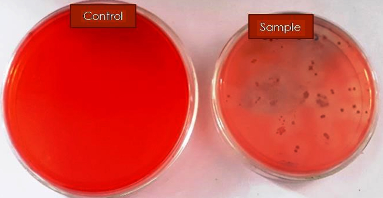
Figure 1. Isolation of Congo red decolorizing bacteria on MSA medium
Biochemical, morphological, and molecular characterization
The Congo red dye-decolorizing bacterium was identified by assessing its biochemical and morphological properties in our laboratory, with the results presented in Table 3.
Table (3):
Morphological and Biochemical characteristics of Pseudomonas aeruginosa MT-2 isolate
Character |
Observation |
|---|---|
Shape |
Small rods |
Motility |
– |
Gram’s reaction |
Negative |
Catalase |
+ |
Spore |
– |
Production of H2S |
– |
Glucose utilization by bacterium |
+ |
Sucrose utilization by bacterium |
+ |
Starch utilization by bacterium |
+ |
+: for positive reactions, -: for negative reactions
The identification of the MT-2 isolate was confirmed through 16S rRNA sequence analysis. genomic DNA was extracted, and the 16S rRNA gene was amplified using PCR with specific primers.34 The sequence was submitted to the DNA Data Bank of Japan (DDBJ), where it received The accession number LC720406. The isolate was identified as Pseudomonas aeruginosa MT-2. The Neighbor Joining (NJ) method was used to construct a phylogenetic tree (Figure 2).
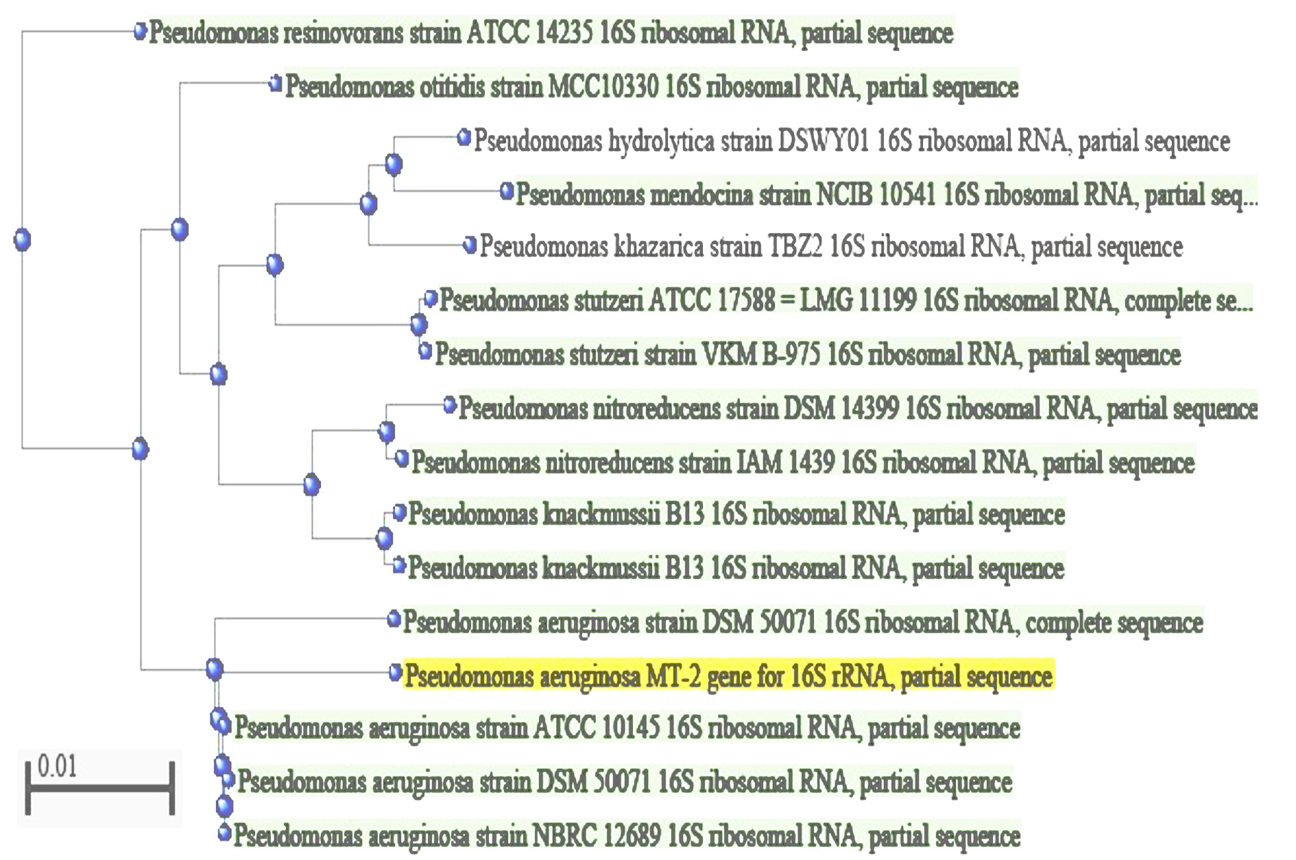
Figure 2. Phylogenetic tree construction of MT-2 strain using Neighbour Joining method
The 16S rRNA gene is The most widely used marker for bacterial classification and identification.35,36 This highly conserved gene is suitable for phylogenetic analysis at higher levels of taxonomy. In contrast, the internally transcribed spacer (ITS) region, which is highly variable, is used to differentiate isolates at lower levels of taxonomy.34 Techniques based on 16S rRNA gene sequencing are considered more robust, reproducible, precise, and provide objective results.35,36
Effect of different process parameters for effective dye decolorization
Impact of aeration
The findings indicate that bacterial isolates achieved more efficient decolorization under stationary conditions compared to shaking culture conditions. A maximum of 98.2% Congo red dye decolorization was achieved at 48 hours under static culture conditions at an initial concentration of 50 mg/L. In contrast, a maximum of 73.8% decolorization was observed under shaking culture conditions. The percentage of dye decolorization was significantly higher under static conditions. Similarly, Xie et al.37 reported 95% decolorization of RB5 dye under stationary culture conditions for 24 hours. Lakshmaiah et al.38 also found that the B5 strain achieved 61% decolorization of reactive blue 222 dye in 24 hours under stationary conditions. Other studies have also reported more efficient dye decolorization under stationary conditions compared to shaking conditions.32,39 In aerobic environments, oxygen can act as a terminal electron acceptor instead of the azo dyes, potentially leading to lower decolorization rates.40
Effect of dye concentration
The concentration and type of dye can significantly impact its remediation capability. It was observed that the extent of Congo red dye decolorization varied with different incubation times. The results revealed that increasing dye concentrations (50-300 mg/L) inhibited the extent of decolorization at each incubation period (24-96 hours). Maximum decolorization of 98.2% was achieved at 50 mg/L, followed by 95% at 100 mg/L, and 87.0% at 150 mg/L. the extent of dye decolorization decreased sharply as the dye concentration increased from 150 to 300 mg/L. However, there was only a marginal increase in the percentage of color removal as the concentration decreased from 150 to 50 mg/L. Consequently, further studies were conducted at an optimized initial concentration of 150 mg/L of Congo red dye. Microorganisms play a crucial role in the effective remediation of pollutants, offering a sustainable and environmentally friendly approach.41-45 Researchers have reported that dye concentration affects the extent of biodecolorization. For instance, Pham et al.23 noted that dye decolorization efficiency declined with increasing dye concentration. High dye concentrations can inhibit reductase enzymes responsible for dye degradation.46
Effect of carbon and nitrogen sources
Microorganisms require specific concentrations of carbon (C) and nitrogen (N) sources for optimal growth and metabolic activity. The effect of various carbon sources (glucose, sucrose, and starch) and nitrogen sources (ammonium sulfate, ammonium nitrate, and peptone) on dye decolorization was evaluated under optimized conditions: an initial 150 mg/L Congo red dye concentration, static culture conditions, 4.0% (v/v) inoculum, pH 7.0, and 35°C over 48 hours of incubation. Among the carbon sources tested, the maximum dye decolorization (87.5%) was achieved using glucose as a co-substrate at a concentration of 4 g/L, followed by sucrose and starch. Among the nitrogen sources, ammonium sulfate proved to be the most effective, resulting in the highest dye decolorization (91.8%). The other nitrogen sources were ranked based on their dye-decolorization efficiency as follows: ammonium nitrate (89%) >peptone (68.2%). In a related study, Nasrin et al.47 investigated the impact of various nitrogen sources, including peptone, yeast extract, and ammonium chloride, on the decolorization of four dyes by Pseudomonas taiwanensis. They observed improved results when yeast extract and peptone were used in combination with ammonium chloride, compared to using yeast extract and peptone individually. This suggests that combined nitrogen sources might enhance dye decolorization effectiveness.
Dyes with complex structures can be particularly challenging for microbes to degrade; hence, additional carbon sources are often necessary to meet their carbon requirements.48 Liu et al.49 also reported that using glucose as a carbon source enhances dye degradation due to its easy uptake and rapid metabolism by bacteria, which supports bacterial growth and activity.
Effect of inoculum dose
The results indicated that dye decolorization improved with increasing inoculum dose (1.0-6.0% v/v) over the 48 hour incubation period. Maximum decolorization was achieved with a 4% v/v inoculum size. As the inoculum size increased from 1.0% to 4.0% (v/v), the extent of dye decolorization also increased correspondingly at each time point. However, further increasing the inoculum size from 4.0% to 5.0% did not significantly enhance the percentage of Congo red dye decolorization. An increase in inoculum size generally promotes microbial growth, as a higher number of cells can more effectively utilize enzymes and substrates.50-52 However, beyond a certain point, additional increases in inoculum size do not necessarily lead to further improvements in decolorization, possibly due to reduced enzyme production or substrate availability.53
Effect of pH and temperature
The influence of pH (6.0-9.0) and temperature (25-40°C) on dye decolorization was assessed. The MT-2 isolate achieved a maximum of 94.0% dye decolorization at 35°C and pH 8.0 under optimized conditions with an initial 150 mg/L dye concentration and no shaking. Deviations from these optimal values resulted in reduced decolorization efficiency. Both pH and temperature are crucial parameters as they directly affect the enzymatic activity of the microorganism. The optimal growth temperature for effective bio-decolorization of azo dyes is typically between
35°C and 45°C. However, decolorization efficiency declines above this range, likely due to enzyme denaturation.54 Similarly, deviations from the optimal pH and temperature can reduce dye degradation efficiency due to enzyme inactivation and decreased bacterial growth.55,56 Elevated temperatures can denature bacterial enzymes, rendering them ineffective.
Biodegradation of Congo red dye
The degradation of Congo red dye and other azo dyes led to the formation of non-toxic intermediates and products. The control sample exhibited maximum absorption at 495 nm, with a distinct peak observed at this wavelength (Figure 3). In contrast, no peak was detected in the inoculated, decolorized samples, indicating that Congo red dye was degraded by the Pseudomonas aeruginosa MT-2 strain. Several studies have reported similar findings for dye biodegradation. For instance, Dutta et al.57 documented the degradation of brilliant green dye by the bacterium Achromo bacterinsolitus, isolated from forest soil in the Biosphere Reserve of Odisha. Pinontoan et al.58 observed efficient decolorization of Trypan Blue dye by a bacterial isolate (TB2) from dye-contaminated sewage water, identified as Aeromonas caviae. Additionally, Xie et al.59 reported that the bacterial strain MS-S2, found in the intestine of termites feeding on wood, was effective in bioremediating malachite green dye. Ullah et al.60 also reported the remediation of Brown 703 azo dye by Pseudomonas aeruginosa from dye-containing effluent.
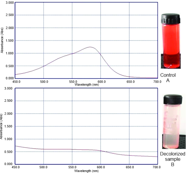
Figure 3. Biodegradation study of Congo red Dye: (A) UV-vis spectral analysis of control dye (B) UV-vis spectral analysis of bio-decolorized dye
FT-IR analyses of decolorized dye
To confirm the decolorization of Congo red dye, Fourier transform infrared (FT-IR) spectroscopy was employed. A comparative analysis of the FT-IR spectra of the degraded and non-degraded dye samples was conducted. The FT-IR spectra of the Congo red dye control and the decolorized dye revealed differences in the presence of functional groups. The control dye exhibited peaks around 1600 cm-1, 3300 cm-1, and smaller peaks near 1400 cm-1 (Figure 4). In contrast, the FT-IR spectrum of the decolorized sample showed the disappearance of the peak near 1400 cm-1. Sarkar et al.29 also investigated FT-IR spectra for Congo red dye and observed several peaks in the control sample at 645 cm-1, 1446 cm-1, and 1740 cm-1. However, the FT-IR spectra of the decolorized dye showed the absence of peaks at 1584 cm-1, which correspond to the azo bond stretching vibration (-N=N-). This indicates the reduction and cleavage of the azo bond in Congo red dye.
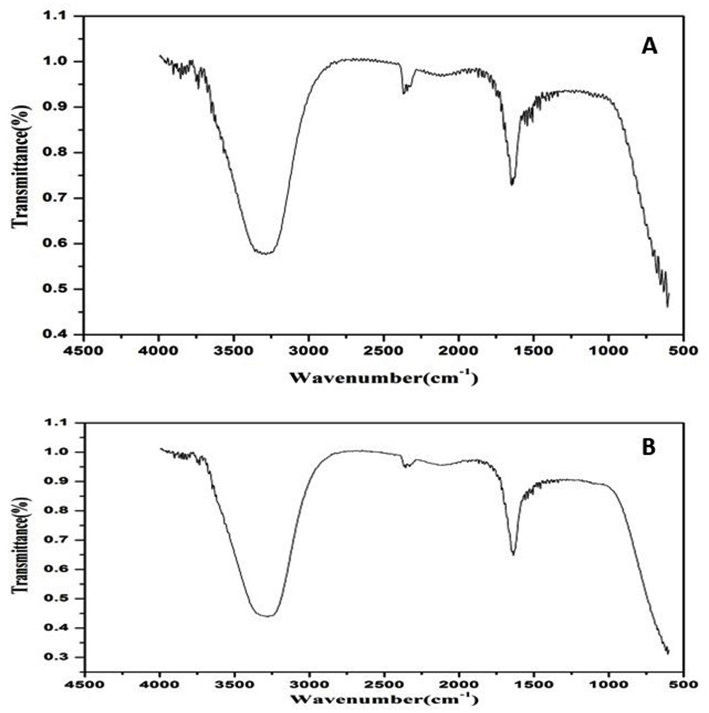
Figure 4. FTIR spectra of (A) Control dye, (B) Decolorized dye
Multi-heavy metal tolerance
The MT-2 isolate demonstrated resistance to arsenic (As), chromium (Cr), and lead (Pb) at an initial concentration of 50 mg/L. However, it was unable to grow on nutrient agar supplemented with mercury (Hg), even at a 25 mg/L concentration. The isolate showed tolerance to a maximum of 100 mg/L of arsenic and 125 mg/L of lead. These findings suggest that the dye-decolorizing bacterium MT-2 has potential for use in remediating sites contaminated with heavy metals. Kishor et al.25 investigated Congo red dye decolorization by the bacterial strain Bacillus cohnni (RKS9), which achieved 99% decolorization in 12 hours and removed 59.76% of cadmium, 40.51% of chromium, 52.71% of lead, and 26.51% of nickel.
Microbial remediation of dyes offers a cost-effective, eco-friendly solution for treating textile dye effluents. This study aimed to explore the dye remediation potential of microbes and enhance the efficiency of dye decolorization to mitigate environmental contamination. The MT-2 strain of Pseudomonas aeruginosa, isolated from colored wastewater, proved highly effective in decolorizing Congo red dye, achieving up to 94% discoloration under optimal conditions. This indicates that Pseudomonas aeruginosa MT-2 may be a promising candidate for in situ dye decolorization in textile industry wastewater. Additionally, the MT-2 strain’s heavy metal tolerance makes it suitable for use in sites contaminated with heavy metals. Although various bacterial strains have demonstrated dye decolorization potential, further research on optimizing bioremediation processes and applying advanced strategies is essential for broader-scale implementation and success.
ACKNOWLEDGMENTS
None.
CONFLICT OF INTEREST
The authors declare that there is no conflict of interest.
AUTHORS’ CONTRIBUTION
All authors listed have made a substantial, direct and intellectual contribution to the work, and approved it for publication.
FUNDING
None.
DATA AVAILABILITY
All datasets generated or analyzed during this study are included in the manuscript.
ETHICS STATEMENT
Not applicable.
- Oyekanmi AA, Ahmad A, Setapar SHM, et al. Sustainable Durio zibethinus-derived biosorbents for Congo Red removal from aqueous solution: statistical optimization, isotherms and mechanism studies. Sustainability. 2021;13(23):13264.
Crossref - Hamad KH, Yasser AM, Nabil R, et al. Nylon fiber waste as a prominent adsorbent for Congo red dye removal. Sci Rep. 2024;14(1):1088.
Crossref - Singh G, Dwivedi SK. Decolorization and degradation of Direct Blue-1 (Azo dye) by newly isolated fungus Aspergillus terreus GS28, from sludge of carpet industry. Environ Technol Innov. 2020;18:1-12.
Crossref - Benkhaya B, Harfi SE, Harfi AE. Classifications, properties and applications of textile dyes: a review. Appl J Environ Eng Sci. 2017;3(3):311-320.
Crossref - Benkhaya S, M’rabet S, El Harfi A. A review on classifications, recent synthesis and applications of textile dyes. Inorg Chem Commun. 2020;115:107891.
Crossref - Mansour D, Alblawi E, Alsukaibi AKD, Al Shammari B. Removal of Congo red dye by electrochemical advanced oxidation process: optimization, degradation pathways, and mineralization. Sustain Water Resour Manag. 2024;10(1):41.
Crossref - Singh PK, Singh P, Singh RP, Singh RL. Biodecolorization of Azo Dye acid blue 113 by soil bacterium Klebsiella variicola RMLP1. J EcophysiolOccup Health. 2021;21(2):64-71.
Crossref - Vikrant K, Giri BS, Raza N, et al. Recent advancements in bioremediation of dye: Current status and challenges. Bioresour Technol. 2018;253:355-367.
Crossref - Harja M, Buema G, Bucur D. Recent advances in removal of Congo Red dye by adsorption using an industrial waste. Sci Rep. 2022;12(1):6087.
Crossref - Siddiqui SI, Allehyani ES, Al-Harbi SA, Hasan Z, Abomuti MA, Rajor HK, Oh S. Investigation of congo red toxicity towards different living organisms: a review. Processes 2023;11(3):807.
Crossref - Kim H, Park C, Choi N, Chao K. Congo red dye degradation using Fe-containing mineral as a reactive material derived from waste foundry dust. Environ Sci Pollut Res. 2024;31(19):1-11.
Crossref - Markandeya,Mohan D, Shukla SP. Hazardous consequences of textile mill effluents on soil and their remediation approaches. Clean Eng Technol. 2022;7:100434.
Crossref - de Vasconcelos GMD, Mulinari J, Souza S, de Souza A, de Oliveira D, de Andrade CJ. Biodegradation of azo dye-containing wastewater by activated sludge: a critical review. World J MicrobiolBiotechnol. 2021;37(6):101.
Crossref - Das A, Mishra S. Decolorization of different textile azo dyes using an isolated bacterium Enterococcus durans GM13. Int J Curr Microbiol App Sci. 2016;5(7):675-686.
Crossref - Semwal N, Mahar D, Chatti M, Dandapat A, Arya MC. Adsorptive removal of Congo Red dye from its aqueous solution by Ag-Cu-CeO2 nanocomposites: Adsorption kinetics, isotherms, and thermodynamics. Heliyon. 2023;9(11):e22027.
Crossref - Singh RL, Singh PK, Singh RP. Recent advances in decolorization and degradation of dyes in textile effluent by biological approaches. Baca Raton New York: CRC Press. 2020.
Crossref - Hua Z, Pan Y, Hong Q. Adsorption of Congo red dye in water by orange peel biochar modified with CTAB. RSC Adv. 2023;13(18):12502-12508.
Crossref - Garg SK, Tripathi M, Microbial strategies for discoloration and detoxification of Azo dyes from textile effluents. Res J Microbiol. 2017;12(1):1-19.
Crossref - Shindhal T, Rakholiya P, Varjani S, et al. A critical review on advances in the practices and perspectives for the treatment of dye industry wastewater. Bioengineered. 2021;12(1):70-87.
Crossref - Li HX, Xu B, Tang L, Zhang JH, Mao ZG. Reductive decolorization of indigo carmine dye with Bacillus sp. MZS10. Int Biodet Biodeg. 2015;103:30-37.
Crossref - Aryee AA, Dovi E, Li Q, Han R, Li Z, Qu L. Magnetic biocomposite based on peanut husk for adsorption of hexavalent chromium, Congo Red and phosphate from solution: Characterization, kinetics, equilibrium, mechanism and antibacterial studies. Chemosphere. 2022;287(Pt 1):132030.
Crossref - Javed T, Thumma A, Uddin AN, et al. Batch adsorption study of Congo Red dye using unmodified Azadirachta indica leaves: isotherms and kinetics. Water Prac. Technol. 2024;19(2):546-566.
Crossref - Pham VHT, Kim J, Chang S, Chung W. Biodegradation of methylene blue using a novel lignin peroxidase enzyme producing bacteria, named bacillus sp. react3, as a promising candidate for dye-contaminated wastewater treatment. Fermentation. 2022(c);8(5):190.
Crossref - Upadhyay R, Przystas W, Dave B. Myco-remediation of synthetic dyes: a comprehensive review on contaminant alleviation mechanism, kinetic study and toxicity analysis. Int J Environ Sci Technol. 2024:1-18.
Crossref - Kishor R, Purchase D, Saratale GD, et al. Environment friendly degradation and detoxification of Congo red dye and textile industry wastewater by a newly isolated Bacillus cohnni (RKS9). Environ Technol Innov. 2021a;22:101425.
Crossref - Kishor R, Purchase D, Saratale GD, et al. Ecotoxicological and health concerns of persistent coloring pollutants of textile industry wastewater and treatment approaches for environmental safety. J Environ Chem Eng. 2021b;9(2):105012.
Crossref - Ikram M, Naeem M, Zahoor M, et al. Biological degradation of the Azo dye basic orange 2 by Escherichia coli: a sustainable and ecofriendly approach for the treatment of textile wastewater. Water. 2022;14(13):2063.
Crossref - Hanis KKA, Nasri ARM, Farahiyah WKW, Rabani MYM. bacterial degradation of Azo dye Congo red by Bacillus sp. J Phys: Conf Ser. 2020;1529:022048.
Crossref - Sarkar S, Echeverria-Vega A, Banerjee A, Bandopadhyay R. Decolourisation and biodegradation of textile di-azo dye Congo red by Chryseobacteriumgeo carposphaerae DD3. Sustainability, 2021;13(19):10850.
Crossref - APHA/AWWA/WEF. Standard Methods for the Examination of Water and Wastewater, 20th Edn, American Public Health Association/American Water Works Association/Water Environment Federation, Washington DC, USA, 1998, ISBN:0-87553-235-7.
- Upadhyay R, Khan HIU, Przystas W. An evaluation of decolorization mechanism of synthetic dyes belonging to the Azo, anthraquinone, and triphenylmethane group, as a sustainable approach, by immobilized CB8 strain (Trametes versicolor). Desalin Water Treat. 2023;284:268-277.
Crossref - Garg SK, Tripathi M, Singh SK, Tiwari JK. Biodecolorization of textile dye effluent by Pseudomonas putida SKG-1 (MTCC 10510) under the conditions optimized for monoazo dye orange II color removal in simulated minimal salt medium. Int BiodetBiodeg. 2012;74:24-35.
Crossref - Asses N, Ayed L, Hkiri N, Hamdi M. Congo Red Decolorization and Detoxification by Aspergillus niger: Removal Mechanisms and Dye Degradation Pathway. Biomed Res Int. 2018;2018(1):3049686.
Crossref - Khan S, Joshi N. Molecular identification of dye degrading bacterial isolates and FT-IR analysis of degraded products. Environ Eng Res. 2020;25(4):561-570.
Crossref - Leelakriangsak M. Molecular approaches for bacterial azoreductases. Songklanakarin J Sci Technol. 2013;35(6):647-657.
- Woo PCY, Lau SKP, Teng JLL, Tse H, Yuen KY. Then and now: use of 16S rDNA gene sequencing for bacterial identification and discovery of novel bacteria in clinical microbiology laboratories. Clin Microbiol Infect. 2008;14(10):908-934.
Crossref - Xie XH, Zheng XL, Yu CZ, et al. High-efficient biodegradation of refractory dye by a new bacterial flora DDMY1 under different conditions. Int J Environ Sci Technol. 2020;17:1491-1502.
Crossref - Lakshmaiah VV, Krishna SBN, More SS, Jayanna SK. Bio-Decolorization and Degradation of Reactive Blue 222 by a Novel Isolate Kucoria marina CU2005. Curr Trends Biotechnol Pharm. 2023;17(1):637-648.
Crossref - Hamad MTMH. Biodegradation of diazinon by fungal strain Apergillus niger MK640786 using response surface methodology. Environ Technol Innov. 2020;18:100691.
Crossref - Srivastava A, Dangi LK, Kumar S, Rani R. Microbial decolorization of Reactive Black 5 dye by Bacillus albus DD1 isolated from textile water effluent: kinetic, thermodynamics & decolorization mechanism. Heliyon. 2022;8(2):e08834.
Crossref - Bala S, Garg D, Thirumalesh BV, et al. Recent strategies for bioremediation of emerging pollutants: a review for a green and sustainable environment. Toxics. 2022;10(8):484.
Crossref - Tripathi M, Singh P, Singh R, et al. Microbial biosorbent for remediation of dyes and heavy metals pollution: a green strategy for sustainable environment. Front Microbiol. 2023;14:1168954.
Crossref - Tripathi M, Singh S, Pathak S, et al. Recent strategies for the remediation of textile dyes from wastewater: a systematic review. Toxics. 2023;11(11):940.
Crossref - Tripathi M, Pandey R, Kumar S. Biodecolorization of Orange II dye by native Bacillus sp. and Staphylococcus sp. in simulated medium. J Pharmacogn Phytochem. 2018;7(1S):1366-1368.
- Singh P, Singh R, Singh S, et al. Microbial engineering for a greener ecosystem and agriculture: recent advances and challenges. J Pure Appl Microbiol. 2024;18(2):797-807.
Crossref - Azam A, Muhammad G, et al. Aerobic Biological Units in Dye Removal. In Biological Approaches in Dye-Containing Wastewater; Springer: Singapore, 2022:57-94.
Crossref - Nasrin T, Saha AK, Mohanta MK, Reza A, Haque MF. Effect of various carbon and nitrogen sources on decolorization of textile dyes by Pseudomonas taiwanensis Strain TNZ3. J Adv Microbiol. 2023;23(2):27-42.
Crossref - Sihag S, Pathak H, Jaroli DP. Factors affecting the rate of biodegradation of polyaromatic hydrocarbons. Int J Pure Appl Biosci. 2014;2(3):185-202.
- Liu K, Yang Y, Sun F, Liu Y, Tang M, Chen J. Rapid degradation of Congo red wastewater by Rhodopseudomonas palustris intimately coupled carbon nanotube-Silver modified titanium dioxide photocatalytic composite with sodium alginate. Chemosphere. 2022;299:134417.
Crossref - Ayed L, Bekir K, Achour S, Cheref A, Bakhrouf A. Exploring bioaugmentation strategies for azo dye CI Reactive Violet 5 decolourization using bacterial mixture: dye response surface methodology. Water and Environ J. 2017;31(1):80-89.
Crossref - Palanivelan R, Rajakumar S, Raja SSS, Ayyasamy PM. Optimization of prime parameters for textile dye decolorization by design of experiments (DOEs) using Lysinibacillus fusiformis M1. Desalination and Water Treatment. 2015;56(4):1077-1089.
Crossref - Roy DC, Biswas SK, Saha AK, et al. Biodegradation of Crystal Violet dye by bacteria isolated from textile industry effluents. Peer J. 2018;6:e5015.
Crossref - Bonugli-Santos RC, Vieira GAL, Collins C, et al. Enhanced textile dye decolorization by marine-derived basidiomycete Peniophorasp. CBMAI 1063 using integrated statistical design. Environ Sci Poll Res. 2016;23(9):8659-8668.
Crossref - Pearce CI, Lloyd JR, Guthrie JT. The removal of colour from textile wastewater using whole bacterial cells: a review. Dyes and Pigments. 2003;58(3):179-196.
Crossref - Wu K, Shi M, Pan X, et al. Decolourization and biodegradation of methylene blue dye by a ligninolytic enzyme-producing Bacillus thuringiensis: Degradation products and pathway. Enzyme Microb Technol. 2022;156:109999.
Crossref - Singh AL, Chaudhary S, et al. Biodegradation of Reactive Yellow-145 azo dye using bacterial consortium: A deterministic analysis based on degradable Metabolite, phytotoxicity and genotoxicity study. Chemosphere. 2022;300:134504.
Crossref - Dutta S, Rath S, Sarangi A, Thatoi H. Brilliant green decolorization by dye decolorizing peroxidase producing bacterium, Achromobacter insolitus isolated from Forest soils of Similipal Biosphere Reserve, Odisha. Bioremediation J. 2024:1-14.
Crossref - Pinontoan R, Susanto TSR, Lucy J, et al. Trypan blue dye decolorization by Aeromonas caviae isolated from water sewage in Jakarta, Indonesia. Biodiversitas J Biolog Diver. 2024;25(4):1631-1637.
Crossref - Xie R, Danso B, Sun J, Schagerl M, Al-Tohamy R, Ali SS. Harnessing the potential of a novel lignin-degrading Streptomyces sp. MS-S2 from wood-feeding termite for malachite green decolorization and detoxification. Process Saf Environ Prot. 2024;186:189-199.
Crossref - Ullah KA, Zahoor M, Ur Rehman M, et al. bioremediation of azo dye brown 703 by pseudomonas aeruginosa: an effective treatment technique for dye-polluted wastewater. Microbiol Res. 2023;14(3):1049-1066.
Crossref
© The Author(s) 2024. Open Access. This article is distributed under the terms of the Creative Commons Attribution 4.0 International License which permits unrestricted use, sharing, distribution, and reproduction in any medium, provided you give appropriate credit to the original author(s) and the source, provide a link to the Creative Commons license, and indicate if changes were made.