


ISSN: 0973-7510
E-ISSN: 2581-690X
Exopolysaccharides (EPS) particularly, from Lactic acid bacteria have received increasing attention in food, medical, and pharmaceutical applications. The present work aims to isolate, characterize and identify exopoly saccharide-producing bacteria from fermented fruits and vegetables and dairy products. A total of 55 isolates were isolated from fermented fruits, vegetables, and dairy products depending on the mucoid appearance of the colonies. Based on total EPS production, the most promising nine strains were selected, phenotypically and genotypically characterized. They were facultative anaerobe, arranged in pairs/chains (cocco bacillus), oxidase, and catalase-negative, non-spore forming and non-motile Gram-positive bacteria. All the strains were capable of growing at optimum pH between 5-7, tolerate to NaCl up to 7% (w/v), growing at 20-37°C with optimum growth at 30°C, no growth was observed at 45°C. In addition they could utilize small range of organic compounds, except isolate S1 was differ from the others by their ability to utilize a varied range of organic compounds. Construction of phylogenetic tree, on the basis of partial 16S rRNA gene sequences indicated that isolate S1 was similar to Leuconostoc citreum with similarity of 91.3%, while, isolates S2 and S3 were similar to Leu. fallax and Leu. mesenteroides with similarity of 99.40 % and 97.73%, respectively. Isolates S4, S5, S7, S8, and S9 were similar to Leu. holzaapfelii with similarity of 98.3, 98.7 and 99.8, 98.5 and 98.1, respectively, while isolate S6 was similar to Leu. lactis with similarity of 97.9%. None of sugars such as lactose, glucose, and fructose except sucrose were support EPS production from these strains. The highest yield of EPS was recorded for isolates S6, S1 and S7 which were 61.90, 61.80 and 60 gl-1, respectively, followed by isolates S4, S9, S5 and S8 which were 58.40, 53.06, 51.61 and 33.53 gl-1, respectively. Although, the lowest yield was observed for the isolates S3 and S2 which were 22.08 and 18.80 g l-1, respectively. Finally, it could be concluded that EPS production from these strains in the current study, considering them to be the alternative choice for enhancing production of EPS with increased yields, with promising realistic importance in food, pharmaceutical, as well as dairy industries.
Lactic acid bacteria, Leuconostoc, Exopoly saccharids, Genotypic and Phenotypic characterization, Food, dairy, medical, pharmaceutical applications.
Lactic acid bacteria (LAB) are microorganisms widely used for along time particularly in fermented dairy industry and pharmaceutical applications worldwide. LAB is generally recognized as safe (GRAS) in which no reports of any illness or toxicity recorded or any harmful compounds produced by them1. Recently, lactic acid bacteria (LAB) stirred up a sensation for their ability to produce many valuable products in particular extracellular polysaccharides (EPS)2. EPS from LAB have acknowledge dincreasing consideration because of their budding importance in various fields3, 4, 5. In food and beverage industry, EPS used as stabilizers, gelling, thickeners, suspending, film-forming, coagulating, emulsifiers agents, and water-binding agents. In medicine, EPS from LAB used as antitumor, a cholesterol-lowering6, immune stimulators7, and depollution agents, they exhibit antioxidant and prebiotic activities8.
LAB are able to generate a wide range of different EPS with different composition and functionality, and this led to widening their industrial applications. Some of LAB are capable of producing EPS either it is released to the extracellular environment or attached to the cell wall of the bacteria (capsular EPS)3. In LAB, EPS are classified into two types on the basis of their chemical composition, homo- and hetero-EPS. Single type of monosaccharide (e.g. fructose) are present in Homo-EPS called fructans (levan and inulin-type) while those containing glucose are called: a-glucan (as dextran, mutan, and alternan), b-glucans these types produced by Leuconostoc mesenteroides, and various streptococci9. Hetero-EPS, composed of varying numbers (from tri- to octal) of diverse monosaccharides and the majority of them comprises combination of D-galactose, D-glucose, also, in rare cases, N-acetylgalactosamine, N-acetylglucosamine, glucuronic acid, fucose, and non-carbohydrate substituent (glycerol, phosphate, and acetyl)10. Also, EPS is characterized depending on the nature of mucous colonies as “ropy” colonies that produce viscous strands when pulled with an inoculating loop or resistance of liquid EPS to flow through serological pipettes and produce long filament and go down from the pipette tip11. While the”nonropy”colonies that are slime or mucoid colonies but not able to form a long filament when extended with an inoculation loop2, 12.
Homo-EPS and hetero-EPS biosynthesis are completely dissimilar; homo-EPS are mainly synthesized from combining sucrose either levansucrase or glucansucrase. While, the hetero-EPS synthesis involves four main reactions: sugar transporting, sugar nucleotide synthesizing, synthesis of repeating unit, and finally their polymerization13, 14. Most commonly the LAB capable of producing EPS belong to the genera- Lactobacillus, Leuconostoc, Streptococcus, Lactococcus and Pediococcus15, 16, 17. The enhancement of EPS production to diminish the industrial costs remains a challenge. So, the searching for new highly efficient isolates, optimization of fermentation condition, use of economical and cheap precursors by using metabolic and genetic engineering approaches played an essential role in this respect and sought after. Thus, the present work focused on the isolation, phenotypic and genotypic characterization of EPS producing bacteria with highly efficient EPS production ability.
Samples collection and preparation
Different types of environmental fermented foods (26 samples from fermented fruits, diluted molasses, sugar cane, plants, yogurt, labneh, and fermented vegetables) were collected from various Saudi Arabian and Egyptian markets and were kept in sterile bottles for isolation of EPS producing bacteria. For preparation of the samples, 10 grams of each fermented samples have been taken by aseptic techniques and were suspended individually into Erlenmeyer flask containing 90 ml sterilized saline phosphate buffer and were shaken thoroughly to make the microorganism migrate to the solution. Serial dilutions were made starting from 10-1 to 10-7 in sterile saline solution.
Isolation and screening of EPS producing bacteria from fermented fruits and vegetables
For this, fermentation medium containing (g l-1): sucrose (80 g), sodium gluconate (20 g), yeast extract (3g), peptone (2g), glycerol (3g), mannitol (10g), was prepared with adjusted pH 7.0 and sterilization was by autoclaving at 121°C for 20 minutes18. Subsequently, the medium was cooled at 45°C, it was poured in sterile petri plats and left for solidifying, and then 0.3 ml of each serial dilution was taken and spread over the agar plate medium. Finally, the plates were incubated at for 48h at 30°C. The EPS producing isolates were detected by forming slimy colonies. To get single colonies, to check the purity of the cultures they were microscopically observed and ensured through streaking on the modified double strength plate count agar medium supplemented with 0.4g sucrose. All bacterial strains were maintained for long term preservation at –70°C after mixing with sterile glycerol with final concentration 20%(v/v). The most promising EPS producing strains were selected for detailed taxonomic studies.
Phenotypic characterization of EPS producing isolates
All isolated strains were tested for EPS production using sodium gluconate medium and then were partially characterized based on Gram stain, motility, sporulation, and cell shape. Based on the quantity of EPS produced, the most promising EPS-producing isolates were selected and were subjected for phenotypic and genotypic characterizations. Phenotypic characterization was performed according to Gerhardt et al.19 and further biochemical tests were determined using biomיrieux vitek® 2GP(BioMיrםeux, Lyon, France)20. The growth at different temperature (20-45°C), pH (pH 4-9) and salt concentrations (0-9% NaCl), were determined using sodium gluconate broth medium. The bacterial growth was monitored by assessing optical density (OD) at 650 by using a JASCO V-530 UV-VIS spectrophotometer.
Hemolytic activity test
The selected strains capable of producing EPS were checked for blood hemolysis by spotting the bacterial culture on blood agar plates and incubated at 37oC for 24 h.
Antibiotic susceptibility testing
The antibiotic susceptibility profiling of the selected EPS producing strains was performed according to Bauer et al.21 and EFSA22. fresh cultures in tryptic Soy Bean broth(TSB) were swabbed on onto the surface of Tryptic soy agar plates, which were allowed to stand at room temperature before placing antibiotic discs for about 3h. Commercial antibiotic disks (Mast Diagnostic GmbH, Germany) containing Amikacin (30µg), Ceftazidime (30µg), Aztreonam (30µg), Piperacillin (100µg), Imipenem (10µg), Ciprofloxacin (5µg), Cephalothin (30µg), Clindamycin (2µg), Oxacillin (5µg), Cotrimoxazole (25µg), Erythromycin (15µg), Gentamicin (10µg), Oxytetracycline (30µg), Penicillin (10µg), Chloramphenicol (30µg) and Tetracycline (µg 30) were used. Plates were allowed to incubate aerobically at 30°C for 24 h. The sensitivity profile of selected EPS producing strains towards different antibiotics were expressed in terms of resistance or sensitive.
Genomic DNA isolation, PCR amplification, and 16s rRNA gene sequencing
For the assessment of genomic DNA isolation, the selected EPS producing strains were allowed to grow in 50 ml of TSB medium at 30°C for 24h. The cells from the culture were harvested by centrifugation at 4000 rpm for 20 min in a Megafuge 1.OR (Fa. Heraeus Sepatech GmbH, Osterode) and then obtained pellets were washed one time with TE-buffer. Genomic DNA was isolated according to the method described by Elbanna et al.23. By employing the usage of universal primers for the eubacteria, amplification of the 16S rDNA gene was done by PCR. The PCR products (~1540 bp) were confirmed by means of electrophoresis and purified by using the PCR extraction kit (Macherey-Nagel, D ren, Germany). The sequencing of 16S rRNA fragments were performed with ABI 3730 X1 automated DNA sequencer (Applied biosystems USA) through Lab of Macrogen Company, South Korea. The obtained sequence of selected isolated strains and other representatives strains of Leuconostoc (retrieved from the NCBI database) were aligned by using the computer-assisted tool ClustalX24. The phylogenetic tree was reconstructed and displayed with Tree View25. The phylogenetic tree was calculated using the neighbor-joining method26 with boots trap values intended from 100 replicate runs, and Pseudomonas putida strain ATCC 17390 (gi|10567508) was used as out group.
EPS production by the selected isolates
For this, pre-cultures of selected isolates were prepared by inoculating each bacterial isolate in 250 of Erlenmeyer flasks containing ml 20 ml sterile TSB medium (g l-1: Tryptone (17 g), glucose (2.5 g), dipotassium phosphate (2.5g), sodium chloride (5g), soy peptone (3 g) and pH was maintained at pH 7.3± 0.2) and incubated at 30°C for 48 h. Subsequently, Erlenmeyer flasks containing 50 ml of the sterile fermentation medium (pH 7.0) supplemented individually with different concentrations of sucrose or lactose, glucose, and fructose. All the inoculated flasks were allowed to incubate on a rotary shaker at 30°C for 3 days.
Extraction, purification, and estimation of EPS
or extraction of EPS, each fermented culture was diluted with deionized water to reduce the viscosity, as well as, the bacterial cells embedded in the viscous mixture can be removed easily by centrifugation. The diluted culture broth was centrifuged at 10, 000 xg for 20 minutes, cell pellet was discarded, and in cell-free supernatant two volumes of cold isopropanol gradually along the sidewall of the conical flask was added and then allowed to settle down for about 2 hours at room temperature for precipitation and collection of EPS27. EPS produced was washed with cold ethanol three times and dried at 50oC. For further purification, removal of proteins was employed by the hydrochloric acid method according to Huang28. For this, the dried-crude EPS redissolved in sterilized deionized water and shacked strongly until complete dissolving. The pH of EPS solution was maintained to 3 by adding 2 Mhydrochloric acid and left for overnight. To obtain proteins free solution, it was centrifuged at 5, 000 rpm for 15 min, and the precipitate was discarded. The EPS were precipitated from the aqueous phase by adding 2 volumes of isopropanol and allowed to settle down at room temperature for 2 hours, followed by washing with ethanol three times, lyophilized, and then the weight of the dried EPS was gravimetrically measured.
Isolation and screening of EPS producing bacteria
In the current work, a total number of 55 bacterial isolates were recovered from fermented fruits, vegetables, and dairy products basis of the mucoid colony characteristics. All isolates were partially characterized based on Gram staining, sporulation, motility and EPS amount produced. Most of these isolates were non-motile, non-spore for mingand Gram-positive bacteria (Table 1). Most of the isolates were EPS producers using the broth medium containing 8% sucrose, except isolates S18, 19, 34, 46 and 51 exhibited mucoid appearance colonies in agar medium but were unable to produce EPS using the broth medium. EPS amount produced from these isolates was varied from 0.8 to 62.15 g l-1. It was manifested that, viscosity and the amount of polysaccharide increased of most the isolates when the plates were kept in refrigerator at 8°C, while some isolates such as S15, S16, S17, and S31 were consumed EPS when kept in the same condition. The most promising strains on the based of amount of EPS production, were; S1, S2, S3, S4, S5, S6, S7, S8, and S9 were selected and subjected to subsequent evaluation of their phenotypic and genotypic characterizations.
Table (1):
Partial characterization of EPS producing isolates isolated from different sources
| Isolate Code | Source of Strains | EPS (g/l)* | Isolate Code | Source of Strains | >EPS (g/l)* | ||||||||
|---|---|---|---|---|---|---|---|---|---|---|---|---|---|
| Gram | Motility | Sporulation | Gram | Motility | <Sporulation | ||||||||
| Stain | KOH< | Stain | KOH | ||||||||||
| S1
S2 S3 S4 S5 S6 S7 S8 S9 S10 S11 S12 S13 S14 S15 S16 S17 S18 S19 S20 S21 S22 S23 S24 S25 S26 S27 |
Orange A
Potato Gawava Gelatin A Gelatin B Molokhai Apricot A Kiwi Labneh A Orange B Avocado Melon Melon Melon Sugar cane Sugar cane Sugar cane Strawberries Pineapple pineapple Watermelon watermelon watermelon watermelon Molasses Molasses Molasses |
61.80
18.08 22.08 58.40 51.61 61.90 60.0 33.53 53.06 9.65 3.11 8.45 10.12 5.45 6.32 2.25 11.65 NA NA 2.65 NA 3.58 8.64 7.12 6.20 6.4 5.52 |
+
+ + + + + + + + – + + + + + + + + + – + + + + + + + |
–
– – – – – – – – + – – – – – – – – – + – – – – – – – |
–
– – – – – – – – – – – – – – – – + + – + – – – – – – |
–
– – – – – – – – – – – – – – – – + + – + – – – – – – |
S28
S29 S30 S31 S32 S33 S34 S35 S36 S37 S38 S39 S40 S42 S43 S44 S45 S46 S47 S48 S49 S50 S51 S52 S53 S54 S55 |
Mango
Onion Onion Apricot B Apricot B Tomato Tomato tomato Peache Peache Sucrose Date pomegranate Mandarin Mandarin mandarin Apple Apple Apple beet Banana Banana Grape Grape Grape Pickled Labneh B |
4.85
4.6 5.6 0.8 9.56 9.6 NA 2.65 2.94 1.65 3.51 3.0 1.15 2.51 6.82 9.54 7.58 NA 2.35 21.6 12.85 8.30 NA 2.4 0.89 2.53 15.8 |
+
+ + + + + + + + + + + – + + + – + + + + + + + – + + |
–
– – – – – – – – – – – + – – – + – – – – – – – + – – |
–
– – – – – + – – – – – – – – – – + – – – – + – – – – |
–
– – – – – + – – – – – – – – – – + – – – – + – – – – |
Notes: * Exopolysaccharide (g l-1) produced using sucrose fermentation medium (18) supplemented with 80 (g l-1) sucrose.
Phenotypic characterization of selected EPS producing isolates
Biochemical and phenotypic characterization profiles of selected EPS isolates analyzed using Biomיrieux Vitek 2 GP were summarized in Tables 2 and 3. Colonies of all nine isolates on nutrient agar were up to 1 mm indiameter, smooth and convex with opaque white color. When they grow on EPS agar medium, all isolates exhibited very slimy colonies, while S3 isolate had the feature of a ropy colony during picking up using inoculating loop. All isolates were facultative anaerobe, oxidase and catalase-negative, coccobacillus (pairs/chains), non-motile, non-spore forming and Gram-positive bacteria. They were able to with stand at pH ranging between 4-9 with optimum growth was between pH 5-7, tolerance to NaCl was observed up to 7% (w/v), temperatures for growth were found between 20-40°C with optimum growth was recorded at 30°C, no growth was observed at 45°C. In addition the isolates were capable of using a narrow range of organic substrates, except isolate S1 differ from the others by its ability to use a wide range of organic substrates that include D-amygdalin, D-xylose, cyclodextrin, D-sorbitol, mannitol, salicin, and raffinose. However, the phenotypic characteristics that make the isolated strains distinguishable as compared to the reference strains were summarized in Tables 3 and 4. Moreover, all EPS-producing isolated strains were susceptible to tetracycline, imipenem, piperacillin, and chloramphenicol, whereas they were resistant to gentamicin, amikacin, aztreonam, ciprofloxacin, cephalothin, oxacillin, penicillin G and cotrimoxazole. None of isolates exhibited haemolytic activities on blood agar plates, and they were negative for arginine dihydrolase, phosphatidylinositol phospholipase, beta-galactosidase, Ala-the-pro arylamidase, Alpha- mannosidase, L-aspartate arylamidase, beta-glactopyranosidase, phosphatase, Beta-glucuronidase, Alpha- galactosidase, tyrosine arylamidase, and gelatin liquefaction. According to the analytical profile index using Biomיrieux Vitek 2 GP database (Table 3), isolate S1 was identified as Enterococcus columbae with probability 87%, while isolates S2, S4, S5, S6 and S9 were identified as Leucocnostoc mesenteroides ssp dextranicum with similarity of 95, 93, 92, 90 and 90%, respectively. Isolates S3 and S8 were identified as leu. pseudomesenteroides with similarity of 95 and 89%, respectively, while isolate S7 were identified as leu. citreum with similarity 91%.
Table (2):
Morphological andpartial characterization ofthe most promising EPS producing isolates
| Characteristics | The most promising EPS producing isolates | ||||||||
|---|---|---|---|---|---|---|---|---|---|
| S1 | S2 | S3 | S4 | S5 | S6 | S7 | S8 | S9 | |
| Colony morphology | |||||||||
| Form | Spindle | Circular | Circular | Spindle | Spindle | Spindle | Spindle | Spindle | Spindle |
| Color | White | White | White | White | White | White | White | White | White |
| Elevation | Flat | Flat | Flat | Flat | Flat | Flat | Flat | Flat | Flat |
| Opacity | Opaque | Opaque | Opaque | Opaque | Opaque | Opaque | Opaque | Opaque | Opaque |
| Consistency | Mucoid | Mucoid | Mucoid | Mucoid | Mucoid | Mucoid | Mucoid | Mucoid | Mucoid |
| Cellmorphology | |||||||||
| Shape | Cocco-bacillii | Cocco- bacillii | Cocco-bacillii | Cocco- bacillii | Cocco- bacillii | Cocco- bacillii | Cocco- bacillii | Cocco- bacillii | Cocco- bacillii |
| Gram stain | + | + | + | + | + | + | + | + | + |
| Catalase | – | – | – | – | – | – | – | – | – |
| Sporulation | – | – | – | – | – | – | – | – | – |
| Motility | – | – | – | – | – | – | – | – | – |
| Growth on NaCl | |||||||||
| 0 % | + | + | + | + | + | + | + | + | + |
| 2 % | + | + | + | + | + | + | + | + | + |
| 4 % | + | + | + | + | + | + | + | + | + |
| 6.5 % | + | + | + | + | + | + | + | + | + |
| % 7.0 | +/– | +/– | +/– | +/– | +/– | +/– | +/– | +/– | +/– |
| 8 % | – | – | – | – | – | – | – | – | – |
| pH values | |||||||||
| 4 | –/+ | –/+ | –/+ | –/+ | –/+ | –/+ | –/+ | –/+ | –/+ |
| 5 | + | + | + | + | + | + | + | + | + |
| 6 | + | + | + | + | + | + | + | + | + |
| 7 | + | + | + | + | + | + | + | + | + |
| 8 | +/– | +/– | +/– | +/– | +/– | +/– | +/– | +/– | +/– |
| 9 | +/– | +/– | +/– | +/– | +/– | +/– | +/– | +/– | +/– |
| Temperature | |||||||||
| 20 oC | +/– | +/– | +/– | +/– | +/– | +/– | +/– | +/– | +/– |
| 25oC | + | + | + | + | + | + | + | + | + |
| 30 oC | + | + | + | + | + | + | + | + | + |
| 37oC | + | + | + | + | + | + | + | + | + |
| 45 oC | – | – | – | – | – | – | – | – | – |
| Oxygen requirement | Facult-ative | Facult-ative | Facult-ative | Facult-ative | Facult-ative | Facult-ative | Facult-ative | Facult-ative | Facult-ative |
| Growth on Blood agar | – | – | – | – | – | – | – | – | – |
| Gelatin liquefaction | – | – | – | – | – | – | – | – | – |
Notes: (+)Symbols and abbreviations: +, growth; -, no growth; (+/– or (-/+) weak growth. Other physiological tests were determined according to Gerhardt et al. (19).
Table (3):
Biochemical testsaccording to Biomérieux Vitek for the selected promising EPS producing isolates
| Sr No. | Different carbon source utilization, enzymatic activities and resistance. | Conc. (mg)
|
Polysaccharide isolates / identification results | ||||||||
|---|---|---|---|---|---|---|---|---|---|---|---|
| S1 | S2 | S3 | S4 | S5 | S6 | S7 | S8 | S9 | |||
| 1. | D-amygdalin (AMY) | 0.1875 | + | – | – | – | – | – | – | – | – |
| 2. | Phosphatidylinositol phospholipase c (PIPLC) | 0.015 | – | – | – | – | – | – | – | – | – |
| 3. | D-xylose (dXYL) | 0.30 | + | – | + | – | – | – | – | – | – |
| 4. | Arginine dihydrolase (ADH1) | 0.111 | – | – | – | – | – | – | – | – | – |
| 5. | Beta-galactosidase (BGAL) | 0.036 | – | – | – | – | – | – | – | – | – |
| 6. | Alpha-glucosidase (AGLU) | 0.036 | + | – | – | – | – | – | + | + | – |
| 7. | Ala-phe-pro arylamidase (APPA) | 0.0384 | – | – | – | – | – | – | – | – | – |
| 8. | Cyclodextrin (CDEX) | 0.30 | + | – | – | – | – | – | – | – | – |
| 9. | L-aspartate arylamidase(AspA) | 0.024 | – | – | – | – | – | – | – | – | – |
| 10. | Beta galactopyranosidase(BGAR) | 0.00204 | – | – | – | – | – | – | – | – | – |
| 11. | Alpha-mannosidase (AMAN) | 0.036 | – | – | – | – | – | – | – | – | – |
| 12. | Phosphatase (PHOS) | 0.0504 | – | – | – | – | – | – | – | – | – |
| 13. | Leucine arylamidase (LeuA) | 0.0234 | – | – | – | – | – | – | (+) | – | – |
| 14. | L-proline arylamidase(PrOA) | 0.0234 | – | – | – | – | – | – | – | – | – |
| 15. | Beta glucuronidase (BGURr) | 0.0018 | – | – | – | – | – | – | – | – | – |
| 16. | Alpha-galactosidase (AGAL) | 0.036 | – | – | + | – | – | – | – | – | – |
| 17. | L-pyrrolydonyl-arylamidase(PyrA) | 0.018 | – | – | – | – | – | – | – | – | – |
| 18. | Beta-glucuronidase (BGUR) | 0.0378 | – | – | – | – | – | – | – | – | – |
| 19. | Alanine arylamidase(ALaA) | 0.0216 | + | – | – | (+) | + | + | + | + | + |
| 20. | Tyrosine arylamidase(TYrA) | 0.0276 | – | – | – | – | – | – | – | – | – |
| 21. | D-sorbitol (dSOR) | 0.1875 | + | – | – | – | – | – | – | – | – |
| 22. | Urease (URE) | 0.15 mg | – | – | – | – | – | – | – | – | – |
| 23. | Polymixin b resistance (POLYB) | 0.00093 | + | – | + | + | + | + | + | + | + |
| 24. | D-galactose (dGAL) | 0.3 | – | – | – | – | – | – | – | – | – |
| 25. | D-ribose (dRIB)_ | 0.3 | – | – | – | – | – | – | – | – | – |
| 26. | L-lactate alkalinization(ILATk) | 0.15 | – | – | – | – | – | – | – | – | – |
| 27. | Lactose (LAC) | 0.96 | – | – | – | – | – | – | – | – | – |
| 28. | N-acetyl-d-glucosamine (NAG) | 0.30 | + | + | + | + | + | + | + | + | + |
| 29. | D-maltose (dMAL) | 0.30 | + | – | + | – | – | – | – | + | – |
| 30. | Bacitracin resistance (BACI) | 0.0006 | + | + | + | + | + | + | + | (+) | + |
| 31. | Novobiocin resistance (NOVO) | 0.000075 | + | + | + | + | + | + | + | + | + |
| 32. | Growth in 6.5% NaCl (NC6.5) | 1.68 | + | + | + | + | + | + | + | + | + |
| 33. | D-mannitol (DMAN) | 0.1875 | + | + | – | + | + | + | + | – | + |
| 34. | D-mannose (DMNE) | 0.3 | + | + | + | + | + | + | + | – | + |
| 35. | Methyl-b-d-glucopyranoside (MBdG) | 0.3 | + | – | – | – | – | – | + | – | – |
| 36. | Pullulan (PUL) | 0.3 | + | – | – | – | – | – | – | – | – |
| 37. | D-raffinose (dRAF) | 0.3 | + | – | + | – | – | + | – | – | – |
| 38. | O/129 resistance (comp.vibrio.) O129R | 0.0084 | + | + | – | – | (-) | + | – | – | + |
| 39. | Salicin(SAL) | 0.30 | + | + | + | + | + | + | + | + | + |
| 40. | Saccharose/sucrose (SAC) | 0.30 | + | + | + | + | + | + | + | + | + |
| 41. | D-trehalose(dTRE) | 0.30 | + | – | + | – | – | – | + | – | – |
| 42. | Arginine dihydrolase(ADH2s) | 0.27 | – | – | – | – | – | – | – | – | – |
| 43. | Optochin resistance (OPTO) | 0.000399 | + | + | + | + | + | + | + | + | + |
Notes: +, growth; -, no grwth; (+/-) weak growth).Positive reaction +, negative reaction -, weak reactions for both (+) and (-)
Genotypic characterization of selected isolates
The 16S rRNA gene sequences of EPS producing selected strains were determined and deposited in the Gene bank database under accession (MN396397), (MN251606), (MN251629), (MN251658), (MN251661), (MN251663), (MN251672), (MN251740) and (MN251741) for S1, S2, S3, S4, S5, , S6, S7, S8 and S9, respectively. Phylogenetic tree that relies on the partial 16S rRNA gene sequences (Fig. 1) indicated that strain S1was similar to Leu. citreum with similarity of 91.3%. Isolate 2 and 3 were similar to Leu. fallax and Leu. mesenteroides with similarity of 99.40% and 97.73%, respectively. Isolates 4, 5, 7, 8 and 9 were similar to Leu. holzaapfelii with similarity of 98.3, 98.7 and 99.8, 98.5 and 98.1, respectively, while isolate 6 was similar to Leu.c lactis with similarity of 97.9%.
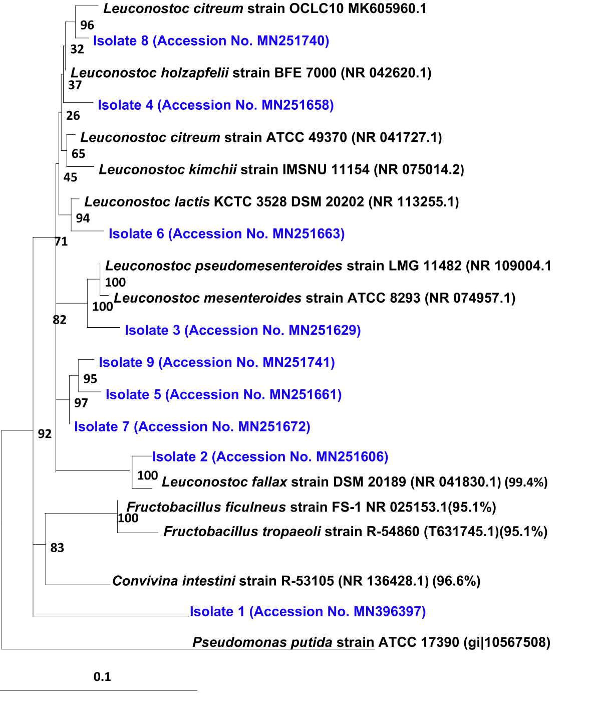 Fig. 1. Neighbor-joining tree showing the estimated phylogenetic relationship of the Exopolysaccharide producing bacterial isolates (shown in blue) and other closely related strains of the genus Leuconostoc. Bootstrap values out of 100 are given at the nodes. Pseudomonas putida strain ATCC 17390 (gi|10567508) was used as out group,
 Table (4):
Differential carbohydrate fermentation of the selected EPS isolates and related species
| Carbon sources |
Selected EPS producing bacteria |
References strains* | |||||||||||||
|---|---|---|---|---|---|---|---|---|---|---|---|---|---|---|---|
| S1 | S2 | S3 | S4 | S5 | S6 | S7 | S8 | S9 | A | B | C | D | E | F | |
| Ribose | – | – | – | – | – | – | – | – | – | – | – | – | – | – | + |
| D-Xylose | + | – | + | – | – | – | – | – | – | – | – | – | – | – | + |
| Galactose | – | – | – | – | – | – | – | – | – | – | – | + | – | +/- | + |
| Glucose | + | + | + | + | + | + | + | + | + | + | + | + | + | + | + |
| Fructose | + | + | + | + | + | + | + | + | + | + | + | + | + | – | + |
| Mannose | + | + | + | + | + | + | + | – | + | + | + | – | – | – | + |
| Mannitol | + | + | – | + | + | + | + | – | + | – | – | – | + | – | – |
| Sorbitol | + | – | – | – | – | – | – | – | – | – | – | – | – | – | – |
| α –Methyl-D-Glucopyranoside | + | – | – | – | – | – | + | – | – | + | – | – | + | – | – |
| N-acetyl-glucosamine | + | + | + | + | + | + | + | + | + | + | + | + | + | – | – |
| Amygdalin | + | – | – | – | – | – | – | – | – | + | – | – | – | – | – |
| Salicin | + | + | + | + | + | + | + | + | + | + | – | – | – | – | – |
| Maltose | + | – | + | – | – | – | – | + | – | + | – | – | – | + | + |
| Lactose | – | – | – | – | – | – | – | – | – | – | – | – | – | + | – |
| Sucrose | + | + | + | + | + | + | + | + | + | + | + | + | + | +/- | + |
| Trehalose | + | – | + | – | – | – | + | – | – | + | + | + | – | – | + |
| Raffinose | + | – | + | – | – | + | – | – | – | – | – | – | – | – | + |
Symbols and abbreviations: +, growth; -, no growth;A,leuconostoc citreum;B, leuconostoc mesenteroides subsp. dextr anicum;C, leuconostoc holzapfel;D, leuconostoc fallax;E, leuconostoc lactis;F, leuconostoc pseudomesenteroides.
Biochemical and physiological characteristics were determined using an API 50 CHL kit (BioMerieux, Lyon, France) and Vitek. Data of the reference strains were obtained from Antunes et al. (46).
Table (5):
Antibiotic susceptibilityofthe selected promising EPS producing isolates using antibiotic discs
| Antibiotic group | Antibiotics / Disc potency | S1 | S2 | S3 | S4 | S5 | S6 | S7 | S8 | S9 |
|---|---|---|---|---|---|---|---|---|---|---|
|
Aminoglycosides |
Amikacin 30 µg (AK) | R | S | R | R | R | R | R | R | R |
| Gentamicin 10 µg (GM) | R | R | R | R | R | R | R | R | R | |
| Tetracyclines | Tetracycline 30 µg (T) | S | S | S | S | S | S | S | S | S |
| Monobactams | Aztreonam 30 µg (ATM) | R | R | R | R | R | R | R | R | R |
| Fluoroquinolones | Ciprofloxacin 5 µg (CIP) | R | R | R | R | R | R | R | R | R |
| Lincosamide | Clindamycin 2 µg (CD) | R | R | R | S | S | R | R | S | S |
| Cephalosporins | Cephalothin 30 µg (Kf) | R | R | R | R | R | R | R | R | R |
| Ceftazidime30 µg (CAZ) | R | R | R | R | R | R | R | S | R | |
| Carbapenem | Imipenem 10 µg (IMI) | S | S | S | S | S | S | S | S | S |
|
Penicillins
|
Oxacillin 5µg (OX) | R | R | R | R | R | R | R | R | R |
| Piperacillin 100 µg (PRL) | S | S | S | S | S | S | S | S | S | |
| Penicillin G 10 units (PG) | R | R | R | R | R | R | R | R | R | |
| Macrolides | Erythromycin (15 µg) E | R | S | S | S | S | S | R | S | S |
| Sulfonamides | Co-trimoxazole 25 µg (TS) | R | R | R | R | R | R | R | R | R |
| Fenicols | Chloramphenicol 30 µg(C)
|
S | S | S | S | S | S | S | S | S |
Notes: R, Resistant (0-6 mm*); S, Sensitive (≥7 mm*)
EPS production from the selected EPS isolates
Capability of the selected isolates for EPS production using different sugars (1-8% w/v) such as sucrose, lactose, glucose, and fructose were investigated individually. Except sucrose, none of those sugars supported EPS production. EPS production from sucrose by the selected isolates was illustrated in Fig. 2. It was observed that the maximum yield of EPS was recorded for isolates S6, S1 and S7 which were 61.90, 61.80 and 60 (g l-1), respectively, followed by isolates S4, S9, S5 and S8 which were 58.40, 53.06, 51.61 and 33.53(gl-1), respectively. Though, the lowest yield was noticed for the isolates S3 and S2 which were 22.08 and 18.80(g l-1), respectively. However, as sucrose concentration supplemented to the fermentation media raised up to 8%, the yield of EPS from all selected isolates was increased.
 Fig. 2. Yield of EPS produced fromthe most promising isolates.To determine the EPS yield produced fromthe selected strains, fresh pre-cultures were inoculated in 250 mlErlenmeyer flasks containing 50 ml of the sterile fermentation medium(pH 7.0) supplemented individually with different concentrations of sucrose or lactose, glucose and fructose. All flasks were incubated at 30C for 3 days on a rotary shaker. The cultures were centrifuged and the EPS was extracted, purified, lyophilized, and then the weight of the dried EPS was gravimetrically measured according to Kumar et al.(27), Huang (28).
 Microbial exopolysaccharides from LAB are receiving a renewed interest and attention because of their pronounced applications in food, cosmetic, agriculture and pharmaceutical industries1, 2, 4. However, searching for the most efficient EPS-producing strains and upgrading of EPS production to minimize industrial costs remains a serious challenge. In the current study, a total of 55 bacterial strains were isolated from fermented fruits, dairy products, and vegetables and screened for their EPS production. In the first screening experiment, it was noticed that, viscosity and amount of the polysaccharide of most isolates were increased when the plates were kept in refrigerator at 8°C, while some isolates such S15, S16, S17, and S31 were consumed EPS when kept in the same condition, indicating that these isolates possess exopolysaccharide depolymerase enzymes27 that is able to hydrolyze wide variety of O-glycosidic linkages found in the polysaccharides, so the slim is destroyed and the viscosity decreases progressively upon prolonged incubation9. Additionally, it was noticed that some of the isolated strains were unable to produce exopolysaccharides in liquid fermentation medium although being able to form very slimy colonies when grown in the same agar medium. With respect to the susceptibility of antibiotics, selected EPS producing strains were found to show sensitivity towards clinically important antibiotics such as b-lactams (Piperacillin and Imipenem) but were resistance for Penicillin G also for Ciprofloxacin and this in accordance with the observation of Agarwal et al.30 who mentioned that Leu. mesenteroides VC236, exhibit resistance to 8 antibiotics (penicillin G, oxacillin, ciprofloxacin, levofloxacin, vancomycin, streptomycin, rifampicin, and nitrofurantoin). All isolates were susceptible to tetracycline, and these findings were found to be similar with those as reported by Morandi et al.31. Isolates S1, S2, S3, S4, S5, S6, S7, S8 and S9 were selected and characterized based on the produced amount of EPS. Phenotypic characterizations analyzed by Vitek, indicated that isolates S2, S4, S5, S6, S7, S8, and S9 were identified as Leuconostoc with different species, while isolate S1 was identified as Enterococcus columbae with probability 87%. However, species-level characterization of some lactic acid bacteria through solely biochemical methods is not as reliable as molecular level characterization; therefore the identification of unknown microorganism based on the molecular approaches is considered as the best choice because of the considerable variations in biochemical attributes between strains currently considered to belong to the same species. In the current study, aphylogenetic tree constructed based on 16S rRNA gene sequences Fig. 1 revealed that all selected EPS producing isolated strains represented a member of the genus Leuconostoc in the family of Leuconostoc aceae. Isolates S2, S3, S4, S5, S6, S7, S8, and S9 are very similar to each other, and they showed similarity ranged 97.9 – 99.4 of the genus Leuconostoc with different species. In this perspective, Collwell et al. 32 have found that “a bacterial species is generally considered to be a collection of strains that show a high degree of overall similarity and differ considerably from related strain groups with respect to many independent characteristics”. However, the 16S rRNA gene sequence was not generally as adequate to make a distinction between species. Thus, when the researchers are differentiating between closely related bacterial species of the same genus, fatty acid analysis, DNA-DNA hybridization and G+C ratio, involving housekeeping genes sequences should be considered as the methods of choice, in agreement with the proposed molecular identification of species33, 34, 35. Worth mentioning that, the distinguishing characteristics of the strain S1 from related reference strains in a separate node with similarity of 91% to Leuconostoc strains proposed that this isolate could be classified as gen. nov. sp. nov. This result was supported by the phenotypic features of their ability to utilize of sorbitol, mannitol, amygdalin, salicin and raffinose and a –methyl-D-Glucopyranosid. Because of this reason, subsequent characterization of this strain could not only direct the identification and characterization of a novel phylogenetic genus within the lactic acid bacteria groups but also make a lead to the production of highly promising and economically significant EPS yield. However, further work such as DNA-DNA hybridization, fatty acid analysis, and G+C ratio are proposed to ascertain if this promising strain can be identified as gen. nov. sp. nov. However, Lactic acid bacterial strains particularly those belonging to the genus Leuconostoc display the most efficient prospective to convert varied range of fermentable sugars through the biosynthesis of functional exopolysaccharide. Thus, Patel et al.3 mentioned that production of EPS from LAB strains can be economically significant if the parameters and other important factors which influences the EPS production could be optimized such as cheaper organic substrates and money-spinning fermentation conditions. In this study, all selected isolated strains were tested for production of EPS from different sugars such sucrose, lactose, glucose, and fructose. Except sucrose, none of those sugars support EPS production by these isolates. Similar finding was carried out by Antunes et al.36, Holzapfel et al.37 who mentioned that most of the leuconostocs do not ferment these types of sugars. Cerning9 reported that production of dextrans, mutans, and levans (type of exocellular homo- polysaccharides) requires an exacting substrate, typically sucrose and this point to the acting of enzymes with high specificity on oligosaccharide carbon sources. Vijayendra & Sharath Babu38 reported that the amount of EPS produced was insufficient by usage of sucrose contrary to this glucose could have stimulatory effect on the growth. Significant amount of EPS production was observed only with sucrose, this observation could help in exploiting the sucrose-rich industrial byproducts for EPS production. Futhermore, Harutoshi39 reported that homo EPS are synthesized by LAB using extracellular enzymes (fucosyltransferase (FTF) enzymes “fructansucrases” or glycosyltransferase (GTF) “glucansucrases”enzyme) outside the cell and sucrose cause induction for the GTF enzymes of Leuconostoc species and the production of GTF by Leu. mesenteroidesis low when using carbon sources other than sucrose. Of special interest, isolates S1, S6, and S7 in this work produced significant amount of EPS which were 61.80, 61.90 and 60 (g l-1), respectively. These results were higher than that produced by most other workers, who reported that the maximum EPS produced from different Leuconostoc species using sucrose medium were ranged 30 gl-1 38, 25.4 g l-1 40, 41, 2.39 gl-1 42, 0.34 g l-1 43, 0.004 gl-1, 1.6-9.2 gl-1 44, and 15 gl-1 45.
It could be concluded that EPS production from the strains reported in the current study, to make them the candidates of interest in enhanced production of EPS with maximum yields, and their practical exploitations in food, pharmaceutical, and dairy industries. The chemical composition of the growth medium (carbon and nitrogen substrate) as well as optimum growth conditions, i.e., pH, temperature, and incubation time) are the crucial parameters influencing the total yield of EPS production by Leuconostoc species. Thus, further investigations in progress to realize the full structure and chemical composition as well as the factors affecting EPS production and other possible biotechnological potential applications.
Acknowledgements
None.
Conflict Of Interest
The authors declare that there is no conflict of interest.
Authors’ Contribution
All authors have made substantial, direct and intellectual contribution to the work and approved it for publication.
Funding
None.
Data Availability
All datasets generated or analyzed during this study are included in the manuscript.
Ethics Statement
This article does not contain any studies with human participants or animals performed by any of the authors.
- Yadav V, Prappulla SG, Jha A, PooniaA.A novel exopolysaccharide from probiotic Lactobacillus fermentum CFR 2195: Production, purification, and characterization. Biotechnol. Bioinf. Bioeng., 2011; 1: 415-21.
- Benhadria MK, Touil MAT, Meddah B. Optimization of production of Microbial Exopolysaccharides (EPS) with essential oils from two medicinal plants. J. Appl. Biosci., 2017; 111: 10925-10933.
Crossref - Patel S, Majumder A, Goyal A. Potentials of exopolysaccharides from lactic acid bacteria. Ind. J. Microbiol., 2012; 52: 3-12.
Crossref - Patel A, Prajapati JB. Food and health applications of exopolysaccharides produced by lactic acid bacteria. Adv. Dairy Res., 2013; 1: 107.
Crossref - Elbanna K, Metry W, Elgarhy H. Exopolysaccharide from Lactobacillus pentosus Strain H2 and Its Impact on Rheological Properties and the Sensory Evaluation of Low Fat Yoghurt and UF-Soft Cheese. Int. J. Nutri. Food Sci., 2015; 4: 555-564.
Crossref - Kitazawa H, Toba T, Itoh T, Kumano N, Adachi S, Yamaguchi T. Antitumoral activity of slime-forming, encapsulated Lactococcus lactis ssp. cremoris isolated from Scandinavian ropy sour milk, ‘‘viili’’. Anim. Sci. Technol., 1991; 62: 277–283.
Crossref - Surayot U, Wang J, Seesuriyachan P, Kuntiya A, Tabarsa M. Exopolysaccharides from lactic acid bacteria: structural analysis, molecular weight effect on immunomodulation. Int. J. Biol. Macromolec., 2014; 68:233–240.
Crossref - Liu C, Lu J, Lu L, Liu Y, Wang F, Xiao M. Isolation, structural characterization and immunological activity of an exopolysaccharide produced by Bacillus licheniformis 8-37-0-1. Bioresour. Technol., 2010; 101: 5528–5533.
Crossref - Cerning J. Exocellular polysaccharides produced by lactic acid bacteria. FEMS Microbiol. Rev., 1990; 7: 113-130.
Crossref - Low D, Ahlgren JA, Horne D, McMahon DJ, Oberg CJ, Broadbent JR. Role of S. thermophilus MR.1C capsular exopolysaccharide in cheese moisture retention. Appl. Environ. Microbiol., 1998; 64: 2147–2151.
- Vedamuthu ER, Neville JM. Involvement of a plasmid in production of ropiness (mucoidness) in milk cultures by Streptococcus cremoris MS. Appl. Environ. Microbiol., 1986; 51: 677–682.
- Ruas-Madiedo P, De Los Reyes-Gavilבn CG. Methods for the screening, isolation, and characterization of exopolysaccharides produced by lactic acid bacteria. J. Dairy Sci., 2005; 88: 843-856.
Crossref - de Vuyst L, de Vin F, Vaningelgem F, Degeest B. Recent developments in thebiosynthesis and applications of heteropolysaccharides from lactic acid bacteria. Int. Dairy J., 2001; 11: 687-707.
Crossref - Kralj S, van Geel-Schutten GH, Dondorff MMG, Kirsanovs S, van der Maarel MJEC, Dijkhuizen L. Glucan synthesis in the genus Lactobacillus: isolation and characterization of glucansucrase genes, enzymes and glucan products from six different strains. Microbiol., 2004; 150: 3681-3690.
Crossref - Jin Q, Li L, Kim YJ, Han NS. Construction of a dextran free L euconostoc citreum mutant by targeted disruption of the dextransucrase gene. J. Appl. Microbiol., 2014; 117: 1104-1112.
Crossref - Frantzen CA, Kot W, Pedersen TB, Ardצ YM, Broadbent JR, Neve H, Vogensen FK. Genomic characterization of dairy associated Leuconostoc species and diversity of Leuconostocs in undefined mixed mesophilic starter cultures. Front. Microbiol., 2017; 8: 132.
Crossref - Ho JCK, Sze YL. Isolation, identification and characterization of enzyme-producing lactic acid bacteria from traditional fermented foods. Biosci. Hori.: Int. J. Stud. Res, 2018; 11: hzy004.
Crossref - Jakob F, Mei ner D, Vogel RF. Comparison of novel GH 68 levansucrases of levan-overproducing Gluconobacter species. Acetic Acid Bact., 2012; 1: e2.
Crossref - Gerhardt P, Murray RGE, Wood WA, Krieg NR. Methods for General and Molecular Bacteriology. American Society for Microbiology, Washington D.C., 1994.
- Yang C, Wang D, Zhou Q, Xu J. Bacteremia due to vancomycin resistant Leuconostoc lactis in a patient with pneumonia and abdominal infection. Am. J. Med. Sci., 2015; 349: 282-283.
Crossref - Bauer AW, Kirby WMM, Sherris JC, Turck M. Antibiotic susceptibility testing by a standardized single disk method. Am. J. Clin. Pathol., 1966; 45: 493-496.
Crossref - EFSA. Scientific opinion on the maintenance of the list of QPS biological agents intentionally added to food and feed. EFSA J., 2012; 10: 3020.
Crossref - Elbanna K, Hassan G, Khider M, Mandour R. Safe Biodegradation of Textile Azo Dyes by Newly Isolated Lactic Acid Bacteria and Detection of Plasmids Associated With Degradation. J. Bioremed. Biodeg., 2010; 1: 1-6.
- Thompson J, Gibson TJ, Plewinak F, Jeanmougin F, Higgins DG. The Clustal X windows interface: flexible strategies for multiple sequence alignment aided by quality analysis tools. Nuc. Acids Res., 1997; 25: 4867-488.
Crossref - Page RDM. Tree View: An application to display phylogenetic trees on personal computers. Comput. Appl. Biosci., 1996; 12: 357-358.
Crossref - Saitou N, Nei M. The neighbour-joining method: A new method for reconstructing phylogenetic trees. Mol. Biol. Evol., 1987; 4: 406-425.
- Kumar MA, Anandapandian KTK, Parthiban K. Production and characterization of exopoly-saccharides (EPS) from biofilm forming marine bacterium. Braz. Arch. Biol. Technol., 2011; 54: 259-265.
Crossref - Huang G, Yang Q, Wang ZB. Extraction and deproteinization of mannanoligosaccharides. Z. Naturforsch. C., 2010; 65: 387-390.
Crossref - Stone BA, Svensson B, Collins ME, Rastall RA. Polysaccharide degradation. Glycoscience: Chemistry and Chemical Biology, 2008, 2325-2375.
Crossref - Agarwal M, Garg FC, Negi, YK. Antibiotic resistance and plasmid profile of Leuconostoc spp. isolated from carrot. Afri. J. Bacteriol. Res., 2014; 6: 7-12.
Crossref - Morandi S, Cremonesi P, Silvetti T, Brasca M. Technological characterisation, antibiotic susceptibility and antimicrobial activity of wild-type Leuconostoc strains isolated from north Italian traditional cheeses. J. Dairy Res., 2013; 80: 457-466.
Crossref - Colwell RR, Clayton RA, Ortiz-Conde BA, Jacobs D, Russek-Cohen E. The microbial species concept and biodiversity. In: Microbial Diversity and Ecosystem Function. Allsopp, D., Colwell, R. and Hawksworth, L. (eds.), 1995; pp. 3-15. Wallingford, UK.
- Murray E, Brenner J, Colwell R, De Vos P, Goodfellow M, Grimont D, Pfennig N, Stackebrandt E, Zavarzin A. International Committee on Systematic Bacteriology. Report of the ad hoc committee on approaches to taxonomy within the Proteobacteria. Int. J. Syst. Bacteriol., 1990; 40: 213-215.
Crossref - Berkum P, Beyene D, Eardly BD. Phylogenetic relationships among Rhizobium species nodulating the common bean (Phaseoli vulgaris L.), Int. J. Syst. Bacteriol., 1996; 46: 240-244.
Crossref - ElbannaK, Elbadry M, Gamal-eldin H., Genotypic and phenotypic characterization of rhizobia that nodulate Snap bean (Phaseolus vulgaris L.) in Egyptian soils. Syst. Appl. Microbiol., 2009; 32: 522–530.
Crossref - Antunes A, Rainey FA, Nobre MF, Schumann P, Ferreira AM, Ramos A, Santos H.. Leuconostoc ficulneum sp. nov., a novel lactic acid bacterium isolated from a ripe fig, and reclassification of Lactobacillus fructosus as Leuconostoc fructosum comb. nov. Int. J. Syst. Evol. Microbiol., 2002; 52: 647-655.
Crossref - Holzapfel WH, Bjצrkroth JA, Dicks LM. Leuconostoc. Bergey’s Manual of Systematics of Archaea and Bacteria, 2015; 1-23.
Crossref - Vijayendra SVN, Sharath Babu RS. Optimization of a new heteropolysaccharide production by a native isolate of Leuconostoc sp. CFR 2181. Lett. Appl. Microbiol., 2008; 46: 643-648.
Crossref - Harutoshi T. Exopolysaccharides of lactic acid cacteria for foodand colon health applications. biochemistry, genetics and molecular biology. In: Lactic Acid Bacteria: R & D for Food, Health and Livestock Purposes. Kongo M (ed), 2013; pp 222-38.
Crossref - Vijayendra SVN, Palanivel G, Mahadevamma S, Tharanathan RN. Physico-chemical characterization of an exopolysaccharide produced by a non-ropy strain of Leuconostoc sp. CFR 2181 isolated from dahi, an Indian traditional lactic fermented milk product. Carbohyd. Polym., 2008, 72: 300-307.
Crossref - Khue NTH, Ngoc NH. Exopolysaccharide in Lactobacillus rhamnosus Pn04 after co-culture with Leuconostoc mesenteroides Vtcc-B-643. J. App. Pharmaceut. Sci., 2013; 3: 14.
- Heperkan D, Daskaya-Dikmen C, Bayram B. Evaluation of lactic acid bacterial strains of boza for their exopolysaccharide and enzyme production as a potential adjunct culture. Process Biochem., 2014; 49: 1587–1594.
Crossref - Joshi SR, Koijam K. Exopolysaccharide production by a lactic acid bacteria, Leuconostoc lactis isolated from ethnically fermented beverage. Nat. Acad. Sci. Lett., 2014; 37: 59-64.
Crossref - Khudair AY, Salman JAS, Ajah HA. Production of Levan from Locally Leuconostoc mesensteroides isolates. J. Pharmaceut. Sci. Res., 2018; 10: 3372-3378.
- Yan M, Wang B-hX, der Meister TJ, Tabyac H-T, Hwang F, Liu Z. Extrusion of dissolved oxygen by exopolysaccharide from Leuconostoc mesenteroides and its implications in relief of the oxygen stress. Front. Microbiol., 2018; 9: 2467.
Crossref - Antunes A, Rainey F, Nobre F, Schumann P, FerreiraAM, Ramos A, Helena Santos H, da Costa M.. Leuconostoc ficulneum sp. nov., a novel lactic acid bacterium isolated from a ripe fig, andreclassification of Lactobacillus fructosus as Leuconostoc fructosum comb. nov.Int. J. Syst. Evol. Microbiol., 2002; 52: 647–655.
Crossref
© The Author(s) 2019. Open Access. This article is distributed under the terms of the Creative Commons Attribution 4.0 International License which permits unrestricted use, sharing, distribution, and reproduction in any medium, provided you give appropriate credit to the original author(s) and the source, provide a link to the Creative Commons license, and indicate if changes were made.