


ISSN: 0973-7510
E-ISSN: 2581-690X
 and Umar Farooq
The development of methicillin-resistant Staphylococcus aureus (MRSA) and its ability to confer cross-resistance to MLSB group of antibiotics, i.e. macrolide-lincosamide-streptogramin B, have made difficult to treat MRSA infections. Routine use of clindamycin susceptibility testing is unable to identify inducible MLSB (iMLSB) phenotype which frequently leads to treatment failure and requires the use of a simple D-test for detection of this type of resistance. This cross-sectional observational study was performed in the Department of Microbiology, TMMC & RC Moradabad, India. Two hundred S. aureus strains have been identified in various clinical samples, including blood, pus, body fluids, and respiratory samples. S. aureus strains were subjected to antibiotic susceptibility testing using the Kirby–Bauer disc diffusion (D-test) technique. Cefoxitin-resistant S. aureus (MRSA) isolates were subjected to mecA gene detection using polymerase chain reaction. A total of 200 S. aureus isolates were obtained from various specimens, of which 134 (67%) were MRSA and 66 (33%) were MSSA. All MRSA strains possessed mecA. The constitutive MLSB (cMLSB), iMLSB, and MS phenotype prevalence was found to be 37.3%, 22.4%, and 40.3% in MRSA and 27.7%, 12.1%, and 65.2% in MSSA, respectively. In this study, S. aureus isolates exhibited a significant frequency of iMLSB in this region. Therefore, the D-test can be regularly used for clinical isolates to prevent clindamycin treatment failure.
Constitutive Macrolide-Lincosamide-Streptogramin B, Inducible Macrolide-Lincosamide-Streptogramin B, Methicillin-resistant Staphylococcus aureus, Staphylococcus aureus
Staphylococcus aureus is the most common pathogenic bacterium of the genus Staphylococcus. It is considered a normal flora and opportunistic bacterium that colonizes the vast majority of humans.1,2 S. aureus causes both hospital and community-acquired infections globally, and staphylococcal resistance to methicillin is an increasing problem.3 The rise in antibiotic resistance in staphylococci highlights the need for new treatment options for these diseases. Despite their structural differences, macrolide-lincosamide-streptogramin B (MLSB) group antibiotics are most commonly used to treat infections by inhibiting protein synthesis.4 Clindamycin (lincosamide) is a convenient choice for doctors because of its parenteral and oral availability, tissue circulation, and bacteriostatic properties against S. aureus.5
Indiscriminate use and misuse of MLSB group antibiotics for the treatment of serious staphylococcal diseases have led to an increased frequency of resistant isolates.6 In S. aureus, the MLSB resistance phenotype may be constitutive MLSB (cMLSB), inducible MLSB (iMLSB) or MS phenotype. Staphylococci with ribosomal methylase (erm) genes may be resistant to MLSB antibiotics such as erythromycin and clindamycin in vitro. This type of resistance refers to cMLSB trait. An inciting agent is required for some staphylococci with erm genes to produce a methylase enzyme that confers clindamycin resistance, which is known as the iMLSB phenotype. In vitro, these bacteria show deceptive susceptibility to clindamycin and resistance to erythromycin.7 In Staphylococcus species, macrolide streptogramin resistance (msrA) causes active efflux. Before macrolides attach to their target sites on the ribosome, this energy-dependent pump effectively eliminates them from bacterial cells. Consequently, it causes resistance to erythromycin and azithromycin, but not to clindamycin, which is known as MS phenotype. There is no chance of treatment failure, and clindamycin remains effective against these strains.8,9
The goal of the research was to evaluate the prevalence of methicillin-resistant S. aureus (MRSA) and iMLSB isolated from different types of clinical specimens using an antibiotic sensitivity test (AST) with various antibiotics.
Study setting and design
This cross-sectional study was conducted after obtaining approval from the TMU Institutional Ethics Committee (Ethical Approval Number: TMU/IEC/2024-25/PG/003) from March 2025 to February 2026 in the Microbiology Department of TMMC & RC Moradabad, India. A total of 200 S. aureus strains were identified from different specimens, including respiratory samples, pus, blood, and other body fluids.
Inclusion and exclusion criteria
All the S. aureus isolated from IPD and OPD patients of the hospital were included in the study and coagulase-negative Staphylococci (CoNS) were excluded from the study.10
Identification and isolation of bacteria
The samples were cultured on appropriate culture media and aerobically incubated for 24 hrs at 37 °C. β-hemolytic colonies on blood agar and yellow color colonies on mannitol salt agar were observed. Isolates were identified using standard microbiological tests, such as Gram staining, catalase, oxidation-fermentation (OF), coagulase (free and bound), DNase, and gelatin hydrolase tests.11
AST
AST was performed using the Kirby–Bauer disc diffusion technique on Mueller–Hinton agar (MHA) plates. The following antibiotics were tested against S. aureus: azithromycin (15 µg), cefoxitin (30 µg), ciprofloxacin (5 µg), co-trimoxazole (25 µg), clarithromycin (15 µg), tetracycline (30 µg), clindamycin (2 µg), doxycycline (30 µg), linezolid (30 µg), erythromycin (15 µg), gentamycin (10 µg), levofloxacin (5 µg), minocycline (30 µg), ofloxacin (5 µg), according to CLSI guidelines 2025.12 S. aureus isolates with a zone of inhibition (ZOI) around the cefoxitin disc ≥22 mm were sensitive to methicillin (MSSA), whereas ZOI around the cefoxitin disc ≤21 mm were resistant to methicillin (MRSA).13
mecA gene detection for confirmation of MRSA by polymerase chain reaction (PCR)
DNA was extracted from the MRSA strains using the QIAGEN DNeasy blood and tissue extraction kit. Isolated DNA was amplified using the primers described by Azimian et al., who previously used the primers for amplification of mecA.14
Forward primer sequence: AGAAGATGGTATGTGGAAGTTAG
Reverse primer sequence: ATGTATGTGCGATTGTATTGC
Clindamycin resistance detection through D-test
Clindamycin resistance was identified using D-test. On the MHA plate that had been pre-inoculated with 0.5 McFarland standard bacterial inoculum, clindamycin and erythromycin discs were placed 15 mm apart. The plate was incubated at 37 °C for 18-24 hrs after that a flattening zone (D-shape) surrounding clindamycin disc shows inducible clindamycin resistance.12 Three distinct phenotypes were identified using D-test, which are subsequently analyzed. This analysis was performed only for S. aureus strains that were resistant to erythromycin; all S. aureus strains sensitive to erythromycin were excluded from the study.15
MS Phenotype
This phenotype describes S. aureus isolates that had a circular ZOI surrounding clindamycin, and clindamycin was sensitive (ZOI ≥21 mm) but erythromycin was resistant (ZOI ≤13 mm) (Figure 1A).15
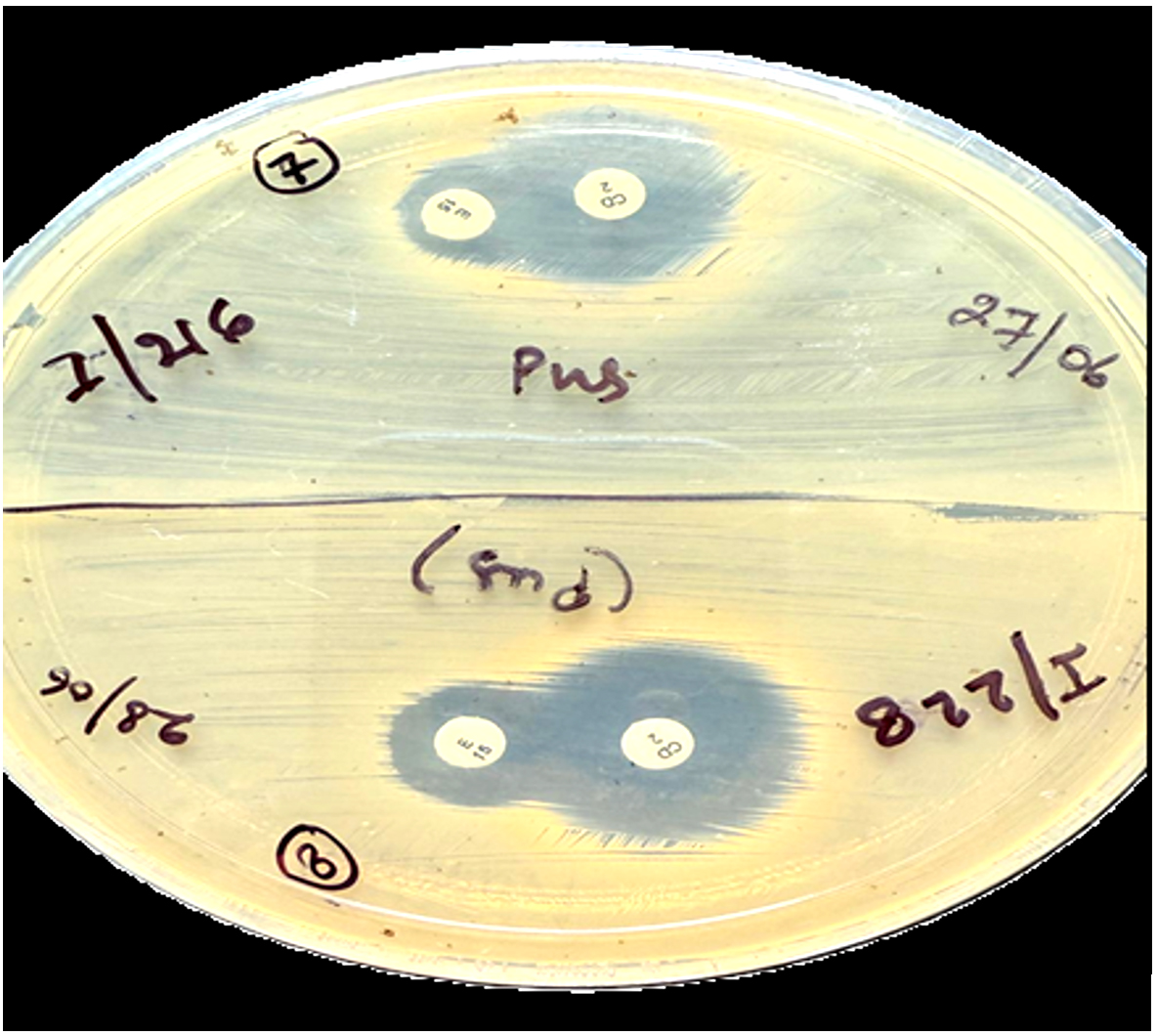
Figure 1A. Negative D-test (MS phenotype)
iMLSB
S. aureus strains with clindamycin susceptible (ZOI ≥21 mm) and erythromycin resistant (ZOI ≤13 mm); this isolate also displayed a D-shaped ZOI around the clindamycin, which flattened towards the erythromycin, indicating the presence of this phenotype (Figure 1B).15
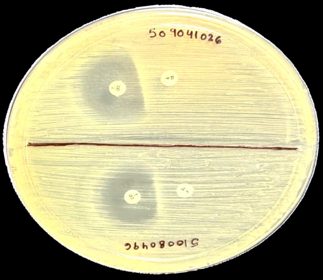
Figure 1B. D-test Positive (iMLSB phenotype)
cMLSB
This type of clindamycin resistance refers to S. aureus strains that was resistant to both erythromycin and clindamycin with ZOI ≤13 mm and ≥14 mm, respectively (Figure 1C).15
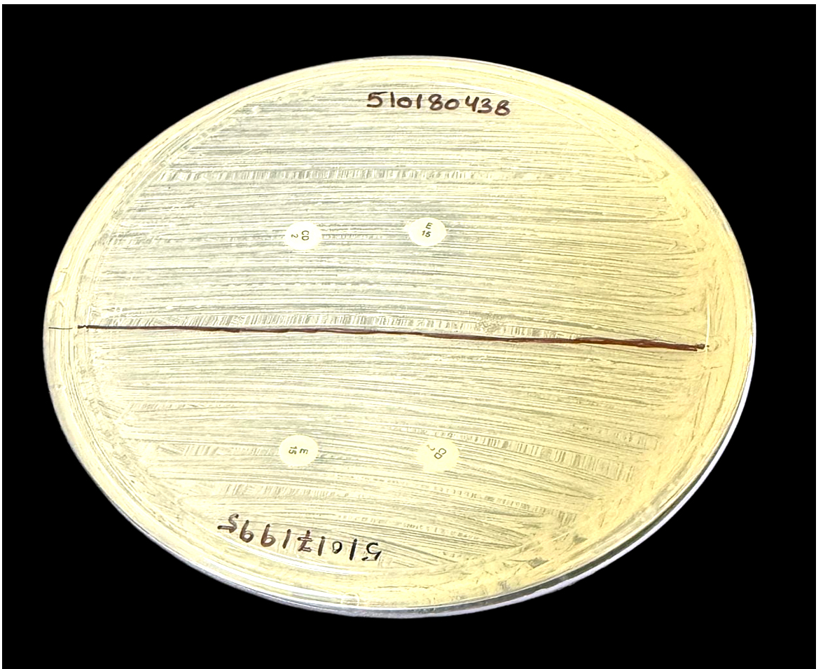
Figure 1C. Constitutive resistance (cMLSB phenotype)
The erythromycin and clindamycin discs were subjected to quality control using S. aureus ATCC 25923 following the standard disc diffusion protocol.16
Statistical analysis
The prevalence of iMLSB, cMLSB and MS phenotypes in S. aureus was represented as a percentage. Chi-squared and Fisher’s exact tests were used to statistically assess the results. Statistical significance was set at P-value <0.05. SPSS version 25.0 (IBM Corp., Chicago, Illinois, USA) was used for this analysis.
A total of 320 staphylococci were isolated from IPD and OPD patients at the TMU Hospital between March 2025 and February 2026. A total of 200 S. aureus and 120 CoNS were identified from different types of clinical specimens. Of the 200 S. aureus strains, 134 (67%) were MRSA and 66 (33%) were MSSA. DNA was extracted from the MRSA strains, and mecA gene was amplified using PCR. In this study, all MRSA strains possessed mecA.
A total of 200 S. aureus isolates were distributed based on the source and clinical samples. Of the 200 S. aureus strains, 162 (81%) and 38 (19%) were isolated from IPD and OPD patients, respectively (Table 1). The maximum S. aureus was detected in pus samples 114 (57%), followed by blood 70 (35%), tissue 5 (2.5%), and HVS 3 (1.5%), and least from body fluid, ear swab, ET secretion, and sputum 1 (0.5%). A statistically significant correlation between the specimens and sources was found at P < 0.05. (χ2 = 266.815, df = 26, P-value < 0.001) (Table 1).
Table 1. Source and samples wise distribution of S. aureus
| Samples name | Sources | Total | χ2, df, P-value | |
|---|---|---|---|---|
| IPD | OPD | |||
| Ascitic fluid | 1 | 0 | 1 | 266.815, 26, <0.001 |
| Bronchoalveolar lavage | 1 | 0 | 1 | |
| Blood | 70 | 0 | 70 | |
| Cerebrospinal fluid | 1 | 0 | 1 | |
| Ear swab | 1 | 0 | 1 | |
| Endotracheal aspirate | 1 | 0 | 1 | |
| High vaginal swab | 3 | 0 | 3 | |
| Pleural fluid | 1 | 0 | 1 | |
| Pus | 76 | 38 | 114 | |
| Sputum | 1 | 0 | 1 | |
| Synovial fluid | 1 | 0 | 1 | |
| Tissue | 5 | 0 | 5 | |
| Total | 162 | 38 | 200 | |
χ2 = Chi-square, df = Degree of freedom, P-value = Probability-value
Clindamycin resistance was categorized into cMLSB, iMLSB, and MS phenotypes based on the sensitivity patterns of erythromycin and clindamycin. The maximum cMLSB (14/65) was isolated from 51-60 years age group, followed by 31-40 years age group (12/65). The maximum iMLSB (10/38) was isolated from >60 years age group, followed by the 21-30, 31-40, 41-50 years age groups (7/38 iMLSB isolated from each group). Among the 97 MS phenotypes, 20 were isolated from the 51-60 years age group, followed by 17 isolated from the 31-40 years age group. The association between age group and clindamycin resistance was statistically significant (P < 0.05). (χ2 = 242.837, df = 21, P-value < 0.001) (Table 2).
Table 2. S. aureus distribution on the basis of age group among clindamycin resistance
| Age group | Clindamycin resistance | Total | χ2, df, P-value | ||
|---|---|---|---|---|---|
| cMLSB | iMLSB | MS Phenotype | |||
| 0-10 | 8 | 1 | 10 | 19 | 242.837, 21, <0.001 |
| 11-20 | 3 | 3 | 14 | 20 | |
| 21-30 | 11 | 7 | 13 | 31 | |
| 31-40 | 12 | 7 | 17 | 36 | |
| 41-50 | 6 | 7 | 11 | 24 | |
| 51-60 | 14 | 3 | 20 | 37 | |
| >60 | 11 | 10 | 12 | 33 | |
| Total | 65 | 38 | 97 | 200 | |
Of the 200 S. aureus, 134 (67%) were MRSA isolates and 66 (33%) were MSSA isolates from various clinical samples. Among the 134 MRSA strains, 50 cMLSB, 30 iMLSB, and 54 MS phenotypes were isolated. Among the 66 MSSA strains, 15 cMLSB, 8 iMLSB and 43 MS phenotypes were isolated. A statistically significant correlation was observed among MRSA, MSSA, and clindamycin resistance (P < 0.05) (χ2 = 10.980, df = 2, P-value = 0.004) (Table 3).
Table 3. S. aureus distribution on the basis of MRSA, MSSA and clindamycin resistance
| MRSA/MSSA strains | Clindamycin resistance | Total | χ2, df, P-value | ||
|---|---|---|---|---|---|
| cMLSB | iMLSB | MS Phenotype | |||
| MRSA | 50 | 30 | 54 | 134 | 10.980, 2, 0.004 |
| MSSA | 15 | 8 | 43 | 66 | |
| TOTAL | 65 | 38 | 97 | 200 | |
S. aureus isolates were highly sensitive to linezolid (99.5%), minocycline (99%), followed by tetracycline (83.5%), co-trimoxazole (62%), and doxycycline (58%), and least sensitive to ciprofloxacin (8%) (Figure 2).
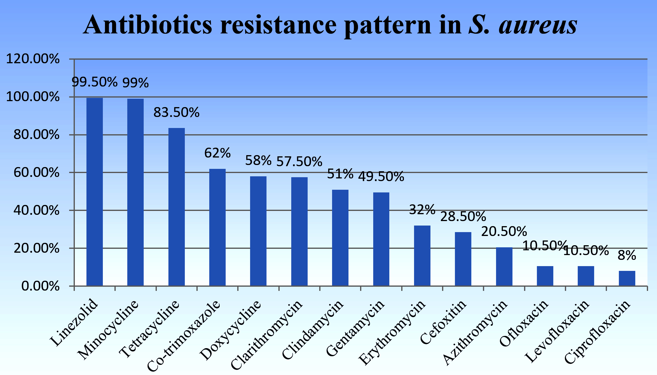
Figure 2. Antibiotic sensitivity test for S. aureus isolates
Microbiology laboratories must precisely identify and document S. aureus isolates exhibiting clindamycin susceptibility and erythromycin resistance. To determine the actual susceptibility status, iMLSB can be detected using the D-test, which is a simple disk-diffusion test.17
Antimicrobial resistance in micro-organisms that cause hospital-acquired illnesses is a significant global problem. The common skin colonizer S. aureus causes many community-onset and healthcare-related infections.18 S. aureus infections are treated with clindamycin antibiotics because of its benefits, including low cost, few side effects, and easy tissue penetration. Despite being an effective substitute for β-lactam antibiotics in patients with allergy.19
In this study, 200 S. aureus strains were identified from different types of clinical specimens collected from the IPD and OPD of the TMU Hospital during the study period. Of the 200 S. aureus isolates, 134 (67%) were MRSA and 66 (33%) were MSSA. All MRSA strains possessed mecA gene mentioned in Figure 3. Our findings were similar to the findings of other researchers who reported a high prevalence of MRSA, such as Jahanbakhshi et al.20 reported the prevalence of MRSA and MSSA to be 65.9% and 34.1%, respectively, in Iran, and in all the MRSA strains, the mecA gene was detected. Mohanty et al.11 also reported the prevalence of MRSA (62.18%) and MSSA (37.82%) in West Bengal, India. Other researchers found lower prevalence of MRSA as compared to our study, such as Mansouri and Sadeghi21 found the prevalence of MRSA and MSSA in Iran to be 56.8% and 43.2%, respectively. Alam et al.22 conducted a study in Uttar Pradesh, India, and reported the prevalence of MRSA and MSSA as 55.5% and 44.5%, respectively; and Chudasama et al.23 from Ahmedabad, India, reported MRSA and MSSA prevalence of 54.78% and 45.22%, respectively.
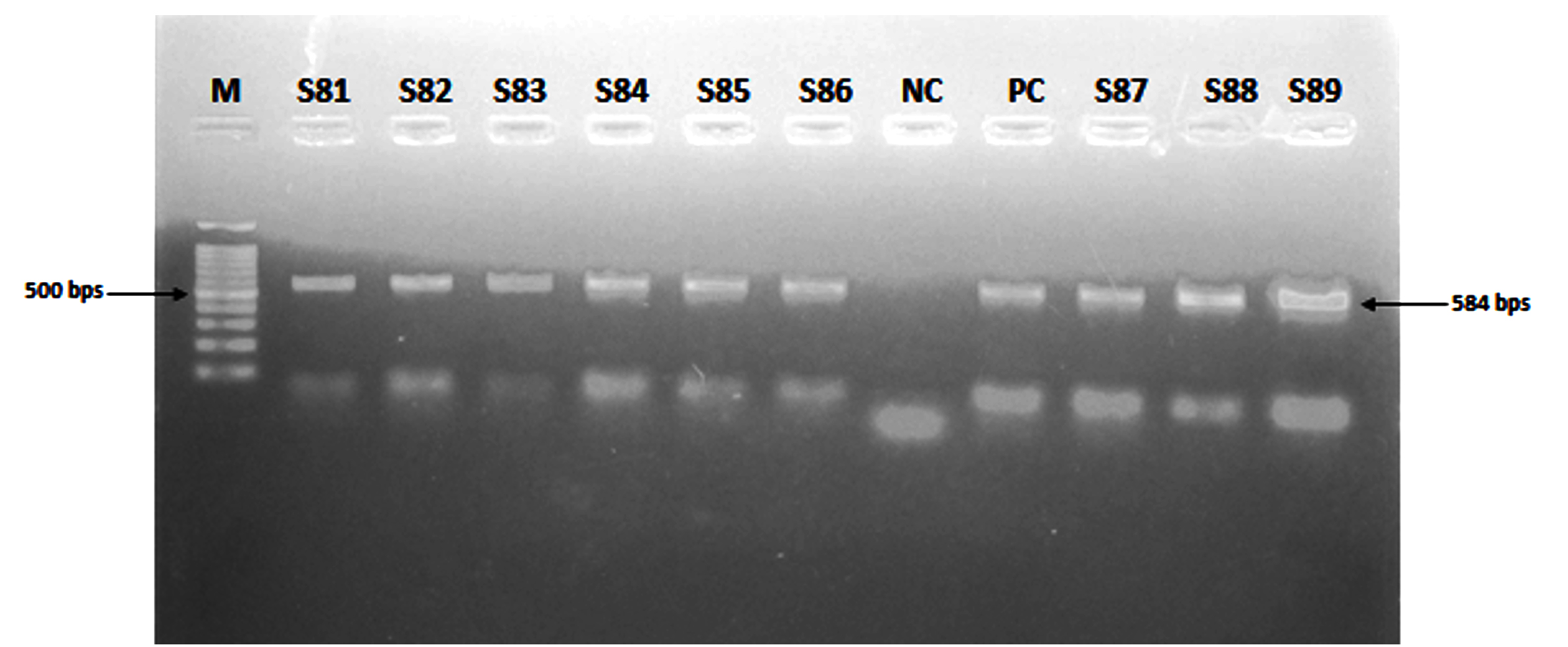
Figure 3. Representative gel image of the mecA gene PCR (584 bp). Lanes: M, molecular weight marker; ATCC-43300 (S. aureus) was used as PC = Positive control; ATCC 25923 (S. aureus) was used as NC = Negative control; the MRSA isolates S81-S89 possess mecA gene
S. aureus causes different clinical infections, and specimens were collected from the site of infection. Therefore, different types of specimens were collected and processed to identify S. aureus in the laboratory.24 In this study, highest S. aureus was detected in pus sample 114 (57%), followed by blood 70 (35%), and the least in body fluids. Our findings are comparable to those of Alam and Farooq.25 reported that the maximum number of S. aureus isolates was from pus 130 (41.4%), followed by blood 85 (27.07%) and urine 34 (10.83%).
Traditionally viewed as a hospital acquired pathogen resistant to multiple antibiotic classes beyond β-lactams, MRSA has increasingly infected people without obvious risk factors in recent years, leading to community-associated cases. Staphylococcal infections responsive to various β-lactam antibiotics are typically treated with first-line antibiotics from the MLSB group.20 iMLSB, cMLSB, and MS phenotypes may be detected by using D-test, which is routinely used in clinical laboratories. In the present study, the prevalence of iMLSB was 19%. The study conducted globally reported the emergence of iMLSB and other researchers reported a higher prevalence of iMLSB compared to our study, such as Majhi et al.,26 Timsina et al.,13 Peshattiwar et al.,27 and Moosavian et al.,17 reported a high prevalence of iMLSB (22%, 23.4%, 31.37%, and 32.3%, respectively). Some researchers conducted studies and reported a prevalence of iMLSB similar to our study, such as Gupta et al.,28 and Gupta et al.29 reported the prevalence of iMLSB (18.18% and 18%, respectively). Jahanbakhshi et al.,20 and Mansouri and Sadeghi21 reported a lower prevalence of iMLSB than in our study (15.9% and 11.95%, respectively).
The prevalence of the MS phenotype is high in studies conducted in different parts of the world. In the present study, the prevalence of the MS phenotype was 48.5%. Regmi et al.30 conducted a study in Nepal and reported the prevalence of the MS phenotype (56.8%), which was higher than the prevalence of the MS phenotype in this study. Gupta et al.,31 and Sreekanth and Dattaraya.32 reported that the prevalence of MS phenotypes to be 47.2% and 47%, respectively, which is similar to the results of our study. Mohanty et al,11 Chaudhary et al,33 and Bose et al.34 reported that the prevalence of MS phenotypes was lower than that reported in this study (42.85%, 31.62%, and 26%, respectively).
In this study, the prevalence of cMLSB was 32.5%. The findings of cMLSB were reported in a study conducted by Pradhan et al.35 in Nepal (32.2%), which were similar to our findings. Other authors have reported the prevalence of cMLSB to be lower than that in our study, such as Adhikari et al.,36 and Ghanbari et al.19 who reported the prevalence of cMLSB to be 29.25% and 26.9%, respectively. However, Nikbakht et al.37 Jahanbakhshi et al.20 and Bose et al.34 reported that the prevalence of cMLSB was higher than that in our study (34.42%, 38.9%, and 40%, respectively).
Limitations
The limitations of this study were the inability to detect erm genes and staphylococcal cassette chromosome mec (SCCmec) typing of S. aureus owing to a lack of resources and funding.
The treatment of S. aureus infections has become a major challenge for clinicians. When treating severe and resistant S. aureus infections, clindamycin should be considered while keeping in mind the pharmacokinetics, side effects, and mode of action of some antibiotics, such as vancomycin and linezolid. According to several studies conducted worldwide, the prevalence of iMLSB and MRSA varies from place to place. Therefore, we recommend that when clindamycin is used for the treatment of S. aureus infections, clinical microbiology laboratories should test the isolated S. aureus for iMLSB using a D-test before reporting clindamycin sensitivity. Clinicians will be able to select a suitable treatment with the help of this study, which provides the magnitude of clindamycin resistance among S. aureus isolates from this part of the country.
ACKNOWLEDGMENTS
The authors would like to express their heartfelt thanks to the academic and technical staff of Teerthanker Mahaveer Medical College and Research Centre, Moradabad, and to all healthcare professionals who participated in this study for their support in completing this study.
CONFLICT OF INTEREST
The authors declare that there is no conflict of interest.
AUTHORS’ CONTRIBUTION
Both authors listed have made a substantial, direct and intellectual contribution to the work and approved it for publication.
FUNDING
None.
DATA AVAILABILITY
All datasets generated or analyzed during this study are included in the manuscript.
ETHICS STATEMENT
This study was approved by the Institutional Ethics Committee (IEC), Teerthanker Mahaveer Medical College and Research Centre, Moradabad, under Ethical Approval Number TMU/IEC/2024-25/PG/003.
INFORMED CONSENT
Written informed consent was obtained from the participants before enrolling in the study.
- Bitrus AA, Peter OM, Abbas MA, Goni MD. Staphylococcus aureus: A review of antimicrobial resistance mechanisms. Vet Sci Res Rev. 2018;4(2):43-54.
Crossref - Tong SYC, Davis JS, Eichenberger E, Holland TL, Fowler Jr VG. Staphylococcus aureus infections: epidemiology, pathophysiology, clinical manifestations and management. Clin Microbiol Rev. 2015;28(3):603-661.
Crossref - Yilmaz G, Aydin K, Iskender S, Caylan R, Koksal I. Detection and prevalence of inducible clindamycin resistance in staphylococci. J Med Microbiol. 2007;56(3):342-345.
Crossref - Fiebelkorn KR, Crawford SA, McElmeel ML, Jorgensen JH. Practical disk diffusion method for detection of inducible clindamycin resistance in Staphylococcus aureus and coagulase -negative staphylococci. J Clin Microbiol. 2003;41(10):4740-4744.
Crossref - Gorbach SL, Barlett JG, Blacklow NR, editors. Infection diseases, Lippincott Williams & Wilkins. 2004.
- Deotale V Mendiratta DK, Raut U, Narang P. Inducible clindamycin resistance in Staphylococcus aureus isolated from clinical samples. Indian J Med Microbiol. 2010;28(2):124-126.
Crossref - Juyal D, Shamanth AS, Pal S, Sharma MK, Prakash R, Sharma N. The prevalence of inducible clindamycin resistance among staphylococci in tertiary care hospital a study from the Garhwal hills of Uttarakhand, India. J Clin Diagn Res. 2012;7(1): 61-65.
Crossref - O’ Sullivan MVN, Cai Y, Kong F, Zeng X, Gilbert GL. Influence of disk Separation Distance on Accuracy of Disk Approximation Test for detection of inducible clindamycin resistance in Staphylococcus spp. J Clin Microbiol. 2006;44(11):4072-4076.
Crossref - Lewis JS, Jorgensen JH. Inducible clindamycin resistance in staphylococci: should clinicians and microbiologists be concerned? Clin Infect Dis. 2005;40(2):280-285.
Crossref - Mokta KK, Verma S, Chauhan D, et al. Inducible clindamycin resistance among clinical isolates of Staphylococcus aureus from sub Himalayan region of India. J Clin Diagn Res. 2015;9(8):20-23.
Crossref - Mohanty A, Mohapatra KC, Pal BB. Isolation and identification of Staphylococcus aureus from skin and soft tissue infection in sepsis cases, Odisha. J Pure Appl Microbiol. 2018;12(1):419-424.
Crossref - CLSI Performance standards for Antimicrobial Susceptibility Testing. 35th Edition. CLSI Standard M100TM. Clinical and Laboratory Standards Institute. 2025.
- Timsina R, Shrestha U, Singh A, Timalsina B. Inducible clindamycin resistance and erm genes in Staphylococcus aureus in School children in Kathmandu, Nepal. Future Sci OA. 2021;7(1):1-9.
Crossref - Azimian A, Havei SA, Fazeli H, et al. Genetic characterization of a vancomycin-resistant Staphylococcus aureus isolated from the respiratory tract of a patient in university Hospital in northeastern Iran. J Clin Microbial. 2012;50(11):3581-3585.
Crossref - Krithikaa S, Rangachari RK, Priyadharsini IR. Prevalence of Clindamycin Resistance among Staphylococcus aureus in a tertiary care hospital in South India. Indian J Microbiol Res. 2016;3(2):151-7.
Crossref - Prabhu K, Rao S, Rao V. Inducible clindamycin resistance in Staphylococcus aureus isolated from clinical samples. J Lab Physicians. 2011;3(1):25-27.
Crossref - Moosavian M, Shoja S, Rostani S, Torabipur M, Farshadzadeh Z. Inducible clindamycin resistance in clinical isolates of Staphylococcus aureus due to erm genes, Iran. Iran J Microbiol, 2014;(6):421-427.
- Lall M, Sahni AK. Prevalence of inducible clindamycin resistance in Staphylococcus aureus isolated from clinical samples. Med J Armed Forces India. 2014;70(1):43-47.
Crossref - Ghanbari F, Ghajavand H, Havei R, et al. Distribution of erm genes among Staphylococcus aureus isolates with inducible resistance to clindamycin in Isfahan, Iran. Adv Biomed Res. 2016;5(1):1-5.
Crossref - Jahanbakhshi Z, Nowroozi J, Kahrarian Z, Gilani AT, Ahmadvand M, Sohrabi N. Inducible Clindamycin Resistance in Staphylococcus aureus Isolates in Kermanshah, Iran. J Clin Res Paramedical Sci. 2023;12(2):1-6.
Crossref - Mansouri S, Sadeghi J. Inducible clindamycin resistance in methicillin resistant and susceptible Staphylococcus aureus isolated from South East of Iran. Jundishapur J Microbiol. 2014;7:1-3.
Crossref - Alam S, Verma D, Jafar H, Agarwal S, Sharma VP. Minimum inhibitory concentration of vancomycin among methicillin-resistant Staphylococcus aureus isolates from a tertiary care centre. Int J Pharm Sci & Res. 2025; 16(2): 496-501.
Crossref - Chudasama V, Solanki H, Vadsmiya M, Vedsmiya M, Vegad MM. Prevalence of inducible resistance of Staphylococcus aureus from various clinical specimens by D test in tertiary care hospital. IOSR J Dent Med Sci. 2014;13(3):29-32.
- Alam S, Farooq U. Detection of Heterogeneous Vancomycin Intermediate Resistance in Methicillin-Resistance Staphylococcus aureus Clinical isolates. J Health Sci Med Res. 2025;21:20251263.
Crossref - Alam S, Farooq U. Detection of mecA, vanA and vanB Genes Among Methicillin- Resistant Staphylococcus aureus Isolates from Various Clinical Specimens in Moradabad, India. Iran J Med Microbiol. 2025;19(5):354-359.
Crossref - Majhi S, Dash M, Mohaptra D, Mohapatra A, Chavani N. Detection of Inducible and constitutive clindamycin resistance among Staphylococcus aureus isolates in a tertiary care hospital, Eastern India. Avicema J Med. 2016;6(03):75-80.
Crossref - Peshattiwar P, Choudhary BL, Panwar S, Kumar D. Inducible clindamycin resistant among Methicillin-Resistant Staphylococcus aureus isolated from Clinical Specimens. J Cardiovascular dis Res. 2024;15(4):1547-51
- Gupta A, Sahu M, Saxena A, Jain R, Rai AS. Phenotypic Detection of Inducible Clindamycin Resistance in Staphylococcus Aureus: A Study in Central India. Int J Toxicol Pharmacol Res. 2023;13(10):25-29
- Gupta V, Datta P, Hena B, Chander J. Inducible clindamycin resistance in Staphylococcus aureus: a study from North India. J Postgraduate med. 2009;55(3):176-179.
Crossref - Regmi RS, Khadka S, Sapkota S, et al. Phenotype detection of inducible clindamycin resistance among clinical isolates of Staphylococcus aureus in Bharatpur Hospital. J college Med Sci-Nepal. 2020;16(3):178-183.
Crossref - Gupta DK, Pandey A, Thakuria B et.al. Occurrence of inducible clindamycin resistance in clinical isolates of Staphylococcus aureus in a tertiary care hospital. Int J Health Sci Res, 2019; 9(8):71-77.
- Sreekanth B, Dattaraya GS. Detection of Inducible Clindamycin Resistance in Staphylococcus aureus isolates. Indian J Applied Res. 2014;4(6):442-3
- Chaudhary AB, Shah KK, Parmar RJ, Kavthia GU, Goswami YS. Inducible clindamycin resistance in Staphylococcus aureus isolated from clinical samples. Int J Applied Res. 2015;1(9):922-4
- Bose S, Chanda P, Chattopadhyay S, Chaudhuri SJ, Mondal J. Prevalence of inducible clindamycin resistance amongst methicillin resistant and sensitive strains of Staphylococcus aureus from a tertiary care hospital, West Bengal, India. Int J Endorsing Health Sci Res. 2024;12(1):46-51.
Crossref - Pradhan S, Regmi SM, Shrestha N. Inducible Clindamycin and methicillin resistance Staphylococcus aureus in tertiary care centre: A descriptive cross-sectional study. J Nepal Med Associat. 2021;59(243):1111-1115.
Crossref - Adhikari RP, Shrestha S, Barakoti A, Amatya R. Inducible clindamycin and methicillin resistant Staphylococcus aureus in a tertiary care hospital, Kathmandu, Nepal. BMC Infect Dis. 2017;17(1): 1-5.
Crossref - Nikbakht M, Ahangarzadeh RM, Hasani A, Nahaei MR, Sadeghi J, Seifi SJ. Phenotypic and genotypic study of inducible clindamycin resistance in clinical isolates of Staphylococcus aureus in Tabriz, Northwest Iran. Judishapur J Microbiol. 2017:10(1):1-8.
Crossref
© The Author(s) 2026. Open Access. This article is distributed under the terms of the Creative Commons Attribution 4.0 International License which permits unrestricted use, sharing, distribution, and reproduction in any medium, provided you give appropriate credit to the original author(s) and the source, provide a link to the Creative Commons license, and indicate if changes were made.