


The recurrence and relapse of vaginal infections in women is a major issue and a challenging pathway to identify and develop new approaches to treatment. In the case of antibiotic therapy, contraceptives, and dietary changes, the recurrence of vaginitis is more common these days. Anaerobic bacteria, Candida spp., and trichomonas in the vaginal microflora cause both symptomatic and asymptomatic vaginitis, which includes vaginal inflammation. It changes the vaginal microbiota and decreases Lactobacilli spp. growth, which is maintaining the vaginal pH (3.5-4.5) through lactic acid production, antimicrobial peptides, bacteriocin, and bacteriocin-like inhibitory substances. The remarkable antimicrobial activity of plant’s producing metabolites like alkaloids, tannins, phenolic compounds, flavonoids and terpenoids for several vaginal infections have been reported in previous studies. Presented review focuses on the pivotal role of monoterpenes, providing a detailed description of thymol, geraniol, limonene, eugenol, eucalyptol, and α-terpineol as antimicrobial molecules in the treatment of vaginal infections. These monoterpenes are very good at killing E. coli, Staphylococcus aureus, Pseudomonas aeruginosa, Aspergillus niger, Trichomonas vaginalis and Candida albicans which are the main microbes that cause vaginitis. Future research could explore the latent combinations of such monoterpenes as synergistic antimicrobial agents to treat bacterial and fungal vaginal infections, trichomoniasis, among other conditions.
Vaginal Infections, Bacterial Vaginosis, Vulvovaginal Candidiasis, Trichomoniasis, Monoterpenes, Antimicrobial Molecule
Vaginal microbiota contains several healthy microbes that remain in a mutually regulated relationship with the human body.1 The body produces immunoglobulins (secretory IgA and IgG), mucins, neutrophil gelatinase-associated lipocalin, secretory leucocyte protease inhibitor and β-defensins, as well as other proteins possess antimicrobial action. Collectively, these substances create a strong initial defense against infection.2 Lactobacilli spp. fortifies the defensive mechanistic pathway against the colonization of unhealthy microbes.3 The vaginal milieu becomes more prone to this kind of invasion because of continuous transformations in the vaginal microbiota during the menstrual cycle, menopause, and hormonal changes during the pregnancy.4 The DNA sequencing method describes different types of dominant Lactobacilli spp. in the vaginal microenvironment. These include four Lactobacillus spp. (L. gasseri, L. crispatus, L. iners, and L. jensenii) which are also known as Community State types (Type-I, II, III, and V) along with some anaerobic bacteria linked to bacterial vaginosis (BV) that can cause vaginal infections but remain dormant due to Lactobacilli acting as their defense.5 Lactobacilli spp. is associated with the production of L-isomers and D-isomers of lactic acid in the vagina. Lactobacillus iners produces L-isomer of lactic acid because it does not constitute the gene coding for hybridization of D-isomer.6 To regulate MMP-8 (matrix metalloproteinase) production, D-isomer modulates metalloproteinase-inducing activity. The MMP-8 is having the potential of degrading cervical plugs in vagina that prevent bacteria to reach to the upper part of genitals.7 The microbiota of vagina comprises immune-responsive specialized cells known as (natural killer cells, neutrophils, macrophages, cytotoxic cells and helper cells. It also contains specialized receptors known as NLRs and TLRs, which can detect invasive pathogens and initiate the release of interleukins and TNF-a. These immune responses are regulated by the MAPK signaling pathway.8,9 The development of resistance by microorganisms in several antibiotic therapies sparked the interest of researchers in antimicrobial plant extracts and their secondary metabolites.10 Traditional studies reported a huge number of antimicrobial plants with their secondary metabolites, which act as antimicrobial molecules against bacteria, fungi, and disease-causing pathogens.11 Plant secondary metabolites included alkaloids, saponins, monoterpenes, sesquiterpenes, and flavonoids, exhibit remarkable in-vitro activity against those pathogens that causes vaginitis.12
Vaginitis
Vaginitis reflects the changes in vaginal microbial community that is frequently correlated with several vaginal infections like as candidiasis, trichomoniasis, bacterial vaginosis, cytolytic vaginosis and aerobic vaginitis.13 Higher vaginal pH is directly associated with increased vulvovaginal infections due to the decrease in lactic acid concentration and the number of Lactobacilli spp. It results in a spectrum of symptoms such as irritation, foul odor, burning sensation, frequent urination, vaginal discharge, abdominal pain, and preterm delivery.14 Certain types of contraceptives, improper hygiene, dietary changes, lifestyle, and antibiotic resistance in the body have caused women to experience recurrent vaginitis episodes.15 The synchronous occurrence of these infections not only misleads the diagnosis, but it may also increase the probable risk of longer-term persistence of these microbes in the body, which may lead to overlapping of the symptoms and failure to identify an adequate treatment regimen.16 Major vaginal infections, including VVC vulvovaginal candidiasis, BV bacterial vaginosis, TV trichomoniasis, AV aerobic vaginitis and CV cytolytic vaginosis, with their associated changes in vaginal microflora, common causes, symptoms, and associated risk factors are listed in Table 1.17,18
Table (1):
Causes, symptoms with associated risk factors of different types of vaginitis
Types of vaginitis |
Changes in vaginal microflora |
Common causative reasons |
Symptoms |
Associated risk factors |
|---|---|---|---|---|
Vulvovaginal Candidiasis (VVC) |
Increased number of Candida albicans, higher vaginal pH |
Colonization of Candida albicans, Antibiotic resistance, Hormonal replacement therapies |
Vaginal edema and inflammation with vulvar excoriations |
Uncontrolled diabetes, Multiple sex partners, oral contraceptives, douching and smoking |
Bacterial Vaginosis (BV) |
Decreased number of Lactobacillus, higher vaginal pH, excessive growth of anaerobic bacteria |
Colonization of Gardnerella vaginalis, Mycoplasma hominis and BV associated bacteria |
Pelvic inflammation, cervicitis, discharge with foul odour |
Frequent use of antibiotics, overlapping infections, multiple sex partners |
Trichomoniasis (TV) |
Destruction of vaginal and urethral epithelial lining, higher vaginal pH, Decreased number of Lactobacillus |
Colonization of trophozoite Trichomonas vaginalis, promote in growth of anaerobic bacteria |
Dysuria, Vulvar pruritis, pelvic inflammation, UTI |
Sexually transmitted disease, HIV, smoking and douching |
Aerobic Vaginitis (AV) |
Thinning of vaginal epithelium, Decreased number of Lactobacillus, vaginal pH over 6 |
Increased number of aerobic bacteria |
Green vaginal discharge with foul smell, vaginal mucosal inflammation, and ulcer formation |
Lower level of oestrogen, |
Cytolytic Vaginosis (CV) |
Lysis of vaginal epithelium, Increased number of Lactobacillus, vaginal pH lesser than 3.5, excessive H2O2 |
Overgrowth of Lactobacillus spp. |
White frothy vaginal discharge, dyspareunia, vulvar dysuria and pruritis, vulvodynia |
Frequent use of antifungals |
Vulvovaginal Candidiasis (VVC)
Candidiasis is a commonly occuring inflammatory condition caused by Candida spp., which majorly affects about 70-75% women in their reproductive age.19 The clinical symptoms of candidiasis are characterized by itching and redness of the vulva and vagina, a burning sensation, clumpy white discharge, and a frequent urge for urination.20 Onset of VVC is associated with many of the triggering factors, such as antibiotic resistance (after long term antibiotic therapy), oral contraceptives (which increase estrogen levels), hormonal replacement therapies, tight fitting clothes, multiple sex partners, and uncontrolled diabetes. Gene polymorphism is a predisposing factor for the occurrence of VVC, includes single nucleotide polymorphism (SNP in that gene, which codes for mannose binding lectin), dectin-1 receptor, IL-4, and variants of genes of IL-22 with enzymes of regulatory T cells.21 It is recently reported that out of 284 women, 78% had the history of one or more VVC previous episode, 65% had three VVC annual episodes, and 35% had four or more recurrence annual episodes.22
Etiology and pathophysiology
Candida albicans is a main factor in the development of VVC. This fungus changes shape from a normal ‘Y’ yeast cell to a mycelial ‘H’ hyphae organism. This process is known as the dimorphic transition. Normally, its ‘Y’ form remains in commensalism with the host, while the ‘H’ form found in tissue’s samples collected from women, had VVC. When the ‘Y’ form does not remain in homeostasis with the host and its tolerance becomes defective for the body, it changes into ‘H’ form. Furthermore, the ‘H’ form makes a firmly attached biofilm to the vaginal epithelium. The clinical sign of VVC, vaginal discharge is the result of detached ‘H’ form, cell debris, particularly inflammatory cells, and vaginal fluid.23,24 The ability of Candida albicans to infect other organisms is mostly linked to the Sap family of proteins (ten genes that code for ten proteins, Sap1-Sap10), which are expressed differently in different hosts. This Sap gene differential expression depends on the number of factors, like vaginal pH, the number of ‘Y’ forms present in the host, and growth stages and types of Candida albicans.25 The natural defensive mechanism of the vaginal environment responds to eliminate the opportunistic pathogen. Cell activation or the secretion of immune mediators by vaginal epithelial cells triggers inflammation during infection.26 Inflammasomes are the vaginal intracellular multiprotein complexes of NOD like receptors (complexed with ASC protein and procaspase-1 proenzyme) that translate the encountered signals, cause immune activation, and encase the delivery of β-defensin after pathogenic invasion. It results in production of cytokines or interleukins such as IL-1β, IL-18 and lymphocyte activation (T-1 and T-17), which are traced to anti-Candida activity.27,28
Bacterial Vaginosis or Vaginosis (BV)
Vaginosis caused by specific bacteria, is one of the prevalent vaginal infections that are affecting about 60% of women at the ages of 16 to 45 years. It leads into an abrupt increase in the colonies of etiological microorganisms such as Gardnerella vaginalis, Mycoplasma hominis, Megasphaera phylotypes 1 and 2, Atopobium vaginae, Peptostreptococcus, Sneathia, Bacteroides spp., Prevotella spp. and bacterial vaginosis-associated bacteria also classified as (BVAB1, BVAB2 and BVAB3).29 BV is commonly associated with several complications, like vaginal discharge with foul odor, pelvic inflammation, and infertility, susceptibility to STDs, cervicitis, miscarriages, preterm labour, postpartum infections, and fetal low birth weight. This inflammatory disease is the result of an imbalance in the vaginal microflora, increased vaginal pH, fewer Lactobacilli spp., frequent use of antibiotics, and an excessive number of anaerobic bacteria.30 Amsel criteria are the standardized method used to evaluate the condition of BV microscopically. This criterion includes four characteristics, i.e. pH of vagina greater than 4.5, presence of uniform vaginal discharge (white or grey), presence of clue cells (epithelial cells that layered by bacteria), and whiff’s test (characterized by presence of the fishy odor, when discharge added to KOH solution) found positive. The Nugent’s score is an evaluating system used to assess the presence of Gram-positive morphotypes in numbers i.e. normal (0-3), intermediate (4-6) and (7-10) categorized as containing BV.31,32 Other methods such as, targeted qPCR assays have been used to spot commonly present bacterial species in the discharge of BV patients using 16S rRNA sequencing. Further taxon-directed PCR assays and DNA hybridization studies have also been reported for sensitive detection of BVAB.32,33
Etiology and pathophysiology
An excessive number of anaerobic bacteria replace the normally present Lactobacilli spp. in BV, resulting in a reduced level of lactic acid, antimicrobial hydrogen peroxide, and some organic acids.34 BV is mostly asymptomatic until the vaginal discharge is found to have a fishy odor that is primarily caused by amine production by anaerobic bacteria. This odor is more common after intercourse (due to the presence of semen) and in menstruation (due to the presence of blood), which results in increased alkalinity of the vaginal flora.35 In both symptomatic and asymptomatic conditions, BV increased the risk of HIV transmission, developing preterm birth, pelvic inflammatory disease and premature membrane rupture.36 Gardnerella vaginalis is the widely described BV causative agent. It is the only bacteria that express virulence factors and the rest are virulent opportunistic pathogens in BV. It is likely associated with the ability to colonize the urogenital tract and form biofilms. It produces mucin-degrading enzymes, resulting in a decrease in the viscosity of vaginal fluid. Cytolysins are known as virulence factors that produced by Gardnerella vaginalis that leads into cell death in epithelial cells of vagina after trigger of protein kinase pathway. These cytolysins act as vaginolysins (pore forming toxins) that lyses RBC and vaginal epithelial cells. The presence of cytolysins increases the nutrient availability of Gardnerella vaginalis. It also produces some mucosal degrading factors like prolidase, putrescine, and sialidase, resulting in the exfoliation of vaginal membrane. The peptidases of Gardnerella vaginalis release amino acids and peptides in the vaginal environment, which stimulates bacterial growth and provides the nutrient facility for other BVABs.37,38
Trichomoniasis (TV)
Trichomonas vaginalis causes Trichomoniasis (TV), a protozoan infection that primarily contributes to most sexually transmitted diseases such as STDs, HIV, vaginitis, and urethritis.39 Trichomoniasis can be asymptomatic for several months, and it is mainly characterized by yellowish-green diffused malodorous thin vaginal discharge with a pH greater than 4.5 and irritation and itchiness in the vulva. It is majorly associated with many reproductive complications, like dysuria, UTIs, vulvar pruritis, painful intercourse, pelvic pain, underweight babies, preterm birth, infertility, and cervical cancer. TV is a white blood cell type flagellated motile protozoan that can exist in more than one cellular form. It can reside in the lumen of the urogenital tract of a woman for a longer time. It releases some cytotoxic proteins that destroy the vaginal and urethral epithelial linings. This parasite decreases Lactobacillus colonization, promotes the growth of anaerobic bacteria, and damages the vaginal epithelium, which predisposes women to other urogenital infections.40,41
Etiology and pathophysiology
The trophozoite form of TV has unique patterns of protein phosphorylation and a specific signaling mechanism that only works in certain protozoan cell types and the right conditions.41 The binding of parasites to the host cell is adhesin mediated through TV lipoglycans (TVLG) (the surface molecule of parasites). In humans, galectin-1 is the receptor for TV. TVLG modulates the macrophages and inflammatory response of epithelial cells in the host body.42 Studies found 11 adhesion proteins on the surface of parasites that cause a high level of binding to epithelial cells. Trichomonas vaginalis phagocytoses epithelial cells, fungi, and bacteria in the human host to get nutrients. TV neutralizes host defense proteins by receptor mediated endocytosis or phagocytosis.43 Phagocytosis or endocytosis of the vaginal microbiota induces dysbiosis (imbalance in microbial’s community) that resulted in other vaginal infections like BV due to activation of BVABs. The major cause of this type of imbalance is continuous use of antibiotics which actively trigger human microbe interaction. TV remains in symbiosis with Mycoplasma hominis and causes TV-Mycoplasma infections. These types of co-infections consume large amount of arginine that leads to cause reduction in nitric oxide produced by natural macrophages in vagina that ultimately interfere with defensive mechanism in human body.44,45
Aerobic Vaginitis (AV)
Aerobic vaginitis is also called vaginal dysbiosis or desquamative inflammatory vaginitis and results from an increased number of bacteria such as Streptococcus (Group-B), Streptococcus viridans, Staphylococcus aureus, Staphylococcus epidermidis, Escherichia coli, Klebsiella spp., Enterococcus faecalis, Pseudomonas spp., and Citrobacter spp. and remains asymptomatic in around 10-30% of the cases.46 AV is associated with a reduced number or absence of Lactobacilli spp. in the vaginal microflora. Symptoms associated with AV included yellowish green putrid vaginal discharge with a foul smell. AV gives a negative Whiff test. Vaginal pH increased over 6, causing burning and inflammation in the vaginal mucosa with severe ulceration. AV is diagnosed by the increased number of parabasal cells, increased interleukins (IL-8, IL-6, IL-1β), and numerous leucocytes, which is most likely the condition in cervical neoplasias in women.47
Etiology and pathophysiology
In AV, different types of pathogenic mechanisms are involved that trigger the release of inflammatory cytokines, i.e., interleukins and inhibitory factors (about three folds increase in comparison to BV).48 Interleukins (IL-6 and IL-8) are linked with an increase in prostaglandins and their delivery. Interleukins activate the leucocytes, which results into thinning of vaginal mucosa and increases the desquamation of epithelial cells. Kallikrein relative peptidases increased in AV resulted in desquamation. Sialidases are hydrolytic enzymes that increase in AV and degrade IgA (the host’s defensive molecule). Another reason for AV or DIV is the lower level of oestrogen that might induce the development of DIV in lactating women and in menopause.49-51
Cytolytic Vaginosis (CV)
Cytolytic vaginosis or Doderlein’s cytolysis or lactobacillus overgrowth syndrome is diagnosed by the presence of an increased number of Lactobacillus spp. that resulted in vaginal epithelial cell lysis.52 It is characterized by abundant white and frothy vaginal discharge, dyspareunia, vulvar dysuria, and pruritis. These symptoms are mostly occured in the luteal phase and during pregnancy. CV is also associated with the presence of false clue cells, in which increased Lactobacilli covered the vaginal epithelial cells. More likely CV has symptoms like BV except the pH of vagina (lower than 3.5), absence of Candida, Gardnerella, and Trichomonas and Whiff’s test results.53
Etiology and pathophysiology
Hormones play a vital role in vaginal infections, including CV development. Due to lower pH and elevated glycogen degradation by an increased number of Lactobacillus spp. cytolysis in CV occurs. Lactobacilli produce isomers of lactic acid. An imbalance in the ratio of these two isomers leads to CV development causing an alteration in extracellular matrix (loproteinase) expression.54 Patients who have previously received any antifungal treatment often experience CV mimicking the symptoms of VVC. CV is commonly associated with localized vulvodynia. Exposure of the vestibule to an increased number of Lactobacilli, low pH, and excessive amount of H2O2 often leads to vulvodynia. Researchers have reported over-acidification, an elevated level of l-lactic acid, and numerous fragmented bare nuclei with cytoplasm debris in a wet smear of the affected woman.55,56
Monoterpenes: naturally present antimicrobial compounds
Terpene compounds (C10H6) are also classified as isoprenoid’s units and their derivatives. When it contains an excessive oxygen element, then it termed as terpenoids. They can exist as monoterpenoids, diterpenoids, triterpenoids, and sesquiterpenoids in nature. The antibacterial, anti-trichomonal and antifungal activity of tea tree oil (TTO) against Candida, Gardenella, and Trichomonas spp. have been widely reported due to the presence of monoterpenoids such as eucalyptol and terpinen-4-ol.57 Thymol and carvacrol are the main extracted monoterpenoids from Thymus spp. that have been reported to have interesting anti-Candida activity in VVC.58 Plants and their associated secondary metabolites such as geraniol, linalool, limonene, and menthol widely reported to possess remarkable antimicrobial activity against a broad spectrum of microbes.59,60 In a murine model of VVC, nanoemulsions containing eucalyptol and limonene were used. The developed nanoemulsion showed superior anti-Candida activity than miconazole cream. In a further study, 16 essential oils were tested against 21 fungi isolates, of which the major monoterpenes citronellol, geraniol, thymol and carvacrol were the monoterpenoids that have been reported for their antifungal effect against all fungi isolates.61 Monoterpenes such as eugenol, carvacrol, thymol, p-cymene, a-pinene, terpinolene, eucalyptol, and a–terpineol against Gardenella spp. have shown therapeutic success up to 80% for BV.62 Monoterpenes are the phenolic compounds of plant’s essential oils. Phenolic monoterpenes contributed to intrinsic antimicrobial properties in vaginitis. Several monoterpenes and their role in the management of vaginitis with their mechanism of action in treatment of vaginal infections are represented in Figure.63,64
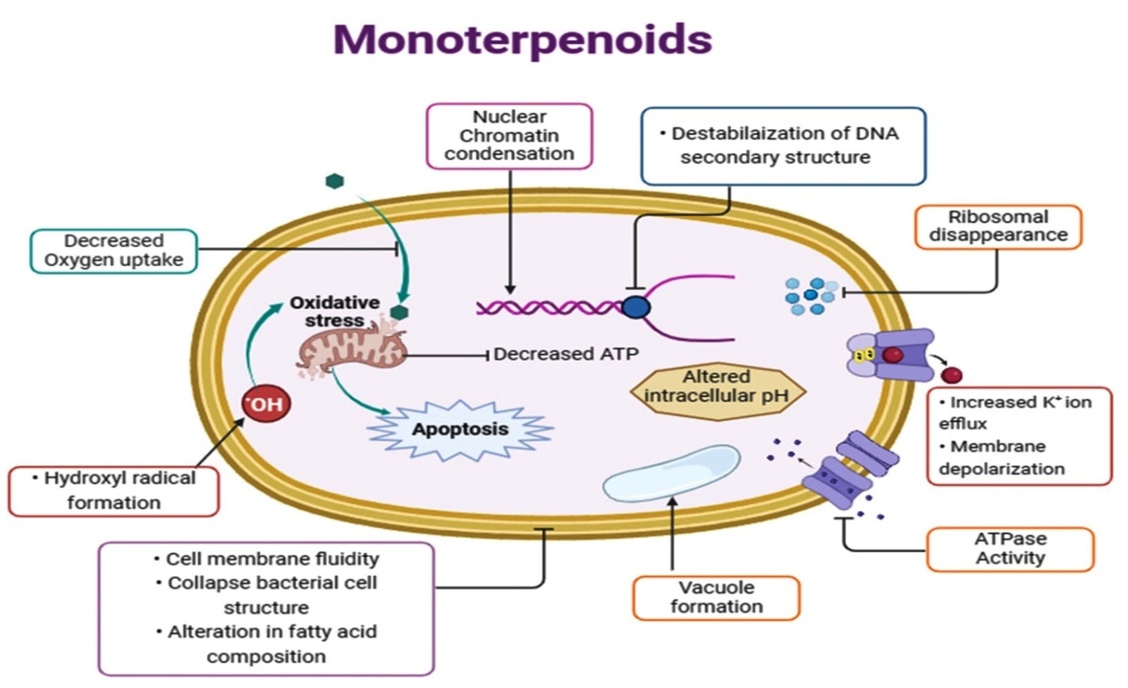
Figure. Schematic representation of mechanism of action of monoterpenes against causative agents in vaginitis
Thymol
Thymol or 2-isopropyl-5-methylphenol or hydroxyl cymene is a monoterpene phenolic compound and an isomer with carvacrol, majorly present in extracted oils obtained from plants that belong to families Apiaceae, Scrophulariaceae, Lamiaceae, Ranunculaceae, and Verbenaceae.65 Thymol (12-65%) is the major constituent obtained from the plant Thyme (Thymus vulgaris and other Thymus spp. of the Lamiaceae family) and many other plants. Thymol has been reported for different pharmacological activities like antiseptic, anti-inflammatory, antinociceptive, antioxidant, local anaesthetic, and most commonly for possessing antifungal and antibacterial properties.66-70
Mechanism of action of thymol
Thymol has good antimicrobial action. The antibacterial action of thymol has been evaluated in various in-vitro studies that included Salmonella typhimurium, E. coli, Yersinia enterocolitica, Pseudomonas fluorescens, Proteus mirabilis, E. coli, Sarcina flava, Listeria innocua, Staphylococcus aureus, Micrococcus spp., and Bacillus licheniformis.71,72 Its antibacterial activity mainly results from changes in the bacterial lipid bilayer and interactions with bacterial DNA. At very low concentrations, thymol can alter fatty acids composition (specially branched 14-methylhexadecanoic acid and 12- methyltetradecanoic acid) of lipid membrane in bacteria and at high concentration it can alter cell membrane integrity and reduces the viability of bacterial cells. The interaction with bacterial genomic DNA included the formation of a junction between bacterial DNA (minor groove) and thymol, which causes destabilization of secondary structures in DNA and results in DNA disaggregation.73 Another study revealed that in bacterial cell membrane thymol causes uncontrolled intracellular material release, resulting in disturbances in the metabolic actions of bacteria. Thymol is also associated with a reduction in glutathione and nitric oxide production in bacteria.74 In a microbial cell model, thymol distended DPPS (dipalmitoylphosphotidylserine) monolayers resulted in decreased surface elasticity and a change in the rheological properties of bacterial membrane films. Through this alteration in the cell membrane, thymol can easily permeate into the membrane, resulting in changes in membrane morphology, membrane structural order, membrane fluidity and lipid loading density.75,76 An antifungal activity study of thymol against Candida spp. concluded that it inhibits the ergosterol synthesis in fungal cell membrane and leads into cell impairment. It acts as a chemo sensitizing agent with interfering agents of cell wall. After penetration it reaches to fungal DNA and interferes with DNA synthesis. Thymol also disrupts the oxidative stress response system in fungi when used in combination therapy.77 A further study reported that thymol inhibits biofilm formation in Candida albicans by preventing further biofilm development or adherence. The morphological characteristics of thymol-treated biofilms change due to cell wall shrinkage and leakage in the intracellular matrix In Candida biofilms; thymol also inhibits sessile cells.78 Thymol has superior anti-trichomonia activity.79 The mode of action against Trichomonas spp. includes cell wall lysis and changes in the permeability. Thymol causes apoptosis in Trichomonas spp. cells and alters protozoan mitochondrial membrane permeability. It is also reported to interfere with Trichomonas metabolism by inhibiting enzyme dihydrofolate reductase, which leads to cell death.80,81
Antimicrobial activity of thymol
A study in 1994 reported noteworthy activity of thymol against wide range of bacteria in an effective range, with the lowest minimum inhibitory concentration of 175 µg/mL for three different strains of Staphylococcus spp. and 125-245 µg/mL for E. coli.82 Sasso et al. identified the antibacterial activity of thymol on S. aureus and E. coli. In that study it was reported that the ½ MIC shows the highest inhibition for E. coli and 1/16 for Staphylococcus aureus on vaginal epithelial cells. For thymol, efficient inhibition for adhesion was reported between ½ and 1/32 MIC values.83 Thymol containing vaginal cream has been reported to show a better response to vaginal and vulvar edema in comparison to the metronidazole-containing vaginal gel.84 Antifungal activity of thymol in vaginal infections has been reported with highest MIC ≤0.0038% and 0.0078, <0.015%, v/v for three strains of Candida spp. Thymol effectively decreased the number of Candida albicans and its biofilm production in the dose range of 64-120 µg/mL. These studies concluded that thymol could suppress the transformation of Candida from its yeast to hyphae form.85 In 2018, a study on five essential oils, including thymol, with seven E. coli spp. and twelve Candida spp., resulted in promising antibacterial and antimycotic activity with lower MIC values.86 Cordoba et al., evaluated antifungal activity of thymol against four Candida spp., including other essential oils and concluded that thymol is an active essential oil at MIC values of 0.8-0.16 mg/L.87 De Castro et al., reported antifungal activity of thymol with MICs range of 39-78 µg/mL for three strains of Candida spp. Researchers also reported synergistic antimicrobial activity with fluconazole and amphotericin B.88 Thymol-loaded nanoparticles and nystatin combination have been found to have significant anti-Candida activity in comparison to the nystatin group with the best MIC of 0.158 µg/mL.89 Metronidazole was not as effective against Trichomonas vaginalis as thymol, eugenol, and carvacrol extracts. Their hexane extract and methanol extract were more effective.90 Researchers investigated the anti-trichomonal activity of pure thymol and two nanopreparations, namely nanoliposomes and nanoparticles of thymol, and compared them with metronidazole. The developed formulation resulted in the best activity with a lower inhibitory effect.91
Eugenol
Eugenol or 4-Allyl-2-Methoxyphenol is a hydroxyphenyl propene found in volatile oils extracted from a variety of plants, such as Eugenia cariophylata, Myristica fragrans, and Ocimum basilicum, of the family Myristicaceae, Lauraceae, Lamiaceae, and Myrtaceae.92 It is a mainly present in clove oil (45-90%), obtained from Syzygium aromaticum, Myrtaceae and widely used for flavouring in the food and cosmetic industry. Traditional medicine uses it for its many pharmacological activities such as antibacterial, anti-inflammatory, antifungal, anticancer, and antioxidant.93 It has antibacterial, antifungal, and antitrichomonal properties, either alone or in combination.
Mechanism of action of eugenol
A free hydroxyl group present in eugenol is mainly responsible for its antimicrobial activity. The hydroxyl group is attached to bacterial cell proteins and prevents enzymatic action. In Gram-negative bacteria, eugenol penetrates the lipopolysaccharide of cell membrane and causes an alteration in cell structure.94,95 Eugenol damages the bacterial cytoplasmic membrane, causing potassium ions to leak out and a significant loss of cellular substances, ultimately leading to cell death.96 Eugenol mainly changes the portrayal of principal fatty acids and increases fatty acids with a low molecular weight.97 In bacterial cells, eugenol causes cell cytotoxicity by creating reactive oxygen species (ROS). These ROS stop cell growth, damage cell membranes, and damage bacterial DNA, which ultimately causes cell death.98 It also exhibits noticeable activity against bacterial enzymes like histidine carboxylase, protease, amylase, ATPase.99 Eugenol causes diminish in production of pyocyanin, violacein elastase (some virulence factors in Pseudomonas aeruginosa) and biofilm formation. It also inhibits biofilm production by 65% the gene expression level; especially the QS synthase genes.100 Eugenol changes the shape of the envelopes of bacteria and fungi due to presence of free hydroxyl group. It is highly effective in reducing colonies of Candida albicans and increasing the amount of damaged cells. Candida albicans envelope modification is relative to its pathogenicity, so it can cause compromises in host cell adhesion and some morphological transitions.101 Eugenol affects ion transportation in fungi. Eugenol reduces H+-ATPase activity in Candida albicans and inhibits H+ extrusion, leading to glucose stimulation. There is a high level of superoxide dismutase in Candida albicans that can cause an oxidative stress response. This leads to lipid peroxidation in the cytoplasmic membrane and then cell death.102,103 Antitrichomonas activity was reported for eugenol in many of the recent studies.104,105 The mechanism of action against Trichomonas vaginalis includes cell wall lysis and cell membrane permeability alteration. Metabolism interference is also one of the main causes for the cell disruption in Trichomonas spp.106
Antimicrobial activity of eugenol
Eyambe et al. determined the antimicrobial activity of eugenol against a number of bacteria such as Staphylococcus aureus. The MBC value indicated that it could kill about 99% of bacteria at a minimum concentration. The MIC value was at 115 µg/mL and MBC value was found out at 230 µg/mL.107 The activity of eugenol against Staphylococcus aureus, E. coli, and Candida spp. is well documented. The study estimated the MIC value of eugenol against E. coli to be 1.6 mg/L, for Staphylococcus aureus to be 128 mg/mL, and for Candida spp. to be 0.88 mg/mL. The study concluded that eugenol exhibited remarkable antimicrobial activity against the causative agents of vaginitis.108 Saugella probiogel, which mainly contained eugenol as an active ingredient, has been tested on a group of 209 women who suffered from BV, VVC, and RVVC. Researchers discovered that eugenol interferes with the biofilms, leading to lower the number of bacterial and fungal pathogens. This resulted in a normal state for 80% of BV cases, 62.5% of VVC cases, and 100% of RVVC cases.109 Banks and Taghread determined the antimicrobial activity of eugenol and extract of cloves against E. coli, Staphylococcus aureus (that were resistant to cefotaxime) and Candida spp. by disc diffusion sensitive method. It was found that both eugenol and non-polar clove extracts showed a significant inhibition of Staphylococcus aureus, E. coli, and Candida spp. with p ˂ 0.001.110 The antitrichomonal activity of eugenol showed that the extracts containing eugenol as an active compound cause growth inhibition of Trichomonas vaginalis with MIC range of 0.156 mg/mL.111 Yassin et al., used three strains of Candida spp. and tested with ethyl acetate extract of clove containing eugenol 60% as an active ingredient. In the disc diffusion method, the zones of inhibition were 14.9, 20.9 and 30.7 for Candida glabrata, Candida albicans and Candida tropicalis.112 In a study of 2022, it was found out that eugenol shows better antimicrobial effect than cinnamaldehyde against 18 Candida strains that was derived from vaginal strains cultures of infected women. MIC value for eugenol was 455.42 mg/L and MFC value was 690.09 mg/L and all the viable fungal cells were lost within 1 hour.113 In 2023, another study reported the anti-trichomonal vaginalis activity of eugenol. In this study the IC50 value for eugenol was 1.21 mg/mL which was better than metronidazole.114
Eucalyptol (1, 8-Cineole)
Eucalyptol or (1,3,3-trimethyl-2- oxabicyclo[2.2.2]octane) is a naturally produced monoterpene majorly obtained (70%) from leaves of Eucalyptus globulus as eucalyptus oil and other Eucalyptus spp. of the family Myrtaceae.115 Eucalyptol is widely reported to be used in the pharmaceutical, cosmetic and chemical industries. Its pharmacological activities are reported for anti-inflammatory, antiseptic, and antioxidant effects as well as having a strong antimicrobial activity in a broad spectrum.116 Numerous studies have documented its antimicrobial and antiprotozoal activity in vaginitis.
Mechanism of action of eucalyptol
Eucalyptol was reported to interfere with signaling in quorum sensing in bacteria. Quorum sensing is the technique of communication through chemical signals in microorganisms for promoting their growth, spread, and invasion. Biofilm formation is the result of quorum sensing, which protects the microbes from their external environment. Eucalyptol blocks those receptors that receive signals through different autoinducers.117 Eucalyptol changes the size and shapes of bacteria cells. In bacterial cells, it causes apoptosis in Staphylococcus aureus; when treated, it causes a nuclear chromatin condensation with necrosis in E. coli results into a reduction of nuclear chromatin and nucleoplasm.118 Eucalyptol makes Gram-negative bacteria more likely to have their cell walls damaged, which is a clinical benefit that can cover a wide range of antibacterial activity. Its activity is due to its hydrophobicity, which can synergistically increase the antimicrobial action of other agents.119 Eucalyptol affects cell membrane permeability, and it enhances the fluidity of the membrane, which results in membrane protein topology and lipid peroxidation. This inhibits bacterial cell respiration. It also increases the oxidative stress and ROS (reactive oxygen species) that inhibit the biological processes of bacterial cells.120 Eucalyptol was reported to alter membrane fluidity and cell wall permeability of Candida albicans by modifying the properties and functions of membrane. Eucalyptol also caused distortion in the fungal hyphae wall, followed by cell membrane disruption. Eucalyptol and other agents can interfere with the activity of mitochondrial dehydrogenase, which is ultimately involved in fungal ATP biosynthesis. The effectiveness of eucalyptol on the H+-ATPase of fungal membrane interferes with intracellular pH and fungal cell nutrient uptake. It finally leads to the acidification of intracellular material and cell death. Eucalyptol acts as a potent antifungal for Candida albicans and biofilms due to metabolic interference in comparison to fluconazole.121,122 Eucalyptol hydrophobicity and lipophilic nature allow it to enter the cell membrane of Trichomonas vaginalis. It increases cytoplasmic material permeability, which leads to impaired function in Trichomonas. Eucalyptol can interfere with the lipid composition leading to protein denaturation, cytoplasmic substance leakage, membrane disintegration, and cell death. Enzymatic inhibition and immune modulation are other modes of action of eucalyptol in protists.123
Antimicrobial activity of eucalyptol
Trinh et al. found that eucalyptol and α-terpineol are effective at killing two vaginal bacteria: Candida albicans and Gardnerella vaginalis. The combination of eucalyptol and α-terpineol potently inhibits the growth of these microorganisms at MIC values (0.125 and 0.065% v/v) respectively.124 In a study, eucalyptol lowered the number of Trichomonas vaginalis in a culture media at the effective concentration of 12.54 mg/mL after 48 hours.125 Rosemary oil containing eucalyptol showed potent activity against Pseudomonas aeruginosa and E. coli, with MIC 50 mg/mL and 25 mg/mL, respectively. The antifungal activity for Candida reported in this same study, with MIC/MFC value of 1.26 mg/mL.126 Bogavac et al., evaluated the antibacterial and antifungal activity of eucalyptus oil against two strains of of E. coli, Staphylococcus aureus and Candida albicans. The MIC values were reported to be 12.5-25, 6.255 and 6.254 µL/mL for E. coli, Staphylococcus aureus, Candida albicans, respectively. The study concluded that eucalyptol can be a possible potent antimicrobial agent in vaginal infections.127 Zhou et al., demonstrated that eucalyptus oil containing eucalyptol as major active constituent having antimicrobial activity against Candida albicans, E. coli and Pseudomonas aeruginosa with MICs range from 5.5-40.2 µL/mL.128 In a study of 2021, Sang-Youn et al., reported that eucalyptol possessed good antimicrobial and antioxidant effect against Candida albicans at MIC value 1.25 mg/mL.129 In a recent study of 2020, it was demonstrated that eucalyptol is having an efficient anti-trichomonal activity against Trichomonas vaginalis at the concentration of 21 mg in a culture media that was found out to be similar as metronidazole.130 In a further study of 2021, eucalyptol was tested against Gram-positive bacteria, methicillin resistant strains, Gram-negative bacteria, vancomycin resistant strains and Candida spp. It showed the inhibition in growth of these microbes in a very prominent range of MIC (0.25-8% v/v).131
R-Limonene
Limonene or 1-methyl-4-1-methyl phenyl cyclohexene is a naturally occurring monoterpene (R-Limonene or S-Limonene) containing two isoprene units with a lemon-like odour and is mainly found in citrus oil extracted from lemon, orange, and mandarin peels and in lemongrass oil, i.e., extracted from plants of the family Rutaceae.132 It is reported to possess different pharmacological activities like as antimicrobial, antioxidant, anticarcinogenic, antidiabetic and chemopreventive.133-135 Limonene has been prominently reported to have strong antimicrobial activities in vaginal infections.
Mechanism of action of R-limonene
The antimicrobial activity and mechanism of action of R-limonene have been well documented. Limonene damages lipopolysaccharide, which is associated with barrel proteins. It also changes membrane permeability and inactivates the bacterial cell envelope.136 At an optimum concentration, it was found that it leads to the formation of hydroxyl radicals (Fenton-mediated), which result in the oxidation of bacterial DNA and alter the membrane permeability.137 Collapse in bacterial cell structure (Gram-positive and Gram-negative) was seen when treated with limonene-containing nanoemulsion, which further lead to cell lysis, cell deformation, exudation of cellular material and cell death.138 The efficacy of antibacterial agents might differ for bacterial cells (Gram-positive and Gram-negative) because of differences in their targeted sites. Limonene also showed difference in its activity for both as Gram-negative bacterial cells are having efficient membrane homeostasis. Limonene is hydrophobic and Gram-negative bacterial cells are prone to develop a hydrophilic barrier to protect themselves, due to which these cells are lightly sensitive for limonene.139,140 The antifungal activity of limonene included the overexpression of different genes that are associated with cell signaling pathway. It was observed that Candida cell deteriorated after treatment with limonene due to altered cell structure and accumulation of reactive oxygen species or ROS that ultimately leads to cell destruction.141 About 80% of the changes in membrane permeability observed in Candida isolates resulted from rupture of cell wall. Researchers found that limonene interferes with pectin methyl esterase and cellulase in fungus cells.141 Inhibition of biofilm formation by limonene is also reported in various studies against several microbes. Limonene was reported to have better anti-trichomonal activity by causing changes in the morphology of protists. After exposure to limonene, it was observed that it might cause ribosomal disappearance, organelle disintegration, and formation of vacuoles, endoplasmic reticulum dilation, and leakage in cytoplasmic material. It also causes damage to the cytoplasmic membrane and appearance of cytoplasmic vacuoles. Limonene developed holes and projections in cytoplasmic membrane, which finally caused cell death of Trichomonas vaginalis. The attachment capacity of protist was also interfered by the presence of limonene.142,143
Antimicrobial activity of R-limonene
Vuuren and Viljoen assessed the antibacterial and antifungal efficiency of limonene and eucalyptol in a 1:1 ratio against wide range of bacteria and a fungus isolate. It was resulted into the MIC value range of 2-27 mg/mL for limonene enantiomers for Pseudomonas spp., Staphylococcus aureus and Cryptococcus neoformans.144 Bassole et al., estimated that limonene and its epoxides possessed better antibacterial and antifungal activity against vaginal isolates such as E. coli, Pseudomonas spp., Staphylococcus aureus, and Candida albicans, with MIC range of 2.4-3.2%.145 Essential oil containing lemongrass oil (active constituent limonene) shows a remarkable inhibitory effect against Streptococcus spp. with a mean value of 10.07 of the zone of inhibition after 48 hours. Limonene was found to be the second most active antibacterial against Streptococcus spp.146,147 Zahi et al. determined the antimicrobial activity of limonene loaded in nanoemulsion (organogel-based) form against Staphylococcus aureus, Pseudomonas spp., and Candida albicans with a MIC range of 3.14-12.56 µg/mL. The microbial cell’s constituent release increased about two folds after the application of limonene organogel.138 Gundel et al. estimated in vitro antifungal effect of limonene and eucalyptol in a murine model of vulvovaginal candidiasis. Nanoemulsion containing limonene and eucalyptol showed better antifungal activity in comparison to miconazole cream with p ˂ 0.05. The nanoemulsion showed better results due to the smaller size of the globules and larger surface area.61,148 Another study of 2005 tested the activity (in vitro and in vivo) of limonene against different specimens of Candida spp. on vulvovaginal candidiasis model. It decreased cell viability due to treatment with limonene at EC 50% (444 ± 35 µM) after 8 hr. In vivo result was showing significant decrease in number of fungal cells at the concentration of 500 µM of limonene.139 In another study, an anti-trichomonal activity was examined by using tea tree oil. It was found out that due to presence of limonene the number of protozoan Trichomonas vaginalis decreased at IC50 0.06 µL/mL.149 Nanoemulsion containing limonene with resveratrol showed prominent inhibition of Staphylococcus aureus due to the increased skin permeation. Limonene showed synergistic antibacterial activity.150
Alpha-Terpineol (α-Terpineol)
α–terpineol or 2-(4-methylcyclohex-3-en-1-yl)propan-2-ol is one of the isomers of terpineols (α, β, γ, and δ) naturally present as monoterpenoid alcohol in essential oils, mainly obtained from Mentha spp., Origanum spp., Artemisis spp., Thymus spp., Pinus spp., Salvia spp., Melaleuca spp., and Narcissus spp. in high fractions and in small fractions from many more plants. It is characterized by the floral fragrance and flavour.151 It has been widely known and documented to possess several biological properties as being anticarcinogenic, antioxidant, antimicrobial, anti-inflammatory, antiulcer, skin penetration enhancer, and anticonvulsant.152,153
Mechanism of action of α-terpineol
Monoterpenes have been reported to cause hydrocarbon partitioning in microbial cell membranes and disrupt vital functions. It causes lysis of bacterial cell membrane and resulted in to extrusion of cytoplasmic matter with associated ions and respiration inhibition.154 It disrupts microbial potassium homeostasis, eliminates propidium iodide, forms mesosomes, and modifies the glucose-dependent respiration mechanism in Staphylococcus aureus and Pseudomonas spp.155 It also changes the structural functions of the bacterial cellular membrane. Alpha-terpineol resulted to cell shape irregularities, formation of edges in the bacterial nuclear part, condensation of cytoplasm, plasmolysis, and formation of vacuoles in E. coli bacterial cells.156 In Candida spp. and Saccharomyces spp., it alters membrane permeability and inhibits cell respiration. It also affects the mitochondrial membrane of fungi by inhibiting medium acidification, which in turn interferes with the energy production of the fungi cells. Alpha-terpineol inhibits the germ tube formation; thereby obstructing the conversion of fungi into their mycelial form.157 Treatment with a-terpineol also causes abnormal hyphae formation due to disruption in the fungi cell wall. It down regulates fungi metabolic pathway and disrupts energy formation.158 Monoterpene extract contained a-terpineol, which is reported to block acetylcholinesterase in Trichomonas vaginalis. Due to acetylcholinesterase blockage, the level of acetylcholine increases, which increases the activation of nicotinic acetylcholine receptors, ultimately leading to muscular contraction and membrane depolarization.159
Antimicrobial activity of Alpha-terpineol
Alpha-terpineol has been documented to have activity in vaginitis against Staphylococcus aureus and E. coli at MICs 0.065 and 0.252% v/v, respectively. In a similar study, anti-candida activity was also reported in the range of 0.064-0.2568% v/v.160 Alpha-terpineol inhibited biofilms produce by Candida spp. after 2 h of exposure at 0.1256% v/v.157 The activity of α-terpineol in bacterial vaginosis and vulvovaginal candidiasis has been reported against G. vaginalis and C. albicans at MIC 0.065% and 0.1254% v/v, respectively, by increasing cytokines (interleukins) expressions and NF-kB activation.124 Alpha-terpineol has shown antifungal activity, along with other constituents.161 Alpha-terpineol showed prominent inhibitory activity against Candida albicans isolates that were resistant to fluconazole with MIC range of 300-3000 µg/mL.162 A study in 2023, reported to have antibacterial, anti-candida and anti-trichomonal activity of tea tree oil due to its main active constituent α-terpineol in a very effective way.163 Alpha-terpineol is the majorly present in tea tree oil and possesses antibacterial and antifungal activities against ten bacterial isolates and Candida albicans isolates. The Rideal-Walker coefficient for α-terpineol was found to be 16, which was the highest among all the constituents of tea tree oil. The anti-inflammatory activity also possessed by α-terpineol in vaginal inflammation occurs in vaginal candidiasis. This study also demonstrated anti-trichomonal activity, concluding that -terpineol effectively combats Trichomonas vaginalis.164,165 Alpha-terpineol is also effective against Aspergillus niger and effectively inhibits fungal mycelial growth and spore germination.165 Alpha-terpineol has antibacterial activity against fifty Staphylococcus spp.166
Geraniol
Geraniol or 3-7dimethylocta2-6-dien1-ol is a monoterpene found in a mixture of nerol and geraniol (cis and trans forms). It is mostly obtained from Rosa rubiginosa, Cymbopogon martini, Cymbopogon nardus, Pelargonium graveolens, and Monarda fistulosa. It has a sweet floral or rose-like fragrance, so it is widely used as a fragrance material.167 It possesses various pharmacological activities, including antitumor, insecticidal, antioxidant, antimicrobial and anti-inflammatory properties. It has efficient antibacterial, antifungal, and antitrichomonal properties for vaginal infections.168
Mechanism of action of geraniol
The antimicrobial activity of geraniol has been described against Staphylococcus aureus and E. coli. Geraniol induces DNA damage and increases osmotic stress in bacterial cells.169 It disrupts the microbial cell membrane lipid structure, interacts with bacterial cellular components, and makes the bacterial cell more permeable to the outer compounds. It penetrates the cell interior and ultimately limits bacterial cell growth. It also disturbs the membrane efflux pumps in bacterial cells and reduces the level of ergosterol, leading to an alteration in ATPase activity in the membrane.170,171 The activity of geraniol has been reported against a broad range of Candida and non-Candida spp. In fungal cells, it causes impaired iron homeostasis with mitochondrial dysfunction. It also alters the functioning of calcineurin signaling pathway in a fungal cell that is responsible for the antifungal activity of geraniol.172 In Candida albicans spp., membrane disruption is the major mechanism of action of geraniol that affects fungal cells. Geraniol changes the ATPase activity of the plasma membrane and alters intracellular pH. Geraniol damages DNA repair in Candida albicans. Geraniol is a potent inhibitor of hyphae formation in yeast cells. This inhibits biofilm formation.173 In vaginal infections, geraniol has better anti-trichomonal activity. The treated cells of Trichomonas vaginalis showed morphological changes, formation of vacuoles, ribosomal disappearance, and dilation in the rough endoplasmic reticulum. After treatment with geraniol, the protozoan nucleus membrane damages and leads to leakage of cytoplasmic content, accumulation of chromatin in the cytoplasm, disintegrated organelles, and damaged cell membrane, which resulted into cell death.174,175
Antimicrobial activity of geraniol
An in vitro study reported antifungal activity of geranium oil containing geraniol against Candida albicans, and it was found that vaginal washing with geranium oil inhibited fungal growth (IC50 25-26 mg/mL). The study concluded that geraniol is a potent antifungal agent in vaginal candidiasis.176 Antimicrobial activity of geraniol has been accounted against a wide range of bacteria and fungi. It has remarkable bactericidal activity, with BA50 value of 1.5 for E. coli. In its gaseous state, it has potent activity against Staphylococcus aureus.177 Researchers have reported the activity of geraniol with carvacrol and fluconazole against seven different strains of Candida albicans. Geraniol showed a MIC value range 0.5-2 mg/mL for those tested strains. The synergism of geraniol has been reported with fluconazole against resistant fungal species.177 A study involved antifungal activity of geraniol at the MIC90 value was 16 µg/mL and it was recommended that geraniol as a potent anti-Candida agent.178 Another study reported anti-trichomonal activity of geraniol against two isolates of Trichomonas vaginalis. Geraniol showed MLC values for TV1 and TV2 isolates as 45 mg/mL and 89.95 mg/mL, respectively. The IC50 values were found out to be 22.5 mg/mL and 45 mg/mL, respectively.174 The antimicrobial activity of rose oil containing geraniol as a major constituent against Staphylococcus aureus (methicillin-resistant) and mycelial growth of Candida reduced 99 percent of Staphylococcus aureus and inhibited the mycelial growth of Candida albicans within 1 h of geraniol treatment with an IC50 value of 0.00045%.179 Geraniol’s detailed antibacterial and antifungal activity with related mode of action in vaginitis have been well documented.173 Antifungal activity of nanoemulsion containing geraniol with volatile oil and hydrogel containing essential oil with geraniol has been reported against five strains. The hydrogel containing geraniol showed about 64 times better anti-Candida activity.180 Antibacterial activity of geraniol has been reported for a range of bacteria.181
Researchers have reported several monoterpenes, either individually or in combinations, for their intrinsic antimicrobial activities in vaginal infections, as well as their other pharmacological activities. This review reported a collective number of monoterpenes so that they can further be utilized in combinations to demonstrate their synergistic activities. These antimicrobial activities are against a broad range of micro-organisms, including fungi, bacteria (Gram-positive and Gram-negative bacteria) and other pathogens (Table 2).
Table (2):
Monoterpenes and their antimicrobial activity in vaginal infections (MIC/MBC)
Monoterpene |
Source |
Family |
Activity in vaginal infections |
MIC/MBC |
Reference |
|---|---|---|---|---|---|
Citral |
Cymbopogon citratus |
Gramineae |
Anticandida in VVC S. aureus E. coli |
0.05% 0.06% 0.12% |
Onawunmi GO182 Naik et al.183 |
Eucalyptol |
Eucalyptus globulus |
Myrtaceae |
Anticandida in VVC Against Gram-ve bacteria in BV Antitrichomonal activity |
0.7 mg/mL 6.2 mg/mL 12.5 mg/mL |
Quatrin et al.184 Klančnik et al.185 Hashemi et al.130 |
Carvacrol |
Thymus vulgaris |
Lamiaceae |
Anti-Gardnerella vaginalis activity Anticandida in VVC Antitrichomonal activity |
0.16 µL/mL 103 mg/L MLC-100 µg/mL |
Can Baser186 Chami et al.187 Karami et al.90 |
Thymol |
Thymus vulgaris |
Lamiaceae |
Anticandida in VVC Staphylococcus aureus E. coli Antitrichomonal activity |
0.16 mg/L 175.5 µg/mL 125.23 µg/mL 2000 µg/mL |
Shu et al.86 Sasso et al.83 Jalili et al.91 |
Eugenol |
Eugenia cariophylata |
Myrtaceae |
Anticandida in VVC Staphylococcus aureus E. coli Antitrichomonal activity |
455.52 mg/L 128 mg/mL p ˂ 0.001 0.156 mg/mL |
Chami et al.187 Fontenelle et al.188 Marchese et al.108 Filippo et al.109 Jafari et al.114 |
Borneol |
Rosmarinus officinalis |
Lamiaceae |
Anticandida in VVC Against bacteria in BV |
1.2 mg/mL p ˂ 0.005 |
Chen et al.189 Bogavac et al.126 |
α-terpineol |
Origanium vulgare |
Lamiaceae |
Anti-Gardnerella vaginalis activity Staphylococcus aureus E. coli Anticandida in VVC Antitrichomonal activity |
0.06% 6.4 mg/mL 5.6 cfu/ mL 0.1254% 0.06% |
Li et al.118 Swamy et al.190 Yang et al.191 Andrine et al.192 |
α-pinene |
Rosmarinus officinalis |
Lamiaceae |
Anticandida in VVC Against bacteria in BV |
3125 µg/mL p˂ 0.001 |
Silva et al.193 |
R-Limonene |
Citrus Limon |
Rutaceae |
Gardnerella activity Staphylococcus aureus Anticandida in VVC Antitrichomonal activity |
1 µL/mL 20 mL/L 2 µL/mL 150 µg/mL |
Schwiertz et al.194 Han et al.195 Jamshidi et al.196 |
Geraniol |
Cymbopogon nardus |
Poaecea |
Staphylococcus aureus E. coli Anticandida in VVC Antitrichomonal activity |
11200 µg/mL 5600 µg/mL 30-130 µg/mL 342.96 µg/mL |
Bhattamisra et al.197 Sharma et al.198 de Brum et al.199 |
Camphene |
Chrysanthemum morifolium |
Asteraceae |
Anticandida in VVC Against bacteria in BV |
128 µg/mL 2-31.2 µg/mL |
Thakre et al.200 de Freitas et al.201 |
Linalool |
Lavendula angustifolia |
Lamiaceae |
Anti-Gardnerella vaginalis activity Staphylococcus aureus Anticandida in VVC Antitrichomonal activity |
1.25 µL/mL 12.8 mg/mL 16-32 µg/mL 25.34 µg/mL |
Sousa et al.202 Hsu et al.203 Maria et al.204 |
Fenchone |
Foeniculum vulgare |
Umbelliferae |
Anticandida in VVC Staphylococcus aureus Antitrichomonal activity |
62.51 µg/mL 6.256 µg/mL 100 µg/mL |
Bozovic et al.205 Ghasemian et al.206 Maria et al.204 |
Trans-anethole |
Foeniculum vulgare |
Umbelliferae |
Anticandida in VVC Staphylococcus aureus |
123.50 mg/mL 6-10% |
Dabrowska et al.207 Kwiatkowski et al.208 |
Menthol |
Mentha piperita |
Lamiaceae |
Staphylococcus aureus Anticandida in VVC |
1.11 mg/mL 500 µg/mL |
Kifer et al.209 Samber et al.210 |
Menthone |
Mentha piperita |
Lamiaceae |
Staphylococcus aureus Anticandida in VVC |
3540 µg/mL p˂ 0.005 |
Zhao et al.211 Iraji et al.212 |
β-Myrcene |
Cannabis sativa |
Cannabiaceae |
Anticandida in VVC |
39-78 µg/mL |
Cecchini et al.213 |
Terpinen-4-ol |
Origanium vulgare |
Lamiaceae |
Anti-Gardnerella vaginalis activity Anticandida in VVC Antitrichomonal activity |
0.13% 0.06% 0.003% |
Mondello et al.214 Menezes et al.105 |
p-cymene |
Artemesia vulgaris |
Asteraceae |
Anticandida in VVC Staphylococcus aureus, E. coli Antitrichomonal activity |
4 mg/mL >8% >0.01% |
Randelovic et al.215 Balahbib et al.216 Maria et al.204 |
Carvone |
Carum carvi |
Apiaceae |
Anticandida in VVC Staphylococcus aureus Antitrichomonal activity |
0.312 mg/mL 500 µg/mL 72.11 µg/mL |
Mun et al.217 Moro et al.218 Dominguez et al.219 |
Carvacrol |
Origanium vulgare |
Lamiaceae |
Anticandida in VVC Staphylococcus aureus |
256 µg/mL 400 µg/mL |
Lima et al.220 Rua et al.221 |
α-phellanderene |
Eucalyptus phellandra |
Myrtaceae |
Anticandida in VVC |
1.7 mL/L |
Thangaleela et al.222 |
Geranyl acetate |
Callitris spp. |
Cuprassaceae |
Anticandida in VVC Staphylococcus aureus |
0.3 mg/mL 2.5 mg/mL |
Humeriah et al.223 |
Nerol |
Cymbopogon flexuosus |
Poaceae |
Anticandida in VVC Staphylococcus aureus |
0.77 µL/mL 40 µg/mL |
Wang et al.224 Togashi et al.225 |
Sabinene |
Quercus ilex |
Fagaceae |
Staphylococcus aureus |
6.25 µg/mL |
Mahizaan et al.226 |
Researchers have extensively studied the vaginal microbiome and its associated infections over the past decade. An imbalance in the vaginal microbiota leads to severe infections. The recurrence of these infections with longer persistence affects a woman in many ways, which can diminish or interfere with her quality of life. The resistance caused by the vaginitis causing microorganisms for conventional drugs led to further research for those antimicrobial agents who can act potently on these microorganisms and their resistant species. Plant secondary metabolites have been mainly focused and examined for their antimicrobial properties as well as other therapeutic effects. Terpenes are the most commonly reported antimicrobial agents in a number of conditions that are associated with microbial infections, including vaginal infections because of their antimicrobial, antioxidant and anti-inflammatory effects. Monoterpenes like linalool, geraniol, eugenol, thymol, limonene, eucalyptol, carvacrol, and terpineol are widely reported to have potent antimicrobial activity in vaginal infections. They have been reported either individually or in many of the combinations present as plant’s volatile oils and in combination with conventional drugs. Vaginal infections are caused by some specific strains of microorganisms that attack the vaginal healthy microbiota and cause an imbalance there. For this purpose, certain combinations of plant secondary metabolites are required, which have antimicrobial activities in different dimensions with anti-inflammatory and antioxidant effects. This review summarizes the types of vaginal infections that affect women in today’s life and their overlapping persistent effects. Further, this review reported the commonly tested monoterpenes for vaginal infections and their potent effects. We can envision these monoterpenes and their combinations working together in the future due to their strong antimicrobial qualities in vaginal infections, making them the ideal treatment for curing bacterial, fungal, and trichomonal vaginal infections.
ACKNOWLEDGMENTS
None.
CONFLICT OF INTEREST
The authors declare that there is no conflict of interest.
AUTHORS’ CONTRIBUTION
PS and NA conceptualized the study. SS performed supervision. NA performed visualization. PS and SKY wrote the original draft. HC, SS and KD wrote, reviewed and edited the manuscript. All authors read and approved the final manuscript for publication.
FUNDING
None.
DATA AVAILABILITY
All datasets generated or analyzed during this study are included in the manuscript.
ETHICS STATEMENT
Not applicable.
- Gupta S, Kakkar V, Bhushan I. Crosstalk between vaginal microbiome and female health: a review. Microbial pathogenesis. 2019;136:103696.
Crossref - Yildirim S, Yeoman CJ, Janga SC, et al. Primate vaginal microbiomes exhibit species specificity without universal Lactobacillus dominance. ISME J. 2014;8(12):2431-2444.
Crossref - Ravel J, Gajer P, Abdo Z, et al. Vaginal microbiome of reproductive-age women. Proc Natl Acad Sci U S A. 2010;108(Suppl 1):4680-4687.
Crossref - Amabebe E, Anumba DOC. The Vaginal Microenvironment: The Physiologic Role of Lactobacilli. Front Med. 2018;5:181.
Crossref - Stumpf RM, Wilson BA, Rivera A, et al. The primate vaginal microbiome: Comparative context and implications for human health and disease. Am J Phys Anthropol. 2013;152(57):119-134.
Crossref - Zozaya-Hinchliffe M, Lillis R, Martin DH, Ferris MJ. Quantitative PCR Assessments of Bacterial Species in Women with and without Bacterial Vaginosis. J Clin Microbiol. 2010;48(5):1812-1819.
Crossref - Witkin SS, Mendes-Soares H, Linhares IM, Jayaram A, Ledger WJ, Forney LJ. Influence of Vaginal Bacteria and d- and l-Lactic Acid Isomers on Vaginal Extracellular Matrix Metalloproteinase Inducer: Implications for Protection against Upper Genital Tract Infections. Blaser MJ, ed. mBio. 2013;4(4):e00460-13.
Crossref - Aldunate M, Srbinovski D, Hearps AC, et al. Antimicrobial and immune modulatory effects of lactic acid and short chain fatty acids produced by vaginal microbiota associated with eubiosis and bacterial vaginosis. Front Physiol. 2015;6:164.
Crossref - Smith SB, Ravel J. The vaginal microbiota, host defence and reproductive physiology. J Physiol. 2016;595(2):451-463.
Crossref - Van Kessel K, Assefi N, Marrazzo J, Eckert L. Common Complementary and Alternative Therapies for Yeast Vaginitis and Bacterial Vaginosis: A Systematic Review. Obstet Gynecol Surv. 2003;58(5):351-358.
Crossref - Vaou N, Stavropoulou E, Voidarou C, Tsigalou C, Bezirtzoglou E. Towards Advances in Medicinal Plant Antimicrobial Activity: A Review Study on Challenges and Future Perspectives. Microorganisms. 2021;9(10):2041.
Crossref - Riaz M, Khalid R, Afzal M, et al. Phytobioactive compounds as therapeutic agents for human diseases: A review. Food Sci Nutr. 2023;11(6):2500-2529.
Crossref - Vieira-Baptista P, Bornstein J. Candidiasis, Bacterial Vaginosis, Trichomoniasis and Other Vaginal Conditions Affecting the Vulva. Vulvar Disease. 2019:167-205.
Crossref - Ceccarani C, Foschi C, Parolin C, et al. Diversity of vaginal microbiome and metabolome during genital infections. Sci Rep. 2019;9(1):14095.
Crossref - Thulkar J, Kriplani A, Agarwal N, Vishnubhatla S. Aetiology & risk factors of recurrent vaginitis & its association with various contraceptive methods. Indian J Med Res. 2010;131:83-87.
- Cotch MF, Hillier SL, Gibbs RS, Eschenbach DA. Epidemiology and outcomes associated with moderate to heavy Candida colonization during pregnancy. Am J Obstet Gynecol. 1998;178(2):374-380.
Crossref - Anderson MR, Klink K, Cohrssen A. Evaluation of Vaginal Complaints. JAMA. 2004;291(11):1368.
Crossref - Chee WJY, Chew SY, Than LTL. Vaginal microbiota and the potential of Lactobacillus derivatives in maintaining vaginal health. Microb Cell Fact. 2020;19(1):203.
Crossref - Sobel JD, Faro S, Force RW, et al. Vulvovaginal candidiasis: Epidemiologic, diagnostic, and therapeutic considerations. Am J Obstet Gynecol. 1998;178(2):203-211.
Crossref - Sobel JD. Pathogenesis and Treatment of Recurrent Vulvovaginal Candidiasis. Clin Infect Dis. 1992;14(Suppl 1):S148-S153.
Crossref - Babula O, Lazdane G, Kroica J, Ledger WJ, Witkin SS. Relation between Recurrent Vulvovaginal Candidiasis, Vaginal Concentrations of Mannose-Binding Lectin, and a Mannose-Binding Lectin Gene Polymorphism in Latvian Women. Clin Infect Dis. 2003;37(5):733-737.
Crossref - Yano J, Sobel JD, Nyirjesy P, et al. Current patient perspectives of vulvovaginal candidiasis: incidence, symptoms, management and post-treatment outcomes. BMC Women’s Health. 2019;19(1):48.
Crossref - Chandra J, Kuhn DM, Mukherjee PK, Hoyer LL, McCormick T, Ghannoum MA. Biofilm Formation by the Fungal Pathogen Candida albicans: Development, Architecture, and Drug Resistance. J Bacteriol. 2001;183(18):5385-5394.
Crossref - Peters BM, Palmer GE, Nash AK, Lilly EA, Fidel PL, Noverr MC. Fungal Morphogenetic Pathways Are Required for the Hallmark Inflammatory Response during Candida albicans Vaginitis. Deepe GS, ed. Infect Immun. 2013;82(2):532-543.
Crossref - Cassone A, Flavia De Bernardis, Mondello F, Ceddia TL. Agatensi. Evidence for a Correlation Between Proteinase Secretion and Vulvovaginal Candidosis. J Infect Dis. 1987;156(5):777-783.
Crossref - Hise AG, Tomalka J, Ganesan S, et al. An Essential Role for the NLRP3 Inflammasome in Host Defense against the Human Fungal Pathogen Candida albicans. Cell Host Microbe. 2009;5(5):487-497.
Crossref - Zelante T, De Luca A, Bonifazi P, et al. IL-23 and the Th17 pathway promote inflammation and impair antifungal immune resistance. Eur J Immunol. 2007;37(10):2695-2706.
Crossref - Muzny CA, Balkus J, Mitchell C, et al. Diagnosis and Management of Bacterial Vaginosis: Summary of Evidence Reviewed for the 2021 Centers for Disease Control and Prevention Sexually Transmitted Infections Treatment Guidelines. Clin Infect Dis. 2022;74(Suppl 2):S144-S151.
Crossref - Coleman JS, Gaydos CA. Molecular Diagnosis of Bacterial Vaginosis: an Update. J Clin Microbiol. 2018;56(9):e00342.
Crossref - David AE, Patton Dorothy L, Thomas MH, et al. Effects of Vaginal Intercourse with and without a Condom on Vaginal Flora and Vaginal Epithelium. J Infect Dis. 2001;183(6):913-918.
Crossref - Banupriya M, Geetha N. Comparative Study of Amsel Criteria Vs Nugent CriteriaScoring System In Vaginal Discharge in Gmkmch, Salem. IOSR Journal of Dental and Medical Sciences. 2016;15(07):05-09.
Crossref - van den Munckhof EHA, van Sitter RL, Boers KE, et al. Comparison of Amsel criteria, Nugent score, culture and two CE-IVD marked quantitative real-time PCRs with microbiota analysis for the diagnosis of bacterial vaginosis. Eur J Clin Microbiol Infect Dis. 2019;38(5):959-966.
Crossref - Yoshimura K, Morotomi N, Fukuda K, et al. Intravaginal microbial flora by the 16S rRNA gene sequencing. Am J Obstet Gynecol. 2011;205(3):235.e1-235.e9.
Crossref - Boggess KA, Trevett TN, Madianos PN, et al. Use of DNA hybridization to detect vaginal pathogens associated with bacterial vaginosis among asymptomatic pregnant women. Am J Obstet Gynecol. 2005;193(3):752-756.
Crossref - Ness RB, Hillier SL, Kip KE, et al. Bacterial Vaginosis and Risk of Pelvic Inflammatory Disease. Obstet Gynecol. 2004;104(4):761-769.
Crossref - Martin HL, Richardson BA, Nyange PM, et al. Vaginal Lactobacilli, Microbial Flora, and Risk of Human Immunodeficiency Virus Type 1 and Sexually Transmitted Disease Acquisition. J Infec Dis. 1999;180(6):1863-1868.
Crossref - Olmsted SS, Meyn LA, Rohan LC, Hillier SL. Glycosidase and Proteinase Activity of Anaerobic Gram-Negative Bacteria Isolated From Women With Bacterial Vaginosis. Sex Transm Dis. 2003;30(3):257-261.
Crossref - Marrs CN, Knobel SM, Zhu WQ, Sweet SD, Chaudhry AR, Alcendor DJ. Evidence for Gardnerella vaginalis uptake and internalization by squamous vaginal epithelial cells: implications for the pathogenesis of bacterial vaginosis. Microb Infect. 2012;14(6):500-508.
Crossref - Hirt RP. Trichomonas vaginalis virulence factors: an integrative overview. Sex Transm Infect. 2013;89(6):439-443.
Crossref - Van Der Pol B. Trichomonas vaginalisInfection: The Most Prevalent Nonviral Sexually Transmitted Infection Receives the Least Public Health Attention. Clin Infect Dis. 2007;44(1):23-25.
Crossref - Fichorova RN, Buck OR, Yamamoto HS, et al. The Villain Team-Up or how Trichomonas vaginalis and bacterial vaginosis alter innate immunity in concert. Sex Transm Infect. 2013;89(6):460-466.
Crossref - Johnston VJ, Mabey DC. Global epidemiology and control of Trichomonas vaginalis. Curr Opin Infect Dis. 2008;21(1):56-64.
Crossref - Ryan CM, de Miguel N, Johnson PJ. Trichomonas vaginalis: current understanding of host-parasite interactions. Essays Biochem. 2011;51:161-175.
Crossref - Hirt RP, Natalia de Miguel, Sirintra Nakjang, et al. Trichomonas vaginalis pathobiology new insights from the genome sequence. Adv Parasitol. 2011;77:87-140.
Crossref - Dessi D, Rappelli P, Diaz N, Cappuccinelli P, Fiori PL. Mycoplasma hominis and Trichomonas vaginalis: a unique case of symbiotic relationship between two obligate human parasites. Front Biosci. 2006;11(1):2028.
Crossref - Donders GGG, Bellen G, Grinceviciene S, Ruban K, Vieira-Baptista P. Aerobic vaginitis: no longer a stranger. Res Microbiol. 2017;168(9-10):845-858.
Crossref - Sobel JD, Reichman O, Misra D, Yoo W. Prognosis and treatment of desquamative inflammatory vaginitis. Obstet Gynecol. 2011;117(4):850-855.
Crossref - Donders GGG, Vereecken A, Bosmans E, Dekeersmaecker A, Salembier G, Spitz B. Definition of a type of abnormal vaginal flora that is distinct from bacterial vaginosis: aerobic vaginitis. BJOG. 2002;109(1):34-43.
Crossref - Romero R, Chaiworapongsa T, Kuivaniemi H, Tromp G. Bacterial vaginosis, the inflammatory response and the risk of preterm birth: a role for genetic epidemiology in the prevention of preterm birth. Am J Obstet Gynecol. 2004;190(6):1509-1519.
Crossref - Fan A, Yue Y, Geng N, Zhang H, Wang Y, Xue F. Aerobic vaginitis and mixed infections: comparison of clinical and laboratory findings. Arch Gynecol Obstet. 2013;287(2):329-335.
Crossref - Tansarli GS, Kostaras EK, Athanasiou S, Falagas ME. Prevalence and treatment of aerobic vaginitis among non-pregnant women: evaluation of the evidence for an underestimated clinical entity. Eur J Clin Microbiol Infect Dis. 2013;32(8):977-984.
Crossref - Cerikcioglu N, Beksac MS. Cytolytic Vaginosis: Misdiagnosed as Candidal Vaginitis. Infect Dis Obstet Gynecol. 2004;12(1):13-16.
Crossref - Yang S, Zhang Y, Liu Y, Wang J, Chen S, Li SX. Clinical Significance and Characteristic Clinical Differences of Cytolytic Vaginosis in Recurrent Vulvovaginitis. Gynecol Obstet Invest. 2017;82(2):137-143.
Crossref - Leizer J, Nasioudis D, Forney LJ, et al. Properties of Epithelial Cells and Vaginal Secretions in Pregnant Women When Lactobacillus crispatus or Lactobacillus iners Dominate the Vaginal Microbiome. Reprod Sci. 2017;25(6):854-860.
Crossref - Puri S. Cytolytic vaginosis: A common yet underdiagnosed entity. Ann Trop Pathol. 2020;11(1):29-32.
Crossref - Suresh A, Rajesh A, Bhat R, Rai Y. Cytolytic vaginosis: A review. Indian J Sex Transm Dis AIDS. 2009;30(1):48-50.
Crossref - Hammer KA, Carson CF, Riley TV. Antimicrobial activity of essential oils and other plant extracts. J Appl Microbiol. 1999;86(6):985-990.
Crossref - Farag RS, Shalaby AS, El-Baroty GA, Ibrahim NA, Ali MA, Hassan EM. Chemical and biological evaluation of the essential oils of different Melaleuca species. Phytother Res. 2004;18(1):30-35.
Crossref - Giordani R, Regli P, Kaloustian J, Mikail C, Abou L, Portugal H. Antifungal effect of various essential oils against Candida albicans. Potentiation of antifungal action of amphotericin B by essential oil from Thymus vulgaris. Phytother Res. 2004;18(12):990-995.
Crossref - Azimi H, Fallah-Taf M, Karimi-Dar M, Abdollahi M. A Comprehensive Review of Vaginitis Phytotherapy. Pak J Biol Sci. 2011;14(21):960-966.
Crossref - Gundel S da S, de Godoi SN, Santos RCV, et al. In vivo antifungal activity of nanoemulsions containing eucalyptus or lemongrass essential oils in murine model of vulvovaginal candidiasis. J Drug Deliv Sci Technol. 2020;57:101762.
Crossref - Stevic T, Beric T, Savikin K, et al. Antifungal activity of selected essential oils against fungi isolated from medicinal plant. Ind Crops Prod. 2014;55:116-122.
Crossref - Van Vuuren SF, Kamatou GPP, Viljoen AM. Volatile composition and antimicrobial activity of twenty commercial frankincense essential oil samples. S Afr J Bot. 2010;76(4):686-691.
Crossref - Tomas M, Palmeira-de-Oliveira A, Simoes S, Martinez-de-Oliveira J, Palmeira-de-Oliveira R. Bacterial vaginosis: Standard treatments and alternative strategies. Int J Pharm. 2020;587:119659.
Crossref - Kachur K, Suntres Z. The antibacterial properties of phenolic isomers, carvacrol and thymol. Critical reviews in food science and nutrition. 2020; 60(18):3042-53.
Crossref - Licata M, Tuttolomondo T, Dugo G, et al. Study of quantitative and qualitative variations in essential oils of Sicilian oregano biotypes. J Essent Oil Res. 2015;27(4):293-306.
Crossref - Yanishlieva NV, Marinova EM, Gordon MH, Raneva VG. Antioxidant activity and mechanism of action of thymol and carvacrol in two lipid systems. Food Chem. 1999;64(1):59-66.
Crossref - Mendes SS, Bomfim RR, Jesus HCR, et al. Evaluation of the analgesic and anti-inflammatory effects of the essential oil of Lippia gracilis leaves. J Ethnopharmacol. 2010;129(3):391-397.
Crossref - Haeseler G, Maue D, Grosskreutz J, et al. Voltage-dependent block of neuronal and skeletal muscle sodium channels by thymol and menthol. Eur J Anaesthesiol. 2002;19(08):571-579.
Crossref - Gallucci MN, Carezzano ME, Oliva MM, et al. In vitro activity of natural phenolic compounds against fluconazole-resistant Candida species: a quantitative structure-activity relationship analysis. J Appl Microbiol. 2014;116(4):795-804.
Crossref - Marchese A, Orhan IE, Daglia M, Barbieri R, et al. Antibacterial and antifungal activities of thymol: A brief review of the literature. Food chemistry. 2016;210:402-14.
Crossref - Ceylan E, Fung DY. Antimicrobial activity of spices 1. Journal of Rapid Methods & Automation in Microbiology. 2004;12(1):1-55.
Crossref - Nostro A, Blanco AR, Cannatelli MA, et al. Susceptibility of methicillin-resistant staphylococci to oregano essential oil, carvacrol and thymol. FEMS Microbiol Lett. 2004;230(2):191-195.
Crossref - Wang LH, Zhang ZH, Zeng XA, Gong DM, Wang MS. Erratum to: Combination of microbiological, spectroscopic and molecular docking techniques to study the antibacterial mechanism of thymol against Staphylococcus aureus: membrane damage and genomic DNA binding. Anal Bioanal Chem. 2017;409(11):3055-3055.
Crossref - Cheng YW, Kong XW, Wang N, Wang TT, Chen J, Zhi Qi Shi. Thymol confers tolerance to salt stress by activating anti-oxidative defense and modulating Na+ homeostasis in rice root. Ecotoxicol Environ Saf. 2020;188:109894-109894.
Crossref - Ferreira JVN, Joao HGL, Caseli L. Thymol in cellular membrane models formed by negative charged lipids causes aggregation at the air-water interface. Chem Phys Lett. 2019;717:87-90.
Crossref - Pinto E, Pina-Vaz C, Salgueiro L, et al. Antifungal activity of the essential oil of Thymus pulegioides on Candida, Aspergillus and dermatophyte species. J Med Microbiol. 2006;55(10):1367-1373.
Crossref - Jafri H, Ahmad I. Thymus vulgaris essential oil and thymol inhibit biofilms and interact synergistically with antifungal drugs against drug resistant strains of Candida albicans and Candida tropicalis. J Mycol Med. 2020;30(1):100911.
Crossref - MehriArdestani M, Aliahmadi A, Toliat T, Dalimi A, Momeni Z, Rahimi R. Evaluation of Antimicrobial Activity of Trachyspermum ammi (L.) Sprague Essential Oil and Its Active Constituent, Thymol, against Vaginal Pathogens. Trad Integr Med. 2020;5(2):49-58.
Crossref - Bailen M, Diaz-Castellanos I, Azami-Conesa I, Alonso Fernandez S, et al. Anti-Trichomonas gallinae activity of essential oils and compounds from Lamiaceae and Asteraceae plants. Frontiers in Veterinary Science. 2022;9:981763.
Crossref - Tasdemir D, Kaiser M, Demirc B, Demirci F, Baser KHC. Antiprotozoal Activity of Turkish Origanum onites Essential Oil and Its Components. Molecules. 2019;24(23):4421.
Crossref - Didry N, Dubreuil L, Pinkas M. Activity of thymol, carvacrol, cinnamaldehyde and eugenol on oral bacteria. Pharm Acta Helv. 1994;69(1):25-28.
Crossref - Sasso MD, Culici M, Braga PC, Guffanti EE, Mucci M. Thymol: Inhibitory Activity on Escherichia coli and Staphylococcus aureus Adhesion to Human Vaginal Cells. J Essent Oil Res. 2006;18(4):455-461.
Crossref - Simbar M, Azarbad Z, Mojab F, Alavi Majd H. A comparative study of the therapeutic effects of the Zataria multiflora vaginal cream and metronidazole vaginal gel on bacterial vaginosis. Phytomedicine. 2008;15(12):1025-1031.
Crossref - Mandras N, Nostro A, Roana J, et al. Liquid and vapour-phase antifungal activities of essential oils against Candida albicans and non-albicans Candida. BMC Complement Altern Med. 2016;16(1):330.
Crossref - Shu C, Sun L, Zhang W. Thymol has antifungal activity against Candida albicans during infection and maintains the innate immune response required for function of the p38 MAPK signaling pathway in Caenorhabditis elegans. Immunol Res. 2016;64(4):1013-1024.
Crossref - Ebani VV, Nardoni S, Bertelloni F, Pistelli L, Mancianti F. Antimicrobial Activity of Five Essential Oils against Bacteria and Fungi Responsible for Urinary Tract Infections. Molecules. 2018;23(7):1668.
Crossref - De Castro RD, de Souza TM, Ferreira GL, et al. Antifungal activity and Mode of action of thymol and its synergism with nystatin against Candida species involved with infections in oral cavity: an in vitro study. BMC complementary and alternative medicine. 2015;15:1-7.
Crossref - Azadbakht K, Hadipour S, Rashidipour M, Sepahvand A. Antifungal Effects of Thymol-Loaded Chitosan Nanocomposite Alone and in Combined with Nystatin Against Candida Albicans, a Major Cause of Oral Candidiasis. Global Journal of Medical, Pharmaceutical, and Biomedical Update. 2022;17:7.
Crossref - Karami F, Dastan D, Fallah M, Matini M. In vitro antitrichomonal activity of Satureja khuzestanica and main essential oil components carvacrol, thymol, and eugenol. J Infect Dev Cntries. 2023;17(01):80-85.
Crossref - Siyadatpanah A, Norouzi R, Mirzaei F, et al. Green synthesis of nano-liposomes containing Bunium persicum and Trachyspermum ammi essential oils against Trichomonas vaginalis. Journal of Microbiology, Immunology and Infection. 2023;56(1):150-62.
Crossref - Kamatou GP, Vermaak I, Viljoen AM. Eugenol-from the remote Maluku Islands to the international market place: a review of a remarkable and versatile molecule. Molecules. 2012;17(6):6953-6981.
Crossref - Bakkali F, Averbeck S, Averbeck D, Idaomar M. Biological effects of essential oils – A review. Food Chem Toxicol. 2008;46(2):446-475.
Crossref - Cui H, Zhang C, Li C, Lin L. Antimicrobial mechanism of clove oil on Listeria monocytogenes. Food Control. 2018;94:140-146.
Crossref - Nazzaro F, Fratianni F, De Martino L, Coppola R, De Feo V. Effect of Essential Oils on Pathogenic Bacteria. Pharmaceuticals. 2013;6(12):1451-1474.
Crossref - Devi KP, Nisha SA, Sakthivel R, Pandian SK. Eugenol (an essential oil of clove) acts as an antibacterial agent against Salmonella typhi by disrupting the cellular membrane. J Ethnopharmacol. 2010;130(1):107-115.
Crossref - Di Pasqua R, Betts G, Hoskins N, Edwards M, Ercolini D, Mauriello G. Membrane Toxicity of Antimicrobial Compounds from Essential Oils. J Agric Food Chem. 2007;55(12):4863-4870.
Crossref - Das B, Mandal D, Dash SK, et al. Eugenol Provokes ROS-Mediated Membrane Damage-Associated Antibacterial Activity against Clinically Isolated Multidrug-Resistant Staphylococcus aureus Strains. Infect Dis. 2016;9:11-9.
Crossref - Hyldgaard M, Mygind T, Meyer RL. Essential Oils in Food Preservation: Mode of Action, Synergies, and Interactions with Food Matrix Components. Front Microbiol. 2012;3:12.
Crossref - Bennis S, Chami F, Chami N, Bouchikhi T, Remmal A. Surface alteration of Saccharomyces cerevisiae induced by thymol and eugenol. Lett Appl Microbiol. 2004;38(6):454-458.
Crossref - Braga PC, Sasso MD, Culici M, Alfieri M. Eugenol and thymol, alone or in combination, induce morphological alterations in the envelope of Candida albicans. Fitoterapia. 2007;78(6):396-400.
Crossref - Ahmad A, Khan A, Yousuf S, Khan LA, Manzoor N. Proton translocating ATPase mediated fungicidal activity of eugenol and thymol. Fitoterapia. 2010;81(8):1157-1162.
Crossref - Khan A, Ahmad A, Akhtar F, et al. Induction of oxidative stress as a possible mechanism of the antifungal action of three phenylpropanoids. FEMS Yeast Res. 2010;11(1):114-122.
Crossref - Mahmoud MAEFA, Aminou HA, Hashem HA. Are the fatty acids responsible for the higher effect of oil and alcoholic extract of Nigella sativa over its aqueous extract on Trichomonas vaginalis trophozoites? J Parasit Dis. 2014;40(1):22-31.
Crossref - Menezes SA, Tasca T. Essential Oils and Terpenic Compounds as Potential Hits for Drugs against Amitochondriate Protists. Trop Med Infect Dis. 2023;8(1):37.
Crossref - Palmeira-de-Oliveira A, Silva BM, Palmeira-de-Oliveira R, Martinez-de-Oliveira J, Salgueiro L. Are Plant Extracts a Potential Therapeutic Approach for Genital Infections? Curr Med Chem. 2013;20(23):2914-2928.
Crossref - Eyambe G, Canales L, Banik BK.Antimicrobial activity of eugenol derivatives. Heterocyclic letters, 2011;1(2):154-157.
Crossref - Marchese A, Barbieri R, Coppo E, et al. Antimicrobial activity of eugenol and essential oils containing eugenol: A mechanistic viewpoint. Crit Rev Microbiol. 2017;43(6):668-689.
Crossref - Murina F, Vicariotto F, Francesco SD. Thymol, eugenol and lactobacilli in a medical device for the treatment of bacterial vaginosis and vulvovaginal candidiasis. New Microbiol. 2018;41(3):220-224.
- Pavesi C, Banks LA, Taghread Hudaib. Antifungal and antibacterial activities of eugenol and non-polar extract of Syzygium aromaticum L. J Pharm Sci Res. 2018;10(2):337-339.
- Karami F, Dastan D, Fallah M, Matini M. In Vitro Activity of Foeniculum vulgare and Its Main Essential Oil Component Trans-Anethole on Trichomonas vaginalis. Iran Journal Parasitol. 2020;14(4):631-638.
Crossref - Yassin MT, Mostafa AAF, Al-Askar AA. In vitro anticandidal potency of Syzygium aromaticum (clove) extracts against vaginal candidiasis. BMC Complement Med Ther. 2020;20(1).
Crossref - Saracino IM, Foschi C, Pavoni M, Spigarelli R, Valerii MC, Spisni E. Antifungal Activity of Natural Compounds vs. Candida spp.: A Mixture of Cinnamaldehyde and Eugenol Shows Promising In Vitro Results. Antibiotics. 2022;11(1):73.
Crossref - Jafari M, Amini-Khoei H, Cheshmpanam M, Abdizadeh R. A survey on the anti-Trichomonas vaginalis effect of the hydroalcoholic extract of various medicinal plants in vitro. Journal of Shahrekord University of Medical Sciences. 2022;25(1):1-6.
Crossref - Dhakad AK, Pandey VV, Beg S, Rawat JM, Singh A. Biological, medicinal and toxicological significance of Eucalyptus leaf essential oil: a review. J Sci Food Agric. 2017;98(3):833-848.
Crossref - Hendry ER, Worthington T, Conway BR, Lambert PA. Antimicrobial efficacy of eucalyptus oil and 1,8-cineole alone and in combination with chlorhexidine digluconate against microorganisms grown in planktonic and biofilm cultures. J Antimicrob Chemother. 2009;64(6):1219-1225.
Crossref - Packiavathy IASV, Agilandeswari P, Musthafa KS, Pandian SK, Ravi AV. Antibiofilm and quorum sensing inhibitory potential of Cuminum cyminum and its secondary metabolite methyl eugenol against Gram negative bacterial pathogens. Food Res Int. 2012;45(1):85-92.
Crossref - Li L, Li ZW, Yin ZQ, et al. Antibacterial activity of leaf essential oil and its constituents from Cinnamomum longepaniculatum. Int J Clin Exp Med. 2014;7(7).
- Simsek M, Duman R. Investigation of effect of 1,8-cineole on antimicrobial activity of chlorhexidine gluconate. Pharmacognosy Res. 2017;9(3):234-237.
Crossref - Moo CL, Osman MA, Yang SK, et al. Antimicrobial activity and mode of action of 1,8-cineol against carbapenemase-producing Klebsiella pneumoniae. Sci Rep. 2021;11(1):20824.
Crossref - Nazzaro F, Fratianni F, Coppola R, de Feo V. Essential Oils and Antifungal Activity. Pharmaceuticals. 2017;10(4):86.
Crossref - Ahmad A, Khan A, Kumar P, Bhatt RP, Manzoor N. Antifungal activity of Coriaria nepalensis essential oil by disrupting ergosterol biosynthesis and membrane integrity against Candida. Yeast. 2011;28(8):611-617.
Crossref - Marques-Silva M, Lisboa C, Gomes N, Rodrigues AG. Trichomonas vaginalis and growing concern over drug resistance: a systematic review. J Eur Acad Dermatol Venereol. 2021;35(10).
Crossref - Trinh HT, Lee IA, Hyun YJ, Kim DH. Artemisia princeps Pamp. Essential Oil and Its Constituents Eucalyptol and α-terpineol Ameliorate Bacterial Vaginosis and Vulvovaginal Candidiasis in Mice by Inhibiting Bacterial Growth and NF-kB Activation. Planta Medica. 2011;77(18):1996-2002.
Crossref - Hassani S, Asghari G, Yousefi H, Afsaneh K, Mahmood R, Yousofi DH. Effects of different extracts of Eucalyptus camaldulensis on Trichomonas vaginalis parasite in culture medium. Adv Biomed Res. 2013;2(1):47.
Crossref - Abozid MM, Asker M. Chemical composition, antioxidant and antimicrobial activity of the essential oil of the thyme and rosemary. International journal of academic research. 2013;5(3)186-195.
Crossref - Bogavac MA, Karaman MA, Sudi JJ, et al. Antimicrobial Potential of Rosmarinus officinalis Commercial Essential Oil in the Treatment of Vaginal Infections in Pregnant Women. Nat Prod Commun. 2017;12(1):1934578X1701200.
Crossref - Zhou S, Wei C, Zhang C, Han C, Kuchkarova N, Shao H. Chemical Composition, Phytotoxic, Antimicrobial and Insecticidal Activity of the Essential Oils of Dracocephalum integrifolium. Toxins. 2019;11(10):598.
Crossref - Lee SY, Lee DS, Cho SM, Kim JC, Park MJ, Choi IG. Antioxidant Properties of 7 Domestic Essential Oils and Identification of Physiologically Active Components of Essential Oils against Candida albicans. J. Korean Wood Sci. Technol. 2021;49(1):23-43.
Crossref - Hashemi N, Ommi D, Kheyri P, Khamesipour F, Setzer WN, Benchimol M. A review study on the anti-trichomonas activities of medicinal plants. Int J Parasitol Drugs Drug Resist. 2021;15:92-104.
Crossref - Maczka W, Duda-Madej A, Gorny A, Grabarczyk M, Winska K. Can Eucalyptol Replace Antibiotics? Molecules. 2021;26(16):4933.
Crossref - Golmohammadi M, Borghei A, Zenouzi A, Ashrafi N, Taherzadeh MJ. Optimization of essential oil extraction from orange peels using steam explosion. Heliyon. 2018;4(11):e00893.
Crossref - Crowell PL, Kennan WS, Haag JD, Ahmad S, Vedejs E, Gould MN. Chemoprevention of mammary carcinogenesis by hydroxylated derivatives of d-limonene. Carcinogenesis. 1992;13(7):1261-1264.
Crossref - Murali R, Saravanan R. Antidiabetic effect of d-limonene, a monoterpene in streptozotocin-induced diabetic rats. Biomedicine & Preventive Nutrition. 2012;2(4):269-275.
Crossref - Chikhoune A, Hazzit M, Kerbouche L, Baaliouamer A, Aissat K. Tetraclinis articulata (Vahl) Masters essential oils: chemical composition and biological activities. J Essent Oil Res. 2013;25(4):300-307.
Crossref - Espina L, Gelaw TK, de Lamo-Castellvi S, Pagan R, Garcia-Gonzalo D. Mechanism of Bacterial Inactivation by (+)-Limonene and Its Potential Use in Food Preservation Combined Processes. PLoS ONE. 2013;8(2):e56769.
Crossref - Espina L, Somolinos M, Loran S, Conchello P, Garcia D, Pagan R. Chemical composition of commercial citrus fruit essential oils and evaluation of their antimicrobial activity acting alone or in combined processes. Food Control. 2011;22(6):896-902.
Crossref - Zahi MR, Liang H, Yuan Q. Improving the antimicrobial activity of d-limonene using a novel organogel-based nanoemulsion. Food Control. 2015;50:554-559.
Crossref - Trombetta D, Castelli F, Sarpietro MG, et al. Mechanisms of Antibacterial Action of Three Monoterpenes. Antimicrob Agents Chemother. 2005;49(6):2474-2478.
Crossref - Brennan TCR, Kromer JO, Nielsen LK. Physiological and Transcriptional Responses of Saccharomyces cerevisiae to d-Limonene Show Changes to the Cell Wall but Not to the Plasma Membrane. Appl Environ Microbiol. 2013;79(12):3590-3600.
Crossref - Munoz JE, Rossi DCP, Jabes DL, et al. In Vitro and In Vivo Inhibitory Activity of Limonene against Different Isolates of Candida spp. J Fungi. 2020;6(3):183.
Crossref - Jamshidi-Zad A, Dastan D, Fallah M, Azizi-Jalilian F, Matini M. Essential Oil Components and Antitrichomonal Effects of Piper nigrum L. Journal of Kerman University of Medical Sciences. 2023;30(4):207-12.
Crossref - Moon T, Wilkinson JM, Cavanagh HMA. Antiparasitic activity of two Lavandula essential oils against Giardia duodenalis, Trichomonas vaginalis and Hexamita inflata. Parasitol Res. 2006;99(6):722-728.
Crossref - Vuuren SF van, Viljoen AM. Antimicrobial activity of limonene enantiomers and 1,8-cineole alone and in combination. Flavour Fragr J. 2007;22(6):540-544.
Crossref - Bassole IHN, Lamien-Meda A, Bayala B, et al. Chemical composition and antimicrobial activity of Cymbopogon citratus and Cymbopogon giganteus essential oils alone and in combination. Phytomedicine. 2011;18(12):1070-1074.
Crossref - Chaudhari LKD, Jawale BA, Sharma S, Sharma H, Kumar CM, Kulkarni PA. Antimicrobial Activity of Commercially Available Essential Oils Against Streptococcus mutans. J Contemp Dent Pract. 2012;13(1):71-74.
Crossref - Lin H, Li Z, Sun Y, et al. D-Limonene: Promising and Sustainable Natural Bioactive Compound. Appl Sci. 2024;14(11):4605.
Crossref - Hunde Y, Bachheti A, Chaubey KK, Husen A, Bachheti RK. Nanoemulsions from Essential Oils: Preparation, Characterization, and Their Applications. Current Trends in Green Nanoemulsions. 2023:21-38.
Crossref - Pena EF. Melaleuca alternifolia oil. Its use for trichomonal vaginitis and other vaginal infections. Obstet Gynecol. 1962;19:793-795.
- Szulc-Musioł B, Sarecka-Hujar B. The Use of Micro- and Nanocarriers for Resveratrol Delivery into and across the Skin in Different Skin Diseases-A Literature Review. Pharmaceutics. 2021;13(4):451.
Crossref - Schultes RE. Common Fragrance and Flavor Materials: Preparation, Properties and Uses. Economic Botany. 1987;41(4):493-493.
Crossref - Sales A, Felipe L de O, Bicas JL. Production, Properties, and Applications of a-Terpineol. Food and Bioprocess Technology. 2020;13(8):1261-1279.
Crossref - Khaleel C, Tabanca N, Buchbauer G. a-Terpineol, a natural monoterpene: A review of its biological properties. Open Chemistry. 2018;16(1):349-361.
Crossref - Cox SD, Mann CM, Markham JL, et al. The mode of antimicrobial action of the essential oil of Melaleuca alternifolia (tea tree oil). J Appl Microbiol. 2001;88(1):170-175.
Crossref - Cox SD, Mann CM, Markham JL, Gustafson JE, Warmington JR, Wyllie SG. Determining the Antimicrobial Actions of Tea Tree Oil. Molecules. 2001;6(12):87-91.
Crossref - Li L, Shi C, Yin Z, et al. Antibacterial activity of a-terpineol may induce morphostructural alterations in Escherichia coli. 2014;45(4).
Crossref - Hammer KA, Carson CA, Riley TV. Antifungal effects of Melaleuca alternifolia (tea tree) oil and its components on Candida albicans, Candida glabrata and Saccharomyces cerevisiae. J Antimicrob Chemother. 2004;53(6):1081-1085.
Crossref - Kong Q, Zhang L, An P, et al. Antifungal mechanisms of α-terpineol and terpene-4-alcohol as the critical components of Melaleuca alternifolia oil in the inhibition of rot disease caused by Aspergillus ochraceus in postharvest grapes. J Appl Microbiol. 2019;126(4):1161-1174.
Crossref - Lam NS, Long X, Su X, Lu F. Melaleuca alternifolia (tea tree) oil and its monoterpene constituents in treating protozoan and helminthic infections. Biomedicine & Pharmacotherapy. 130;110624.
Crossref - Hammer KA, Carson CF, Riley TV. Antifungal activity of the components of Melaleuca alternifolia (tea tree) oil. J Appl Microbiol. 2003;95(4):853-860.
Crossref - Marlete BC, Isabel M, Ana RM, et al. Essential oils against Candida spp: in vitro antifungal activity of Origanum vulgare. Afr J Microbiol Res. 2013;7(20):2245-2250.
Crossref - Sharifzadeh A, Shokri H. Antifungal activity of essential oils from Iranian plants against fluconazole-resistant and fluconazole-susceptible Candida albicans. Avicenna J Phytomed. 2016;6(2):215-222.
- Iahtisham-Ul-Haq, Khan S, Sohail M, Iqbal MJ, Awan KA, Nayik GA. Tea tree essential oil. Essential Oils: Extraction, Characterization and Applications. 2023:479-500.
Crossref - Baery N, Amin G, Amin M, et al. Herbal remedies used in treatment of bacterial vaginitis: A mini review. Indo Am J Pharm Sci. 2017;04(11):3830-3839.
Crossref - An P, Yang X, Yu J, Qi J, Ren X, Kong Q. a-terpineol and terpene-4-ol, the critical components of tea tree oil, exert antifungal activities in vitro and in vivo against Aspergillus niger in grapes by inducing morphous damage and metabolic changes of fungus. Food Control. 2019;98:42-53.
Crossref - Kacaniova M, Terentjeva M, Stefanikova J, et al. Chemical Composition and Antimicrobial Activity of Selected Essential Oils against Staphylococcus spp. Isolated from Human Semen. Antibiotics. 2020;9(11):765.
Crossref - Maczka W, Winska K, Grabarczyk M. One Hundred Faces of Geraniol. Molecules. 2020;25(14):3303.
Crossref - Lei Y, Fu P, Jun X, Cheng P. Pharmacological Properties of Geraniol – A Review. Planta Medica. 2019;85(01):48-55.
Crossref - Maruyama N, Takizawa T, Ishibashi H, et al. Protective Activity of Geranium Oil and Its Component, Geraniol, in Combination with Vaginal Washing against Vaginal Candidiasis in Mice. Biol Pharm Bull. 2008;31(8):1501-1506.
Crossref - de Lira MHP, de Andrade FP, Moraes GFQ, da Silva Macena G, de Oliveira Pereira F, Lima IO. Antimicrobial activity of geraniol: an integrative review. J Essent Oil Res. 2020;32(3):187-197.
Crossref - Rosato A, Vitali C, De Laurentis N, Armenise D, Antonietta Milillo M. Antibacterial effect of some essential oils administered alone or in combination with Norfloxacin. Phytomedicine. 2007;14(11):727-732.
Crossref - Dalleau S, Cateau E, Berges T, Berjeaud JM, Imbert C. In vitro activity of terpenes against Candida biofilms. Int J Antimicrob Agents. 2008;31(6):572-576.
Crossref - Singh S, Fatima Z, Hameed S. Insights into the mode of action of anticandidal herbal monoterpenoid geraniol reveal disruption of multiple MDR mechanisms and virulence attributes in Candida albicans. Arch Microbiol. 2016;198(5):459-472.
Crossref - Saravani K, Bameri O, Kayal IB, Nejad PR. A Review of Plants Effective Against Trichomonas vaginalis (Causative Agent of Human Infections) and Their Therapeutic Properties. J Obstet Gynecol Cancer Res. 2020;5(4):131-136.
Crossref - Dai M, Peng C, Peng F, Xie C, Wang P, Sun F. Anti-Trichomonas vaginalis properties of the oil of Amomum tsao-koand its major component, geraniol. Pharm Biol. 2015;54(3):445-450.
Crossref - Chen W, Viljoen AM. Geraniol – A review of a commercially important fragrance material. S Afr J Bot. 2010;76(4):643-651.
Crossref - Silva-Rodrigues G, Castro IM, Borges PH,et al. Geraniol Potentiates the Effect of Fluconazole against Planktonic and Sessile Cells of Azole-Resistant Candida tropicalis: In Vitro and In Vivo Analyses. Pharmaceutics. 2024;16(8):1053.
Crossref - Leite MCA, de Brito Bezerra AP, de Sousa JP, de Oliveira Lima E. Investigating the antifungal activity and mechanism(s) of geraniol against Candida albicans strains. Med Mycol. 2014;53(3):275-284.
Crossref - Maruyama N, Tansho-Nagakawa S, Miyazaki C, Shimomura K, Ono Y, Abe S. Inhibition of Neutrophil Adhesion and Antimicrobial Activity by Diluted Hydrosol Prepared from Rosa damascena. Biol Pharm Bull. 2017;40(2):161-168.
Crossref - dos Santos MK, Kreutz T, Danielli LJ, et al. A chitosan hydrogel-thickened nanoemulsion containing Pelargonium graveolens essential oil for treatment of vaginal candidiasis. J Drug Deliv Sci Technol. 2020;56(Part A):101527-101527.
Crossref - Nourbakhsh F, Lotfalizadeh M, Badpeyma M, Shakeri A, Soheili V. From plants to antimicrobials: Natural products against bacterial membranes. Phytother Res. 2021;36(1):33-52.
Crossref - Onawunmi GO. Evaluation of the antimicrobial activity of citral. Lett Appl Microbiol. 1989;9(3):105-108.
Crossref - Naik MI, Fomda BA, Jaykumar E, Bhat JA. Antibacterial activity of lemongrass (Cymbopogon citratus) oil against some selected pathogenic bacterias. Asian Pac J Trop Med. 2010;3(7):535-538.
Crossref - Quatrin PM, Verdi CM, de Souza ME, et al. Antimicrobial and antibiofilm activities of nanoemulsions containing Eucalyptus globulus oil against Pseudomonas aeruginosa and Candida spp. Microb Pathog. 2017;112:230-242.
Crossref - Klancnik A, Piskernik S, Jersek B, Mozina SS. Evaluation of diffusion and dilution methods to determine the antibacterial activity of plant extracts. J Microbiol Methods. 2010;81(2):121-126.
Crossref - Can Baser KH. Biological and Pharmacological Activities of Carvacrol and Carvacrol Bearing Essential Oils. Curr Pharm Des. 2008;14(29):3106-3119.
Crossref - Chami F, Chami N, Bennis S, Trouillas J, Remmal A. Evaluation of carvacrol and eugenol as prophylaxis and treatment of vaginal candidiasis in an immunosuppressed rat model. J Antimicrob Chemother. 2004;54(5):909-914.
Crossref - Fontenelle ROS, Morais SM, Brito EHS, et al. Alkylphenol Activity against Candida spp. and Microsporum canis: A Focus on the Antifungal Activity of Thymol, Eugenol and O-Methyl Derivatives. Molecules. 2011;16(8):6422-6431.
Crossref - Chen Z, Luo T, Huang F, et al. Kangbainian Lotion Ameliorates Vulvovaginal Candidiasis in Mice by Inhibiting the Growth of Fluconazole-Resistant Candida albicans and the Dectin-1 Signaling Pathway Activation. Front Pharmacol. 2022;12:816290.
Crossref - Swamy MK, Akhtar MS, Sinniah UR. Antimicrobial Properties of Plant Essential Oils against Human Pathogens and Their Mode of Action: an Updated Review. Evid Based Complement Alternat Med. 2016;2016:3012462.
Crossref - Yang X, Zhao S, Deng Y, et al. Antibacterial activity and mechanisms of α-terpineol against foodborne pathogenic bacteria. Appl Microbiol Biotechnol. 2023;107(21):6641-6653.
Crossref - Adrine Maria Innocente, de Brum VP, Amanda Piccoli Frasson, et al. Anti-Trichomonas vaginalis activity from triterpenoid derivatives. Parasitol Res. 2014;113(8):2933-2940.
Crossref - Silva ACR da, Lopes PM, Azevedo MMB de, Costa DCM, Alviano CS, Alviano DS. Biological Activities of a-Pinene and β-Pinene Enantiomers. Molecules. 2012;17(6):6305-6316.
Crossref - Schwiertz A, Duttke C, Hild J, Muller HJ. In vitro activity of essential oils on microorganisms isolated from vaginal infections. Int J Aromather. 2006;16(3-4):169-174.
Crossref - Han Y, Chen W, Sun Z. Antimicrobial activity and mechanism of limonene against Staphylococcus aureus. J Food Saf. 2021;41(5):e12918.
Crossref - Eida OM, El-Shafei HA, Nomeir YA, El Safhi MB. In vivo and in vitro efficacy of Nigella sativa aqueous extracts of Blastocystis hominis. Journal of the Egyptian society of Parasitology. 2016; 46(1):27-34.
Crossref - Bhattamisra SK, Kuean CH, Chieh LB, et al. Antibacterial Activity of Geraniol in Combination with Standard Antibiotics against Staphylococcus aureus, Escherichia coli and Helicobacter pylori. Nat Prod Commun. 2018;13(7):1934578X1801300.
Crossref - Sharma Y, Khan LA, Manzoor N. Anti-Candida activity of geraniol involves disruption of cell membrane integrity and function. J Mycol Med. 2016;26(3):244-254.
Crossref - de Brum Vieira P, Giordani RB, Macedo AJ, Tasca T. Natural and synthetic compound anti-Trichomonas vaginalis: an update review. Parasitol Res. 2015;114(4):1249-1261.
Crossref - Thakre A, Mulange SV, Kodgire SS, Zore GB, Karuppayil SM. Effects of Cinnamaldehyde, Ocimene, Camphene, Curcumin and Farnesene on Candida albicans. Adv Microbiol. 2016;06(09):627-643.
Crossref - de Freitas BC, Queiroz PA, Baldin VP, et al. (-)-Camphene-based derivatives as potential antibacterial agents against Staphylococcus aureus and Enterococcus spp. Future Microbiol. 2020;15(16):1527-1534.
Crossref - Sousa LGV, Castro J, Cavaleiro C, et al. Synergistic effects of carvacrol, a-terpinene, g-terpinene, ρ-cymene and linalool against Gardnerella species. Sci Rep. 2022;12(1):4417.
Crossref - Hsu CC, Lai WL, Chuang KC, Lee MH, Tsai YC. The inhibitory activity of linalool against the filamentous growth and biofilm formation in Candida albicans. Med Mycol. 2013;51(5):473-482.
Crossref - Maria Bailen, Diaz-Castellanos I, Azami-Conesa I, et al. Anti-Trichomonas gallinae activity of essential oils and main compounds from Lamiaceae and Asteraceae plants. Front Vet Sci. 2022;9:981763.
Crossref - Bozovic M, Garzoli S, Sabatino M, et al. Essential Oil Extraction, Chemical Analysis and Anti-Candida Activity of Calamintha nepeta (L.) Savi subsp. glandulosa (Req.) Ball-New Approaches. Molecules. 2017;22(2):203.
Crossref - Ghasemian A, Al-Marzoqi Ali-H, Mostafavi SKS, Alghanimi YK, Teimouri M. Chemical Composition and Antimicrobial and Cytotoxic Activities of Foeniculum vulgare Mill Essential Oils. J Gastroint Cancer. 2020;51(1):260-266.
Crossref - Dabrowska M, Zielinska-Blizniewska H, Kwiatkowski P, et al. Inhibitory Effect of Eugenol and trans-Anethole Alone and in Combination with Antifungal Medicines on Candida albicans Clinical Isolates. Chem Biodivers. 2021;18(5):e2000843.
Crossref - Kwiatkowski P, Grygorcewicz B, Pruss A, et al. The Effect of Subinhibitory Concentrations of trans-Anethole on Antibacterial and Antibiofilm Activity of Mupirocin Against Mupirocin-Resistant Staphylococcus aureus Strains. Microb Drug Resist. 2019;25(10):1424-1429.
Crossref - Kifer D, Muzinic V, Klaric MS. Antimicrobial potency of single and combined mupirocin and monoterpenes, thymol, menthol and 1,8-cineole against Staphylococcus aureus planktonic and biofilm growth. J Antibiot. 2016;69(9):689-696. doi: 10.1038/ja.2016.10
- Samber N, Khan A, Varma A, Manzoor N. Synergistic anti-candidal activity and mode of action of Mentha piperita essential oil and its major components. Pharm Biol. 2015;53(10):1496-1504.
Crossref - Zhao W, Yang C, Zhang N, et al. Menthone Exerts its Antimicrobial Activity Against Methicillin Resistant Staphylococcus aureus by Affecting Cell Membrane Properties and Lipid Profile. Drug Des Dev Ther. 2023;17:219-236.
Crossref - Iraji A, Yazdanpanah S, Alizadeh F, et al. Screening the antifungal activities of monoterpenes and their isomers against Candida species. J Appl Microbiol. 2020;129(6):1541-1551.
Crossref - Cecchini C, Coman MM, Cresci A, et al. Essential oil from fruits and roots of Ferulago campestris (Besser) Grecescu (Apiaceae): composition and antioxidant and anti-Candida activity. Flavour Fragr J. 2010;25(6):493-502.
Crossref - Mondello F, De Bernardis F, Girolamo A, Cassone A, Salvatore G. In vivo activity of terpinen-4-ol, the main bioactive component of Melaleuca alternifolia Cheel (tea tree) oil against azole-susceptible and -resistant human pathogenic Candida species. BMC Infect Dis. 2006;6(1):158.
Crossref - Randelovic M, Dimitrijevic M, Suzana Otasevic, et al. Antifungal Activity and Type of Interaction of Melissa officinalis Essential Oil with Antimycotics against Biofilms of Multidrug-Resistant Candida Isolates from Vulvovaginal Mucosa. J Fungi. 2023;9(11):1080.
Crossref - Balahbib A, El Omari N, Hachlafi NEL, et al. Health beneficial and pharmacological properties of p-cymene. Food Chem Toxicol. 2021;153:112259.
Crossref - Mun SH, Kang OH, Joung DK, et al. In vitro anti-MRSA activity of carvone with gentamicin. Exp Ther Med. 2014;7(4):891-896.
Crossref - Moro IJ, Gondo GDGA, Pierri EG, et al. Evaluation of antimicrobial, cytotoxic and chemopreventive activities of carvone and its derivatives. Braz J Pharm Sci. 2017;53(4).
Crossref - Dominguez-Vigil IG, Mata-Cardenas BD, Esquivel-Ferrino PC, Avalos-Alanis FG, Vargas-Villarreal J, Camacho-Corona MDR. Antigiardial Activity of Foeniculum vulgare Hexane Extract and Some of Its Constituents. Plants. 2022;11(17):2212.
Crossref - Lima IO, Pereira F de O, Oliveira WA de, et al. Antifungal activity and mode of action of carvacrol against Candida albicans strains. J Essent Oil Res. 2013;25(2):138-142.
Crossref - Rua J, del Valle P, de Arriaga D, Fernandez-Alvarez L, Garcia-Armesto MR. Combination of Carvacrol and Thymol: Antimicrobial Activity Against Staphylococcus aureus and Antioxidant Activity. Foodborne Pathog Dis. 2019;16(9):622-629.
Crossref - Thangaleela S, Sivamaruthi BS, Kesika P, Tiyajamorn T, Bharathi M, Chaiyasut C. A Narrative Review on the Bioactivity and Health Benefits of Alpha-Phellandrene. Scientia Pharmaceutica. 2022;90(4):57.
Crossref - A. G. Siti Humeirah, Nor A, M. Mastura, et al. Chemical constituents and antimicrobial activity of Goniothalamus macrophyllus (Annonaceae) from Pasoh Forest Reserve, Malaysia. Afr J Biotechnol. 2010;9(34):5511-5515.
- Wang Z, Yang K, Chen L, et al. Activities of Nerol, a natural plant active ingredient, against Candida albicans in vitro and in vivo. Appl Microbiol Biotechnol. 2020;104(11):5039-5052.
Crossref - Togashi N, Hamashima H, Shiraishi A, Inoue Y, Takano A. Antibacterial Activities Against Staphylococcus aureus of Terpene Alcohols With Aliphatic Carbon Chains. J Essent Oil Res. 2010;22(3):263-269.
Crossref - Mahizan NA, Yang SK, Moo CL, et al. Terpene Derivatives as a Potential Agent against Antimicrobial Resistance (AMR) Pathogens. Molecules. 2019;24(14):2631.
Crossref
© The Author(s) 2024. Open Access. This article is distributed under the terms of the Creative Commons Attribution 4.0 International License which permits unrestricted use, sharing, distribution, and reproduction in any medium, provided you give appropriate credit to the original author(s) and the source, provide a link to the Creative Commons license, and indicate if changes were made.