


ISSN: 0973-7510
E-ISSN: 2581-690X
Strongyloides stercoralis is a nematode typified by a complex replication cycle exhibiting autoinfection, parthenogenesis and a long tenacity of survival in the human host. This infection remains endemic in the humid tropical countries of the world. There exists no gold standard test for the diagnosis of this parasitic infection. However, Inability to clinch the diagnosis can prove fatal among immunosuppressed individuals. We report one such rare case of strongyloidiasis presenting as thrombocytopenic acute gastroenteritis in a patient with chronic kidney disease.
Strongyloides Stercoralis, Chronic Kidney Disease, Acute Gastroenteritis, Immunosuppression
Strongyloidiasis is a globally prevalent helminthiasis of special concern in tropical & subtropical countries. The causative agent of human strongyloidiasis is the intestinal nematode Strongyloides stercoralis. This parasite is notorious for chronic asymptomatic infection which swaps to turn lethal when the host immune response is suppressed. The symptoms associated with manifest strongyloidial parasitosis include; i) Intestinal infective symptoms-intermittent diarrhoea, constipation, fever, abdominal cramps, or ii) hyperinfective symptoms-weight loss, GI haemorrhage, cough, dyspnoea or iii) disseminated disease symptoms-meningitis, skin rash & secondary gram-negative bacteraemia.1,2 Coexistent HIV infection is a predominant predisposing factor.3 The other predisposing factors include pan-hypogammaglobulinemia, ongoing steroid therapy, chemotherapy and chronic liver disease.
The estimated global prevalence of strongyloidiasis is around 8.1%.4 Hyper-infection syndrome is associated with consequential morbidity & definitive mortality (100%) among untreated cases.5 A handful of cases have been reported from the tropical Indian subcontinent.3
We emphasize that misdirected or delayed diagnosis due to closely mirroring gastrointestinal infections is common with this neglected tropical disease. We present a rare case of intestinal strongyloidiasis manifesting with diarrhoea, thrombocytopenia & leukopenia in a stage-5 chronic kidney disease patient on maintenance haemodialysis.
Case report
A 67 year old non diabetic, hypertensive patient with chronic kidney disease stage 5D presented with history of acute gastroenteritis. He complained of multiple episodes of loose stools and a single episode of vomiting and fever spike. On admission patient remained hemodynamically stable, afebrile, with a blood pressure of 130/80 mm Hg and pulse 84 beats/minute. A provisional diagnosis of acute gastroenteritis was made and the treatment was initiated with Injection Ciprofloxacin & Metronidazole.
Routine blood workup revealed a Total leucocyte count (TLC) of 2040 cells/mm3 & a platelet count of 1.19 lakhs/mm3. Dengue & HIV workup was negative. USG abdomen revealed inflammatory changes consistent with colitis. Eosinophils on differential count was marginally raised at 9.5%. Simple yet precise stool microscopic examination was performed by wet mount examination & iodine mount examination. This clinched the diagnosis by revealing the presence of motile rhabditiform strongyloid larvae with short buccal canal, rhabditoid esophagus & prominent genital primordium (Figure).
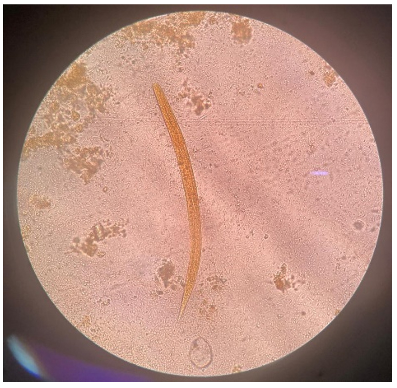
Figure. Iodine mount showing the presence of rhabditiform strongyloid larvae
Patient was treated with 200 mcg/kg of Ivermectin single dose & 400 mg/day of Albendazole for 3 days. Additionally patient underwent hemodialysis as part of his treatment for existing CKD 5D. The condition of the patient drastically improved by day 3 post initiation of treatment and repeat stool microscopy was found to be negative for strongyloid larvae. TLC increased to 6930 and the platelet counts stabilized at 1.35 lakhs. Patient was deemed fit for discharge on day 4 post treatment inception.
S. stercoralis is a soil nematode with a conglomerate biorhythm. This helminth exhibits homogonic development supplementary to a heterogonic life cycle. Although primarily a human bound parasite S. stercoralis infection has been documented in other mammals & primates.6 Across the world S. stercoralis affects approximately 100-370 million people.6 A study by Munishankaar et al, panning between 2013 to 2020 showed a strongyloidial seroprevalence of 33% among South Indian adult population.7 Given the background of asymptomatic strongyloidiasis among immunocompetent individuals, this asymptomatic parasitic infestation can turn fatal when the host Th2 response is suppressed in immunocompromised conditions.
CKD progressing to end stage renal disease results in acquired immune deficiency. The associated gut micro-floral imbalance is attributed to diet, metabolic acidosis, frequent antibiotic & oral iron therapy. This gut flora imparity results in Lipopolysaccharide (LPS) tolerance8 which opens up the possibility towards occurrence of rare nematode infections. Literature reveals a good number of strongyloidiasis cases reported in renal transplant recipients from India.3 However there is sparse documented evidence of CKD patients presenting with disseminated strongyloidiasis alongside augmented mortality reported from Srilanka,9 Thailand,10 and China.11 There is sheer paucity of intestinal strongyloidiasis data among CKD patients from the tropics possibly due to under diagnosis as well as underreporting.
Simple stool microscopy forms the cornerstone for diagnosis of intestinal strongyloidiasis. Unfortunately, the fecal diagnostic modalities inclusive of the Koga agar plate culture method have a low sensitivity despite adoption of varied concentration techniques. Molecular diagnostics have documented sensitivity of a hooting 83.3%-100%,12,13 but are cost ineffective. Serology based diagnostics promise high sensitivity but however at the cost of specificity. The recombinant fecal antigen based ELISA has a sensitivity of about 70% & a specificity of 91%.14 The accuracy of these tests depends on the population assessed and the larval load in the subject assessed. In subjects with decreased larval loads false negative diagnostic mirages may misdirect clinicians. Therefore, the rear-ground of even marginal eosinophilia in an immunocompromised individual may warrant the lookout for a parasitic infestation. Furthermore, the importance of regular screening among immunocompromised adults for early diagnosis & thereby reduced mortality/morbidity is hereby stressed.
This case report brings out the usefulness of timely diagnostic lifesaving intercession towards forbidding mortality in a rare case of strongyloidiasis presenting as thrombocytopenic acute gastroenteritis in a CKD patient. Additionally, authors would like to stress the importance of regular screening among immunocompromised adults for early diagnosis and effective management of strongyloidiasis.
ACKNOWLEDGMENTS
None.
CONFLICT OF INTEREST
The authors declare that there is no conflict of interest.
AUTHORS’ CONTRIBUTION
All authors listed have made a substantial, direct and intellectual contribution to the work, and approved it for publication.
FUNDING
None.
DATA AVAILABILITY
All datasets generated or analyzed during this study are included in the manuscript.
ETHICS STATEMENT
This study was approved by the Institutional Ethics Committee, JSS Medical College, Mysuru, India, with reference number JSSMC/IEC/07112024/06 NCT/2024-25.
INFORMED CONSENT
Written informed consent was obtained from the participant before enrolling in the study.
- Nozais JP, Thellier M, Datry A, Danis M. Disseminated strongyloidiasis. Presse Medicale (Paris, France: 1983). 2001;30(16):813-8.
- Tsai HC, Lee SS, Liu YC, et al. Clinical manifestations of strongyloidiasis in southern Taiwan. J Microbiol Immunol Infect. 2002;35(1):29-36.
- Paul M, Meena S, Gupta P, Jha S, Rekha US, Kumar VP. Clinico-epidemiological spectrum of strongyloidiasis in India: Review of 166 cases. J Family Med Prim Care. 2020;9(2):485-491.
Crossref - Buonfrate D, Bisanzio D, Giorli G, et al. The global prevalence of strongyloides stercoralis infection. Pathogens. 2020;9(6):468.
Crossref - Elzein FE, Alsaeed M, Ballool S, Attia A. Strongyloides hyperinfection syndrome combined with cytomegalovirus infection. Case Rep Transplant. 2016;2016:1786265.
Crossref - Eslahi AV, Badri M, Nahavandi KH, et al. Prevalence of strongyloidiasis in the general population of the world: a systematic review and meta-analysis. Pathog Glob Health. 2021;115(1):7-20.
Crossref - Munisankar S, Rajamanickam A, Balasubramanian S, et al. Seroprevalence of Strongyloides stercoralis infection in a South Indian adult population. PLOS Negl Trop Dis. 2022;16(7):e0010561.
Crossref - Anders HJ, Andersen K, Stecher B. The intestinal microbiota, a leaky gut, and abnormal immunity in kidney disease. Kidney Int. 2013;83(6):1010-1016.
Crossref - Karunathilaka A, Hewageegana A, Perera NY, Kumarasinghe C, Sinharachchi D. Chronic diarrhoea due to severe strongyloidiasis in a Chronic Kidney Disease patient: A case report. Anuradhapura Medical Journal. 2021;15(2):17-19.
Crossref - Hai NT, Hongsrichan N, Intuyod K, et al. Strongyloides stercoralis infection induces gut dysbiosis in chronic kidney disease patients. PLoS Negl Trop Dis. 2022;16(9):e0010302.
Crossref - Qu TT, Yang Q, Yu MH, Wang J. A Fatal Strongyloides Stercoralis Hyperinfection Syndrome in a Patient With Chronic kidney Disease: A Case Report and Literature Review. Medicine (Baltimore). 2016;95(19):e3638.
Crossref - Arndt MB, John-Stewart G, Richardson BA, et al. Impact of helminth diagnostic test performance on estimation of risk factors and outcomes in HIV-positive adults. PloS One. 2013;8(12):e81915.
Crossref - Knopp S, Salim N, Schindler T, et al. Diagnostic accuracy of Kato-Katz, Flotac, Baermann, and PCR methods for the detection of light-intensity hookworm and Strongyloides stercoralis infections in Tanzania. Am J Trop Med Hyg. 2014;90(3):535-545.
Crossref - Bisoffi Z, Buonfrate D, Sequi M, et al. Diagnostic accuracy of five serologic tests for Strongyloides stercoralis infection. PLoS Negl Trop Dis. 2014;8(1):e26.
https://doi.org/10.1371/journal.pntd.0002640
© The Author(s) 2024. Open Access. This article is distributed under the terms of the Creative Commons Attribution 4.0 International License which permits unrestricted use, sharing, distribution, and reproduction in any medium, provided you give appropriate credit to the original author(s) and the source, provide a link to the Creative Commons license, and indicate if changes were made.