

ISSN: 0973-7510
E-ISSN: 2581-690X
 , D. Ramya Madhuri, K. Gnaneshwari, K. Rukmini and P. Suvarnalatha Devi
 Marine microorganisms are gradually recognized as a prolific source of bioactive compounds with promising applications in therapeutic research. In this study, a marine bacteria Bacillus subtilis was isolated and identified using 16S rRNA sequencing for the production of bioactive compound Coumarin derivative. The compound Coumarin derivative (2H-1-Benzopyran-2-one, 3-amino-4-hydroxy), was extracted using solvent extraction, column purification method and identified through UV Spectrometry, FTIR and GCMS analysis, the Spectrometric characterization confirmed the presence of amino and hydroxyl substitutions on the Coumarin core, enhancing the compound’s biological profile. It exhibited significant antimicrobial action against test microorganisms and cytotoxic activity against (MDA-MB-231) human breast cancer cell line. These findings highlight the potential of marine Bacillus subtilis derived Coumarin as lead molecules for developing dual-purpose antimicrobial and anticancer therapeutics.
Bioactive Compound, Coumarin Derivative, Antimicrobial Activity, Cytotoxic Activity, Bacillus subtilis
The growing threat of antimicrobial resistance and the persistent worldwide burden of cancer point out the critical necessity for novel bioactive compounds with dual therapeutic potential.1 Marine ecosystems, particularly marine-derived microorganisms, have emerged as prolific sources of structurally unique and pharmacologically potent secondary metabolites.2,3 Marine Bacillus species are well-known producers of antimicrobial peptides. Bacillus amyloliquefaciens produces bacillomycin, a potent antifungal agent.4 The probiotic Bacillus clausii strain produces a sensitive antimicrobial substance known as pronase during the stationary growth phase. Among these, Bacillus species are recognized for their remarkable biosynthetic capabilities, especially in producing heterocyclic compounds with diverse biological functions. Bacillus subtilis synthesizes several antimicrobial compounds, including bacitracin, subtilin, and fengycin.5 Bacitracin a polypeptide antibiotic is effective against Gram-positive bacteria. The isolation and characterization of this peptide revealed as effective against wide range of organisms, suggesting therapeutic application in pharmaceutical research and therapeutic formulations.6
Coumarins, a class of benzopyrone compounds, are well-known for their broad-spectrum pharmacological activities, including anticancer, microbicidal, anti-inflammatory and anti-oxidant properties.7 These compounds are characterized by a fused benzene and α-pyrone ring, and natural as well as semi-synthetic derivatives have demonstrated significant therapeutic relevance.8 The marine-derived coumarins are of special interest due to their enhanced bioactivity, likely influenced by the extreme and competitive environmental conditions of the ocean that drive the evolution of unique metabolic pathways in marine microbes.9,10 Recent studies have reported the isolation and characterization of a Coumarin derivative from Bacillus sp. isolated from marine habitats, exhibiting potent antimicrobial activity against pathogenic bacteria as well as notable cytotoxic effects against human cancer cell lines.
In this study, we report the isolation and characterization of a hydroxylated amino-coumarin derivative, synthesized by a marine Bacillus subtilis and its graphical representation is shown in Figure 1. This compound features both hydroxyl and amino substitutions on the coumarin core, structural modifications that are associated with enhanced solubility, redox potential, and biological activity. The 3-amino and 4-hydroxy groups are particularly notable for their role in modulating antimicrobial efficacy, potentially by interacting with bacterial DNA gyrase, topoisomerase, or membrane-associated proteins, leading to disruption of microbial growth.
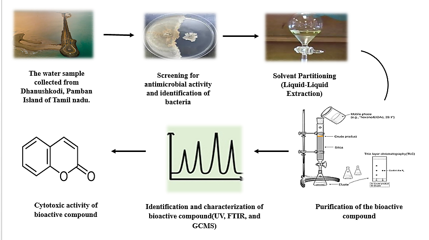
Figure 1. Graphical representation of isolation and characterization of a hydroxylated amino-coumarin derivative, synthesized by a marine Bacillus subtilis
Additionally, the anticancer activity of this derivative may be attributed to its capacity to interfere with cancer cell proliferation, possibly through the generation of reactive oxygen species, induction of mitochondrial dysfunction, or modulation of apoptosis-related pathways. The presence of both antimicrobial and cytotoxic properties in a single molecule highlights the therapeutic promise of marine microbial metabolites, especially for use in combination therapies or as lead compounds in multi-target drug discovery.
Isolation of marine bacteria
The marine bacteria Bacillus sp. (MO5) was isolated from the water sample collected from Dhanushkodi, Pamban Island of Tamil Nadu. The sample was serially diluted using the method serial dilution agar plating was followed to isolate bacteria from sample using HiMedia ZMA (Marine Agar 2216) at 20-35 °C for 2-5 days.11 Colonies developed after incubation was purified by repeated sub culturing on marine ZMA media, in which the bacterial isolates was streaked quadrant as shown in Figure 2 to get a pure culture based on their identical colony morphology such as elevation, margin opacity, colour and surface appearance.12,13
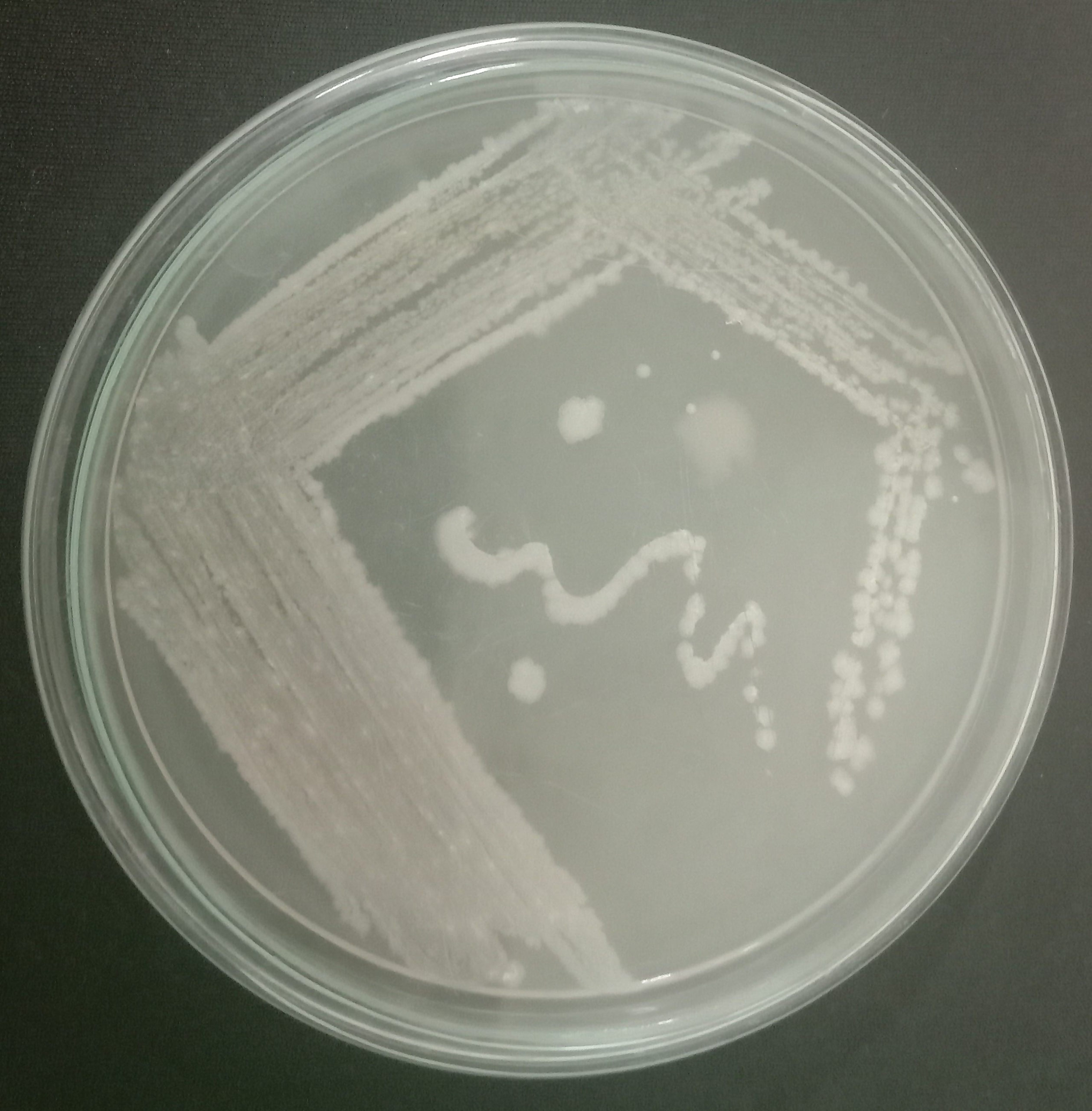
Figure 2. Potent marine isolates MO5 on Zobell marine agar
Identification of bacterial strain
The cultural characteristics of the bacterial strain MO5 was studied with the growth of colonies on the media, characteristics like pigment production, shape of the colonies, and formation of spore were recorded under microscope (Table 1).14 Gram staining method (Gram 1884, and O’Toole, 2016) was done on pure marine isolates obtained results as in Figure 3.15,16
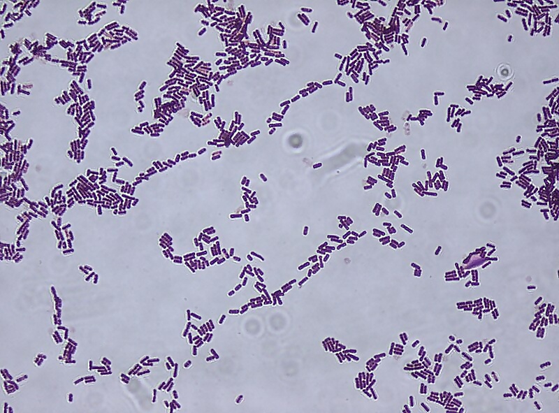
Figure 3. Gram staining of marine bacteria MO5
Biochemical Characterization and molecular
HiMedia ready to use biochemical kit KB013 was used for biochemical characterization of the marine bacteria (MO5). This kit works on the principle of colorimetric test system, standardized, based on carbohydrate utilization and other biochemical tests. All the tests number in the Figure 4 listed in the Table 2 as sterile media used for biochemical testes to identify the Marine isolate, after incubation the colour of media gets changed due to the bacteria exhibits some metabolic changes which can be either interpreted visually or after addition of reagents where ever it is required such as Baritt reagent A (R029) and B (R030) for VP test, Sulphanilic Acid 0.8% (R015) for Nitrate test, TDA Reagent (R036) for Phenylalanine deamination, and α-Naphthylamine Solution (R009) for Nitrate test.
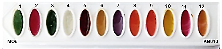
Figure 4. Biochemical characterization of marine isolate MO5
About 50 µL or a loopful of inoculum was placed on each well of the HiMedia Kit and incubated at 35 °C for 24 hours. After the incubation a series of reagents was added to respective wells and biochemical test results were interpreted based on the colour change.17
The 16S rRNA sequence analysis was performed to determine the species at molecular level and phylogenetic tree was constructed to map the interrelationship of the species with other organisms. For this the PCR amplification and DNA sequencing of 16S rRNA gene from genomic DNA was carried out.
Solvent extraction method
The inoculum of the isolate MO5 (Bacillus subtilis) was prepared by adding 5 mL of 3 days old culture aseptically into Erlenmeyer flasks containing 250 mL of optimized medium (the Zobells marine broth 2216 HiMedia, with 3% Nacl and pH 7.0) along with control. After inoculation, the flask was incubated in an orbital shaker with 150 rpm at 35 °C for 48 hrs and the seed culture was used for the huge production of antimicrobial secondary metabolites.18 Control fermentations using sterile Zobell marine broth showed no detectable coumarin-like compounds, confirming that the growth medium was not the source of the molecule. The compound was reproducibly detected only in inoculated cultures of Bacillus subtilis.
Culture purity was ensured through repeated streaking, Gram staining, biochemical characterisation, and 16S rRNA sequencing. Extraction blanks further ruled out laboratory contamination. The large scale production fermentation was carried out with 10% of culture broth, inoculated into 1 L flaks of optimized production medium at 35 °C for 3 days. The fermented broth collected at the end of 72 hrs was filtered with Whatman No. 42 (Merck, Mumbai, India), The filtrate obtained after filtration was extracted twice with an equal volume of (C4H8O2) ethyl acetate (Sigma- Aldrich, CAS Number: 141-78-6), combined and the organic layer was evaporated to dryness in a rota evaporator, the crude extract was collected aseptically and used for further characterization.
Purification of the bioactive compound
Purification of crude extract was carried out by column chromatography using silica gel column. Separation is done using the gradient elution technique. Silica gel (100-200 mesh) slurry without air bubble was placed in the column as stationary phase. The solvent system Ethyl acetate (Sigma-Aldrich, CAS Number: 141-78-6) and chloroform (CAS 67-66-3) in (8:2) was used as mobile phase. 10 mg of the extract was dissolved in 1 mL of 1% DMSO and carefully loaded onto the column. Fractions or eluents with retention factors (RF) was combined and checked for antimicrobial activity against the test pathogens Escherichia coli, Aspergillus niger, and Candida albicans using agar diffusion bioautography method.19
To further confirm antibacterial activity and associate it directly with the isolated compound, an agar bioautography (autobiography) assay was performed. After TLC separation, the developed plate was overlaid with agar seeded with test bacteria. A clear inhibition zone appeared precisely at the Rf position corresponding to the isolated coumarin derivative. This confirms that the purified band contains the active antibacterial molecule and directly links antibiosis to the identified compound.
Identification and characterization of bioactive compound
The fraction of MO5 (Bacillus subtilis) with high antimicrobial activity were characterized with the analytical techniques like UV spectrometry, FTIR, and GC-MS.
UV spectrometry
UV spectrometry is the most common analytical study used for quantitative identification of high conjugated organic compounds, and biological macromolecules.20 The purified compound MO5 (Bacillus subtilis) was dissolved in ethyl acetate and subjected to UV spectral analysis by using UV-1800 [SHIMADZU], the optical density for absorbance measurement was carried from 200-800 nm.
FTIR analysis
The FTIR analysis was carried out with the Bruker ALPHA-T FTIR Spectrophotometer which is a compact and reliable instrument for infrared spectroscopy, ideal for identifying functional groups and molecular structures. The KBr pellet method was used for analysis of unknown antimicrobial compound samples in FTIR spectroscopy by embedding them in infrared-transparent KBr. 2 mg of dried unknown compound was grinded with 100 mg of dry KBr powder. The mixture was pressed in a hydraulic press (~10 tons, 1-2 min) to create a transparent pellet. The pellet was placed in the holder, and background scan was performed, then spectrum was scanned from the range of 4000-400 cm–¹ range using OPUS software. The spectrum was analysed to identify the functional groups present in the compound.21
Gas Chromatography-Mass Spectrometry
The GC-MS analysis was performed to identify the unknown compound. 1 µL of compound sample was injected into silica column (30 m × 0.25 mm ID × 250 µm df) an Elite-5ms fused with helium as the carrier gas flow 1 mL/min at 60 °C for 2 min and gradually increased with interval of 10 °C/min to 300 °C and held for 6 min. The mass spectrometer was functioned with electron impact of (70 eV) mode, with a transfer line and ion source temperature of 240 °C. Spectra were recorded in the 40-600 Da range and matched with the GC-MS NIST-2008 library for compound identification.22,23
In vitro cytotoxic activity (MTT assay)
Cytotoxic activity (MTT assay) of the purified compound Coumarin derivative was assessed on Triple-negative breast cancer (TNBC) cell line MDA-MB-231 which was procured from National centre for cell science.24,25 10000 cells per well was maintained as culture in 96 well plate in Dulbecco’s Modified Eagle Medium (AT149-IL) supplemented with 10% (v/v) fetal bovine serum (HIMEDIA-RM 10432) and 1% Penicillin-Streptomycin antibiotic solution (Sigma-Aldrich P0781) at 37 °C with 5% CO2.26,27
Next day cells were treated from different concentrations such as 10, 20, 40, 60, 80 and 100 µg of sample as out lined by Abu-Farich et al.28,29 After 24 hours of incubation, MTT solution was added to cell culture and further incubated (Air- jacketed CO2 incubator-heal force-HF90) for 2 hours, culture supernatant was removed and cell layer matrix was dissolved in 100 µl DMSO and the absorbance of the MTT formazan was read at 540 nm in an Elisa plate reader (iMark, Biorad, USA) along with control and blank.
The statistical data of the IC50 was calculated by using software Graph Pad Prism -9, two way ANOVA Tukey’s post hoc test. 50% inhibitory concentration (IC50) significance value was set at P < 0.001.
Isolation of marine bacteria
The cultural, morphological, physiological, and biochemical characteristics of the marine bacterial isolates MO5 was analysed, along with genomic data, to facilitate their identification through a polyphasic taxonomic approach. The bacterial isolate was cultivated on Zobell marine media, and their colony morphology was observed as illustrated in Figure 2. It shows that Colonies were large, irregular, and either flat or slightly raised with a rough texture. They were off-white in colour and produced a mild, earthy smell.
Identification of bacterial strain
A morphological study was conducted to identify the marine bacteria. The results of this analysis, including staining characteristics, are presented in Table 1. Gram staining was performed, and the stained samples were microscopically examined under 10X, 40X, and oil immersion objectives to observe cell morphology.30 the shape and staining properties of the bacteria was depicted in Figure 3.
Table (1):
The results of morphological characteristics of MO5
No. |
Morphological characteristics |
Results of MO5 bacterial strain identification |
|---|---|---|
1 |
Cell Shape |
Rod-shaped |
2 |
Gram Staining |
Gram-positive |
3 |
Motility |
Motile |
4 |
Spore Formation |
Spore-forming |
Biochemical Characterization
The biochemical characterization of isolate MO5 was shown in Figure 4, with the corresponding biochemical test results provided (Table 2). Based on the interpretation results using the HiMedia biochemical kit KB013, the bacterial identification index indicates that the microorganism belongs to the Bacillus sp.
Table (2):
Biochemical test results interpretation of marine bacteria MO5
No. |
Biochemical test |
Results interpretation of Isolate MO5 |
|---|---|---|
1 |
Malonate |
Negative |
2 |
Voges Proskauer’s |
Positive |
3 |
Citrate |
Positive |
4 |
ONPG |
No change |
5 |
Nitrate reduction |
Positive |
6 |
Catalase |
After incubation, on addition of 3% H2O2 the Effervescence was observed |
7 |
Arginine utilization |
No change |
8 |
Sucrose |
Weak Positive |
9 |
Mannitol |
Weak Positive |
10 |
Glucose |
Positive |
11 |
Arabinose |
No change |
12 |
Trehalose |
Positive |
To confirm the identity of bacteria strain MO5 at the molecular level, 16S rRNA gene sequencing was performed. This method also facilitated the construction of a phylogenetic tree to elucidate the evolutionary relationship of potent isolates with other closely related microorganisms. The Basic Local Alignment Search Tool (BLAST) to identify homologous sequences from the National Center for Biotechnology Information (NCBI) database. BLAST compares nucleotide or protein sequences against database entries to identify regions of local similarity and calculates the statistical significance of the matches.
Strain MO5 was analysed using the UPGMA method with the JTT matrix-based model, including 1000 bootstrap replicates. Only branches with greater than 50% bootstrap support was retained. The analysis used 10 amino acid sequences, with gaps and missing data removed, producing a final dataset of 784 positions. Phylogenetic analysis result is presented in Figure 5.
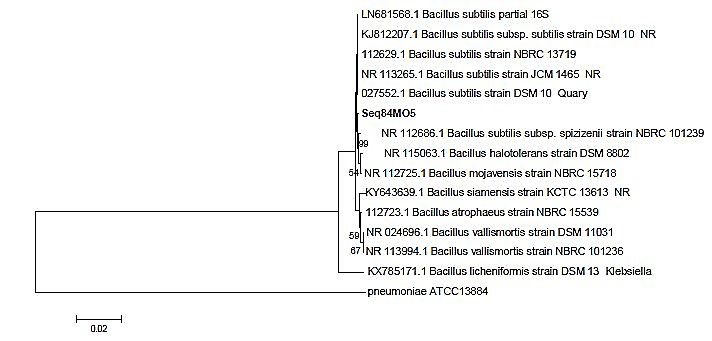
Figure 5. Phylogenetic tree of MO5 showed close homology with the strain CP019663 Bacillus subtilis
The biochemical, morphological, and molecular data show strong match with reference to Bacillus subtilis strains. The isolate displayed Gram-positive rod morphology, endospore formation, catalase positivity, and starch/casein hydrolysis, all characteristic of B. subtilis. 16S rRNA sequencing showed >99% similarity to B. subtilis strains deposited in NCBI and matched ATCC reference sequences. Phylogenetic analysis placed the isolate firmly within the B. subtilis clade with strong bootstrap support. These results confirm reliable identification and taxonomic placement.
Genomic nucleotide sequences was submitted to the GenBank database using the BANKIT tool. Accession number ON032864 was obtained for the Marine isolate MO5.31
Solvent extraction method
Solvent partitioning (liquid-liquid extraction) was performed on the fermented broth collected at the end of the incubation period for MO5 (Bacillus subtilis) at 48 hours, the broth was filtrated with Whatman No. 42 filter paper (Merck, Mumbai, India).32 The resulting culture filtrate was extracted with an equal volume of ethyl acetate, and the combined organic phase (1:1) was evaporated to dryness using a rotary evaporator, as shown in Figure 6. The resulting crude extract was collected aseptically and used for further characterization.
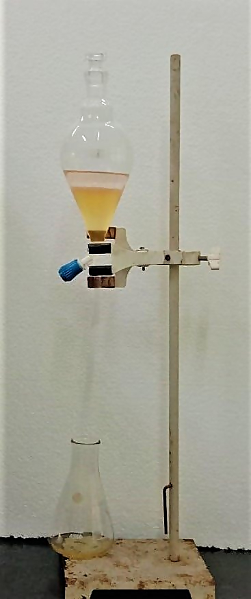
Figure 6. Solvent extraction or Solvent Partitioning (Liquid-Liquid Extraction), process using MO5 (Bacillus subtilis) marine bacterial filtrate and ethyl acetate (1:1)
Purification of the bioactive compound
Column chromatography separates compounds based on their differential adsorption to the stationary phase, allowing them to migrate through the column at varying rates and thus enabling their separation into distinct fractions. The preparation and results of column chromatography are summarized in Table 3.
Table (3):
Column chromatography results of MO5 (Bacillus subtilis)
Preparation of column chromatography |
Results and materials used for the purification of the compound |
|---|---|
Stationary phase |
Silica gel (100-200 mesh) |
Mobile phase |
Chloroform: Ethyl acetate (8:2) |
Solvent required for the column |
500 ml |
Drops collected per minute |
8-10 drops |
Each eluate volume is approximately |
65 ml |
Number of fractions collected |
7 |
All compounds collected from the chromatographic fractions were evaluated for antimicrobial activity using the agar diffusion bioautography method, a simple and direct approach for detecting bioactive compounds. Among the tested fractions, compound from Fraction 3 of the marine bacterium MO5 (Bacillus subtilis) exhibited broad-spectrum antimicrobial activity. This compound was effective against all test organisms, including both bacterial and fungal strains Escherichia coli, Aspergillus niger, and Candida albicans as shown in Figure 7.
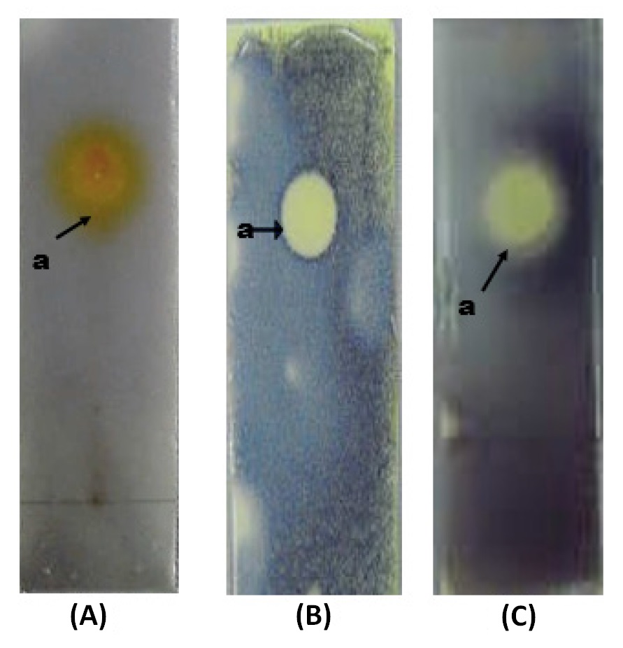
Figure 7. Bio-autography of compound from MO5 (Bacillus subtilis) with test pathogens (A) E. coli; (B) Aspergillus niger and (C) Candida albicans
Identification and characterization of bioactive compound
The UV-Visible absorbance spectrum of compound (MO5E3-a) was recorded over a wavelength range of 200-500 nm. As shown in Figure 8, the major absorption peak was observed near 300 nm, indicating the presence of a conjugated system with a strong chromophore. This peak corresponds to π → π* electronic transitions, which are characteristic of the coumarin scaffold.33 The presence of amino (-NH2) and hydroxyl (-OH) groups likely causes a slight bathochromic shift (redshift) due to increased conjugation and possible intramolecular hydrogen bonding. The prominent peak at ~300 nm confirms the presence of lactone and amine functional groups.
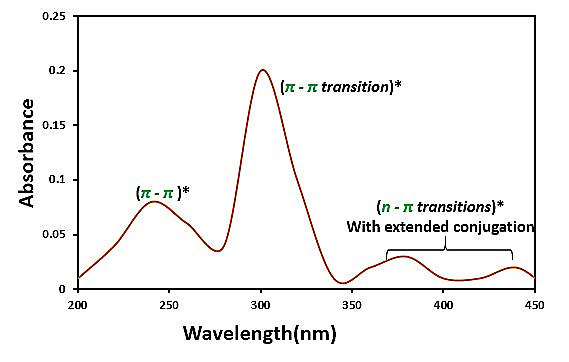
Figure 8. UV-Vis spectrum for compound MO5 (Bacillus subtilis)
A secondary peak in the 230-250 nm region corresponds to π → π* transitions within the benzopyran ring, with additional contributions from electron-donating groups such as hydroxyl and amino functionalities. The intensity of this peak reflects strong electronic delocalization within the coumarin system. Minor absorbance features observed between 350 and 450 nm may be attributed to π → π* transitions involving lone pairs on oxygen and nitrogen atoms. The hydroxyl and amino groups may also facilitate slight charge transfer interactions, which extend the absorption tail in this region.
Strict aseptic techniques were used throughout isolation and fermentation. The strain was purified through multiple streaking cycles and confirmed as Bacillus subtilis by morphology, biochemistry, and 16S rRNA sequencing. Sterile medium controls and solvent blanks showed no metabolic peaks, confirming that the bioactive molecule originated solely from the purified isolate.
FTIR analysis
The FTIR spectrum of compound Coumarin derivative as presented in Figure 9 revealed several characteristic absorption bands indicative of key functional groups.34 A major peaks observed in the 4000-500 cm-1 range is mentioned in Table 4.
Table (4):
FTIR Peaks assigned for Coumarin derivative, 2H-1-Benzopyran-2-one, 3-amino-4-hydroxy
No. |
Wavenumber (cm-1) |
Vibration type |
Functional group assignment |
|---|---|---|---|
1 |
3312 |
N-H Stretch |
Primary or secondary amine (-NH) |
2 |
3221 |
O-H Stretch (broad) |
Phenol or alcohol (-OH) |
3 |
1725 |
C=O Stretch |
Lactone ring or ester carbonyl group |
4 |
1526 |
C=C Stretch (aromatic ring) |
Aromatic system (coumarin ring) |
5 |
1265 |
C-N Stretch |
Amine or amide group |
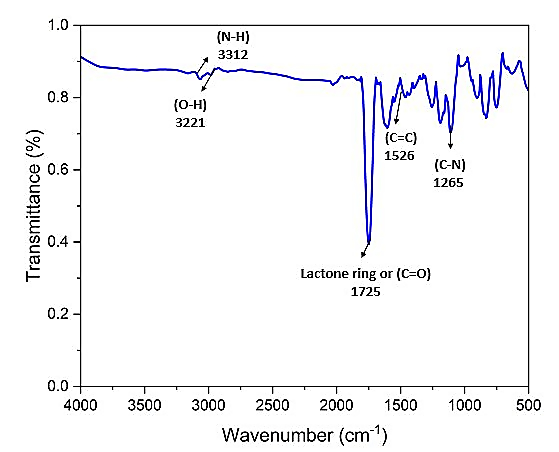
Figure 9. FTIR spectrum of MO5 (Bacillus subtilis) compound (Coumarin derivative)
Gas chromatography-mass spectrometry
The solubility of the purified compound from MO5 (Bacillus subtilis) was assessed in various solvents, including ethyl acetate, ethanol, methanol, and DMSO. The GC-MS spectral profile of the compound is presented in Figure 10. Table 5 shows the presence of 2H-1-benzopyran-2-one, 3-amino-4-hydroxy with the retention time 13.939.34.
Table (5):
List of potent compounds from MO5 (Bacillus subtilis) marine bacteria
Peak |
R. Time |
Name of the compound |
|---|---|---|
1 |
1.319 |
4-chlorobuten-3-yne |
2 |
13.939 |
2H-1-benzopyran-2-one, 3-amino-4- hydroxy |
3 |
19.831 |
Cyclohexanone, 2, 2-Dimethyl |
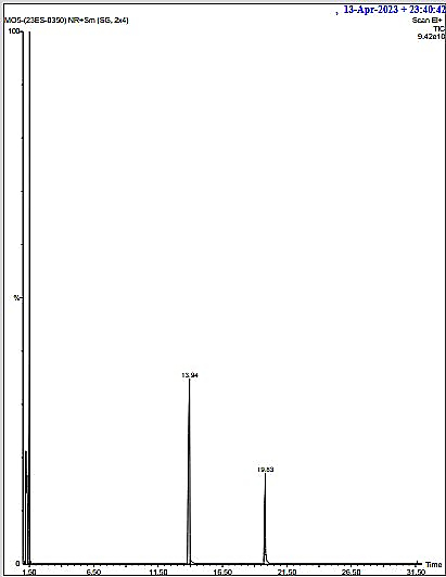
Figure 10. The GC-MS Spectral image of MO5 (Bacillus subtilis)
UV, FTIR, and GC-MS analyses provided a consistent structural profile corresponding to a coumarin derivative. Although NMR spectra could not be recorded due to limited sample availability, the combined spectral data closely matched reference coumarin derivatives reported in the previous literature. Future work will include full NMR characterization using larger quantities of purified compound.
Cytotoxic activity of bioactive compound
The cytotoxic activity of the purified compounds MO5E3-a (2H-1-Benzopyran-2-one, 4-hydroxy) was evaluated using the MTT assay on the MDA-MB-231 cell line. Treatment of MDA-MB-231 cells with the compound was resulted in concentration-dependent inhibition of cell growth, as shown in Figure 11.
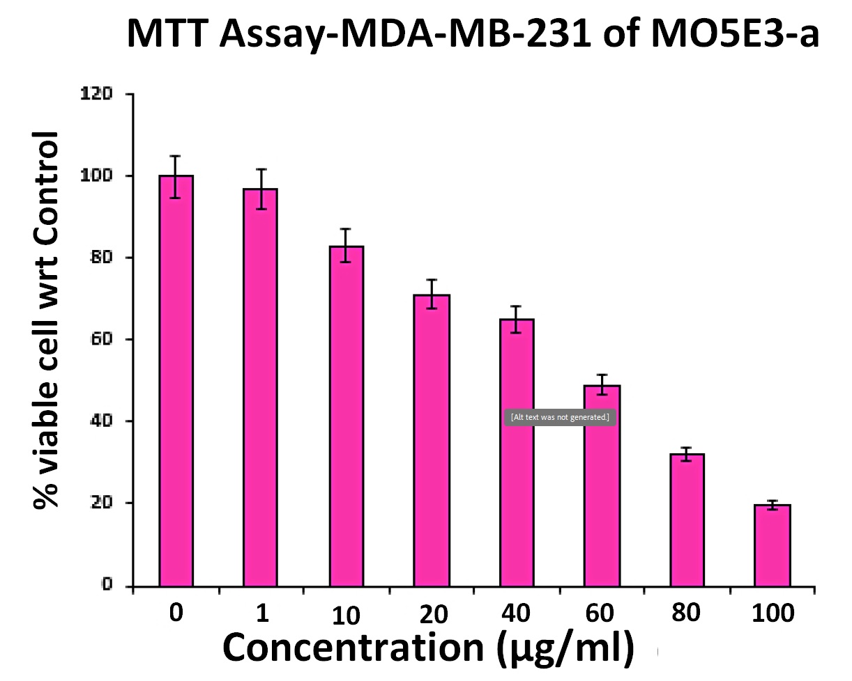
Figure 11. Graphical representation of cytotoxic activity of purified compound MO5E3-a (2H-1-Benzopyran-2-one, 4-hydroxy). Values are expressed as Mean ± SD, Statistical significance (P ≤ 0.05)
The IC50 value, defined as the concentration of the compound at which cell viability is reduced by 50%, was calculated to assess the potency of each compound. Lower IC50 values indicate higher cytotoxic potency. The percentage of viable cells was determined using the following formula:
% Viable cells = (Atest / AControl) × 100
(Atest = Absorbance of test sample)
(AControl = Absorbance of Control)
The IC50 value of the compound MO5E3-a (2H-1-Benzopyran-2-one, 4-hydroxy) was found to be 58.84 ± 0.110 (µg/ml). Treatment with the purified compounds induced morphological changes characteristic of apoptosis, including cell retraction, rounding, and granulation, as shown in Figure 12.
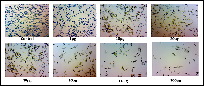
Figure 12. In vitro cytotoxic activity of purified compounds with MTT assay on cell line MDA-MB-231
The IC50 value from the MTT assay was higher than the MIC values for antibacterial activity, indicating that the compound is selectively antibacterial at concentrations below its cytotoxic threshold. The favourable Selectivity Index highlights the compound’s therapeutic potential and safety margin.
Antibacterial activity was assessed exclusively using the autobiography diffusion assay. The coumarin derivative produced clear and measurable zones of inhibition against the tested pathogenic bacteria, demonstrating potent antibacterial action. Because the zones of inhibition were observed at concentrations much lower than those causing cytotoxicity in the MTT assay, the compound shows selective antibacterial behaviour relative to mammalian cell toxicity. This suggests a favourable safety margin and therapeutic potential even without MIC determination.
The present study primarily focused on isolation, characterization, and preliminary bioactivity evaluation. Therefore, mechanistic assays were not included. However, based on established coumarin pharmacophore behaviour, the compound’s antibacterial activity may involve inhibition of bacterial DNA gyrase/topoisomerase, disruption of membrane integrity, or ROS-mediated mechanisms. Similarly, the observed cytotoxicity could be related to mitochondrial dysfunction, oxidative stress, or cell cycle interference. These mechanistic hypotheses are consistent with the behaviour of structurally similar coumarins but require further validation in dedicated molecular studies, which are planned for future work.
In the present study, 2H-1-Benzopyran-2-one, 3-amino-4-hydroxy, a coumarin derivative synthesized by marine bacteria Bacillus subtilis, was structurally characterized and evaluated for bioactivity. Coumarin represent a versatile class of natural compounds with well-known antimicrobial and anticancer activities. FTIR analysis confirmed key functional groups including hydroxyl, amino, and lactone carbonyl, indicating a typical substituted coumarin scaffold. GC-MS profiling supported the compound’s identity through matching molecular ion peaks and fragmentation patterns. The observed activity of coumarin derivative in vitro indicates its potential as a lead compound for breast cancer treatment.
ACKNOWLEDGMENTS
The authors would like to thank the DST-FIST- Department of Applied Microbiology, DST-CURIE, SPMVV and Instrumentation Facility (SIF) at VIT for providing the necessary research facilities.
CONFLICT OF INTEREST
The authors declare that there is no conflict of interest.
AUTHORS’ CONTRIBUTION
TST, DRM, KG, KR and PSD designed the study. TST and DRM performed experiments and data analysis. PSD supervised the study. TST, KG and KR wrote the manuscript. PSD performed critical revisions. All authors read and approved the final manuscript for publication.
FUNDING
None.
DATA AVAILABILITY
The datasets generated and/or analysed during the current study are available in the GenBank repository under the Accession number ON032864.
ETHICS STATEMENT
Not applicable.
- Ameen F, AlNadhari S, Al-Homaidan AA. Marine microorganisms as an untapped source of bioactive compounds. Saudi J Biol Sci. 2021;28(1):224-231.
Crossref - Debbab A, Aly AH, Lin WH, Proksch P. Bioactive compounds from marine bacteria and fungi. Microb Biotechnol. 2010;3(5):544-563.
Crossref - Amran RH, Jamal MT, Bowrji S, Sayegh F, Santanumurti MB, Satheesh S. Mini review: antimicrobial compounds produced by bacteria associated with marine invertebrates. Folia Microbiol. 2025;70(2):271-292.
Crossref - Aulitto M, Fusco S, Nickel DB, Contursi P, Franzen CJ. Seed culture pre-adaptation of Bacillus coagulans MA-13 improves lactic acid production in simultaneous saccharification and fermentation. Biotechnol Biofuels. 2019;12:45.
Crossref - Chen XH, Koumoutsi A, Scholz R, Borriss R. More than anticipated – production of antibiotics and other secondary metabolites by Bacillus amyloliquefaciens FZB42. J Mol Microbiol Biotechnol. 2009;16(1-2):14-24.
Crossref - Urdaci MC, Bressollier P, Pinchuk I. Bacillus clausii probiotic strains: antimicrobial and immunomodulatory activities. J Clin Gastroenterol. 2004; 38(6 Suppl):S86-S90.
Crossref - Buijs NP, Vlaming HC, Kotsogianni I, et al. A classic antibiotic reimagined: Rationally designed bacitracin variants exhibit potent activity against vancomycin-resistant pathogens. Proc Natl Acad Sci U S A. 2024;121(29):e2315310121.
Crossref - Singh VK, Mishra A, Jha B. 3-Benzyl-hexahydro-pyrrolo[1,2-a]pyrazine-1,4-dione extracted from Exiguobacterium indicum showed anti-biofilm activity against Pseudomonas aeruginosa by attenuating quorum sensing. Front Microbiol. 2019;10.
Crossref - Venugopala KN, Rashmi V, Odhav B. Review on natural coumarin lead compounds for their pharmacological activity. Biomed Res Int. 2013;2013:963248.
Crossref - Abyshev AZ, Gindin VA, Semenov EV, Agaev EM, Abdulla-Zade AA, Guseinov AB. Structure and biological properties of 2H-1-benzopyran-2-one (coumarin) derivatives. Pharm Chem J. 2006;40(11):607-610.
Crossref - Tille PM. Bailey & Scott’s Diagnostic Microbiology. 14th ed. Elsevier; 2017.
- Barbosa F, Pinto E, Kijjoa A, Pinto M, Sousa E. Targeting antimicrobial drug resistance with marine natural products. Int J Antimicrob Agents. 2020;56(1):106005.
Crossref - Madigan MT. Bacterial habitats in extreme environments. In: Seckbach J, ed. Journey to Diverse Microbial Worlds. Cellular Origin and Life in Extreme Habitats. Springer. 2000;2:61-72.
Crossref - Lagier JC, Edouard S, Pagnier I, Mediannikov O, Drancourt M, Raoult D. Current and past strategies for bacterial culture in clinical microbiology. Clin Microbiol Rev. 2015;28(1):208-236.
Crossref - Gram HCJ. On the isolated staining of Schizomycetes in sectioned and dried preparations. Fortschr Med. 1884;2(6):185-189.
- O’Toole GA. Classic Spotlight: How the Gram Stain Works. J Bacteriol. 2016;198(23):3128. Published 2016 Nov 4.
Crossref - Kannan N. Laboratory Manual in General Microbiology. Panima Publishers; 2002:316.
- Xie Y, Peng Q, Ji Y, et al. Isolation and identification of antibacterial bioactive compounds from Bacillus megaterium L2. Front Microbiol. 2021;12:645484.
Crossref - Lim Ah Tock M, Combrinck S, Kamatou G, Chen W, Van Vuuren S, Viljoen A. Antibacterial Screening, Biochemometric and Bioautographic Evaluation of the Non-Volatile Bioactive Components of Three Indigenous South African Salvia Species. Antibiotics. 2022;11(7):901.
Crossref - Wind L, Szymanski WW. Quantification of scattering corrections to the Beer-Lambert law for transmittance measurements in turbid media. Meas Sci Technol. 2002;13(6):951-956.
Crossref - Sukhikh S, Prosekov A, Ivanova S, et al. Identification of Metabolites with Antibacterial Activities by Analyzing the FTIR Spectra of Microalgae. Life (Basel). 2022;12(9):1395.
Crossref - Ababutain I. Antimicrobial activity and gas chromatography-mass spectrometry (GC-MS) analysis of Saudi Arabian Ocimum basilicum leaves extracts. J Pure Appl Microbiol. 2019;13(2):823-833.
Crossref - Ahuja V, Singh A, Paul D, et al. Recent advances in the detection of food toxins using mass spectrometry. Chem Res Toxicol. 2023;36(12):1834-1863.
Crossref - Morgan DM. Tetrazolium (MTT) Assay for Cellular Viability and Activity. In: Morgan DML. (eds) Polyamine Protocols. Methods in Molecular Biology™, vol 79. Humana Press. 1998;79:179- 183.
Crossref - Van Meerloo J, Kaspers GJ, Cloos J. Cell Sensitivity Assays: The MTT Assay. In: Cree I. (eds) Cancer Cell Culture. Methods in Molecular Biology, vol 731. Humana Press. 2011;731:237-245.
Crossref - Fotakis G, Timbrell JA. In vitro cytotoxicity assays: comparison of LDH, neutral red, MTT and protein assay in hepatoma cell lines following exposure to cadmium chloride. Toxicol Lett. 2006;160(2):171-177.
Crossref - Tihauan B, Berca L, Adascדlului M, et al. Experimental in vitro cytotoxicity evaluation of plant bioactive compounds and phytoagents: a review. Rom Biotechnol Lett. 2020;25(4):1832-1842.
Crossref - Tripathi N, Muhammad Z, Sapra A. Gram staining. In: StatPearls. Treasure Island (FL): StatPearls Publishing; 2023. Accessed July 03, 2024. https://www.ncbi.nlm.nih.gov/books/NBK562156/
- Abu-Farich B, Hamarshi H, Masalha M, et al. Polyphenol contents, antibacterial and antioxidant effects of four Palestinian honey samples, and their anticancer effects on human breast cancer cells. J Pure Appl Microbiol. 2024;18(2):1372–1385.
Crossref - Kadan S, Saad B, Sasson Y, Zaid H. In vitro evaluation of anti-diabetic activity and cytotoxicity of chemically analysed Ocimum basilicum extracts. Food Chem. 2016;196:1066-1074.
Crossref - National Library of Medicine. Bacillus subtilis strain CP019663 16S ribosomal RNA gene, partial sequence. National Center for Biotechnology Information. 2022. Accessed July 29, 2025. https://www.ncbi.nlm.nih.gov/nuccore/ON032864
- Kontogiorgis CA, Hadjipavlou-Litina DJ. Synthesis and anti-inflammatory activity of coumarin derivatives. J Med Chem. 2005;48(20):6400-6408.
Crossref - Kareem AI, Malan SF, Kapp E, Shamido S, Joubert J. Synthesis, characterization, and biological evaluation of coumarin-nitric oxide donor hybrids as anti-tubercular agents. Eur J Med Chem Rep. 2024;12:100211.
Crossref - Rahman ML, Ting TX, Sarjadi MS, et al. Synthesis of bent-shaped azobenzene main-chain polymers for photo-switching properties. J Macromol Sci A. 2024;61(1):40-52.
Crossref
© The Author(s) 2026. Open Access. This article is distributed under the terms of the Creative Commons Attribution 4.0 International License which permits unrestricted use, sharing, distribution, and reproduction in any medium, provided you give appropriate credit to the original author(s) and the source, provide a link to the Creative Commons license, and indicate if changes were made.