


ISSN: 0973-7510
E-ISSN: 2581-690X
The Minahasa local pig is unique because it is the oldest local pig that has spread to the Philippine islands. Minahasa local pigs have endemic characteristics because they are in the Wallacea zone. Research has been carried out to isolate bacteria from pig intestinal fluid and obtain an antibiotic response profile from pure bacterial isolates. Pig samples were obtained from two locations in North Sulawesi, namely in North Minahasa and North Minahasa. Intestinal fluids are taken immediately when the pig is slaughtered. The liquid was preserved in a sterile container and inoculated directly on the nutrient agar medium by the dilution method. The bacterial isolates obtained were pure cultured and then used for automatic phenotypic identification using Vitek 2 Compact. The results showed that pure culture isolates were obtained from intestinal fluids of local pigs in Minahasa, North Sulawesi, indicating that two isolates (S1 and S2) were Escherichia coli and S3 was Enterobacter aerogenes. Isolate S1 showed resistance to Ampicillin, while isolate S2 showed resistance to Tetracycline, furthermore isolate S3 showed resistance to Tetracycline, Furanes, and Trimethoprim/Sulfonamide. The results confirm that further research is needed to isolate and test the antibiotic resistance of bacteria from pig intestines in several locations and various stages of common local pigs.
Local pig intestinal fluid, Minahasa, antibiotic resistance
Indonesia has the world’s largest pig germplasm because it has five of its eight local pig species. Sulawesi Island has two typical local pigs, namely the local Toraja pig and the local Minahasa pig. The local Minahasa pig is unique because it is the elder of the local pig that spreads to the Philippine islands.1,2 In addition, local Sulawesi pigs have endemic characteristics because they are in the Wallacea zone. Because pigs are all-eating animals, the features of the digestive tract of local pigs are unique. One object of study of the digestive tract in local pigs is the gut microbiome, especially bacteria.2
In recent decades, the gut microbiome has been the subject of research due to its importance in animal health, including human health.3 Digestive bacteria in the gut microbiome play a vital role in the growth and metabolic processes of animals.1,3,4 Uniquely, animals coevolved with the gut microbiome.5 Pigs are monogastric livestock that can convert food ingredients efficiently when supported by the quality of the ration they consume.6,7
Pigs will grow faster and quickly become adults and are prolific, as indicated by having many children per birth, which ranges from 8-14, and can give birth twice.8 As a monogastric animal and consuming everything, the microbes associated with the digestive tract of pigs have tremendous diversity. Pigs have coevolved with digestive tract microbes to be able to grow and develop properly. Based on many research reports, only 1% of bacteria can be isolated and cultured on artificial media.9 The diversity of microbes, primarily bacteria in pig intestinal fluid, is a broad object of study to be investigated.9,10 One aspect of microbial research in pig intestines is the activity of microbial resistance to antibiotics.11 However, pigs are one of the most consumed livestock in the world. The local Minahasa community uses pork as a source of animal protein. Pig intestines are widely used for the manufacture of traditional Minahasa vegetables. Therefore, it is necessary to identify bacteria from pig intestines and test for sensitivity and antibiotic resistance. Knowledge of microbes in pig intestines is essential as a basis for research on nutrient absorption efficiency. Furthermore, the understanding of bacterial isolates and antibiotic resistance is significant for the development of pigs. The profile of bacteria and antibiotic resistance is also crucial for pigs as a source of animal protein.
Sample
Local pig samples were obtained from several locations in North Sulawesi, namely Termal Village, Likupang District, North Minahasa Regency and Poopo Village, Minahasa Regency, North Sulawesi Province (Fig. 1). Local pig samples were identified phenotypically at the research site by local pig experts from the Department of Biology, Manado State University, Prof. Dr. Revolson Mege, MS. Local pigs, after being slaughtered, are then taken small intestine fluid samples (Fig. 2).
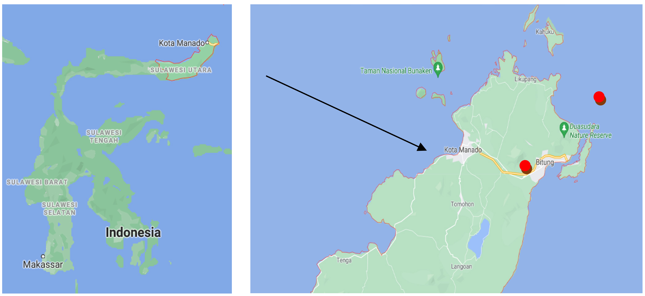
Fig. 1. Map of Origin of local pig intestine fluid samples in North Sulawesi, Indonesia. (map source: https://www.google.com/maps/place/Sulawesi).
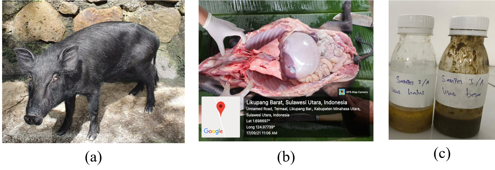
Fig. 2. (a) North Sulawesi Local Pig, (b). Local pig intestine (c). Pig intestine fluid sample.
Bacteria Isolation
Bacterial isolates were isolated from pig intestinal fluid. Pig intestine fluid was taken directly from local pigs after being slaughtered and inoculated on the same day. Isolation of bacteria was carried out by the dilution method in which a dilution of 10-3 to 10-5 was used for bacterial cultivation. On nutritional agar media, bacteria were grown (Merck). Bacterial morphology and pure culture were detected after 1 x 24 hour incubation. In an incubator, the incubation was done at 37°C. The purified isolates were utilized for antibiotic sensitivity testing and identification (Figure 4).
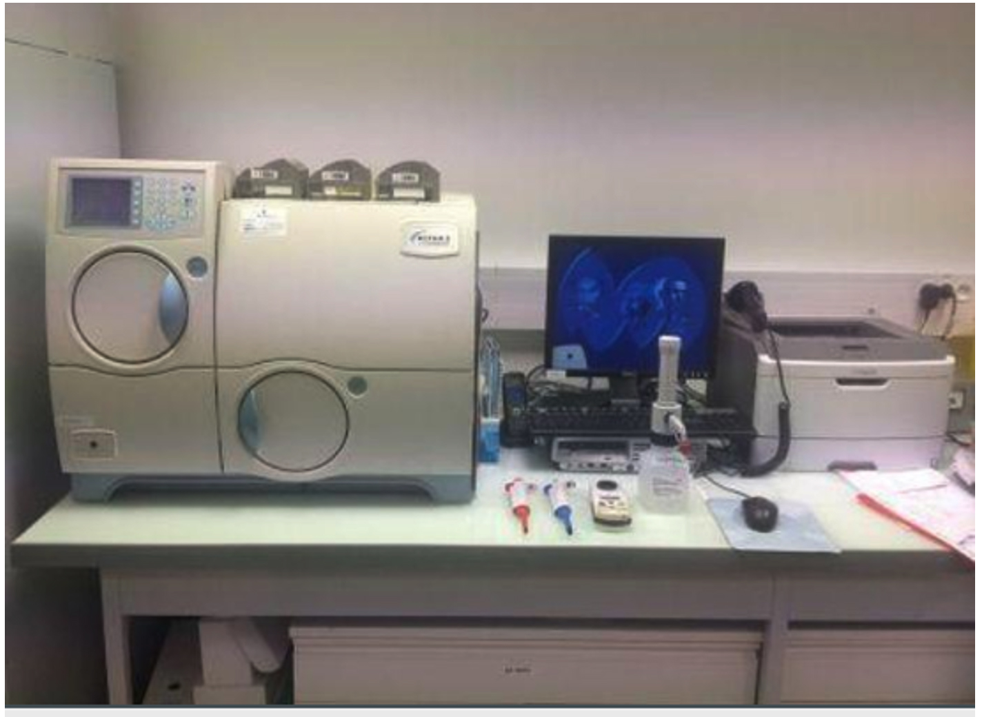
Fig. 3. Vitek 2 Compact in the Laboratory of Dr RSUP. Kandow Malayalang, Manado.
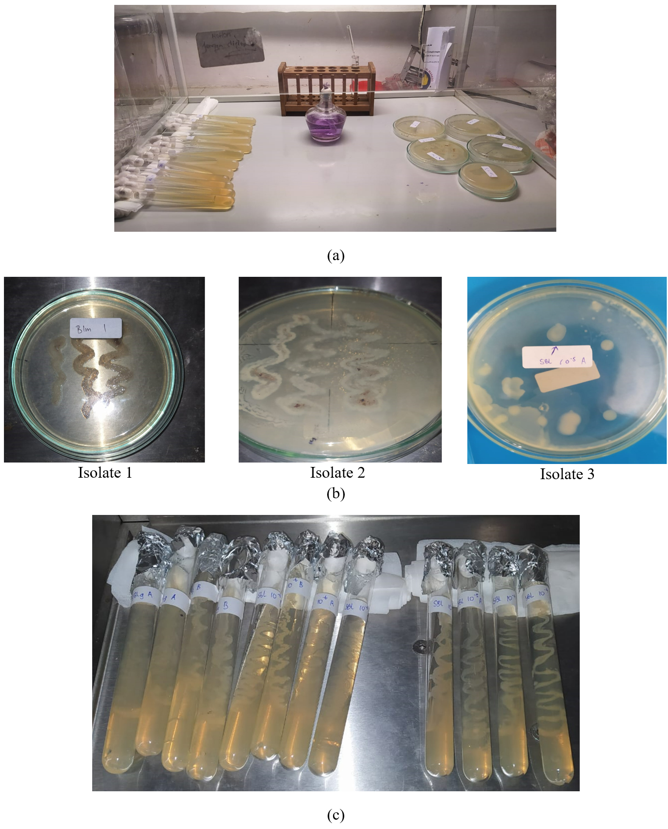
Fig. 4. (a). Bacterial isolation results after 24 hours (b) Pure culture isolate (c). Subculture for phenotypic identification and antibiotic resistance.
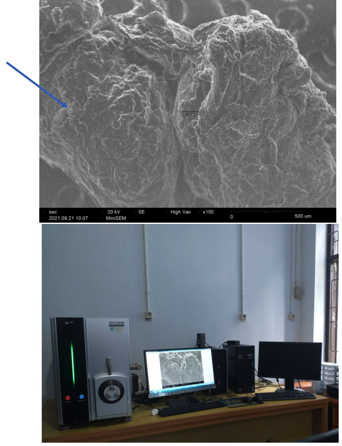
Fig. 5. Observation with Scanning Electron Microscope The inner wall of the pig intestine as a source of intestinal fluid samples
Local pig intestinal wall ultrastructure
Ultrastructural observation of the inner intestinal wall of local pigs using a Scanning electron microscope SNE-4500M Plus Tabletop SEM Nano images. Samples were prepared by cutting finely / tip[is then coated using palladium. Furthermore, it is placed in the sample holder to be observed with SEM.
Biochemical analysis of isolates using Vitek 2 Compact
The Vitek 2 Compact automated identification equipment, which is accessible at the. Regional General Hospital Laboratory Prof. Dr. Kandow, Manado, North Sulawesi, was used for biochemical identification (Fig. 3). Vitek 2 Compact is a microorganism identification system that is fully automated. It is simple to use because to the newest technology used by Vitek 2 Compact. There are just three phases of the test. It will be simple to acquire findings of antibiotic identification and sensitivity that have been verified and interpreted in accordance with Clinical Laboratory Standard International.12,13
The three processes are preparation and standardization of inoculum turbidity, data entry via a barcode system, and card insertion onto the device. Furthermore, the tool will automatically carry out the entire inoculation process, incubation, reading, validation, and interpretation of the results. Moreover, the finished inspection will generate a printout, while the system will instantly delete the ID/AST (Identification/Antimicrobial Sensitivity Test) card. The use of an identity card is the basis of automated identification. A well or a biochemical test medium that has been modified to be performed for fast bacterial identification may be found on the card (Table 1). According to McFarland standards and title, the gram test, card selection, and creating a bacterial suspension, the testing method using the Vitek 2 Compact equipment proceeds until the identification result sheet is printed.12,14
Table (1):
Vitek 2 Compact Output Analysis Standards.
Confidence Level |
Choice |
% Probability |
|---|---|---|
Excellent |
1 |
96 to 99 |
Very Good |
1 |
93 to 95 |
Good |
1 |
89 to 92 |
Acceptable |
1 |
85 to 88 |
Analysis of sensitive drugs and resistance to bacteria
The identification of bacteria was combined with the analysis of sensitive and resistant medications to bacterial isolates detected in indigenous pigs from Minahasa, North Sulawesi. This research employs a biochemical procedure that is fully automated (Vitek 2 Compact).
Bacteria was isolated and identified from pig intestinal fluid. Intestinal fluid was taken directly from local pigs slaughtered and immediately preserved in a sample box filled with ice. Inoculation of bacteria on nutrient agar media was carried out on the same day when the intestinal fluid sample was taken. Bacterial inoculation was carried out by the dilution method in which a dilution of 10-3 to 10-5 was used for the cultivation of bacteria on nutrient agar media. Three consistent bacterial isolates were obtained after incubation for 2 x 24 hours at room temperature in the incubator. The three bacterial isolates had a round colony shape, white colony colour and stem cell shape, and Gram-negative identification results (Table 2).
Table (2):
Morphological Characteristics of Bacterial Isolates from a Pigs intestinal fluid.
No |
Isolate |
Colony shape |
The shape of the edge of the colony |
Colony colour |
Cell shape |
Gram Stain |
|---|---|---|---|---|---|---|
1 |
S1 |
Round |
Flat |
Yellow white |
Rod |
Negative |
2 |
S2 |
round |
Flat |
White |
Rod |
Negative |
3 |
S3 |
round |
Flat |
White |
Rod |
Negative |
Inner pig intestines were observed with a Scanning Electron Microscope showing droplet colonization of bacteria on the intestinal wall
(Fig. 5).
Identification
The results of phenotypic identification obtained two species of bacteria, namely Escherichia coli and Enterobacter aerogenes. Based on the output of Escherichia coli analysis for sample 1, the confidence level is acceptable, while the second sample has an excellent confidence level. The S3 sample, namely Enterobacter aerogenes, showed a very good confidence level (Table 3).
Table (3):
Results of Isolate Biochemical Identification with Vitek 2 Compact.
No |
Sample code |
Confidence Level |
% Probability |
Identification results |
|---|---|---|---|---|
1 |
S1 |
Acceptable |
87 |
Escherichia coli |
2 |
S2 |
Very Good |
92 |
Escherichia coli |
3 |
S3 |
Very Good |
96 |
Enterobacter aerogenes |
Escherichia coli is a gram-negative, peritrichously flagellated bacterium belonging to the family Enterobacteriaceae. It is the causative agent of various diseases in pigs, including neonatal diarrhoea and postweaning diarrhoea (PWD), which are important causes of death in suckling pigs.14. Antibiotic resistance was found from isolates of E. coli bacteria in crossbreed pigs.15
The majority (>90%) of bacteria in the pig gut microbiome belong to two phyla: Firmicutes and Bacteroidetes. Disturbances in the microbiome respond to many factors, including stress, treatment with antibiotics, and diet.3 The phyla Firmicutes and Bacteroidetes dominated the pig feces microbiome, according to taxonomic analysis of metagenomic data. At better phylogenetic resolution, Prevotella spp. dominate the porcine fecal metagenome.16 The two most common phyla, Firmicutes and Bacteroidetes, account for more than 80% of all sequences. Fecal microbial variety increases as the pig grow, and the proportion of Firmicutes rises while the amount of Bacteroidetes falls.17 There are many types of E. coli, some are normal inhabitants of the intestine, but other types cause various colibacillosis disease syndromes. These pathogenic E. coli bacteria generally have fimbriae (pili) for attachment, enterotoxigenic exotoxins, endotoxins and capsules. There are various ways to classify E. coli infection in pigs.18 The main clinical syndromes due to E. coli in pigs will include neonatal colibacillosis, postweaning colibacillosis, diarrhoea, edematous disease, and colisepticaemia coliform mastitis, and urinary tract infections.19
Antibiotic resistance test isolates
Based on the resistance test, sample S1 showed susceptibility to 16 types of antibiotics and one type of antibiotic, namely Ceftazidime, was moderate (I). The largest minimum inhibitory concentration was Trimethoprim/Sulfamethoxazole (<=20) (Table 4). Based on the results of the identification of the isolate S1 is Escherichia coli. The results of the analysis of phenotypic detection, sample S1 was resistant to Tetracyclines.
Table (4):
S1 Isolate Resistance Test Results.
| Susceptibility Information | Analysis Time. 13.58 hours | Status: | Final | ||
|---|---|---|---|---|---|
| Antimicrobial | MIC | Interpretation | Antimicrobial | MIC | Interpretation |
| ESBL | NEG | – | Aztreonam | <=1 | S |
| Ampicillin | <=2 | S | Ertapenem | <=0.5 | S |
| Ampicillin/Sulbactam | <=2 | S | Meropenem | <=0.25 | S |
| Piperacillin/Tazobactam | <=4 | S | Amikacin | <=2 | S |
| Cefazolin | <=4 | S | Gentamicin | <=1 | S |
| Ceftazidime | <=1 | I | Ciprofloxacin | <=0.25 | S |
| Ceftriaxone | <=1 | S | Tigecycline | <=0.5 | S |
| Gentamicin | <=1 | S | Nitrofurantoin | <=16 | S |
| Cefepime | <=1 | S | Trimethoprim/
Sulfamethoxazole |
<=20 | S |
: = Deduced drug *=ACO modified **= User modified
Phenotypes
Antibiotic family |
Detected Phenotypes |
|---|---|
Beta Lactams |
Wild |
Aminoglycosides |
Wild |
Quinolones |
Decreases susceptibility, wild |
Tetracyclines |
Resistant, Wild |
Furanes |
Wild |
Trimethoprim/Sulfonamides |
Trimethoprim Resistant, Wild |
Based on the resistance test, sample S2 showed resistance to two types of antibiotics, namely Ampicillin and Trimethoprim/Sulfamethoxazole. Isolate S2 was susceptible to 13 kinds of antibiotics, and two types of antibiotics, namely Ceftazidime and Ampicillin/Sulbactam, were moderate (I). The largest minimum inhibitory concentration was Trimethoprim/Sulfamethoxazole (<=320) (Table 5). Based on the results of the identification of isolate S2 is Escherichia coli. The results of the phenotypic detection analysis showed that sample S1 was resistant to Tetracyclines and Trimethoprim/ Sulfonamides.
Table (5):
S2 Isolate Resistance Test Results.
| Escherichia coli Susceptibility Information |
Analysis Time. 13.58 hours | Status: | Final | ||
|---|---|---|---|---|---|
| Antimicrobial | MIC | Interpretation | Antimicrobial | MIC | Interpretation |
| ESBL | NEG | – | Aztreonam | <=1 | S |
| Ampicillin | <=32 | R | Ertapenem | <=0.5 | S |
| Ampicillin/Sulbactam | 16 | 1 | Meropenem | <=0.25 | S |
| Piperacillin/Tazobactam | <=4 | S | Amikacin | <=2 | S |
| Cefazolin | <=4 | S | Gentamicin | <=1 | S |
| Ceftazidime | <=1 | I | Ciprofloxacin | <=0.25 | S |
| Ceftriaxone | <=1 | S | Tigecycline | <=0.5 | S |
| Gentamicin | <=1 | S | Nitrofurantoin | <=16 | S |
| Cefepime | <=1 | S | Trimethoprim/
Sulfamethoxazole |
<=320 | R |
: = Deduced drug *=ACO modified **= User modified
Phenotypes
Antibiotic family |
Detected Phenotypes |
|---|---|
Beta Lactams |
Acquired pase + case (AMpC), Acquired Penicillinase, Cephalosporinase (AmpC) |
Aminoglycosides |
Wild |
Quinolones |
Decreases susceptibility, wild |
Tetracyclines |
Resistant, Wild |
Furanes |
Wild |
Trimethoprim/Sulfonamides |
Resistant |
Based on the resistance test, the S3 sample showed resistance to three types of antibiotics, namely Ampicillin, Ampicillin/Sulbactam and Cefazolin. The S3 isolate was susceptible to 13 kinds of antibiotics and two types of antibiotics, namely Nitrofurantoin which was moderate (I). The largest minimum inhibitory concentration is Trimethoprim/Sulfamethoxazole (64) (Table 6). Based on the identification of isolate S3 is Entereobacter aerogenes, the results of the phenotypic detection analysis showed that sample S1 was resistant to Tetracyclines, Furanes and Trimethoprim/Sulfonamides.
Table (6):
S3 Isolate Resistance Test Results.
| Entereobacter aerogenes Susceptibility Information |
Analysis Time. 13.58 hours | Status: | Final | ||
|---|---|---|---|---|---|
| Antimicrobial | MIC | Interpretation | Antimicrobial | MIC | Interpretation |
| ESBL | NEG | – | Aztreonam | <=1 | S |
| Ampicillin | <=32 | R | Ertapenem | <=0.5 | S |
| Ampicillin/Sulbactam | *R | Meropenem | <=0.25 | S | |
| Piperacillin/Tazobactam | <=4 | S | Amikacin | <=2 | S |
| Cefazolin | <=64 | R | Gentamicin | <=1 | S |
| Ceftazidime | <=1 | S | Ciprofloxacin | <=0.25 | S |
| Ceftriaxone | <=1 | S | Tigecycline | 1 | S |
| Cefepime | <=1 | S | Nitrofurantoin | 64 | I |
| Cefepime | <=1 | S | Trimethoprim/
Sulfamethoxazole |
<=20 | S |
: = Deduced drug *=ACO modified **= User modified
Phenotypes
Antibiotic family |
Detected Phenotypes |
|---|---|
Beta Lactams |
Wild (cephalosporins) acquired penicillinase |
Aminoglycosides |
Wild |
Quinolones |
Decreases susceptibility, wild |
Tetracyclines |
Resistant, Wild |
Furanes |
Wild, resistant |
Trimethoprim/Sulfonamides |
Trimethoprim Resistant, wild |
Two types of bacterial isolates from pig intestinal fluid showed resistance to the same antibiotic, namely Ampicillin. Ampicillin is a type of penicillin class of antibiotics that is reported to be resistant to Escherichia coli. E. coli isolated from pigs showed higher resistance than E. coli from goats.20,21,22 Furthermore, isolate two and isolate 3 showed resistance to Tetracycline. Tetracycline is used to treat a wide variety of bacterial infections. Tetracyclines are bacteriostatic. Tetracyclines are broad-spectrum antibiotics that can manage infections by Chlamydiacease, Mycoplasma spp., Rickettsia spp., spirochetes, various gram-positive and gram-negative pathogenic bacteria, and several protozoa.23,24
Furthermore, bacterial isolates from pig intestines also showed resistance to trimethoprim/sulfonamides. Sulfamethoxazole + trimethoprim is a combination of two types of antibiotics: sulfamethoxazole and trimethoprim. Sulfamethoxazole competes against bacteria by inhibiting the use of para-aminobenzoic acid during the synthesis of dihydrofolate by bacteria.25 This ability gives rise to a bacteriostatic mechanism. Trimetropin reversibly inhibits the enzyme dihydrofolate reductase, an enzyme that activates the metabolic pathway of folic acid by converting dihydrofolate to tetrahydrofolate.26 Therefore trimethoprim and sulfamethoxazole inhibit two steps in the biosynthesis of purines that are important in nucleic acid formation and DNA synthesis in bacteria.27 This study indicates that the bacterial isolates obtained from local pigs have been resistant to several standard antibiotics that are still widely used by the community. Further research using pig samples from multiple locations and based on the age of the pigs needs to be done.
Pure culture isolates obtained from local pig intestine fluid in Minahasa, North Sulawesi, showed that two isolates (S1 and S2) were Escherichia coli and isolate S3 was Enterobacter aerogenes. Isolate S1 showed resistance to Ampicillin, while isolate S2, 3 showed resistance to Tetracycline, while isolate S3 showed Tetracyclines, Furanes and Trimethoprim/Sulfonamides. The study results confirm that further research is needed to isolate and test antibiotic resistance of bacteria from pig intestines at multiple locations and various general stages of local pigs.
ACKNOWLEDGMENTS
The authors would like to thank the DRPM of the Ministry of Education, Culture, Research and Higher Education, for funding this research through the Basic Research Scheme for Higher Education Excellence in 2021. We also thank the leadership and staff of the microbiology laboratory, Central General Hospital, Prof. Dr. R. D. Kandow Manado, North Sulawesi, Indonesia, for carrying out laboratory analysis.
CONFLICT OF INTEREST
The authors declare that there is no conflict of interest.
AUTHORS’ CONTRIBUTION
RAM conceived, designed the study. MYS structured the work order. MYS and NM conducted experiments. EHA collected the data. NM wrote the draft manuscript. MYS and RAM revised the manuscript. All authors read and approved the final manuscript for publication.
FUNDING
This research was funded by Directorate of Research and Community Service, Ministry of Education, Culture, Research and Universities of the Republic of Indonesia through the Basic Research Scheme for Colleges Education in the 2021 fiscal year with contract number 1401/UN.41/KP/2021.
ETHICS STATEMENT
This study was approved by the Institutional Ethics Committee, Department of Biology, Faculty of Mathematics and Natural Science, Manado State University, Indonesia, with reference number 021/REC/05/2021.
AVAILABILITY OF DATA
All datasets generated or analyzed during this study are included in the manuscript.
- Bondoc OL, Dominguez JMD, Peñalba FF. DNA barcoding of domestic swine breeds and crossbreeds (Sus scrofa) in the Philippines. Philipp J Vet Anim Sci. 2013, 39(1): 31-42
- Mege RA, Semuel MY, Mokosuli YS. DNA barcoding of local pigs in Minahasa. North Sulawesi, Indonesia. J Adv Zool. 2017;38(2):110-120.
- Mege RA, Mokosuli YS, Rayer DJJ, Adil HE, Rompas C, Manampiring N, Montolalu M. Philogenic Relationship of Wild Pigs and Local Pig from North Sulawesi Based on the Growth Hormone Gene (GH Gene). In Materials Science Forum. 2019; 967:71-72.
Crossref - Isaacson R, Kim HB. The intestinal microbiome of the pig. Anim Health Res Rev. 2012;13(1):100-109.
Crossref - Backhed F, Ley RE, Sonnenburg JL, Peterson DA, Gordon JI. Host-bacterial mutualism in the human intestine. Science. 2005;307(5717):1915-1920.
Crossref - Greenblum S, Turnbaugh PJ, Borenstein E. Metagenomic systems biology of the human gut microbiome reveals topological shifts associated with obesity and inflammatory bowel disease. Proc Natl Acad Sci USA. 2012;109(2):594-599.
Crossref - Rauw WM, Rydhmer L, Kyriazakis I, et al. Prospects for sustainability of pig production in relation to climate change and novel feed resources. J Sci Food Agric. 2020;100(9):3575-3586.
Crossref - Hennessy DP, Shalloo L, Van Zanten HHE, Schop M, De Boer IJM. The net contribution of livestock to the supply of human edible protein: the case of Ireland. J Agric Sci. 2021;159(5-6):463-471.
Crossref - Edwards S, Grand N. Behavioral Biology of Pigs and Minipigs. Behavioral Biology of Laboratory Animals. 2021:243-259.
Crossref - Greenblum S, Turnbaugh PJ, Borenstein E. Metagenomic systems biology of the human gut microbiome reveals topological shifts associated with obesity and inflammatory bowel disease. Proc Natl Acad Sci USA. 2012;109(2):594-599.
Crossref - Wang W, Hu H, Zijlstra RT, Zheng J, Ganzle MG. Metagenomic reconstructions of gut microbial metabolism in weanling pigs. Microbiome. 2019;7(1):1- 11.
Crossref - Lauridsen C, Matte JJ, Lessard M, Celi P, Litta G. Role of vitamins for gastro-intestinal functionality and health of pigs. Anim Feed Sci Technol. 2021;273:114823.
Crossref - Barry J, Brown A, Ensor V, et al. Comparative evaluation o f the VITEK-2 Advanced Expert System (AES) in the five UK hospitals. J Antimicrob Chemother. 2003;51(5):1191-1202.
Crossref - Kanan M, Salaki C, Mokosuli YS. Molecular Identification of Bacterial species from Musca domestica L. and Chrysomya megachepala L. In Luwuk City, Central Sulawesi, Indonesia. J Pure Appl Microbiol. 2020;14(2):1595-1607.
Crossref - Larone DH, Tucci LJ, Samide DO. Time study of three Automated Systems for the identification and Susceptibility of Bacteria: The Microscan WalkAway 96,VITEK, and VITEK-2, Annual Meeting of the American Society for Microbiology Meeting Los Angeles, CA. 2000;279.
- Prihatini, Aryati, Hetty. Identifikasi Cepat Mikroorganisme Menggunakan Alat VITEK-2. Indonesian Journal Of Clinical Pathology And Medical Laboratory. 2007;13(3):129-132.
Crossref - Luppi A. Swine enteric colibacillosis: diagnosis, therapy and antimicrobial resistance. Porcine Health Manag. 2017;3:16.
Crossref - Sun Y, Kim SW. Intestinal challenge with enterotoxigenic Escherichia coli in pigs, and nutritional intervention to prevent postweaning diarrhea. Anim Nutr. 2017;3(4):322-330.
Crossref - Lamendella R, Domingo JW, Ghosh S, Martinson J, Oerther DB. Comparative fecal metagenomics unveils unique functional capacity of the swine gut. BMC Microbiol. 2011;11:103.
Crossref - Shao M, Wang Z, He Y, Tan Z, Zhang J. Fecal microbial composition and functional diversity of Wuzhishan pigs at different growth stages. AMB Express. 2021;11(1):88.
Crossref - Aasmäe B, Häkkinen L, Kaart T, & Kalmus P. Antimicrobial resistance of Escherichia coli and Enterococcus spp. isolated from Estonian cattle and swine from 2010 to 2015. Acta Veterinaria Scandinavica, 2019: 61(1), 1-8.
- Sayah RS, Kaneene JB, Johnson Y, Miller R. Patterns of antimicrobial resistance observed in Escherichia coli isolates obtained from domestic-and wild-animal fecal samples, human septage, and surface water. Appl Environ Microbiol. 2005;71(3):1394-1404.
Crossref - Azizah N, Astuti MK, Yudhabuntara D. Resistensi Isolat Lokal Escherichia Coil Pembawa Gena Vt1 Dan Vt2 Asal Babi Dan Domba/kambing Terhadap 6 Antibiotik= Resistance of Local Isolates of Vt1 and Vt2 Genes- bearing Escherichia coil From Sheep/goat and Swine. J Sain Vet. 2002;20(2):46-51.
- Haulisah NA, Hassan L, Bejo SK, Jajere SM, Ahmad NI. High Levels of Antibiotic Resistance in Isolates From Diseased Livestock. Front Vet Sci. 2021;8:652351.
Crossref - Gasparrini AJ, Markley JL, Kumar H, et al. Tetracycline- inactivating enzymes from environmental, human commensal, and pathogenic bacteria cause broad- spectrum tetracycline resistance. Communications Biology. 2020;3(1):241.
Crossref - Li W, Shi C, Yu Y, et al. Interrelationships between tetracyclines and nitrogen cycling processes mediated by microorganisms: A review. Bioresource Technology. 2021;319:124036.
Crossref - Minato Y, Dawadi S, Kordus SL, Sivanandam A, Aldrich CC, Baughn AD. Mutual potentiation drives synergy between trimethoprim and sulfamethoxazole.
Nat Commun. 2018;9(1):1-7.
Crossref
© The Author(s) 2022. Open Access. This article is distributed under the terms of the Creative Commons Attribution 4.0 International License which permits unrestricted use, sharing, distribution, and reproduction in any medium, provided you give appropriate credit to the original author(s) and the source, provide a link to the Creative Commons license, and indicate if changes were made.