

ISSN: 0973-7510
E-ISSN: 2581-690X
 , Pritam Kanti Guha1, Swetha Vallabhaneni1, C. Guru Prasad1, R. Vaishnavi2, Renuka Pallem1, M. Krishna Vamsi1,
Degati Vijayalakshmi1
and D. Vijaya Raghava Prasad1 Pathogenic strains of Staphylococcus species are among the most important agents of nosocomial infections. In the present study, we isolated bacteriophages from hospital sewage of Kadapa, India, that can infect multidrug-resistant Staphylococcus aureus and Staphylococcus ureilyticus. Four phages were isolated, and ΦSU1 was chosen for further investigation because it had a wider host range. Transmission electron microscopy showed that the head of ΦSU1 was 44-45 nm in size with an icosahedral shape, while the tail was noncontractile with a length of 231 nm and adsorption efficiencies of 88.64 and 53.74% on S. ureilyticus and S. aureus, respectively. One-step growth experiments revealed latent periods and burst sizes of 35.02 min and 88.5 PFU/cell for S. ureilyticus and 38.77 min and 58.6 PFU/cell for S. aureus. ΦSU1 was stable at a pH of 4-8 and at a temperature of 28-50 °C, while a drop in titer was found at a pH of 9 and 4 °C, with complete inactivation at a pH of 3 and 10 and at 60 °C. Excess ZnCl2 inhibited ΦSU1 activity on S. ureilyticus. The results indicated that ΦSU1 was lytic and should be further studied.
Wound Pathogens, Adsorption Kinetics, Bacteriophages, One-Step Growth Curve, Host Range
Drug resistance is a major threat to human health because it renders many essential antibiotics ineffective for treating bacterial infections. Wound infections are a substantial burden on the entire healthcare system, and they commonly arise from diabetic foot ulcers, trauma injuries, burns, and surgical wounds.1,2 These infections are important clinically, as they can prolong hospital stays, delay wound healing, raise the cost of care, and increase the likelihood that a patient will develop systemic complications (sepsis).3 In many developing countries, particularly in India, the challenge of wound infections is made significantly more difficult by the large number of patients presenting for care, inappropriate use of antibiotics, and escalating prevalence of AMR.4
Staphylococcus aureus is one of the most clinically important pathogens causing wound infections. It causes skin infections and may result in more serious illnesses such as endocarditis or bacteremia.5 Furthermore, the presence of MRSA makes it even more difficult to treat these types of infections.6
Among the staphylococci, there has been increasing interest in less-characterized staphylococcal species that must be considered the cause of healthcare-associated infections. One such species is Staphylococcus ureilyticus, which belongs to the Staphylococcus saprophyticus group.7 S. ureilyticus can be isolated from multiple clinical samples such as surgical wounds. Recent genomic analyses have revealed multiple virulence factors associated with this species, including characteristics related to pathogen adherence, ability to withstand stressors, and resistance to antibiotics. All of these factors allow S. ureilyticus to persist in a clinical environment and aid the persistence of chronic infections, and increase patient mortality risks.
Bacteriophages have re-emerged as an alternative or a supplement to conventional antibiotics.8,9 Lytic phages show promise against multidrug-resistant bacterial pathogens of major clinical significance, such as Staphylococcal species.10,11
Another important factor in the development of therapeutic bacteriophages is their bacterial host ranges.12 Because therapeutic phage candidates tend to have a broader host range among the tested Staphylococcal isolates, they could be valuable in numerous clinical applications through the development of non-pathogen-specific and non-patient-specific phage products that are used in place of pathogen and patient-specific phage therapies.13
The initial step for a bacteriophage to infect a bacterium is to connect to its surface, which is dependent on numerous parameters such as the type of receptors on the bacterial cell envelope.14 In Staphylococcus, most bacteriophages utilize a polysaccharide termed wall teichoic acid (WTA) as their primary point of entry through the cell wall.15,16 As a result, any structural variation or alteration in the WTA can have a profound impact on the susceptibility of a staphylococcal strain to an array of bacteriophages, thus being responsible for an inconsistent host range of the bacteriophages involved in infecting staphylococcal species.17
Broad host ranges of bacteriophages have significant implications for understanding their ecology and therapeutic applications.18 Researchers have modified bacteriophage attachment proteins to create bacteriophages that infect several bacteria.14
Moreover, previous findings demonstrate that the structural characteristics of molecular components of wall-associated teichoic acid in the several staphylococcal species serve as receptors for corresponding phages, thus aiding in their ability to infect multiple bacteria at once.16,19 As a result, interspecies infectivity among the Staphylococcus genus should be viewed as an ecologically valid option for therapeutic use of phages.
The objective of the present study was to isolate and identify bacteriophages active against multidrug-resistant strains of S. aureus and S. ureilyticus from hospital wastewater. We studied the host range, biological properties, replication kinetics, environmental stability, and adsorption characteristics of the phage for their potential use as antimicrobial agents.
Study setting and Design
The work was conducted from April 2023 to October 2023 at the Department of Microbiology, Yogi Vemana University, Kadapa, after receiving ethical approval from the Institutional Ethical Committee (IEC) (Ref. No.01/IEC/DVR/YVU/2022-23, dated 24.04.2023), and written informed consent were obtained from all the participants prior to sample collection.
Sample collection
Five samples of sewage effluent were taken from hospitals in and around Kadapa’s sewage treatment facilities and transferred into 50 mL Falcon tubes. The samples underwent centrifugation at 10,000 x g for a period of 10 min after collection and were then filtered using a membrane filter (0.22 µm) and collected in a sterile container.20,21
Bacterial isolates
Wound samples were collected from 117 patients with infections from the Government Hospital near Kadapa. Samples, including diabetic foot ulcers, traumatic wounds and post-operative infections, were purified by successive subculturing. Isolates were identified morphologically, followed by Gram staining and confirmed by biochemical tests. The Kirby-Bauer disk diffusion method was used to determine the antimicrobial susceptibility of S. aureus strain SRV05 and S. ureilyticus strain SVV09 on Mueller-Hinton agar (MHA). Comparative profiles were produced from inhibition zone diameters for different classes of antibiotics such as aminoglycosides, fluoroquinolones, phenicols, penicillins, and tetracyclines according to the Clinical and Laboratory Standards Institute (CLSI M100) guidelines. This testing was performed on laboratory strains to assess phenotypic resistance trends, and it was not performed using standard strain controls. The antibiotics used for the study were Ciprofloxacin (5 µg), Gentamycin (10 µg), Oxacillin (1 µg), Tobramycin (10 µg), Chloramphenicol (30 µg), and Tetracycline (30 µg). Diameter measurements were performed after incubation for 18-24 hrs at 37 °C. Multidrug Resistance (MDR) was defined as non-susceptibility to at least one agent in three or more different classes according to international consensus criteria. Identification of Staphylococcus ureilyticus SVV09 and Staphylococcus aureus SRV05 through 16S rRNA gene sequencing, with ~ 1.5 kb fragments obtained by PCR amplification. Sequencing was performed on the ABI 3130xl Genetic Analyzer and the partial 16S rRNA sequences were analyzed using the NCBI BLASTn database.22,23 Further investigation of the bacterial isolates was also done by scanning electron microscopy.
Isolation and enrichment of bacteriophages
Bacteriophages were isolated from overnight cultures of S. aureus and S. ureilyticus (OD600 = 0.6). Filtered sewage and overnight culture were inoculated into double strength Luria Bertani (LB) broth and incubated in shaking incubator (shaking 120 rpm) at 37 °C for 18 hrs. The phage lysate was prepared by centrifugation and filtration through a membrane filter and examined by the double-layer agar method (DLA).21
Purification of bacteriophages
Individual distinct plaques with identifiable characteristics were picked from double layer agar plates using sterile micropipette tips and introduced to SM buffer containing fast growing host cells to elute the phage particles. The plaque purification process was carried out independently in triplicate. The phage lysates were filtered and stored (4 °C) before characterization.
Host range
The ability of some phage strains to infect other bacterial strains was determined by spot test.24 Overnight cultures of bacteria were grown to OD600 = 0.6 and the sample bacteria mixed with soft 0.7% agar before being spread on LB agar plates. 10 µl phage lysate was diluted evenly and incubated (37 °C). Incubation resulted in clear plaque formation. Among four isolated phages (ΦSU1, ΦSU2, ΦSA1 and ΦSA2), phage ΦSU1 was selected for further characterization based on its high lytic spectrum.17
Efficiency of Plating (EOP)
The phages were serially diluted and plated on the test strains (15 strains of Staphylococcus spp., 4 of Klebsiella spp., 3 of Providencia spp., and 3 of Escherichia coli spp.) according to the DLA protocol.25 Each test was repeated three times. EOP was calculated using the following equation:
EOP = PFUs of test strain/PFUs of reference strain.
Phage titre (PFU/mL) data were subjected to log10 transformation prior to any statistical analysis. Host strain effects were analyzed using one-way analysis of variance. The Tukey post-hoc test was used to check mean differences. The significance level was P < 0.05. The homogeneity of variance assumption was tested using the Brown-Forsythe test.
Initially, ΦSU1 showed variable lytic properties in 15 different staphylococcal isolates, as the ability of the phage to infect S. ureilyticus (SVV09), S. aureus (SRV05), and clinical isolates such as SS01-SS13 was confirmed. On the contrary, ΦSU2, ΦSA1, and ΦSA2 phages were less effective, infecting only 40% of isolates for each of the phages and a 13.3% infection rate for ΦSA2. Phages did not show any positive reactions with Gram-negative bacteria (e.g., Klebsiella spp., Providencia spp., and E. coli). Phage ΦSU1 characterization was continued due to its wide host range among staphylococcal strains.
In vitro bacteriolytic activity
The in vitro bactericidal activity of the bacteriophage ΦSU1 was determined by measuring the optical density (OD600) of the bacteria for 24 hrs in three independent biological experiments (n = 3) to assess growth kinetics. Bacterial cultures at mid-log phase (OD600 = 0.3) were infected with phage at an MOI = 0.1, with an uninfected control culture included. To assess overall bacterial growth across the entire time series and to account for potential repeated-measures sphericity issues, we calculated the Area Under the Growth Curve (AUGC) for each biological replicate (n = 3) with a baseline of Y = 0. Statistical comparisons between untreated control and phage-treated cultures were conducted using Welch’s t-test. An MOI of 0.1 (0.1 PFU/CFU) was selected to provide synchronized single-burst infection kinetics with limited secondary infection events during the assay period. Using an MOI of 0.1 (Poisson distribution) ensures that >95% of infected bacterial cells will be infected with one virion only and will therefore not be subject to phage-induced ‘lysis from without’ and will not be subject to competitive interference within the cell that could artificially skew the latent period and burst size calculations.
Phage adsorption assay
The kinetics of ΦSU1 phage adsorption was evaluated by titrating unadsorbed viruses at different time points for 2 strains of S. ureilyticus SVV09 and S. aureus SRV05. The cultures in exponential growth (1.12 × 10⁹ CFU/mL and 1.68 × 10⁸ CFU/mL) were infected with phage ΦSU1 at a MOI of 0.1 and incubated at 37 °C during the experiment. Samples collected at designated intervals (0, 3, 6, 9, 12 and 15 min), were centrifuged and the unbound phages were quantified by DLA.26 The number of bound phages were calculated as a fraction of initial phage concentration at T = 0. Statistical analysis was performed by using one-way ANOVA and Tukey’s test, to assess the differences in unbound phage titer over time.
One-Step growth curve
The kinetics of lytic replication, the duration of the latent period and the size of the burst of the ΦSU1 bacteriophage were studied by comparison of its interactions with the primary host S. ureilyticus SVV09 and the secondary host S. aureus SRV05 through a single-step growth test in biological triplicate with observations on different days.27 Cells of growing bacteria (SVV09: 1.12 × 10⁹ CFU/mL and SRV05: 1.68 × 10⁸ CFU/mL) were exposed to phage ΦSU1 (MOI = 0.1) for 15 min at 37 °C. After centrifugation at 10,000 × g for 5 minutes, the unbound phages were counted using DLA.
Adsorption efficiency = (Pinitial – Punadsorbed)/ (Pinitial) × 100
Infection was synchronized by diluting bacteria 1:1000 in Tryptic Soy Broth (TSB) to remove secondary adsorption and incubating (15 min at 37 °C) with constant stirring (120 rpm). Samples were collected every 5 min and the total titer of phages (PFU/mL) was determined by the DLA method applied on tryptic soy agar (TSA) plates. The number of plaques was transformed to a logarithmic scale (base 10) and fitted to the Gompertz model.
The burst size (number of phages released from one infected cell) was calculated with the parameters of nonlinear regression by the formula:
Burst Size = 10YM – 10Y0 / N0
N0 = B0.MOI.α /1000
where YM and Y0 represents the model-estimated upper plateau and lower baseline titers (log10 PFU/mL) and N0 is the density of infected host complexes post-dilution, determined from initial cell density (B0), MOI (0.1), and physical adsorption efficiency (α) and the 1:1,000 dilution factor of synchronization.
Transmission Electron Microscopy (TEM)
The phage was visualized by transmission electron microscopy (TEM). The sample was spotted on carbon-coated carbon grids and counterstained with 2% uranyl acetate.28 The sample was dried, and the images were captured using a transmission electron microscope at Jamia Hamdard University, New Delhi. However, definitive genomic taxonomy (family and genus assignment) requires whole-genome sequencing as per current International Committee on Taxonomy of Viruses (ICTV) guidelines.
Phage stability assays
The resistance of ΦSU1 phage to heat and pH changes was tested by the DLA method. The data are presented as the mean ± SD of log PFU/mL (n = 3). For the thermal resistance test, phage lysate (1.1 x 10⁸ PFU/mL in SM buffer) were incubated for 1 hrs at different temperatures to test for thermal resistance. The temperatures were
4, 28, 30, 37, 40, 50 and 60 °C. The sample was diluted in a 10-fold dilution on ice to assess the viability of phages. Phage suspensions were taken and diluted 1:9 in sterile SM and incubated at 37 °C for 1 hrs to check the effect of pH inactivation. The DLA was used to determine the phage count in those concentrates.29
Impact of metal Ions on phage attachment
The host strain S. ureilyticus SVV09 (1.15 × 10⁹ CFU/mL) was centrifuged, washed and resuspended in a reaction buffer containing specific divalent cation salts or in buffer alone (untreated control) to investigate the influence of divalent cations on attachment to the host. The host was incubated with phage ΦSU1 suspension (1.1 × 10⁸ PFU/mL) at a MOI of 0.1 and adsorbed for 15 min at 37 °C. Adsorption was performed in a cation-free buffer with 5 mM CaCl2, 5 mM MgSO4, 0.1 mM MnCl2 or 0.5 mM ZnCl2. The untreated control was the host-phage mixture in plain SM buffer without cations or added exogenous salts.
The DLA method was used to count free phages, which were subsequently serially diluted in SM buffer (10-1 to 10-6).30,31 For enumeration, 100 µL aliquots of the 10-6 dilution were plated and incubated overnight at 37 °C.
The efficiency of free (unadsorbed) phages was estimated in triplicate (n = 3 independent replicates) after 15 min at 37 °C. Group comparisons vs. control group (i.e. 37 °C, pH 7 or cation free buffers) were performed by ordinary one-way ANOVA with Dunnett’s post hoc test.
Statistical analysis
Each biological assay was performed three times. Data are expressed as mean ± SD. The data were visualized, settings optimized and basic statistical analysis was performed using GraphPad Prism 11.0.2. The Shapiro–Wilk and Brown–Forsythe tests were used to test normality and homogeneity of variance. Host range EOP titers and pH/thermal stability ranges were compared using normal one-way ANOVA. Post-hoc pairwise comparisons were performed using Tukey’s HSD or Dunnett’s tests, relative to the untreated control group (pH 7.0, 37 °C or cation-free SM buffer). The 24 hrs bacterial suppression was evaluated by the Area Under the Growth Curve (AUGC) and Welch’s t-test was used to compare control and phage-treated groups. Latent duration (1/K) and burst size (PFU/cell) were estimated from infectious phage titers by nonlinear regression with the Gompertz model. Statistical significance was set at α = 0.05, and P-values less than 0.05 (P < 0.05) were considered statistically significant.
A total of 125 bacterial isolates were recovered from 117 wound specimens. The isolates were analyzed and distributed as follows: S. ureilyticus 24 (19.2%), S. aureus 17 (13.6%), Klebsiella pneumoniae 23 (18.4%), Providencia spp. 20 (16.0%), Pseudomonas aeruginosa 14 (11.2%), Escherichia coli 11 (8.8%), Coagulase-negative staphylococci (CoNS) 9 (7.2%), and Micrococcus spp. 7 (5.6%). Multidrug-resistant isolates were identified on the basis of antimicrobial susceptibility testing (Figure 1). S. aureus SRV05 was resistant to CH, CF, TE, GM, OC and S. ureilyticus SVV09 was resistant to CF, OC, CH, GM, TE, and TB. The individual zone of inhibition values (mm) of disk diffusion assays are provided in Supplementary Table S1.
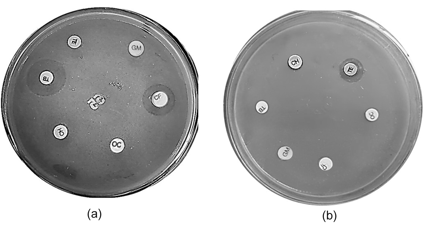
Figure 1. Antibiotic susceptibility testing by disk diffusion showing (a) multidrug resistance profile for S. aureus SRV05, resistant to oxacillin (OC), gentamycin (GM), chloramphenicol (CH), ciprofloxacin (CF), and tetracycline (TE); and (b) multidrug resistance profile for S. ureilyticus SVV09, resistant to oxacillin (OC), chloramphenicol (CH), gentamycin (GM), ciprofloxacin (CF), tetracycline (TE) and tobramycin (TB)
By 16S rRNA sequencing, four bacterial isolates were identified at the species level, and the sequences obtained were deposited in GenBank under the accession numbers of S. ureilyticus strain SVV09 (OR625452.1), S. aureus strain SRV05 (OR636873.1), K. pneumoniae strain KVP07 (OR636877.1), and P. stuartii strain PVK03 (OR636879.1). The 1124 bp sequence for S. ureilyticus SVV09 showed 100% coverage and 98.49% identity to the closest named S. ureilyticus reference (NR_037046.1). The 1412-bp sequence obtained for S. aureus SRV05 had 100% query coverage and 99.58% identity to multiple independent S. aureus reference sequences. Scanning electron microscopy (SEM) illustrated the coccoid morphological characteristics of the multidrug-resistant (MDR) strains (Figure 2) and showed that the Staphylococcus cells in clusters had coccoid morphological characteristics that were confirmed visually by the findings of molecular and biochemical testing.
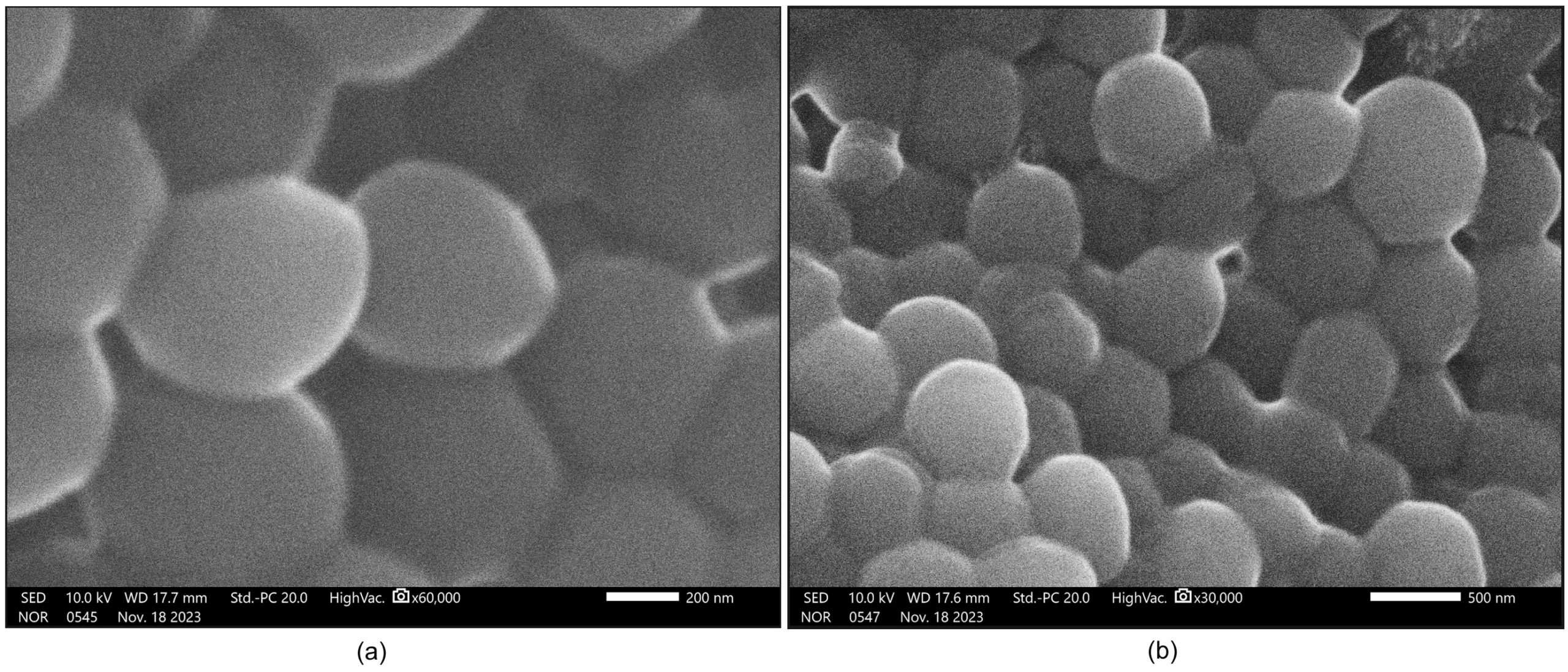
Figure 2. Scanning electron micrographs of the selected multidrug-resistant (MDR) bacterial isolates: (a) S. aureus SRV05; (b) S. ureilyticus SVV09
Phage isolation and assessment of lytic activity
Four phages (ΦSU1, ΦSU2, ΦSA1, and ΦSA2) were isolated and showed lytic activity in vitro against their respective hosts, as confirmed by the presence of clear plaques on DLA containing S. aureus and S. ureilyticus (Figure 3).
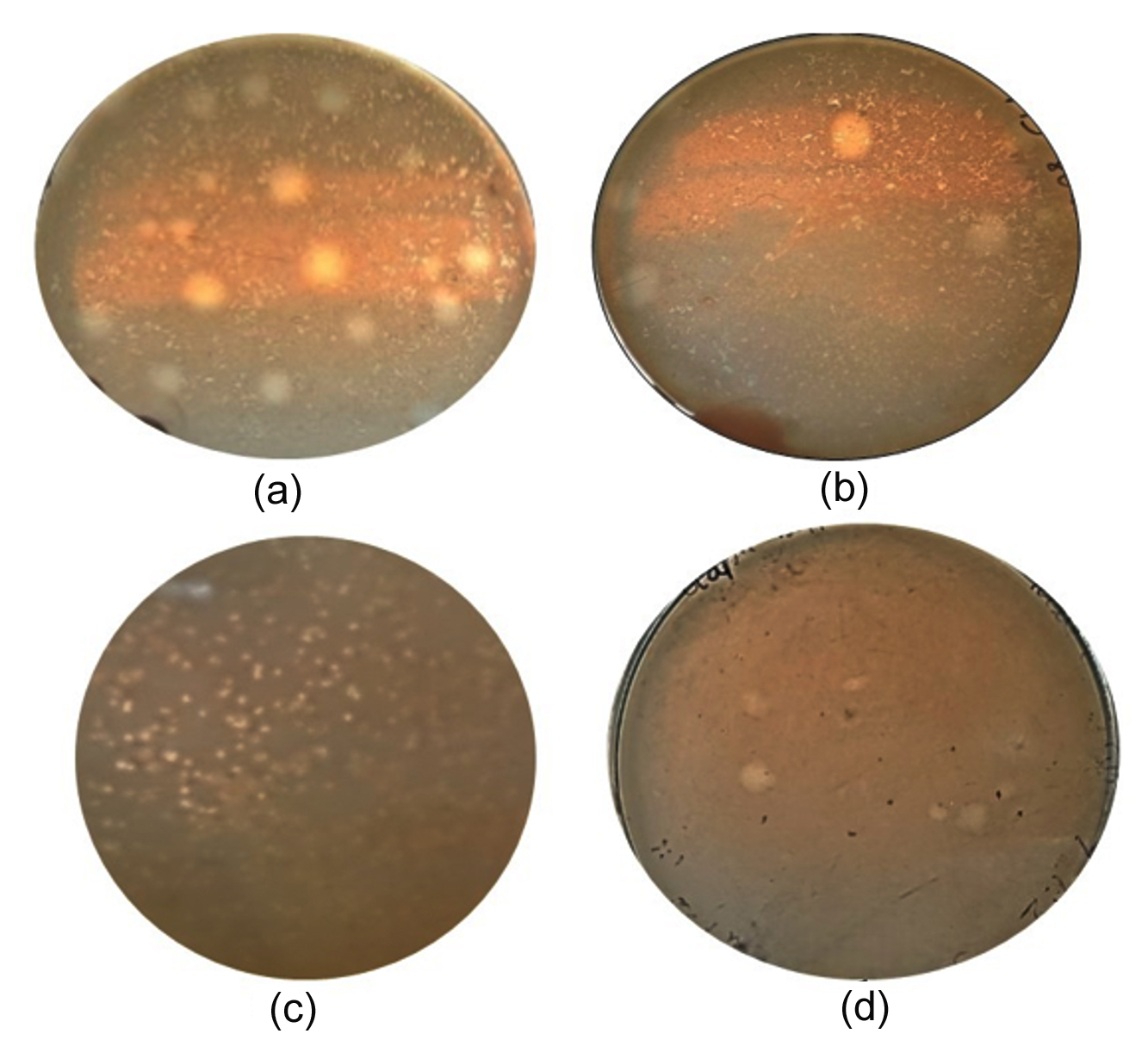
Figure 3. Double-agar plaque assay showing clear plaque formation against host lawns: (a) ΦSU1 (1-4 mm in diameter) on S. ureilyticus SVV09; (b) ΦSU2 (1-3 mm in diameter) on S. ureilyticus SVV09; (c) ΦSA1 (1-2 mm in diameter) on S. aureus SRV05; (d) ΦSA2 (1-2 mm in diameter) on S. aureus SRV05
Host range
The host range of four isolated staphylococcal phages (ΦSU1, ΦSU2, ΦSA1, and ΦSA2) were determined by testing twenty-five bacterial isolates (15 Staphylococcus strains, 4 Klebsiella strains, 3 Providencia strains, and 3 Escherichia coli strains) (Table 1). The phage ΦSU1 had an efficiency of plating (EOP) ranging from 0.12-1.00 on 15 of the 25 isolates, while the other phages had smaller host ranges (Table 2). ΦSU1 was lytic for some of the strains tested of Staphylococcus. The maximum titers obtained for ΦSU1 were 1.19 × 10⁹ PFU/mL for strain SVV09 and 1.05 × 10⁹ PFU/mL for strain SRV05. The lowest titers were 1.53 × 10⁸ PFU/mL and 1.43 ×10⁸ PFU/mL for SS03 and SS04 strains, respectively.
Table 1. Spot test lytic activity of the isolated Staphylococcus phages against the bacterial isolates
| Bacterial isolates | Staphylococcal phage | |||||
|---|---|---|---|---|---|---|
| ΦSU1 | ΦSU2 | ΦSA1 | ΦSA2 | |||
| Test strains | Staphylococcus strains | SVV09 (Host) | + | + | – | – |
| SRV05 (Host) | + | – | + | + | ||
| SS01 | + | – | + | – | ||
| SS02 | + | – | – | – | ||
| SS03 | + | + | – | – | ||
| SS04 | + | – | – | – | ||
| SS05 | + | – | – | – | ||
| SS06 | + | – | + | – | ||
| SS07 | + | – | – | + | ||
| SS08 | + | – | + | – | ||
| SS09 | + | + | – | – | ||
| SS10 | + | + | – | – | ||
| SS11 | + | + | – | – | ||
| SS12 | + | – | + | – | ||
| SS13 | + | – | + | – | ||
| Klebsiella strains (n = 4) | KP1 | – | – | – | – | |
| KP2 | – | – | – | – | ||
| KP3 | – | – | – | – | ||
| KP4 | – | – | – | – | ||
| Providencia strains (n = 3) | PS1 | – | – | – | – | |
| PS2 | – | – | – | – | ||
| PS3 | – | – | – | – | ||
| Escherichia coli strains (n = 3) | EC1 | – | – | – | – | |
| EC2 | – | – | – | – | ||
| EC3 | – | – | – | – | ||
“+” represents visible clearing in the spot test assay “–’’ represents no visible clearing
Staphylococcus spp. (SVV09, SRV05, SS01-SS13); Klebsiella pneumoniae KVP07 (KP1); Klebsiella spp. (KP2-KP4); Providencia stuartii PVK03 (PS1); Providencia spp. (PS2-PS3); Escherichia coli (EC1-EC3)
Table 2. Efficiency of plating of the phage ΦSU1 on susceptible bacterial hosts in the preliminary host range screening
Host strain |
Phage Susceptibility |
EOP (Mean ± SD) |
Tukey group |
EOP category |
|---|---|---|---|---|
SVV09 |
Lysis observed (+) |
1.00 ± 0.00 |
a |
High |
SRV05 |
Lysis observed (+) |
0.89 ± 0.09 |
a |
High |
SS01 |
Lysis observed (+) |
0.45 ± 0.08 |
ab |
Medium |
SS02 |
Lysis observed (+) |
0.21 ± 0.11 |
bc |
Medium |
SS03 |
Lysis observed (+) |
0.13 ± 0.05 |
c |
Medium |
SS04 |
Lysis observed (+) |
0.12 ± 0.03 |
c |
Medium |
SS05 |
Lysis observed (+) |
0.24 ± 0.09 |
bc |
Medium |
SS06 |
Lysis observed (+) |
0.21 ± 0.10 |
bc |
Medium |
SS07 |
Lysis observed (+) |
0.21 ± 0.06 |
bc |
Medium |
SS08 |
Lysis observed (+) |
0.20 ± 0.10 |
bc |
Medium |
SS09 |
Lysis observed (+) |
0.32 ± 0.04 |
bc |
Medium |
SS10 |
Lysis observed (+) |
0.20 ± 0.02 |
bc |
Medium |
SS11 |
Lysis observed (+) |
0.22 ± 0.02 |
bc |
Medium |
SS12 |
Lysis observed (+) |
0.41 ± 0.04 |
ab |
Medium |
SS13 |
Lysis observed (+) |
0.50 ± 0.13 |
ab |
High |
EOP values are reported as mean ± SD. EOP categories were assigned based on the mean EOP value, with high EOP defined as ≥0.5, medium EOP as 0.1 to <0.5 and low EOP as <0.1. Superscript lowercase letters indicate significant differences between different host strains according to Tukey’s HSD post hoc test (P < 0.05) while values with the same letter are not significant. The EOP classification categories, are independent of the Tukey statistical grouping letters. The EOP groups (high, medium, low) were used as operational classifications for convenience in comparing strains and were not intended as universally standardized biological thresholds. The high, medium, low, and inefficient EOP categories are different from the Tukey letter groupings. Tukey letter groupings are pairwise statistical groupings
The plating efficiency (EOP) of ΦSU1 on 15 Staphylococcus strains was tested (Figure 4). EOP values ranged from a low EOP of 0.120 ± 0.031 (SS04) to a high EOP of 1.000 ± 0.000 (S. ureilyticus). S. aureus (0.887 ± 0.089) and S. ureilyticus (1.000 ± 0.000) showed the highest EOP values, whereas SS04 (0.120 ± 0.031) and SS03 (0.128 ± 0.051) showed the lowest.
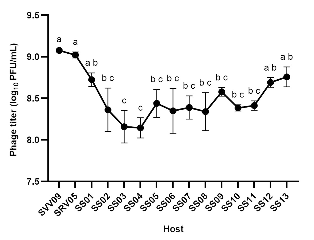
Figure 4. Host susceptibility profile for bacteriophage ΦSU1. Infectious phage titers (log10 PFU/mL) of ΦSU1 tested on 15 staphylococcal host strains (S. ureilyticus SVV09, S. aureus SRV05, and clinical isolates SS01-SS13). (n = 3) are shown as mean ± SD. Significant differences between groups are indicated above the data points by different lowercase letters
The lytic efficiency of phage ΦSU1 was variable across the tested clinical isolates (one-way ANOVA: F(14,30) = 10.86, P < 0.0001, R2 = 0.8352). High efficiency of plating was maintained on clinical S. aureus (EOP = 0.885; P > 0.9999) and isolates S12 (EOP = 0.415; P = 0.1444), S13 (EOP = 0.493; P = 0.3867), and S1 (EOP = 0.451; P = 0.2462), with no statistically significant decrease in titer relative to the primary host strain S. ureilyticus SVV09. In contrast, the EOP values of the strains S2 to S11 were significantly lower, with values between 0.120 and 0.319 (P < 0.05, Tukey’s HSD; Table 2).
Morphology of ΦSU1 by TEM
The morphology of bacteriophage ΦSU1 was examined by TEM. Phage ΦSU1 exhibited a long non-contractile tail of 231 nm in length (Figure 5). The ΦSU1 capsid is icosahedral with a diameter of 44-45 nm. Phage ΦSU1 had a siphovirus-like morphology.
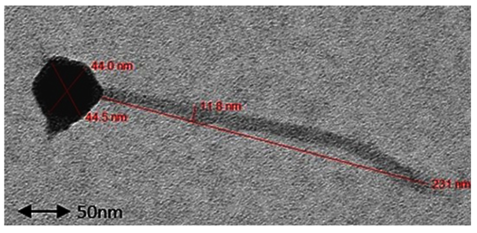
Figure 5. Transmission electron micrograph of bacteriophage ΦSU1 stained negatively with 2% uranyl acetate, showing an icosahedral capsid and a long, flexible, non-contractile tail. Scale = 50 nm
In vitro bacteriolytic activity
The number of S. ureilyticus and S. aureus cells decreased significantly (Figure 6) following treatment with phage ΦSU1. To analyze the long-term bacteriolytic processes, bacterial growth over 24 hrs was determined with the help of the Area Under the Growth Curve (AUGC) method. The results of the phage treatment showed a significant effect on S. ureilyticus propagation compared to the control group (Welch’s t(2.44) = 47.41, P = 0.0001, R2 = 0.9989). The average AUGC decreased from 813.2 OD600 × min for the control group to 357.8 OD600 × min in the presence of phages (Figure 6a), resulting in a 56.0% reduction in bacterial growth (mean difference: -455.4 ± 9.61; 95% CI: [-490.4, -420.4]).
For S. aureus, phage application significantly reduced the bacterial growth over a 24 hrs time period in comparison with the control group (Welch’s t (2.22) = 16.32, P = 0.0023, R2 = 0.9917). Phage treatment resulted in a 42.8% decrease of total AUGC value (Figure 6b) when compared to control AUGC (613.9 OD600 × min and 349.9 OD600 × min, respectively (mean difference: -262.1 ± 16.06, 95% CI: [-324.9, -199.2]).
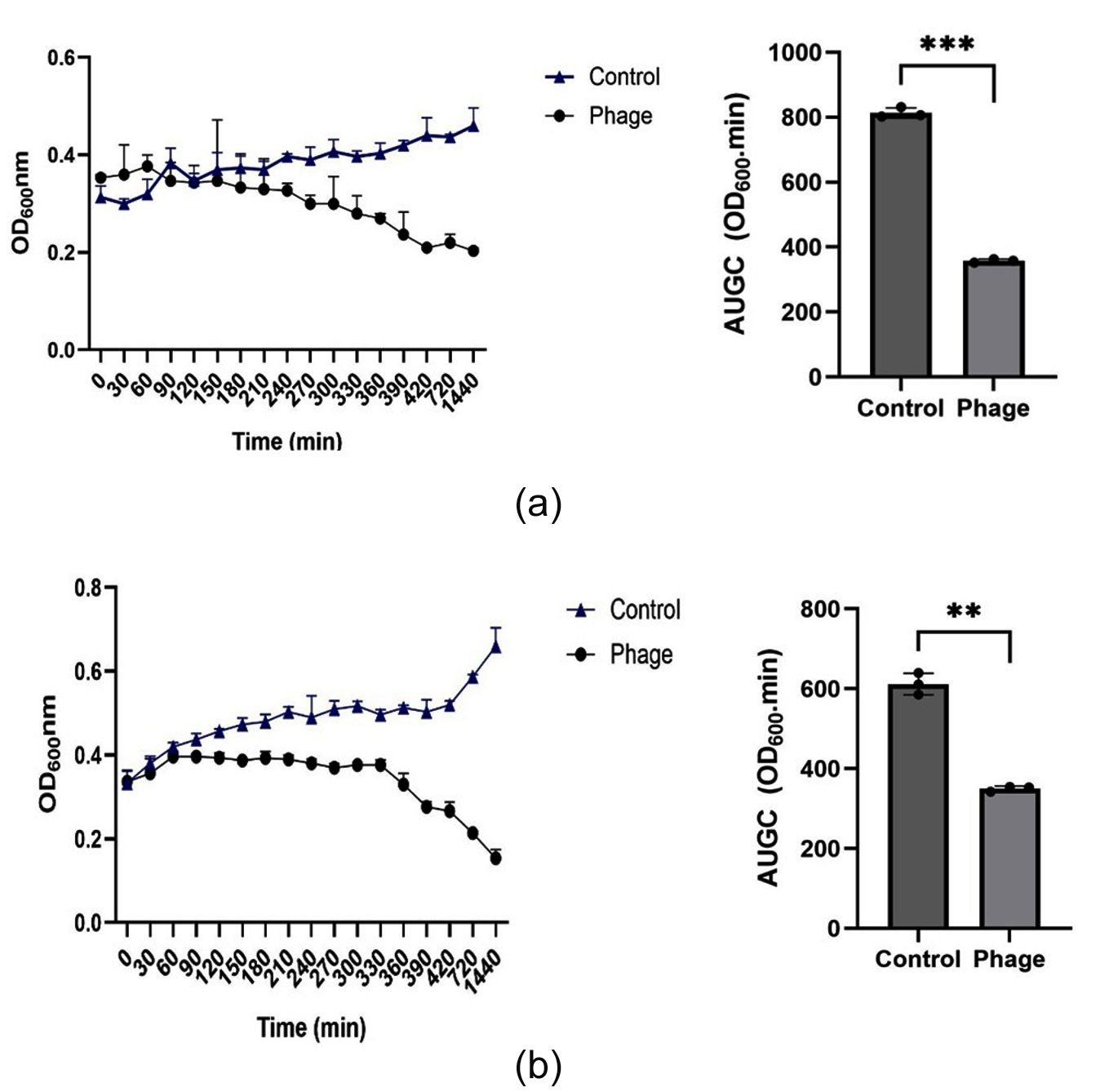
Figure 6. The in vitro bacteriolytic activity of the phage ΦSU1 was examined. OD600 was measured for bacterial samples after infection with phage ΦSU1 at an MOI of 0.1 for 24 hrs (1,440 min) in comparison to uninfected controls for (a) S. ureilyticus SVV09 and (b) S. aureus SRV05
Phage adsorption assay
During the 15 min incubation period at an MOI of 0.1, the rapid time-dependent adsorption kinetics of the ΦSU1 phage were observed against the reference primary host (S. ureilyticus SVV09) and the host strain SRV05 (Figure 7). The statistical analysis confirmed a significant decrease of unadsorbed free phages in the supernatant with time for both SVV09 (F5,12 = 25.99, exact P < 0.0001, R2 = 0.9154) and SRV05 (F5,12 = 12.13, exact P = 0.0002, R2 = 0.8348). The time-dependent virion attachment accounted for 91.54% and 83.48% of the total variance in unadsorbed phage titers of SVV09 and SRV05, respectively.
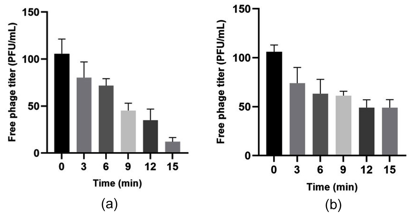
Figure 7. Adsorption kinetics for bacteriophage ΦSU1. (a) S. ureilyticus SVV09 and (b) S. aureus SRV05 during an incubation period of 15 min at 37 °C (MOI = 0.1). The maximum efficiency for adsorption reached 88.64% (S. ureilyticus) and 53.74% (S. aureus) within 12-15 min. The data represents mean ± SD (n = 3)
For the primary propagation host S. ureilyticus SVV09, Tukey’s post-hoc pairwise analysis revealed a fast initial binding of virions, with a statistically significant decrease in unadsorbed phages within 6 min compared with baseline (34.00 PFU decrease, adjusted P = 0.0328). The maximum adsorption efficiency on host SVV09 was 88.64% at 15 min (P < 0.0001). No significant difference was observed between 12 and 15 min (mean difference 23.00 PFU, adjusted P = 0.2163), confirming that adsorption equilibrium on host SVV09 was reached between 12 and 15 min.
Similarly, phage ΦSU1 on host strain SRV05 showed progressive binding kinetics with statistically significant cumulative adsorption at 15 min compared to baseline (53.74%, P = 0.0003). The unadsorbed phage titers at 12-15 min were not affected (mean difference 0.00 PFU, adjusted P > 0.9999), suggesting that adsorption equilibrium on strain SRV05 was also reached within 12-15 min. Both strains showed high affinity and similar equilibrium time courses (< 15 min). However, higher total adsorption efficiencies were observed on the primary host SVV09 (88.64%) compared to SRV05 (53.74%).
One-step growth curve and replication dynamics
The growth kinetics of phage ΦSU1 on S. aureus SRV05 were analyzed by the Gompertz nonlinear regression model (R2 = 0.9631, Sy.x = 0.08418). The latency period (1/K) was estimated to be 38.77 min (95% CI: 32.36-47.91 min). The phage concentration was increased from 7.979 log10 PFU/mL (95% CI: 7.879-8.075) initially to a maximum of 9.754 log10 PFU/mL (95% CI: 9.640-9.913) with a net increase of 1.775 log10, with the estimated burst size being 58.6 PFU per infected host cell (Figure 8a).
The growth kinetics of phage ΦSU1 on S. ureilyticus were modeled by a Gompertz nonlinear regression that fits the experimental data well (R2 = 0.9790, Sy.x = 0.07027). The estimated latent period (1/K) was 35.02 min (95% CI: 30.61-40.67 min). The phage titers increased from the initial level of 7.688 log10 PFU/mL (95% CI: 7.600-7.773) to the level of 9.640 log10 PFU/mL (95% CI: 9.552-9.750), which is an overall increase of 1.952 log10 giving the estimated burst size of 88.5 PFU per infective bacterial cell (Figure 8b).
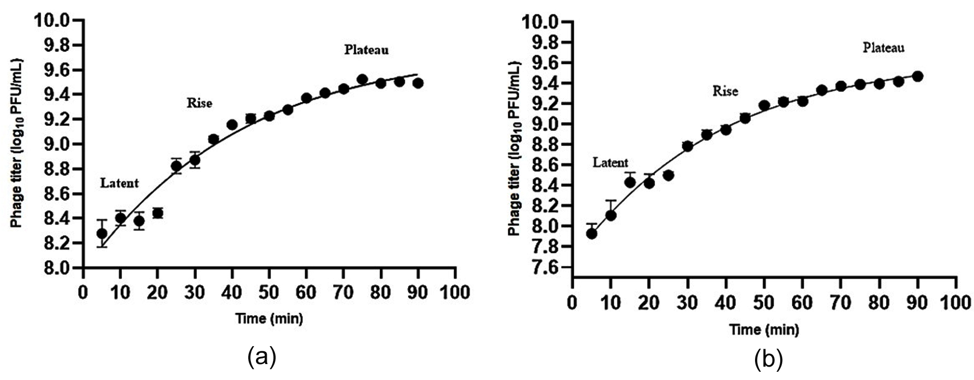
Figure 8. The phage ΦSU1’s one-step growth curve dynamics. The replication kinetics for the indicator host strains are as follows: (a) S. aureus SRV05 (latent period: 38.77 min, burst size: 58.6 PFU/cell) and (b) S. ureilyticus SVV09 (latent period: 35.02 min, burst size: 88.5 PFU/cell). The data points presented are the mean ± SD (n = 3) fitted using the Gompertz nonlinear regression model with phases (latent, rise, plateau) indicated
Environmental stability of ΦSU1
To evaluate the stability of ΦSU1, we tested the infectivity after infection at different pH (Figure 9a) and temperature levels (Figure 9b). Temperature had a significant effect on the infectious titer of phage ΦSU1 (ordinary one-way ANOVA: F(6,14) = 1087, P < 0.0001, R2 = 0.9979). Optimum viability was recorded at 37 °C (9.00 ± 0.11 log10 PFU/mL). Incubation at tested thermal range (4-50 °C) led to small to moderate decreases in titers versus 37 °C (Dunnett’s test: 30 °C, P = 0.0043; 40 °C, P = 0.0017; 28 °C and 50 °C, P < 0.0001), but in general the phage titers were still high (> 8.0 log10 PFU/mL) in this range.
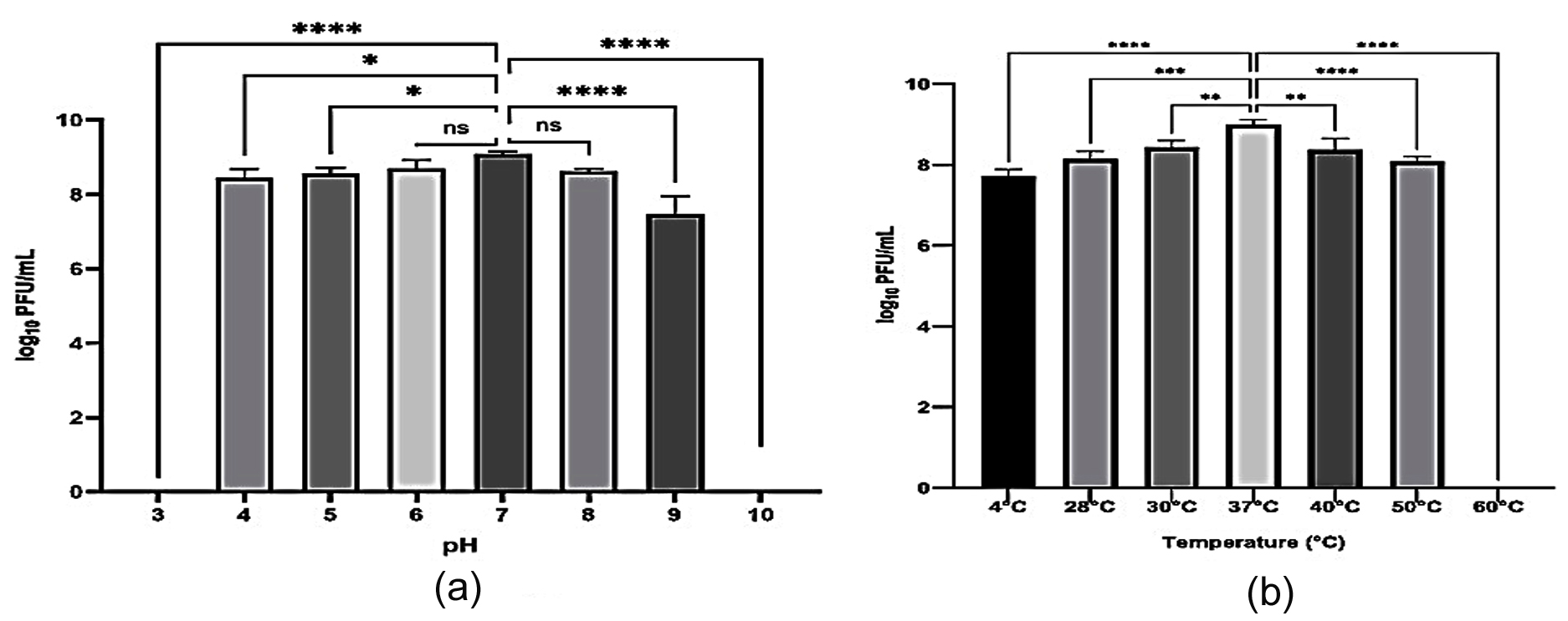
Figure 9. The environmental stability profile of bacteriophage ΦSU1 has been assessed. Stability is given in log10 PFU/mL after being subjected to various environmental conditions for 1 hrs: (a) stability with changing pH ranges between pH 3 and pH 10 measured at 37 °C (control pH 7); and (b) stability with changing temperatures from 4 °C to 60 °C (37 °C is the control value for this test). Results are shown as mean ± SD (n = 3). The asterisks in the plot indicate meaningful differences from the control values, with four levels of significance indicated: *P < 0.05, **P < 0.01, ***P < 0.001, ****P < 0.0001, and ns = not significant
Infectivity decreased by 1.27 log10 after cold incubation at 4 °C (7.73 log10 PFU/mL; P < 0.0001 vs. 37 °C). Thermal inactivation was complete at 60 °C with titers below the detection limit (9.00 log10 reduction; P < 0.0001).
A substantial difference was noted in the infectivity of phage ΦSU1 at all pH values studied (F(7,16) = 1047, P < 0.0001, R2 = 0.9978). The maximum viability was observed at pH 7.0 (9.09 ± 0.07 log10 PFU/mL). Similar trends were maintained by the phage at pH 6.0 and pH 8.0 (P = 0.1865 and P = 0.0882, respectively). At pH 4 and 5, still statistically significant drops in titer were observed (P = 0.0115 and P = 0.0442). At pH 9.0 the alkaline incubation resulted in a dramatic loss of infectivity (1.61 log10 decrease; P < 0.0001). At pH 3 and 10, the phage was completely inactivated to levels below the limit of detection (P < 0.0001).
Divalent cation treatment effects on adsorption of ΦSU1
The effect of different divalent cation treatments on the adsorption kinetics of ΦSU1 was determined (Figure 10). Effect of divalent cation presence on adsorption efficiency of phage ΦSU1 to host S. ureilyticus SVV09 (unadsorbed) phage titers (log10 PFU/mL). There were significant differences between metal ion treatment groups (F (4, 10) = 6.482, P = 0.0077, R2 = 0.7217). Variance homogeneity was tested (P = 0.7087). The multiple comparisons test showed no significant differences in unadsorbed phage levels after Ca+2 (P = 0.1701), Mg+2 (P = 0.8311), or Mn+2 (P = 0.8947) supplementation compared to the control, indicating that adsorption is independent of these cations. In contrast, the addition of Zn+2 resulted in a much higher number of free phages remaining in the supernatant (P = 0.0107, mean difference = 0.2740 log10 PFU/mL), indicating that the exogenous Zn+2 can inhibit the phage adsorption.
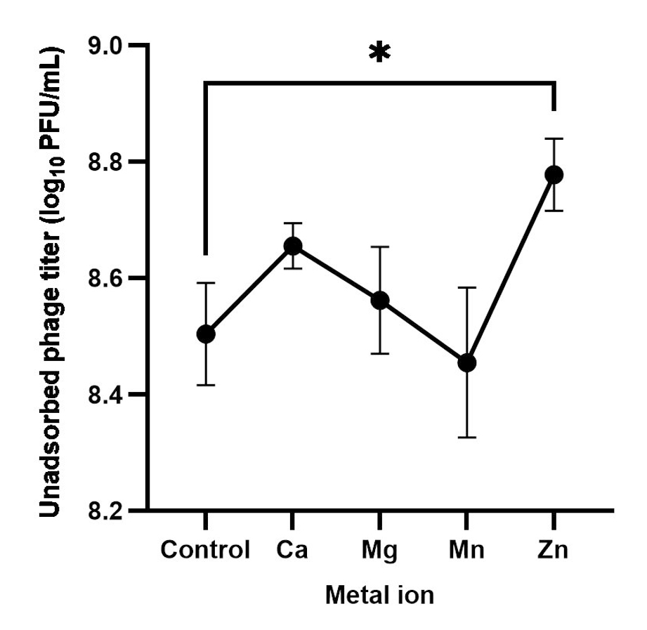
Figure 10. Divalent metal ions’ impact on the ΦSU1 phage’s adsorption. Unadsorbed phages in supernatant (log10 PFU/mL) during the 15 min of the test with S. ureilyticus SVV09 in the buffer without cations (control) and in the presence of CaCl2 (5 mM), MgSO4 (5 mM), MnCl2 (0.1 mM), or ZnCl2 (0.5 mM). Data shown is mean ± SD (n = 3). The asterisk (*) shows that Zn+2 has a statistically significant effect on the adsorption when compared to the control (P = 0.0107)
The emergence of MDR strains of staphylococci has led to investigations into bacteriophages as potential alternative or adjunct therapies to traditional antimicrobial agents.4,32 The results indicate that ΦSU1 has demonstrated lytic activity in vitro against the multidrug-resistant staphylococcal isolates, providing prospects for future study. One of the problems in assessing phage range is that the spot test may grossly overestimate the degree of susceptibility of the host because of the presence of those non-lysis mechanisms referred to as “lysis from without” or bacteriocin-mediated inhibition of host growth.25,33 In this study, the first spot test of ΦSU1 was performed against 15 isolates of staphylococci that showed formation of lysis zones. Further confirmation of the efficiency of plating was done using quantitative assays, showing lytic activity against all 15 strains that were spot-positive by formation of discrete plaques. Bacteriophage ΦSU1 was able to infect all tested strains of the Staphylococcus genus (100% susceptibility), but no activity was observed against 10 strains of non-Staphylococcus bacteria (Klebsiella, Providencia, and Escherichia coli). In addition, the overall assessment was restricted to the panel of 15 staphylococcal strains, although moderate-to-high plating efficiency of ΦSU1 on S. ureilyticus (SVV09) and S. aureus (SRV05/SS01-SS13) was observed. Thus, larger and geographically diverse collections of clinical strains will be required in future studies to better evaluate the capability of ΦSU1 as a broad-spectrum therapeutic agent.34
Based on the one-step growth experiment, the latent periods of S. ureilyticus and S. aureus were 35.02 and 38.77 min, respectively, with burst sizes of 88.5 and 58.6 PFU/cell, respectively. To account for the difference in host cell density (B0) between S. ureilyticus and S. aureus at mid-log phase, burst size calculations were normalized to the density of infected host complexes (N0) after synchronization. Moreover, synchronization via high-fold dilution (1:1,000) reduced the occurrence of secondary adsorption events, thus the derived parameters of replication reflect single-burst kinetics for every single host. Based on the TEM analysis, ΦSU1 exhibits Siphovirus-like morphology. The replication properties of ΦSU1 resemble those of many other lytic phages against Staphylococcus.35 The efficiency of the replication is a major parameter allowing a greater synthesis of the phage components after a successful infection of the bacterial host by the phage.36 The ΦSU1 replication profile suggests that this phage is capable of productive cycles in the two types of hosts mentioned and can therefore be classified as a typical lytic phage. The bacteriolytic activity tests further confirmed the lytic treatment of the ΦSU1 bacteriophage, as it was shown capable of bacteria lysis in vitro and prevention of bacterial proliferation. Other lytic staphylococcal phages had a similar effect and were effective in decreasing bacterial growth. While the in vitro findings in planktonic cells are encouraging, they do not ensure clinical efficacy, and therefore further evaluation is required.37
Infection assays were performed at low multiplicity of infection (MOI = 0.1) to accurately assess single-step infection dynamics. The ratio of phages to host used is sub-saturating, which allows lysis and burst parameters to be determined that can be assigned to single-phage replication cycles and not to the premature disruption of the cell envelope due to high surface phage occupancy.38-40
The adsorption assays showed that ΦSU1 adsorbed to both S. aureus and S. ureilyticus. However, ΦSU1 showed higher affinity toward one host strain as compared to others, with adsorption of 88.64% against Staphylococcus ureilyticus and only 53.74% against Staphylococcus aureus. The ability of the virus to bind may be related to the expression and density of the host cell surface receptors.41,42 Attachment of the phage to the host cell is an important step in initiating the infection cycle.36 In addition, the surface receptors of the host bacteria interacting with phages are different, as well as the site of initial attachment.42 Therefore, it can be inferred that the adsorption level of ΦSU1 to the different strains may be influenced by several factors, such as the cell wall structural features in Staphylococcus spp., the structure of teichoic acids, or the distribution of the receptor.41
Stability of phages in the environment is the key to understanding the characteristics of phages in future applications. The viable states of ΦSU1 under different pH and temperature conditions indicated that its stability was similar to that of other characterized phages infecting staphylococci.35 The stability of phages is crucial for their further application in therapy and biocontrol, since they need to be safe and effective in a range of environments. Phage ΦSU1 is tolerant to a wide range of physiological conditions (pH 4-8, 28-50 °C) but optimal infectivity is strictly maintained at 37 °C and pH 7.0. The significant loss of titer at lower temperatures (4 °C) and alkaline conditions (pH 9.0) suggests a sensitivity to environmental stress, which must be taken into account in the development of storage conditions or clinical application protocols. The observed performance of ΦSU1 with divalent cations is in accordance with previous studies that demonstrated the contribution of ions to phage adherence, phage DNA injection, and stabilization of phage-host interactions. Thus, the ionic conditions under which ΦSU1 is active could affect the efficiency of its use, and this should be considered in the further optimization of its propagation and formulation.
Several siphoviruses need divalent cations for stabilization of their structure or for binding to a receptor on the outer surface of the cell wall. The cation supplementation assays revealed that the attachment of ΦSU1 on S. ureilyticus is independent of Ca+2 (P = 0.1701), Mg+2 (P = 0.8311), or Mn+2 (P = 0.8947) showing no statistical significant with control. Adsorption is cation-independent and can be maintained irrespective of ionic microenvironments of phage-host interactions. Simultaneously, the data about significant inhibition under treatment with Zn+2 suggest a possibility of steric interference or inhibition of enzymatic processes of the tail necessary for the binding of the wall teichoic acid.
While the present work represents an important characterization of the main biological properties of ΦSU1, additional experimental validations remain necessary. A limitation of this study is that standard clinical quality-control reference strains (e.g., S. aureus ATCC 25923) was not used. Antimicrobial sensitivity was performed to determine preliminary phenotypic resistance patterns amongst experimental laboratory isolates for research purposes. Standardized CLSI testing, including cefoxitin surrogate testing and mecA/mecC PCR verification, will be required for future clinical translation of these isolates. While 16S rRNA gene sequence analysis provided high identity for strain identification, it is important to acknowledge that 16S rRNA sequencing alone has inherently limited resolution in resolving closely related species within the Staphylococcus saprophyticus group (including S. saprophyticus, S. xylosus, and S. ureilyticus); therefore, definitive species assignment requires supplementary genomic approaches such as housekeeping gene sequencing (rpoB or tuf) or whole-genome sequence analysis.22,43 WGS is necessary to verify the genomic safety and confirm the absence of lysogeny, bacterial toxins, and antibiotic resistance genes.44,45 Furthermore, as the pathogens responsible for chronic diseases are generally present on extracellular matrices, it is necessary to test the potential of ΦSU1 to remove biofilm formed by staphylococci in the future.46 Finally, topical formulation evaluation and in vivo efficacy testing are also important.47-49
The study demonstrates that bacteriophage ΦSU1 can efficiently lyse in vitro the multidrug-resistant S. aureus and S. ureilyticus strains isolated from clinical wound samples. ΦSU1 exhibits a rapid adsorption rate, with a very short latent period and broad stability at any physiological pH (4-8) and temperature (28-50 °C) and significant loss of titer observed at pH 9 and 4 °C. The adsorption process on S. ureilyticus itself does not depend on Ca+2, Mg+2, and Mn+2 ions, and it is inhibited by excess of Zn+2. Although the results of this study indicate that ΦSU1 is a viable candidate for use in lytic phage therapy, its practical applications requires further research, primarily whole-genome sequencing of ΦSU1 to verify its genomic safety (absence of genes responsible for lysogeny, toxins, and antibiotic resistance); its efficiency against staphylococcal biofilms; and evaluate its safety and efficiency in vivo.
Additional file: Table S1.
ACKNOWLEDGMENTS
The authors would like to acknowledge Rashtriya Uchchattar Shiksha Abhiyan (RUSA) for funding this study. The authors also acknowledge the Department of Microbiology at Yogi Vemana University, Kadapa, for their assistance during the research.
CONFLICT OF INTEREST
The authors declare that there is no conflict of interest.
AUTHORS’ CONTRIBUTION
DVL and DVRP conceptualized, designed and supervised the study. KSLS carried out the experiments and wrote the manuscript. SV, CGP, RP, RV, PKG and MKV reviewed and revised the manuscript. All authors read and approved the final manuscript for publication.
FUNDING
This study was supported by the Rashtriya Uchchattar Shiksha Abhiyan (RUSA) (File No.: RUSA-ANU/YVU/Research project-24/Sanction order/2024).
DATA AVAILABILITY
The datasets generated and/or analysed during the current study are available from the corresponding author upon reasonable request. Gene sequence data generated in this study have been deposited in the NCBI GenBank database under accession numbers OR625452.1 (Staphylococcus ureilyticus strain SVV09), OR636873.1 (Staphylococcus aureus strain SRV05), OR636877.1 (Klebsiella pneumoniae strain KVP07), and OR636879.1 (Providencia stuartii strain PVK03).
ETHICS STATEMENT
This study was approved by the Institutional Ethical Committee (IEC), Department of Microbiology, Yogi Vemana University, Kadapa (Ref. No. 01/IEC/DVR/YVU/2022-23, dated 24.04.2023).
INFORMED CONSENT
Written informed consent was obtained from the participants before enrolling in the study.
- Bjarnsholt T, Kirketerp-Moller K, Jensen PO. et al. Why chronic wounds will not heal: a novel hypothesis. Wound Repair Regen. 2008;16(1):2-10.
Crossref - Malone M, Bjarnsholt T, McBain AJ, et al. The prevalence of biofilms in chronic wounds: a systematic review and meta-analysis of published data. J Wound Care. 2017;26(1):20-25.
Crossref - Gordillo Altamirano FL, Barr JJ. Phage therapy in the postantibiotic era. Clin Microbiol Rev. 2019;32(2):e00066-18.
Crossref - Murray CJ, Ikuta KS, Sharara F, et al. Global burden of bacterial antimicrobial resistance in 2019: a systematic analysis. Lancet. 2022;399(10325):629-655.
Crossref - Tong SY, Davis JS, Eichenberger E, Holland TL, Fowler VG Jr. Staphylococcus aureus infections: epidemiology, pathophysiology, clinical manifestations, and management. Clin Microbiol Rev. 2015;28(3):603-661.
Crossref - Lee AS, de Lencastre H, Garau J, et al. Methicillin-resistant Staphylococcus aureus. Nat Rev Dis Primers. 2018;4:18033.
Crossref - Kwon H, Park SY, Kim MS, Kim SG, Park SC, Kim JH. Characterization of a lytic bacteriophage vB_SurP-PSU3 infecting Staphylococcus ureilyticus and its efficacy against biofilm. Front Microbiol. 2022;13:925866.
Crossref - Takeuchi I, Osada K, Azam AH, Asakawa H, Miyanaga K, Tanji Y. The presence of two receptor-binding proteins contributes to the wide host range of staphylococcal Twort-like phages. Appl Environ Microbiol. 2016;82(19):5763-5774.
Crossref - O’Flaherty S, Coffey A, Meaney W, Fitzgerald GF, Ross RP. The recombinant phage lysin LysK has a broad spectrum of lytic activity against clinically relevant staphylococci, including methicillin-resistant Staphylococcus aureus. J Bacteriol. 2005;187(20):7161-7164.
Crossref - El Haddad L, Harb CP, Gebara MA, Stibich MA, Chemaly RF. A systematic and critical review of bacteriophage therapy against multidrug-resistant ESKAPE organisms in humans. Clin Infect Dis. 2019;69(1):167-178.
Crossref - Liu K, Wang C, Zhou X, et al. Bacteriophage therapy for drug-resistant Staphylococcus aureus infections. Front Cell Infect Microbiol. 2024;14:1336821.
Crossref - Hyman P, Abedon ST. Bacteriophage host range and bacterial resistance. Adv Appl Microbiol. 2010;70:217-248.
Crossref - Gopika O, Sarat N, Manikandan M, et al. Isolation, characterisation and potential applications of a novel bacteriophage targeting beta-lactam-resistant Staphylococcus saprophyticus. Sci Rep. 2026;16(1):7460.
Crossref - Dunne M, Prokhorov NS, Loessner MJ, Leiman PG. Reprogramming bacteriophage host range: design principles and strategies for engineering receptor binding proteins. Curr Opin Biotechnol. 2021;68:272-281.
Crossref - Winstel V, Xia G, Peschel A. Pathways and roles of wall teichoic acid glycosylation in Staphylococcus aureus. Int J Med Microbiol. 2014;304(3-4):215-221.
Crossref - Xia G, Wolz C. Phages of Staphylococcus aureus and their impact on host evolution. Infect Genet Evol. 2014;21:593-601.
Crossref - Beck C, Krusche J, Notaro A, et al. Wall teichoic acid substitution with glucose governs phage susceptibility of Staphylococcus epidermidis. mBio. 2024;15(4):e01990-23.
Crossref - Holtappels D, Alfenas-Zerbini P, Koskella B. Drivers and consequences of bacteriophage host range. FEMS Microbiol Rev. 2023;47(4):fuad038.
Crossref - Azam AH, Tanji Y. Peculiarities of Staphylococcus aureus phages and their possible application in phage therapy. Appl Microbiol Biotechnol. 2019;103(11):4279-4289.
Crossref - Van Twest R, Kropinski AM. Bacteriophage enrichment from water and soil. In: Clokie MR, Kropinski AM, eds. Bacteriophages: Methods and Protocols, Volume 1: Isolation, Characterization, and Interactions. Methods in Molecular Biology. Vol 501. Humana Press; 2009:15-21.
Crossref - Kropinski AM, Mazzocco A, Waddell TE, Lingohr E, Johnson RP. Enumeration of bacteriophages by double agar overlay plaque assay. In: Clokie MR, Kropinski AM, eds. Bacteriophages: Methods and Protocols, Volume 1: Isolation, Characterization, and Interactions. Methods in Molecular Biology. Vol 501. Humana Press; 2009:69-76.
Crossref - Weisburg WG, Barns SM, Pelletier DA, Lane DJ. 16S ribosomal DNA amplification for phylogenetic study. J Bacteriol. 1991;173(2):697-703.
Crossref - Altschul SF, Gish W, Miller W, Myers EW, Lipman DJ. Basic local alignment search tool. J Mol Biol. 1990;215(3):403-410.
Crossref - Kutter E. Phage host range and efficiency of plating. In: Clokie MR, Kropinski AM, eds. Bacteriophages: Methods and Protocols, Volume 1: Isolation, Characterization, and Interactions. Methods in Molecular Biology. Vol 501. Humana Press; 2009:141-149.
Crossref - Khan Mirzaei M, Nilsson AS. Isolation of phages for phage therapy: a comparison of spot tests and efficiency of plating analyses for determination of host range and efficacy. PLoS One. 2015;10(3):e0118557.
Crossref - Moldovan R, Chapman-McQuiston E, Wu XL. On kinetics of phage adsorption. Biophys J. 2007;93(1):303-315.
Crossref - Hyman P, Abedon ST. Practical methods for determining phage growth parameters. In: Clokie MR, Kropinski AM, eds. Bacteriophages: Methods and Protocols, Volume 1: Isolation, Characterization, and Interactions. Methods in Molecular Biology. Vol 501. Humana Press; 2009:175-202.
Crossref - Ackermann HW. Basic phage electron microscopy. In: Clokie MR, Kropinski AM, eds. Bacteriophages: Methods and Protocols, Volume 1: Isolation, Characterization, and Interactions. Methods in Molecular Biology. Vol 501. Humana Press; 2009:113-126.
Crossref - Zheng X, Ma L, Li Y, et al. Isolation and characterization of a novel Staphylococcus phage SPD: genomic insights and its anti-biofilm efficacy against methicillin-resistant Staphylococcus aureus. Front Cell Infect Microbiol. 2026;16:1861467.
Crossref - Ma L, Green SI, Trautner BW, Ramig RF, Maresso AW. Metals enhance the killing of bacteria by bacteriophage in human blood. Sci Rep. 2018;8(1):2326.
Crossref - Shafia F, Thompson TL. Calcium ion requirement for proliferation of bacteriophage Fµ-4. J Bacteriol. 1964;88(2):293-296.
Crossref - Magiorakos AP, Srinivasan A, Carey RB, et al. Multidrug-resistant, extensively drug-resistant and pandrug-resistant bacteria: an international expert proposal for interim standard definitions for acquired resistance. Clin Microbiol Infect. 2012;18(3):268-281.
Crossref - Abedon ST. Lysis from without. Bacteriophage. 2011;1(1):46-49.
Crossref - Hyman P. Phages for phage therapy: isolation, characterization, and host range breadth. Pharmaceuticals. 2019;12(1):35.
Crossref - Wang J, Zhao F, Sun H, et al. Isolation and characterization of the Staphylococcus aureus bacteriophage vB_SauS_SA2. AIMS Microbiol. 2019;5(3):285-307.
Crossref - Ellis EL, Delbruck M. The growth of bacteriophage. J Gen Physiol. 1939;22(3):365-384.
Crossref - Lu TK, Koeris MS. The next generation of bacteriophage therapy. Curr Opin Microbiol. 2011;14(5):524-531.
Crossref - Abedon ST. Phage therapy pharmacology: calculating phage dosing. Adv Appl Microbiol. 2011;77:1-40.
Crossref - Delbruck M. The growth of bacteriophage and lysis of the host. J Gen Physiol. 1940;23(5):643-660.
Crossref - Kasman LM, Kasman A, Westwater C, Dolan J, Schmidt MG, Norris JS. Overcoming the phage replication threshold: a mathematical model with implications for phage therapy. J Virol. 2002;76(11):5557-5564.
Crossref - Moller AG, Lindsay JA, Read TD. Determinants of phage host range in Staphylococcus species. Appl Environ Microbiol. 2019;85(11):e00209-19.
Crossref - Xia G, Corrigan RM, Winstel V, Goerke C, Gr ndling A, Peschel A. Wall teichoic acid-dependent adsorption of staphylococcal siphovirus and myovirus. J Bacteriol. 2011;193(15):4006-4009.
Crossref - Ghebremedhin B, Layer F, Konig W, Konig B. Genetic classification and distinguishing of Staphylococcus species based on different partial gap, 16S rRNA, hsp60, rpoB, sodA, and tuf gene sequences. J Clin Microbiol. 2008;46(3):1019-1025.
Crossref - Turner D, Kropinski AM, Adriaenssens EM. A roadmap for genome-based phage taxonomy. Viruses. 2021;13(3):506.
Crossref - Philipson CW, Voegtly LJ, Lueder MR, et al. Characterizing phage genomes for therapeutic applications. Viruses. 2018;10(4):188.
Crossref - Krusche J, Beck C, Lehmann E, et al. Characterization and host range prediction of Staphylococcus aureus phages through receptor-binding protein analysis. Cell Rep. 2025;44(3):115369.
Crossref - Abedon ST. Information phage therapy research should report. Pharmaceuticals. 2017;10(2):43.
Crossref - Oechslin F. Resistance development to bacteriophages occurring during bacteriophage therapy. Viruses. 2018;10(7):351.
Crossref - Chatterjee A, Duerkop BA. Beyond bacteria: bacteriophage-eukaryotic host interactions reveal emerging paradigms of health and disease. Front Microbiol. 2018;9:1394.
Crossref
© The Author(s) 2026. Open Access. This article is distributed under the terms of the Creative Commons Attribution 4.0 International License which permits unrestricted use, sharing, distribution, and reproduction in any medium, provided you give appropriate credit to the original author(s) and the source, provide a link to the Creative Commons license, and indicate if changes were made.