


ISSN: 0973-7510
E-ISSN: 2581-690X
 , Dhruv Desval2, Mambakkam Jayakanth3 and N. Anoop Kumar4
Oral candidiasis is a common opportunistic fungal infection of the oral mucosa predominantly caused by Candida albicans. This condition frequently develops when local or systemic host defense mechanisms are compromised. Several predisposing factors, including poor oral hygiene, xerostomia, immunosuppression, prolonged antibiotic therapy, and the use of inhaled corticosteroid medications, can disrupt the oral microbial balance and promote fungal colonization. Acute pseudomembranous candidiasis (oral thrush) is the most common clinical presentation, and is characterized by scrapable white plaques in the oral mucosa. A 37 year-old female patient presented with a two-month history of a persistent burning sensation in the oral cavity that was aggravated by spicy foods. The patient had a history of bronchial asthma and reported the use of inhaled bronchodilators containing budesonide and formoterol without routine mouth rinsing post-inhalation. Intraoral examination revealed diffuse curd-like white plaques involving the dorsal tongue, palatal mucosa, and buccal mucosa bilaterally. Gentle scraping of the lesions resulted in partial removal of the plaque, exposing the erythematous mucosal surface. Histopathological examination revealed fungal hyphae with neutrophilic microabscesses. Periodic acid-Schiff staining confirmed the presence of Candida species. The patient was treated with systemic fluconazole and topical antifungal therapy, along with reinforcement of oral hygiene practices and counseling on proper inhalation techniques with post-inhalation mouth rinsing. Complete resolution of the lesions was observed within 10 days, with no recurrence at two-month follow-up. This case emphasizes the need for early detection of inhaler-associated oral candidiasis and highlights the critical role of preventive strategies, including proper inhalation techniques and oral hygiene measures, in reducing the risk of fungal colonization and recurrence.
Bronchial Asthma, Candida albicans, Inhaled Corticosteroids, Oral Candidiasis, Pseudomembranous candidiasis
Oral candidiasis is a common opportunistic fungal infection of the oral mucosa, primarily caused by Candida species, particularly Candida albicans, which normally resides as a commensal organism in the oral cavity, gastrointestinal tract, and genitourinary mucosa.1,2 Candida exhibits dimorphism and exists in both yeast (blastospore) and hyphal (mycelial) forms. Although the yeast form is typically non-pathogenic, the hyphal form is associated with tissue invasion, making this morphological transition an important factor in the diagnosis and management of candidal infections.3
Under normal physiological conditions, Candida growth is regulated by host immune defenses, salivary antimicrobial factors, and the oral microbiota balance. Disruption of these protective mechanisms may facilitate Candida to shift from a commensal organism to an opportunistic pathogen capable of causing mucosal infection.4
Predisposing factors such as immunosuppression, diabetes mellitus, prolonged antibiotic or corticosteroid therapy, xerostomia, denture use, poor oral hygiene, and systemic conditions, including human immunodeficiency virus (HIV) infection or malignancy, can markedly increase the risk of oral candidiasis.5 These factors highlight the need to evaluate both local determinants, such as the surface characteristics of dental prostheses that promote microbial adhesion, as well as systemic host susceptibility.6,7
The pathogenic potential of Candida species is multifactorial and driven by a range of virulence factors that enable colonization, tissue invasion, and persistence within host tissues.8,9
Candida albicans displays pathogenic potential through morphological switching from yeast to hyphal forms, a transition that facilitates epithelial invasion and deeper tissue penetration.10 Moreover, processes such as molecular mimicry and phenotypic switching allow the organism to evade host immune responses and persist within host tissues despite a functional innate immune system.11
Candida produces surface adhesins that promote adherence to epithelial cells, dental surfaces, and prosthetic materials, such as dentures, thereby facilitating colonization and biofilm development. These biofilms shield the organism from the host immune defenses and antifungal therapies, contributing to microbial persistence and treatment resistance.12
Candida secretes hydrolytic enzymes, such as secreted aspartyl proteases, lipases, and phospholipases, which degrade host tissues and extracellular matrix components, thereby facilitating mucosal invasion and inflammation.13 Host immune responses play a crucial role in controlling infection, with both innate and adaptive immune mechanisms, particularly those mediated by neutrophils, macrophages, and T lymphocytes, contributing to antifungal defense.14 The cytolytic peptide toxin candidalysin further intensifies this process by causing epithelial cell damage and activating pro-inflammatory signaling pathways.15
Oral candidiasis may manifest in several clinical forms, including acute pseudomembranous candidiasis (oral thrush), acute erythematous candidiasis, chronic erythematous candidiasis, median rhomboid glossitis, angular cheilitis, and chronic hyperplastic candidiasis, reflecting variations in the host immune status and pathogen-host interactions.3,12
Oral thrush, also known as acute pseudomembranous candidiasis, is the most common clinical presentation of oral candidiasis. It typically manifests as soft, curdy-white plaque-like lesions on the oral mucosa, frequently involving the dorsum of the tongue, buccal mucosa, palate, and oropharynx.16 These plaques comprise fungal hyphae, epithelial debris, inflammatory cells, and fibrin, and can be easily removed to demonstrate an underlying erythematous mucosal surface. Patients may present with burning sensation, dysgeusia, or oral discomfort, although some may remain asymptomatic.17
Acute pseudomembranous candidiasis frequently occurs in infants, older adults, and immunocompromised individuals, and is associated with the use of antibiotics, corticosteroids, chemotherapy, or other immunosuppressive therapies.2
Early diagnosis and timely management are important to prevent disease progression and potential systemic complications. Management typically involves topical or systemic antifungal therapy, along with addressing underlying predisposing factors. Therefore, the effective diagnosis and management of oral candidiasis in dental and medical practice rely on a clear understanding of its pathogenic mechanisms and clinical manifestations.17,18
Recent research has highlighted the potential of medicinal plants and nutraceuticals as cost-effective therapeutic regimens.19 Concurrently, the alarming rise in antifungal resistance necessitates the exploration of novel treatment strategies and probiotic adjuncts, along with a deeper understanding of the interactions between non-albicans Candida species and patient-specific risk factors to optimize clinical outcomes.20,21
Case scenario
A 37 year-old female patient was referred to our outpatient department with the chief complaint of a burning sensation in the oral cavity for the past two months. The patient described the burning sensation as continuous in nature and was aggravated during food intake, particularly with spicy or acidic foods.
The patient’s medical history was significant for bronchial asthma, for which she had been prescribed an inhaled bronchodilator containing low-dose budesonide (corticosteroid) and formoterol (long-acting β-agonist) for the past six months. The medication was administered using a metered-dose inhaler (MDI); however, the patient reported that she had not received proper instructions regarding the inhalation technique and she admitted to frequent inhaler use without routinely rinsing her mouth after inhalation.
The patient denied a history of diabetes mellitus, immunosuppressive therapy, recent systemic illness, prolonged antibiotic use, tobacco use, or alcohol consumption. Her dietary habits were normal and no significant weight loss or systemic symptoms were reported.
The results of the general physical and extraoral examinations were unremarkable. Intraoral examination revealed curdy white plaque-like lesions involving the anterior and middle thirds of the tongue dorsum. The lesions exhibited an irregular distribution pattern, mimicking a coated tongue (Figure 1a). Whitish pseudomembranous patchy plaques interspersed with areas of mild erythema were observed on the hard palate and adjoining palatal mucosa (Figure 1b). The right buccal mucosa exhibited thin irregular white patches over an erythematous mucosal background extending from the commissural region to the posterior buccal mucosa (Figure 1c). Similar white pseudomembranous plaque lesions were observed in the left buccal mucosa and retromolar region (Figure 1d). The lesions were soft, superficial, homogeneous, and loosely adherent. Gentle scraping resulted in partial plaque removal, leaving an erythematous and mildly inflamed mucosal surface.
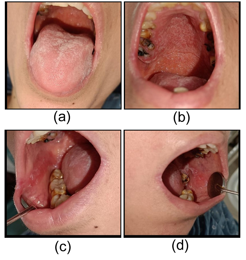
Figure 1. (a) Tongue dorsum showing creamy-white pseudomembranous plaques. (b) Palatal mucosa showing patchy white plaques with interspersed erythema. (c) Right buccal mucosa showing white pseudomembranous lesions over erythematous mucosa. (d) Left buccal mucosa showing creamy-white plaques
The mouth mirror test revealed xerostomia. The patient reported a marked oral burning sensation with a visual analog scale (VAS) score of 7.
In addition, multiple root stumps, grossly decayed teeth, and poor oral hygiene were observed, which could potentially contribute to microbial colonization and opportunistic infections.
The patient’s clinical presentation of multifocal scrapable white plaque lesions, together with a history of corticosteroid inhaler use without proper oral hygiene, strongly suggested acute pseudomembranous candidiasis.
Differential diagnoses included oral lichen planus, oral leukoplakia, coated tongue, oral hairy leukoplakia, and chemical burns. However, unlike these conditions, pseudomembranous candidiasis typically presents as soft, creamy white plaques that can be easily scraped off, leaving beneath an erythematous or occasionally bleeding mucosal surface.
A complete hemogram and rapid human immunodeficiency virus (HIV) test were performed to exclude systemic involvement. These investigations revealed values within normal limits and a non-reactive HIV status.
A smear of the lesion was collected for microscopic evaluation. Hematoxylin and eosin (H&E; 10×) stained sections showed numerous thin-walled filamentous structures suggestive of fungal hyphae (Figure 2a).
At a higher magnification (H&E; 40×), the sections demonstrated multiple neutrophilic microabscesses within the superficial epithelial layers, indicating an acute inflammatory response associated with fungal infection (Figure 2b).
Further confirmation was obtained using periodic acid-Schiff (PAS) staining, which demonstrated magenta-colored tubular hyphal structures consistent with Candida species invading the superficial epithelial layers (Figure 2c). The presence of PAS-positive fungal elements within the epithelium confirmed oral candidiasis.
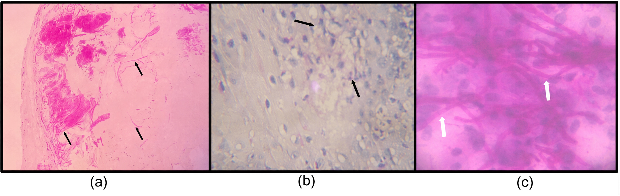
Figure 2. (a) H&E-stained section (×10) showing superficial epithelial changes with thin filamentous fungal structures. (b) H&E-stained section (×40) demonstrating neutrophilic microabscesses within the superficial epithelial layers. (c) PAS-stained section (×40) revealing magenta-colored fungal hyphae consistent with Candida albicans
The patient was prescribed systemic antifungal therapy with oral fluconazole, administered at 200 mg on the first day, followed by 100 mg once daily for the next 10 days, along with topical Clotrimazole therapy (Candid mouth paint 1%) for local application three times daily for 10 days. The patient was also advised to use Coolora mouthwash (Benzydamine Hydrochloride 0.15% w/v) as an adjunct to maintain oral hygiene and reduce the microbial load.
The patient received instructions on the maintenance of oral hygiene, including regular tooth brushing and plaque control. She was advised to rinse her mouth thoroughly after the use of inhaled bronchodilators and to use a spacer with her corticosteroid inhaler to reduce medication deposition in the oropharyngeal region. Dental treatment for multiple carious teeth and root stumps was recommended.
At the 10 day follow-up visit, the patient reported a huge relief from the burning sensation, and clinical examination revealed complete resolution of the lesions (Figures 3a-d).
At the 2 month follow-up, the patient remained asymptomatic and clinical examination revealed no recurrence of candidiasis.
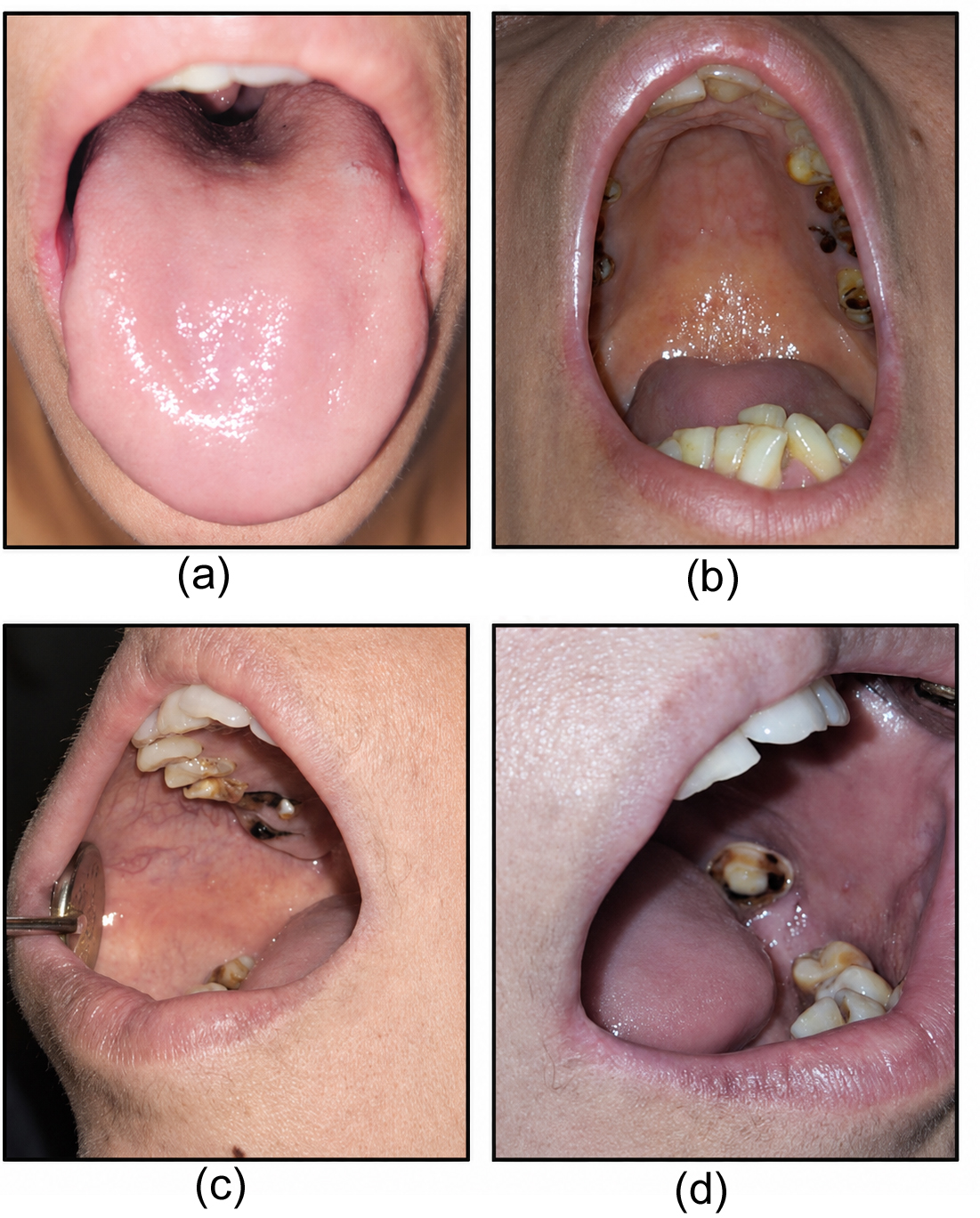
Figure 3. (a-d) Complete resolution of the lesions involving the tongue, palate, and bilateral buccal mucosa
Oral candidiasis is a common fungal infection of the oral mucous membrane, with a multifactorial etiology. It is most commonly associated with Candida species, particularly Candida albicans, which is detected in approximately 90% of the lesions.22,23
The prevalence of Candida albicans in the oral cavity varies among different populations, ranging from approximately 30%-50% in healthy individuals, 50%-65% in denture users, 65%-88% in residents of long-term care facilities, and 90%-95% in patients with HIV infection. Disruption of the normal oral microbiota or impairment of host immune defenses can allow Candida to proliferate excessively, leading to a fungal infection known as oral candidiasis or thrush.2,13
This case highlights the role of long-term inhaler use and poor oral hygiene as key predisposing factors for the development of acute pseudomembranous candidiasis.
Patients with chronic obstructive pulmonary disease (COPD) or asthma often use inhaled corticosteroids and bronchodilators that may serve as potential risk factors for oropharyngeal complications by modifying the oral microenvironment.16,24 Inhaled corticosteroids may promote the phenotypic transition of C. albicans from yeast to hyphal forms, a crucial step in tissue invasion mediated by adhesin proteins such as Agglutination-Like Sequence 3 and Hyphal Wall Protein 1.25 The localized immunosuppressive effects of these medications, along with elevated salivary glucose levels, create a favorable environment that enhances fungal colonization and mucosal penetration.19,21 Furthermore, inhaled corticosteroids may suppress nonspecific inflammatory responses and T-cell-mediated immunity, thereby facilitating the shift of commensal flora toward pathogenic overgrowth.26
Several clinical studies have reported that the prevalence of oral candidiasis in patients using inhaled corticosteroids ranges from 5%-20%. The likelihood of infection varies according to factors, such as the type of inhaler device, inhalation technique, corticosteroid dosage, duration of therapy, and oral hygiene practices. These findings suggest that inhaled corticosteroid therapy represents a significant predisposing factor for fungal colonization of the oral cavity in individuals with asthma or COPD.26-29
This case shares several clinical similarities with previously reported cases of inhaler-associated candidiasis. The patient presented with a persistent oral burning sensation, a commonly reported symptom of oral Candida infections.5,17 The lesions involved multiple oral sites, including the dorsum of the tongue, palate, and bilateral buccal mucosa, a distribution pattern characteristic of pseudomembranous candidiasis.5,16,30 Furthermore, the patient reported inhaler use without post-inhalation mouth rinsing, a practice recognized as a significant contributing factor to oropharyngeal fungal colonization.27
Recent studies have indicated that dry-powder inhalers may cause greater deposition of corticosteroid particles in the oropharyngeal region than MDIs. Higher corticosteroid doses and improper inhalation techniques can increase oral drug retention, leading to localized immunosuppression and facilitating Candida colonization and infection.31-34 These findings underscore the importance of educating patients about the correct inhalation technique and post-inhalation mouth rinsing to minimize local adverse effects.
In the present case, the patient was prescribed an MDI without a spacer, containing low-dose inhaled corticosteroids. However, she reported that she had not received adequate instructions regarding the correct inhalation technique.
The diagnosis of oral candidiasis is primarily clinical and relies on the identification of characteristic oral lesions, which may be further confirmed by the microscopic identification of Candida in oral samples and/or its detection in culture.30 Candida may be obtained from the oral cavity by several techniques, such as a plain swab, the use of a smear, an imprint culture, concentrated oral rinse, collection of whole saliva, and mucosal biopsy.3
Microscopic examination revealed filamentous fungal structures within the superficial epithelial layers along with neutrophilic microabscess formation, indicative of an acute inflammatory response to fungal invasion.
PAS staining revealed magenta-colored fungal hyphae consistent with Candida species within the epithelium, confirming the presence of Candida species. These findings align with the classical histopathological features of oral candidiasis, characterized by hyphal penetration of the superficial epithelium and associated inflammatory responses that contribute to clinical manifestations, such as burning sensation and mucosal erythema.3,30,35
A comprehensive approach involving antifungal therapy along with the elimination of underlying predisposing factors and potential fungal reservoirs, such as contaminated intraoral appliances, is essential for the effective treatment and prevention of recurrence.5,16
Fluconazole remains a widely used antifungal drug owing to its high oral bioavailability and efficacy against Candida albicans.36 Clotrimazole, a topical imidazole antifungal agent, is commonly used in the management of pseudomembranous candidiasis because it inhibits ergosterol biosynthesis in fungal cell membranes, thereby disrupting membrane integrity and inhibiting the growth of Candida species.
In the present case, treatment with systemic fluconazole and topical antifungal therapy resulted in rapid clinical improvement and complete resolution of the lesions.
Additional clinical concerns are the risk of recurrence and the development of antifungal resistance. Persistent exposure to predisposing factors, such as continued inhaled corticosteroid use, poor oral hygiene, xerostomia, or immunosuppression, can increase the susceptibility to recurrent episodes of oral candidiasis by creating a favorable environment for Candida colonization and proliferation.5,36,37
Prolonged or inappropriate antifungal therapy may contribute to the emergence of antifungal-resistant Candida strains, particularly non-albicans Candida species, which often exhibit reduced susceptibility to commonly used azole antifungals. Recent studies have highlighted increasing resistance patterns among clinical Candida isolates, emphasizing that repeated or empirical exposure can select resistant organisms and complicate treatment outcomes in recurrent candidiasis.38-40 Consequently, the exploration and development of new therapeutic agents and alternative regimens, particularly those derived from medicinal plants, have become increasingly important for the effective management of oral candidiasis.40
The favorable outcome observed in the present case, with complete resolution of lesions and no recurrence at two-month follow-up, underscores the importance of integrating antifungal therapy with preventive measures and patient education. Oral candidiasis should be included in the differential diagnosis of patients presenting with scrapable white plaques and persistent oral burning sensations, particularly in those undergoing inhaled therapy for pulmonary ailments.2,5
The integration of comprehensive diagnostic assessments and patient-centered preventive strategies is essential for mitigating the morbidity associated with inhaler-induced oral candidiasis. Future clinical protocols should emphasize the proactive monitoring of respiratory patients, as systematic adherence to post-inhalation oral hygiene serves as a primary defense against opportunistic fungal overgrowth.
Furthermore, the inclusion of molecular diagnostic tools may facilitate the identification of non-albicans species and strains that exhibit innate resistance, thereby guiding more precise therapeutic interventions.41
A limitation of this study was the absence of fungal cultures, species-level molecular identification, and antifungal susceptibility testing, which could provide additional microbiological confirmation.
This case report describes acute pseudomembranous candidiasis in a female patient with inhaler use who presented with a persistent oral burning sensation. The presence of multifocal, white, and scrapable oral plaques, supported by histopathological confirmation, facilitated an accurate diagnosis. Prompt treatment with systemic and topical antifungal therapy, along with correction of underlying predisposing factors, such as poor oral hygiene and improper inhaler practices, resulted in complete clinical resolution.
This case highlights the need for early diagnosis, appropriate antifungal management, and patient education regarding oral hygiene and post-inhaler mouth rinsing to prevent the recurrence of oral candidiasis.
Key Learning Points
- Oral candidiasis should be considered in patients presenting with multifocal scrapable white oral lesions with a persistent oral burning sensation.
- Chronic use of inhaled medications for asthma or other respiratory conditions is an important local predisposing factor for oral candidiasis, particularly in patients who do not rinse their mouths post-inhalation.
- Histopathological evaluation with special stains, such as PAS, plays a valuable role in confirming fungal infections, particularly in cases exhibiting a diagnostic dilemma.
- Successful management entails effective antifungal treatment and elimination of predisposing factors, including improvement of oral hygiene and correction of local irritants such as dental plaque and carious teeth.
- Patient education regarding proper inhalation techniques and routine mouth rinsing after inhaler use is essential to prevent the recurrence of oral candidiasis.
Patient perspective
The patient reported that the persistent burning sensation interfered with her ability to consume spicy foods and caused discomfort during routine daily activities. Following treatment and counseling on proper inhaler use and improved oral hygiene practices, the patient experienced complete relief of symptoms and expressed satisfaction with the treatment outcomes.
ACKNOWLEDGMENTS
None.
CONFLICT OF INTEREST
The authors declare that there is no conflict of interest.
AUTHORS’ CONTRIBUTION
All authors listed have made a substantial, direct, and intellectual contribution to the work and approved it for publication.
FUNDING
None.
DATA AVAILABILITY
All datasets generated or analyzed during this study are included in the manuscript.
ETHICS STATEMENT
Not applicable.
INFORMED CONSENT
Written informed consent was obtained from the patient for publication of this case report.
- Tamai R, Kiyoura Y. Candida infections: The role of saliva in oral health- A narrative review. Microorganisms. 2025;13(4):717.
Crossref - Patel M. Oral cavity and Candida albicans: colonisation to the development of infection. Pathogens. 2022;11(3):335.
Crossref - Saeed S, Hasan S, Kuldeep K, Parmar SS. Conventional and recent diagnostic aids in oral candidal infections: a brief overview. Biomed Pharmacol J. 2017;10(1):419-426.
Crossref - Li H, Miao M, Jia C, et al. Interactions between Candida albicans and the resident microbiota. Front Microbiol. 2022;13:930495.
Crossref - Lu SY. Oral candidosis: pathophysiology and best practice for diagnosis, classification, and successful management. J Fungi. 2021;7(7):555.
Crossref - Kimsa L, Tokarska-Rodak M. Occurrence of Candida spp. in healthy oral microbiota. Health Probl Civ. 2020;14(2):124-129.
Crossref - Patil S, Rao RS, Sanketh DS, Amrutha N. Microbial flora in oral diseases. J Contemp Dent Pract. 2013;14(6):1202-1208.
Crossref - Deorukhkar SC, Roushani S. Virulence traits contributing to pathogenicity of Candida species. J Microbiol Exp. 2017;5(1):00140.
Crossref - Silva KGDS, Nascimento GOD, Da Silva EEM, Cabral LHV, Faria TMR, De Oliveira JR. Candida albicans: virulence factors, pathogenesis, and ways to diagnose and control its infection. Res Soc Dev. 2024;13(1):e44781.
Crossref - Lopes JP, Lionakis MS. Pathogenesis and virulence of Candida albicans. Virulence. 2022;13(1):89-121.
Crossref - Board-Davies EL, Rhys-Williams W, Hynes D, Love WG, Williams DW. Antimicrobial effects of XF drugs against Candida albicans and its biofilms. Front Fungal Biol. 2023;4:1225647.
Crossref - Williams DW, Jordan RP, Wei XQ, et al. Interactions of Candida albicans with host epithelial surfaces. J Oral Microbiol. 2013;5(1):22434.
Crossref - Bras G, Satala D, Juszczak M, et al. Secreted aspartic proteinases: key factors in Candida infections and host-pathogen interactions. Int J Mol Sci. 2024;25(9):4775.
Crossref - Verma A, Gaffen SL, Swidergall M. Innate immunity to mucosal Candida infections. J Fungi. 2017;3(4):60.
Crossref - Juszczak M, Zawrotniak M, Rapala-Kozik M. Complexation of fungal extracellular nucleic acids by host LL-37 peptide shapes neutrophil response to Candida albicans biofilm. Front Immunol. 2024;15:1295168.
Crossref - Acharya S, Lohe VK, Bhowate RR. Diagnosis and management of pseudomembranous candidiasis. J Otolaryngol ENT Res. 2017;8(3):00249.
Crossref - Bouguezzi A, Slim A, Khalifa C, Chokri A, Hentati H, Selmi J. Consensus guidelines for the effective treatment of oral candidosis. SunText Rev Dent Sci. 2025;6(1):184.
Crossref - Sugio CYC, Garcia AAMN, Kitamoto KAA, et al. Mucoadhesive delivery systems for oral candidiasis treatment: a systematic review and meta-analysis. Oral Dis. 2024;30(6):3771-3787.
Crossref - Bhosale A, Dhande MB, Shinde AV, Jadhav P. Oral candidiasis review: pathogenesis, clinical presentation, and treatment strategies. J Adv Zool. 2023;44(S7):260-266.
Crossref - Contaldo M, Di Stasio D, Romano A, et al. Oral candidiasis and novel therapeutic strategies: antifungals, phytotherapy, probiotics, and photodynamic therapy. Curr Drug Deliv. 2023;20(5):441-456.
Crossref - Tkaczyk M, Kuska-Kielbratowska A, Fiegler-Rudol J, et al. The prevalence and drug susceptibility of Candida species and an analysis of risk factors for oral candidiasis: a retrospective study. Antibiotics (Basel). 2025;14(9):876.
Crossref - Ptasiewicz M, Thum-Tyzo K, Matejko A, et al. Clinical and microbiological profile of oral candidiasis: a retrospective study. Pathogens. 2026;15(2):195.
Crossref - Alrayyes SF, Alruwaili HM, Taher IA, et al. Oral candidal carriage and associated risk indicators among adults in Sakaka, Saudi Arabia. BMC Oral Health. 2019;19(1):86.
Crossref - Skoskiewicz-Malinowska K, Pogoda K, Zietek M, Jawor N. Oral health management for post-cancer therapy patients. Nowotwory J Oncol. 2025;75(5):296-304.
Crossref - Salah AN, Doghish YA, Abbass SO, et al. Microbiota-based therapies in oral health and disorders. Folia Microbiol (Praha). 2025;70(6):1217-1240.
Crossref - Arevalo JC, Buban ERD. Prevalence of oral candidiasis in asthma and COPD patients using inhaled corticosteroids in the Philippine General Hospital Department of Out-patient Services. Acta Med Philipp. 2023;57(12):32-38.
Crossref - Patel PM, Gohil PR, Parmar DH, Solanki SJ, Gabhawala YR, Patel JN. Occurrence of oral candidiasis among bronchial asthma patients on inhaled corticosteroids. Natl J Med Res. 2021;11(1):1-3.
- Ahmad S, Iqbal S, Nisa M, Ali S, Khan SA, Ashfaq U. Incidence of oral candidiasis in patients with asthma using inhaled corticosteroids by various inhaler devices. Pak J Med Health Sci. 2022;16(2):1198.
Crossref - Erdogan T, Karakaya G, Kalyoncu AF. The frequency and risk factors for oropharyngeal candidiasis in adult asthma patients using inhaled corticosteroids. Thorac Res Pract. 2019;20(2):136-139.
Crossref - Akpan A, Morgan R. Oral candidiasis. Postgrad Med J. 2002;78(922):455-459.
Crossref - Gani F, Caminati M, Bellavia F, et al. Oral health in asthmatic patients: a review. Clin Mol Allergy. 2020;18(1):22.
Crossref - Fukushima C, Matsuse H, Tomari S, et al. Oral candidiasis associated with inhaled corticosteroid use: comparison of fluticasone and beclomethasone. Ann Allergy Asthma Immunol. 2003;90(6):646-651.
Crossref - Thomas MS, Parolia A, Kundabala M, Vikram M. Asthma and oral health: a review. Aust Dent J. 2010;55(2):128-133.
Crossref - Kurt E, Yildirim H, Kiraz N, et al. Oropharyngeal candidiasis with dry-powder fluticasone propionate: 500 µg/day versus 200 µg/day. Allergol Immunopathol (Madr). 2008;36(1):17-20.
Crossref - Neville BW, Damm DD, Allen CM, Chi AC. Oral and Maxillofacial Pathology. 4th ed. Elsevier; 2016:189-195.
- Lombardi A, Ouanounou A. Fungal infections in dentistry: clinical presentations, diagnosis, and treatment alternatives. Oral Surg Oral Med Oral Pathol Oral Radiol. 2020;130(5):533-546.
Crossref - Khatter NJ, Khan MAB, ed. Clotrimazole. In: StatPearls. StatPearls Publishing. 2024.
- Ghojoghi A, Khodavaisy S, Mahmoudabadi AZ, Fatahinia M. Antifungal susceptibility profile of Candida species and uncommon yeasts from drug abusers with oral candidiasis. BMC Oral Health. 2024;24(1):1555.
Crossref - Kessler SQS, Lang PM, Dal-Pizzol TS, Montagner F. Resistance profiles to antifungal agents in Candida albicans isolated from human oral cavities: systematic review and meta-analysis. Clin Oral Invest. 2022;26(11):6479-6489.
Crossref - Nurdiana, Naorungroj S, Sappayatosok K, Lomlim L, Pahumunto N. Efficacy of botanical antifungals and conventional antifungals in the treatment of oral candidiasis: a systematic review and meta-analysis. Front Pharmacol. 2025;16:1635482.
Crossref - Czajka KM, Verschoor CP, Santi SA, et al. Signs and symptoms of oral candidiasis associated with health factors and resistant Candida infections in a Northern Ontario patient cohort. Front Oral Health. 2025;6:1661524.
Crossref
© The Author(s) 2026. Open Access. This article is distributed under the terms of the Creative Commons Attribution 4.0 International License which permits unrestricted use, sharing, distribution, and reproduction in any medium, provided you give appropriate credit to the original author(s) and the source, provide a link to the Creative Commons license, and indicate if changes were made.