


ISSN: 0973-7510
E-ISSN: 2581-690X
The goal of this study is to evaluate the antifungal activity of terbinafine in its nano form against yeast organisms in vitro and compare its effectiveness to the normal form of terbinafine. Thirty isolated yeast organisms from Thumbay Labs were included in the study. Terbinafine was serially diluted to concentrations of 50 mg, 25 mg, 12 mg, and 6 mg for both the nano and normal forms. After preparing the serial dilutions, fungal suspensions were exposed to these dilutions for 7 minutes. The suspensions were then cultured on Sabouraud dextrose agar and incubated at 37°C overnight. Growth was measured using a colony count. The results for the normal form of terbinafine revealed growth at all concentrations, with varying colony counts with a highest growth was observed at the 6 mg concentration. In contrast, the nano form of terbinafine exhibited distinct development patterns. At the 50 mg concentration, seven samples showed no growth; at the 25 mg concentration, two samples showed no growth; and the remaining samples exhibited limited growth compared to the normal form. According to the results of this study, there were significant differences in the number of colonies between the two forms of terbinafine, with the nano form demonstrating greater efficacy. The most potent antifungal response was observed at the 50 mg concentration of nano terbinafine, which inhibited yeast growth in several samples. In conclusion, terbinafine nanoemulsions are more effective than the normal form against Candida.
Yeast, Candida albicans, Antifungal, Terbinafine, Nanoemulsions
Fungi are a diverse group of organisms, including yeasts and molds, distinct from bacteria, plants, and animals. They play a crucial role in ecosystems as decomposers, recycling nutrients and breaking down organic materials. Fungi also form symbiotic relationships with plants, such as mycorrhizae, which help plants absorb nutrients from the soil. While some fungi can cause diseases in humans, animals, and plants, others have beneficial properties and are used in industry, medicine, and food production. Fungi reproduce through spores, and their filamentous structures, called hyphae, form networks known as mycelium.1
Yeast is a type of single-celled fungus from the kingdom Fungi, commonly used in fermentation, brewing, and baking. Yeasts reproduce asexually through a process called budding, where a small daughter cell forms on the surface of the parent cell and eventually separates to become a new organism. Yeast is a versatile organism with numerous applications in food production, scientific research, and various other fields.2
The human body naturally hosts fungi, such as Candida yeast. These fungi can be found on the skin, in the digestive system, and in the vagina, which is part of the female reproductive system.3
Candida species are the leading cause of fungal infections, with about 90% of infections attributed to five main species: Candida albicans, Candida glabrata, Candida tropicalis, Candida parapsilosis, and Candida krusei. These fungi are among the most common human fungal pathogens, responsible for both superficial (mucosal and cutaneous) and systemic infections. Candida albicans, in particular, is a small, oval, gram-positive yeast that reproduces by budding.4, 5
The microbiome helps regulate the balance of Candida. When this balance is disrupted, Candida can overgrow, leading to candidiasis. Candidiasis is an opportunistic infection caused by Candida yeast that can affect the penis, vagina, oral cavity, and other body areas. Oral candidiasis, commonly known as thrush, is one of the most frequent fungal infections and appears as white spots on the tongue, throat, and other oral tissues. Vaginal candidiasis, or yeast infection, is the second most common infection of the female reproductive system.6
Treatment of candidiasis typically involves antifungal medications, which can be applied topically, taken orally, or administered intravenously, depending on the infection’s location and severity. Antifungal medications are used to treat various fungal infections, including yeast infections, ringworm, and nail and skin diseases. These infections can be caused by fungi present in the soil, air, and on the skin.7,8
Topical antifungals can be used to treat fungal infections on the skin or tongue. They can be applied as creams, powders, or solutions. Terbinafine, an antifungal containing allylamines, functions mainly as a fungicidal agent in vitro.9,10
Oral terbinafine is the recommended treatment for dermatophyte onychomycosis due to its high mycological and clinical cure rates, good tolerability, and minimal risk of drug interactions. It should also be considered the first-choice treatment for cutaneous mycoses requiring systemic treatment, alongside itraconazole. For less extensive mycoses, topical terbinafine is the preferred treatment. While terbinafine is generally less effective against yeasts, it is more effective against dermatophytes compared to azole antifungals like itraconazole.11
Many fungal strains possess genetic and molecular resistance mechanisms, which can be either inherent or developed over time through exposure to antifungal medications. These genetic changes or innate characteristics enable the fungi to resist the effects of the drugs. Understanding these resistance mechanisms can help guide the selection of more effective medications.12
Nano-drugs have garnered significant interest for enhancing the efficacy of antifungal treatments compared to commercial drugs. The effectiveness of current antifungal medications is limited by their toxicity and the drug resistance found in clinical strains. Recent advancements in nanobiotechnology have opened new possibilities for developing innovative formulations with improved therapeutic effectiveness.13-15
The aim of this study was to investigate the antifungal effectiveness of terbinafine in both its conventional and nano-formulations against yeast species, particularly Candida.
A total of 30 yeast organisms were isolated from various samples received in Microbiology Department at Thumby Labs in Ajman, during February and March 2024.
Isolation and identification of the organism
The yeast samples received in Thumby lab GMU were subcultured on Saboruaud
dextrose agar and incubated aerobically at 37°C overnight. Candida species were identified using Gram staining, germ tube test, and API 20 C AUX strip (Biomerieux).
Preparation of terbinafine hydrochloride loaded nanoemulsions
Terbinafine hydrochloride (TF-HCl) loaded nanoemulsions were prepared in the College of Pharmacy at GMU using a modified ultrasonication method as described in the literature.16 A precise quantity of TF-HCl (100 mg) was dissolved in clove oil in a glass vial, and the batch size of the nanoemulsion was 10 mL. The surfactant, Tween-80, and the co-solvent, ethanol, were added at a 1:1 ratio with continuous stirring. Water (5 mL) was added drop by drop to each formulation while stirring continuously until a clear, homogeneous oil-in-water (O/W) emulsion was achieved. After emulsion formation, ultrasonication was performed for a period of 10 minutes.
Droplet size and polydispersity index (PDI) determinations
The droplet sizes and polydispersity values (PDI) values of prepared nanoemulsions were assessed using dynamic light scattering measurements (Zetasizer ZS90 instrument from Nanoseries, Malvern, UK). For the assessments, formulations were diluted tenfold with double-distilled water and placed into a quartz cuvette. The measurements were carried out in triplicate, and average values were calculated (Figure 1).
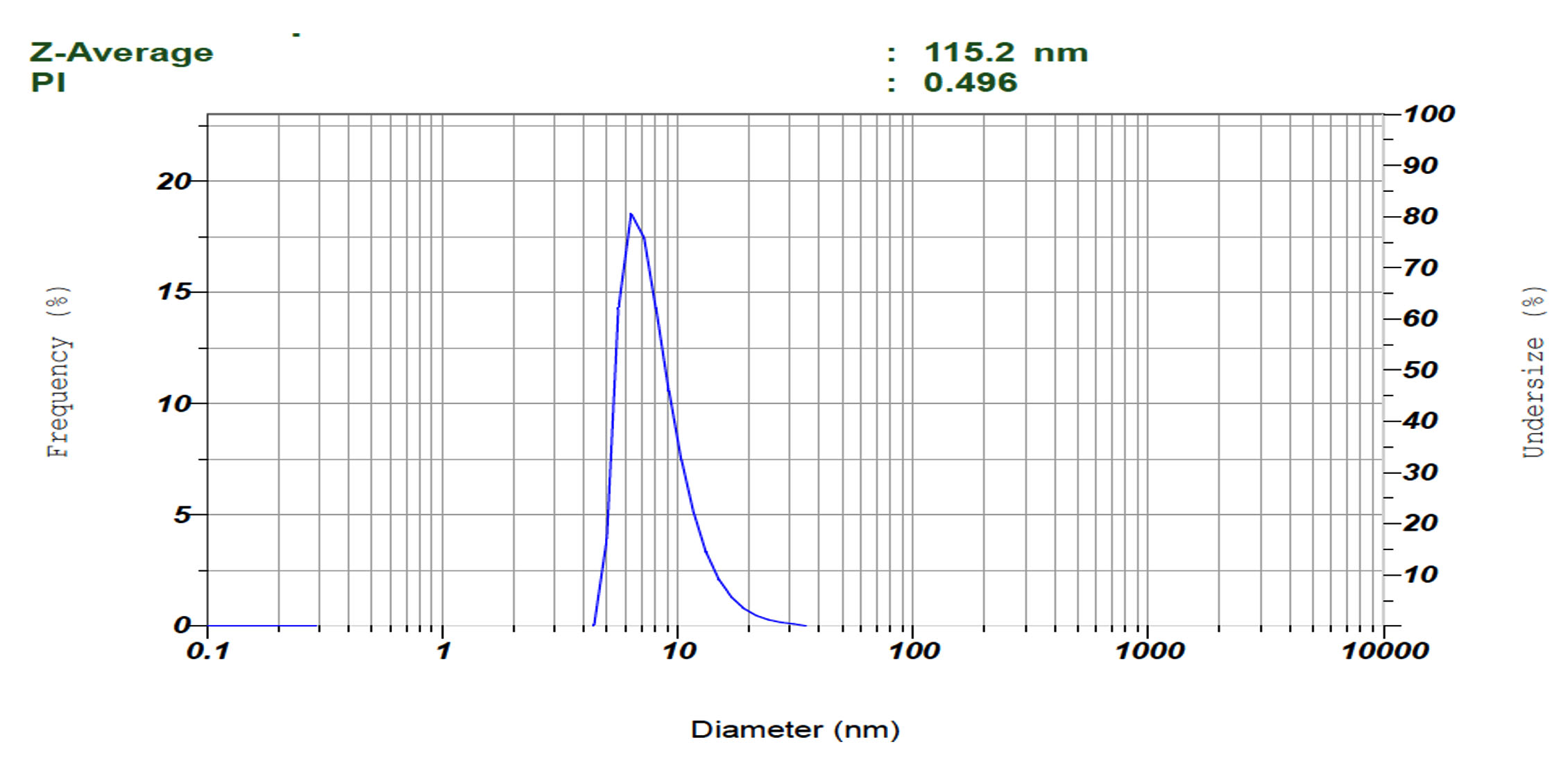
Figure 1. Representative image showing the particle size distribution of terbinafine HCl loaded nanoemulsions
Preparation of Terbinafine antifungal
Both normal terbinafine form (available commercially) and nanoemulsion form were prepared in serial dilutions across four separate glass tubes to achieve four distinct antifungal concentrations: 50 mg, 25 mg, 12 mg, and 6 mg.
Preparation of the organism
A fungal suspension was prepared by adding 3 ml of normal saline to an empty glass tube. One to two yeast colonies were then taken from the Sabouraud dextrose agar and mixed into the saline. The concentration was adjusted to match the McFarland standard 0.5, equivalent to 1.5 × 10⁸ CFU/ml.
The exposure of organism to anti-fungal
100 µL of each terbinafine dilution was added to a separate sterile test tube, followed by 40 µl of the fungal suspension in each tube. This resulted in each tube containing 140 µL of terbinafine dilution mixed with the fungal suspension. A control group containing only the fungal suspension was also prepared in a separate tube. All tubes were then mixed thoroughly and incubated for 7 minutes.
After the incubation period, 100 µl from each tube was transferred to Sabouraud Dextrose Agar (SDA) plates. The lawn culture method was used by spreading the microbial inoculum evenly over the entire surface of the SDA plates. The plates were then incubated aerobically at 37°C overnight. This procedure was applied to both the nano form and the conventional form of the antifungal.
Enumeration of the colonies following exposure to Terbinafine
After overnight incubation, colonies were then counted, and the number of CFUs/mL was plotted against each antifungal concentration (50 mg, 25 mg, 12 mg, 6 mg, and control) using a colony counter machine (Stuart SC6 Colony Counter) to determine the results.
The colonies on four squares of the colony counter surface were counted. The mean of these counts was then calculated and multiplied by 52 to determine the total number of colonies on the entire plate. Subsequently, this total was multiplied by 10 to obtain the CFUs/mL.
When there are too many colonies on the plate, the colonies are indistinguishable as individual colonies. The result considered Too Many To Count (TMTC) or >10000.
The droplet sizes of the prepared nanoemulsion exhibited a particle size of 115.2 nm, and the PDI values were low (0.496), indicating excellent mono-dispersity and stability.
In identification of the organisms, most of the species of the 30 isolates were Candida albicans 28 (93.3%), whereas 2 (6.7%) samples were candida glabrata.
The results for terbinafine in its normal form, presented in Table 1, indicate that all organisms are resistant to the antifungal agent. Fungal colonies appeared at all tested concentrations of the normal form: 50 mg, 25 mg, 12 mg, and 6 mg.
Table (1):
Shows the colony count of the yeast organisms exposed to the terbinafine normal form
Sample No. |
50 mg |
25 mg |
12 mg |
6 mg |
|---|---|---|---|---|
1 |
2028 |
≥10000 |
≥10000 |
≥10000 |
2 |
196 |
209 |
458 |
660 |
3 |
3354 |
5547 |
≥10000 |
≥10000 |
4 |
507 |
1144 |
1872 |
2145 |
5 |
2717 |
3679 |
4796 |
6314 |
6 |
≥10000 |
≥10000 |
≥10000 |
≥10000 |
7 |
3419 |
4518 |
5960 |
7870 |
8 |
819 |
904 |
1950 |
2248 |
9 |
3991 |
6747 |
7750 |
≥10000 |
10 |
2350 |
≥10000 |
≥10000 |
≥10000 |
11 |
2535 |
3623 |
4734 |
≥10000 |
12 |
1404 |
4524 |
≥10000 |
≥10000 |
13 |
1638 |
1677 |
1723 |
1885 |
14 |
4589 |
4940 |
≥10000 |
≥10000 |
15 |
4810 |
5850 |
2223 |
≥10000 |
16 |
1690 |
4290 |
≥10000 |
≥10000 |
17 |
2119 |
2535 |
702 |
715 |
18 |
299 |
689 |
5420 |
6369 |
19 |
3952 |
4927 |
1461 |
≥10000 |
20 |
1378 |
637 |
2119 |
≥10000 |
21 |
≥10000 |
≥10000 |
≥10000 |
≥10000 |
22 |
450 |
501 |
≥10000 |
≥10000 |
23 |
668 |
1500 |
≥10000 |
≥10000 |
24 |
≥10000 |
≥10000 |
≥10000 |
≥10000 |
25 |
≥10000 |
≥10000 |
≥10000 |
≥10000 |
26 |
1352 |
5720 |
2015 |
2550 |
27 |
500 |
512 |
3926 |
2119 |
28 |
589 |
620 |
1560 |
4732 |
29 |
≥10000 |
≥10000 |
≥10000 |
≥10000 |
30 |
≥10000 |
≥10000 |
≥10000 |
≥10000 |
The growth of fungal colonies varies across different concentrations of terbinafine. At the 50 mg concentration, the number of colonies ranges from 196 to over 10,000. In the 25 mg concentration, the growth range extends from 209 to over 10,000 colonies. The 12 mg concentration shows increased growth as well, ranging from 458 to over 10,000 colonies. At the 6 mg concentration, almost all samples exhibited over 10,000 colonies.
Overall, 5 samples (16.6%) exhibited ≥10,000 colonies at all concentrations of the terbinafine in its normal form.
On the other hand, the results for terbinafine in its nano form, presented in Table 2, show different growth ranges compared to the normal form. Seven samples 1, 2, 3, 4, 5, 8, and 21 (Figures 2 to 8) exhibited no growth at the 50 mg concentration of the nanoemulsion. At the 25 mg concentration, two samples, specifically samples 8 and 21 (Figures 7 and 8), also showed no growth.
Table (2):
Shows the colony count of the yeast organisms exposed to the terbinafine nano form
Sample No. |
50 mg |
25 mg |
12 mg |
6 mg |
|---|---|---|---|---|
1 |
0 |
25 |
145 |
257 |
2 |
0 |
30 |
130 |
273 |
3 |
0 |
27 |
≥10000 |
≥10000 |
4 |
0 |
138 |
401 |
450 |
5 |
0 |
75 |
203 |
267 |
6 |
897 |
≥10000 |
≥10000 |
≥10000 |
7 |
297 |
562 |
≥10000 |
≥10000 |
8 |
0 |
0 |
442 |
681 |
9 |
1911 |
≥10000 |
≥10000 |
≥10000 |
10 |
394 |
≥10000 |
≥10000 |
≥10000 |
11 |
1000 |
2000 |
≥10000 |
≥10000 |
12 |
800 |
1000 |
1200 |
1350 |
13 |
≥10000 |
≥10000 |
≥10000 |
≥10000 |
14 |
600 |
1000 |
1200 |
≥10000 |
15 |
409 |
700 |
1500 |
≥10000 |
16 |
≥10000 |
≥10000 |
≥10000 |
≥10000 |
17 |
480 |
800 |
≥10000 |
≥10000 |
18 |
3985 |
5900 |
6700 |
≥10000 |
19 |
140 |
160 |
≥10000 |
≥10000 |
20 |
609 |
653 |
678 |
463 |
21 |
0 |
0 |
41 |
2457 |
22 |
50 |
124 |
1095 |
≥10000 |
23 |
551 |
3172 |
3285 |
3700 |
24 |
2379 |
2938 |
3432 |
≥10000 |
25 |
20 |
129 |
≥10000 |
≥10000 |
26 |
≥10000 |
≥10000 |
≥10000 |
≥10000 |
27 |
609 |
3146 |
3237 |
≥10000 |
28 |
708 |
1781 |
3562 |
3605 |
29 |
5320 |
≥10000 |
≥10000 |
≥10000 |
30 |
649 |
784 |
762 |
910 |
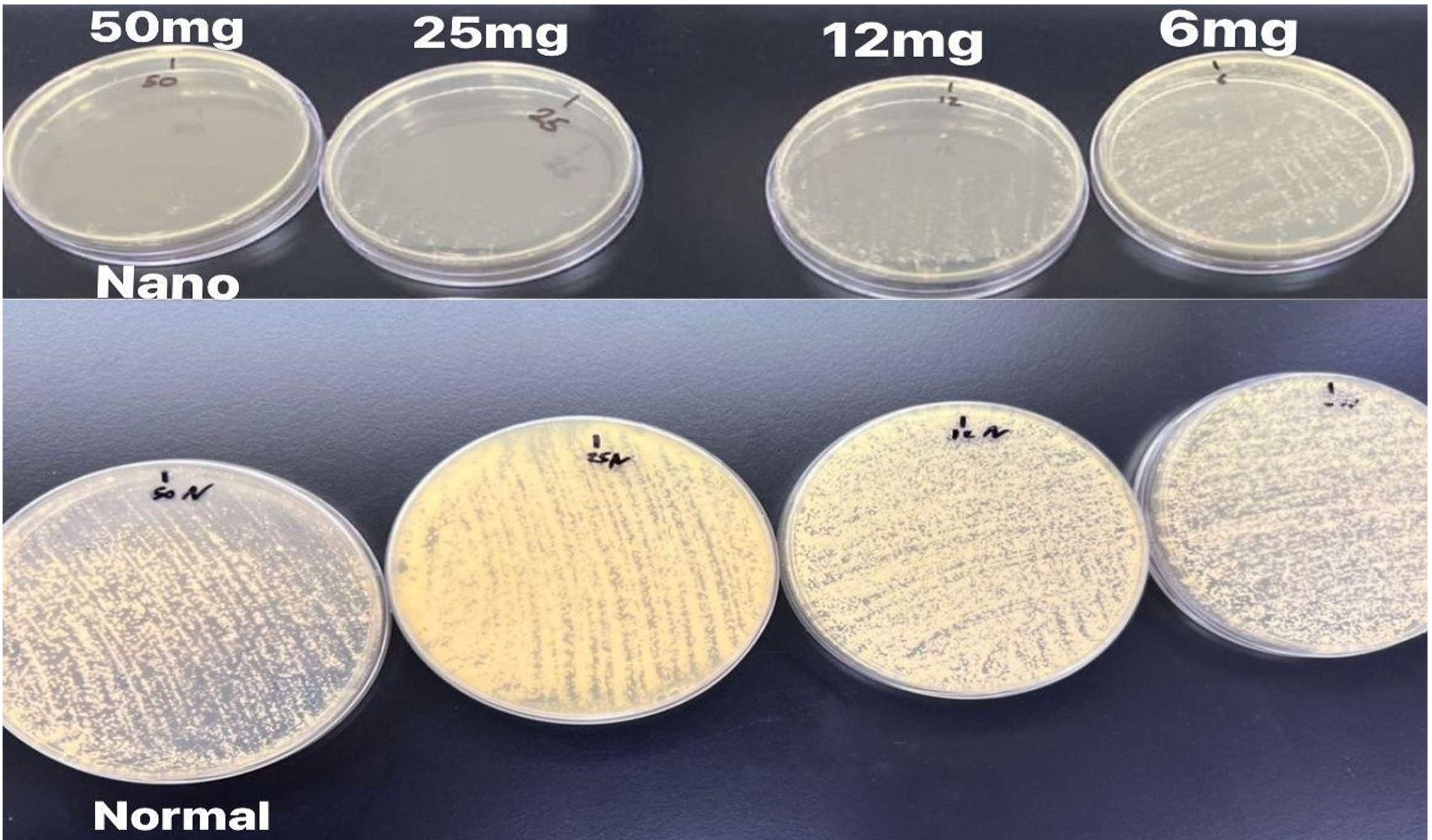
Figure 2. Shows the growth of sample number 1 nano form (upper) which indicates inhibition of growth in 50 mg concentration. Normal form (lower) showed growth in all concentration 50 mg, 25 mg, 12 mg and 6 mg
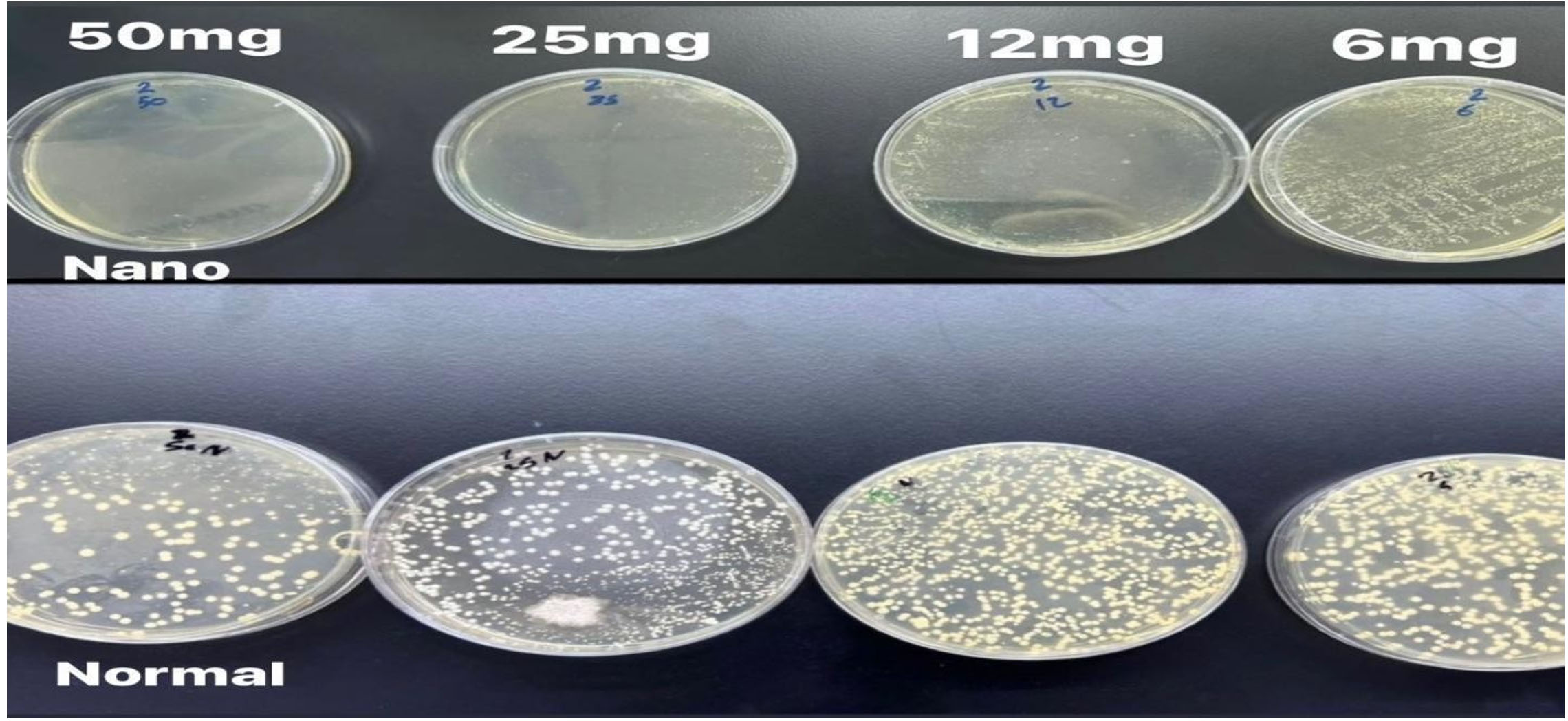
Figure 3. Shows the growth of sample number 2 nano form (upper) which indicates inhibition of growth in 50 mg concentration. Normal form (lower) showed growth in all concentration 50 mg, 25 mg, 12 mg and 6 mg
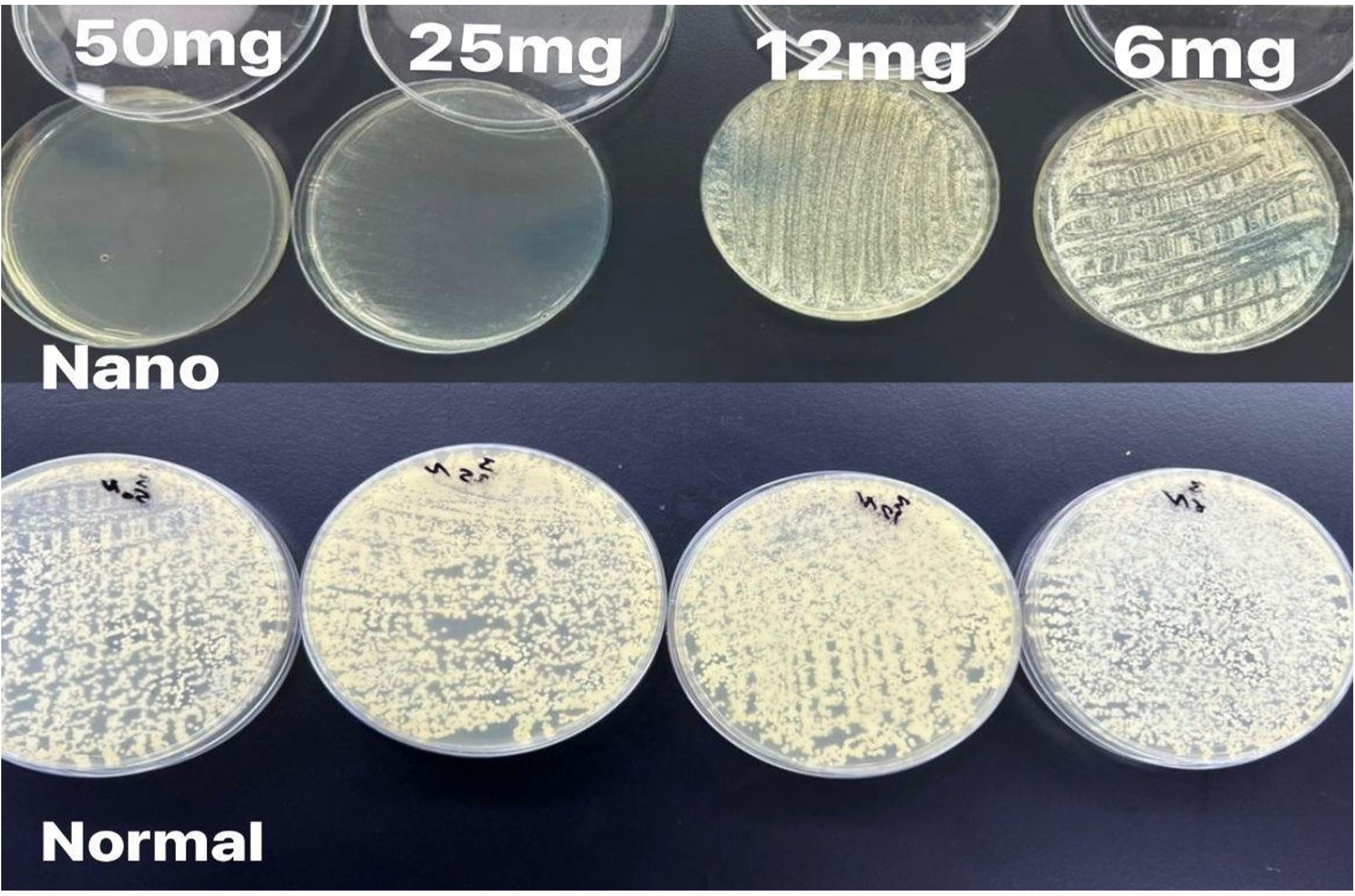
Figure 4. Shows the growth of sample number 3 nano form (upper) which indicates inhibition of growth in 50 mg concentration. Normal form (lower) showed growth in all concentration 50 mg, 25 mg, 12 mg and 6 mg
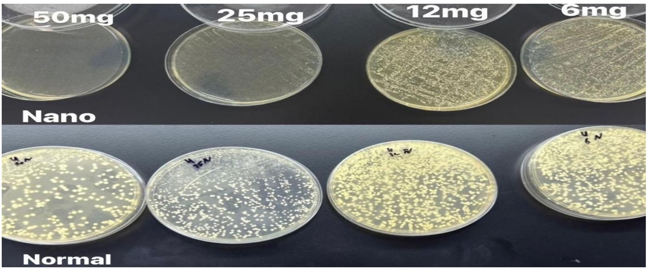
Figure 5. Shows the growth of sample number 4 nano form (upper) which indicates inhibition of growth in 50 mg concentration. Normal form (lower) showed growth in all concentration 50 mg, 25 mg, 12 mg and 6 mg
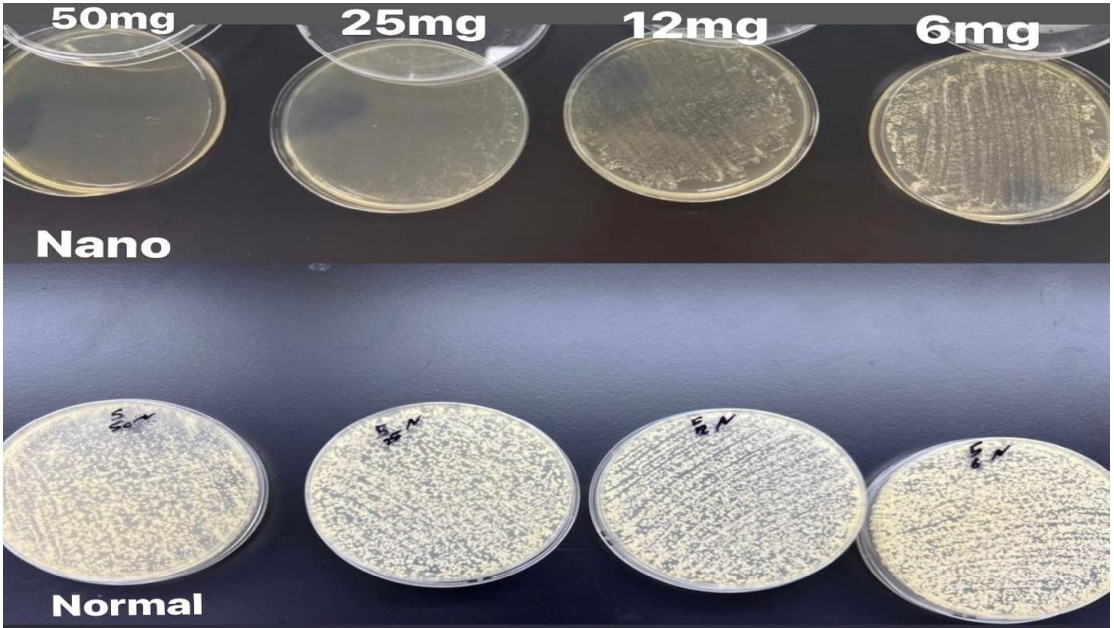
Figure 6. Shows the growth of sample number 5 nano form (upper) which indicates inhibition of growth in 50 mg concentration. Normal form (lower) showed growth in all concentration 50 mg, 25 mg, 12 mg and 6 mg
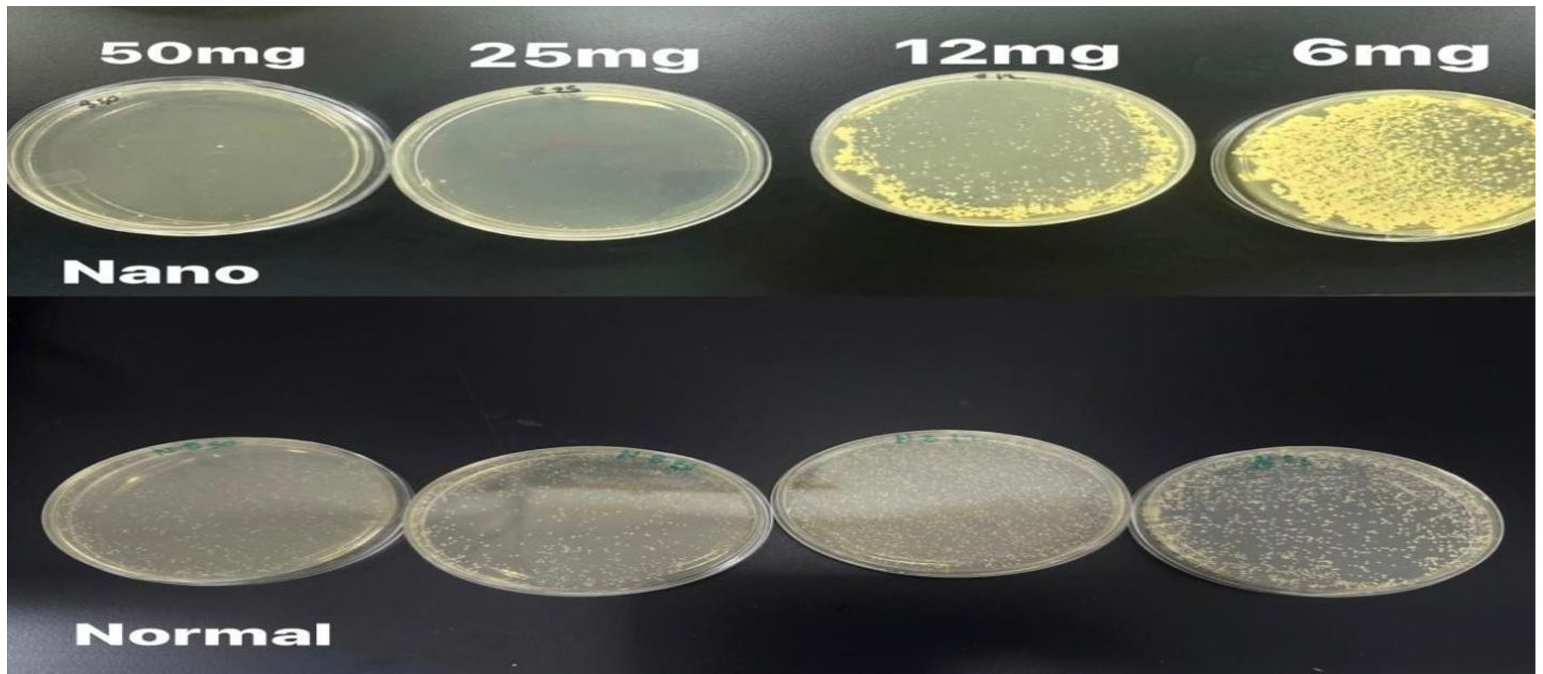
Figure 7. Shows the growth of sample number 8 nano form (upper) which indicates inhibition of growth in 50 mg and 25 mg concentrations. Normal form (lower) showed growth in all concentration 50 mg, 25 mg, 12 mg and 6 mg
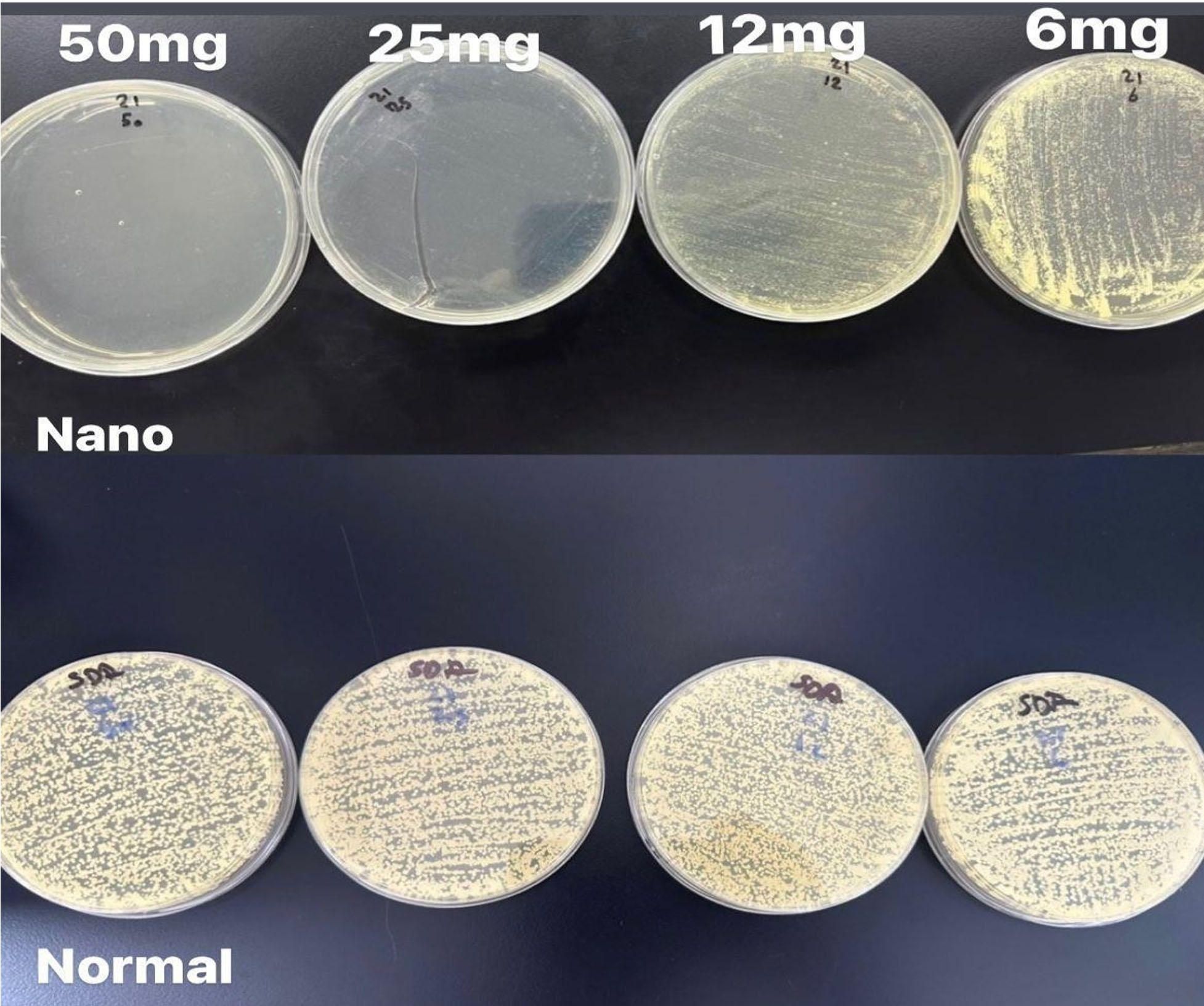
Figure 8. Shows the growth of sample number 21 nano form (upper) which indicates inhibition of growth in 50 mg and 25 mg concentrations. Normal form (lower) showed growth in all concentration 50 mg, 25 mg, 12 mg and 6 mg
Overall, the growth of the organisms exposed to nano terbinafine is less than that observed with the normal form, with colony counts ranging from 25 to over 10,000.
Growth increased as the concentration of the antifungal nanoemulsion decreased. At the 12 mg concentration, the range was from 41 colonies to over 10,000. The 6 mg concentration exhibited the highest growth among all concentrations, with colony counts ranging from 257 to over 10,000.
The observed colony counts reveal a significant difference between the normal-form terbinafine and the nano-form terbinafine. As shown in Table 1, the normal form of terbinafine resulted in fungal growth at all concentrations, including 50 mg. In contrast, Table 2 indicates that the nano-form terbinafine prevented growth in some samples at the 50 mg concentration, specifically in samples numbered 1, 2, 3, 4, 5, 8, and 21 (Figure 9).
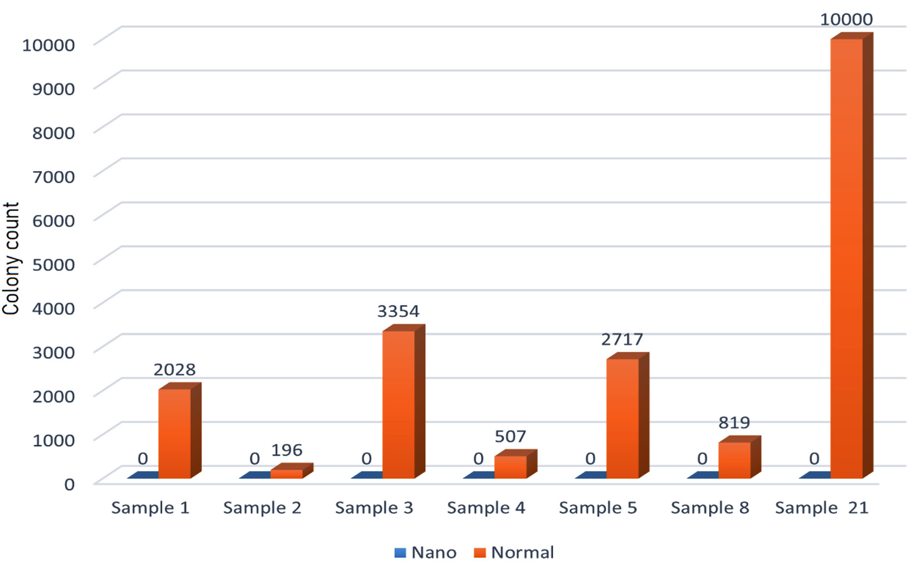
Figure 9. Shows the difference between the colony count for organisms exposed to nano form and normal form terbinafine in 50 mg concentration for samples 1, 2, 3, 4, 5, 8, and 21
The MIC values for samples 1, 2, 3, 4, and 5 were 50 mg. Additionally, 80% of the samples demonstrated a significant decrease in growth compared to the normal form at the 50 mg concentration.
At the 25 mg concentration, 60% of the samples treated with the nano form exhibited decreased growth compared to the normal form. Moreover, samples number 8 and 21 showed no growth at the 25 mg concentration of nano-form terbinafine, with the MIC for these two samples being 25 mg (Figure 10).
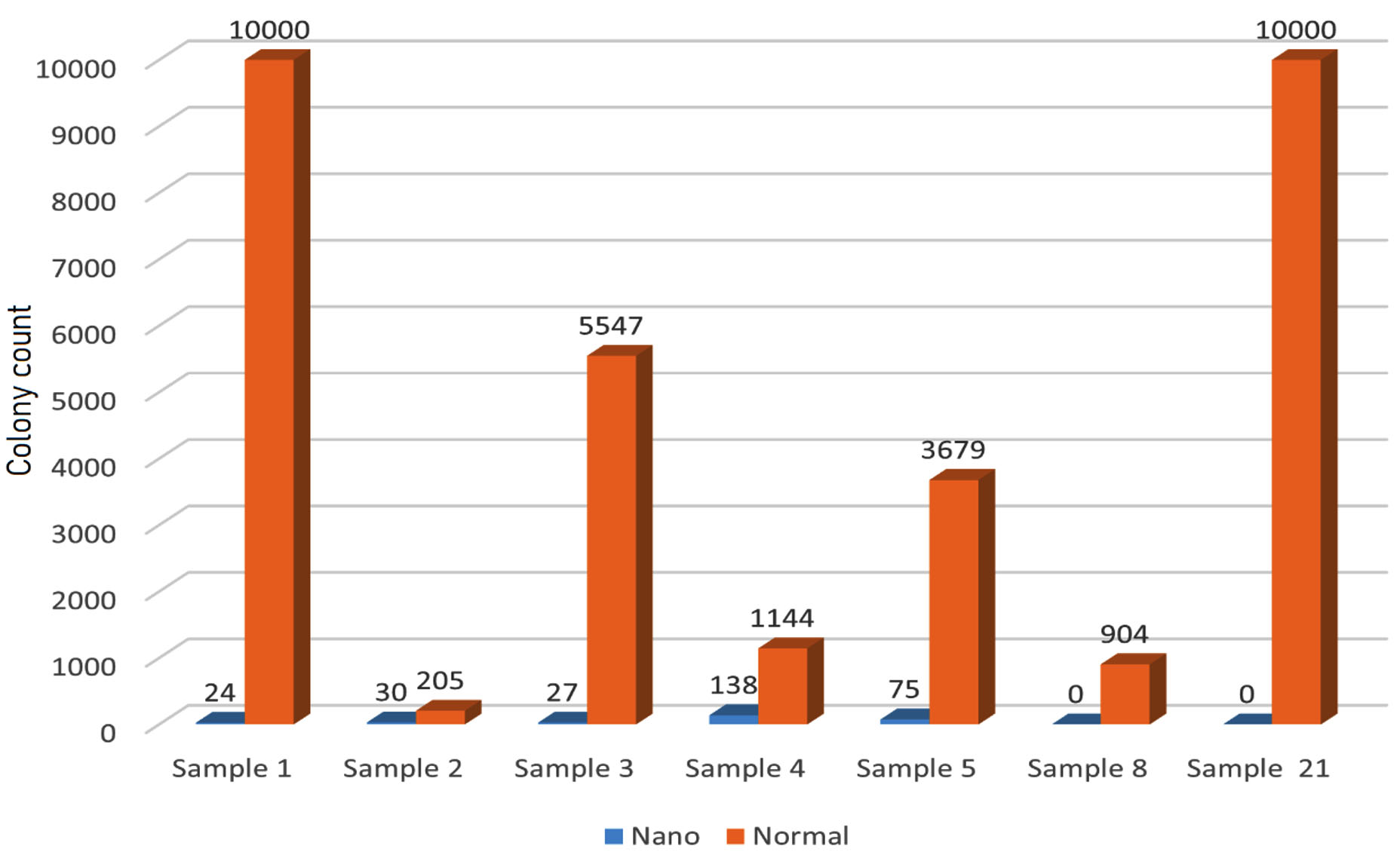
Figure 10. Shows the difference between the Colony count for organisms exposed to nano form and normal form terbinafine in 25 mg concentration for samples 1, 2, 3, 4, 5, 8, and 21
The colony counts for various samples treated with a 12 mg concentration of both the normal form and the nano form of terbinafine against yeast show that 17 samples (57%) exhibit a significant decrease in colony count with the nano form. However, thirteen samples show no difference between the nano and normal forms, with both displaying the maximum colony count of 10,000 (Figure 11).
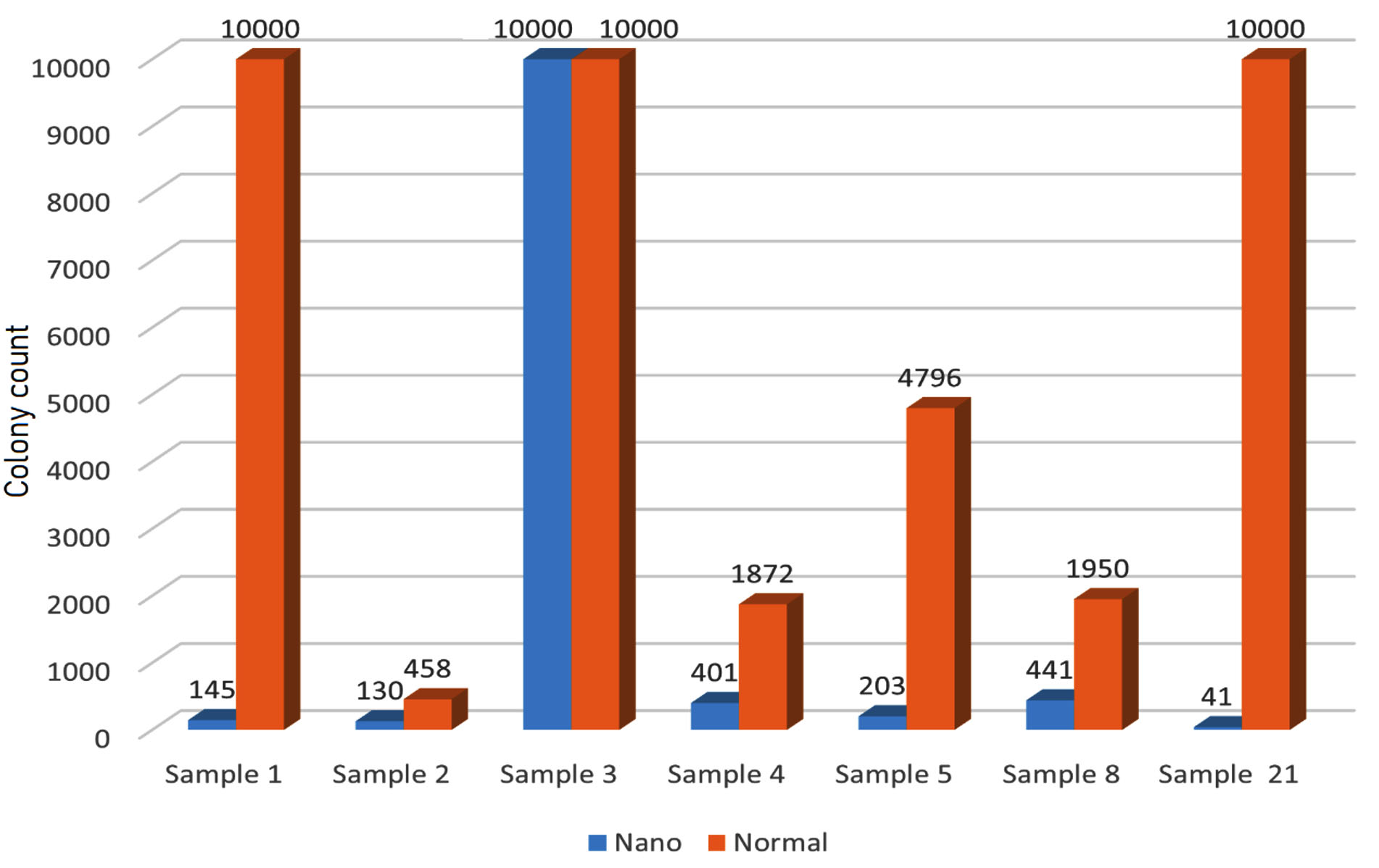
Figure 11. Shows the difference between the Colony count for organisms exposed to nano form and normal form terbinafine in 12 mg concentration for samples 1, 2, 3, 4, 5, 8, and 21
At a 6 mg concentration, both forms of terbinafine showed the highest number of colonies, with most samples exhibiting 10,000 colonies. However, there were exceptions that showed a difference between the normal and nano forms. Only 36.6% of the samples treated with the nano form showed decreased growth compared to the normal form (Figure 12). Generally, growth increases as the concentration of the antifungal decreases.
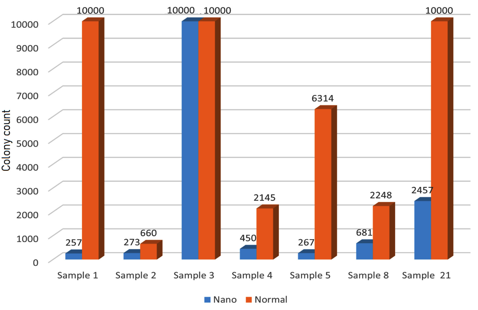
Figure 12. Shows the difference between the Colony count for organisms exposed to nano form and normal form terbinafine in 6 mg concentration for samples 1, 2, 3, 4, 5, 8, and 21
Overall, the nano form of terbinafine appears to be more effective in reducing yeast colony counts at all concentrations across the samples.
This finding aligns with a study conducted in India by Karri et al.17 They found that a nanoemulsion gel (NEG) of Terbinafine HCl (TBH) demonstrated a high zone of inhibition (p < 0.01) in in vitro antifungal experiments against Candida albicans, nearly equal to that of the pure drug solution. When compared to a marketed cream (MC), the NEG formulation of TBH proved to be more effective. Therefore, the NEG formulation may be the most suitable method for delivering poorly soluble medications like TBH for topical antifungal use.17 This study also showed a p-value of <0.01, indicating that the nanoemulsion of terbinafine is more effective against Candida albicans compared to the normal form of terbinafine.
The results of the present study indicate that the nano form of terbinafine is more effective than the normal form against yeast organisms isolated from Thumby Labs, particularly Candida albicans. There was a significant difference in colony counts between the normal and nano forms of terbinafine, with the nano form showing greater efficacy. At a 50 mg concentration, the nano form of terbinafine demonstrated the highest and strongest antifungal activity, inhibiting the growth of yeast organisms in several samples.
ACKNOWLEDGMENTS
None.
CONFLICT OF INTEREST
The authors declare that there is no conflict of interest.
AUTHORS’ CONTRIBUTION
All authors listed have made a substantial, direct and intellectual contribution to the work, and approved it for publication.
FUNDING
None.
DATA AVAILABILITY
All datasets generated or analyzed during this study are included in the manuscript.
ETHICS STATEMENT
This article does not contain any studies on human participants or animals performed by any of the authors.
- Liu D. Classification of medically important fungi. Mol Med Microbiol. 2024:2763-2777.
Crossref - Mukherjee S, Ghorai S. Fungal biology. Current Developments in Biotechnology and Bioengineering. 2023:67-104.
Crossref - Martins N, Ferreira ICFR, Barros L, Silva S, Henriques M. Candidiasis: predisposing factors, prevention, diagnosis and alternative treatment. Mycopathologia. 2014;177(5-6):223-240.
Crossref - Turner SA, Butler G. The Candida pathogenic species complex. Cold Spring Harb Perspect Med. 2014;4(9):19778.
Crossref - Sharma M, Chakrabarti A. Candidiasis and other emerging yeasts. Curr Fungal Infect Rep. 2023;17(1):15-24.
Crossref - Arya NR, Rafiq NB. Candidiasis. StatPearls. Accessed May 29, 2023 https://www.ncbi.nlm.nih.gov/books/NBK560624/
- Ordaya EE, Clement J, Vergidis P. The role of novel antifungals in the management of candidiasis: a clinical perspective. Mycopathologia. 2023;188(6):937-948.
Crossref - Jain A, Jain S, Rawat S. Emerging fungal infections among children: A review on its clinical manifestations, diagnosis, and prevention. J Pharm Bioallied Sci. 2010;2(4):314-320.
Crossref - Borah P, Hazarika S, Sharma D, et al. Systemic and topical antifungal drugs. Medicinal Chemistry of Chemotherapeutic Agents. 2023:285-315.
Crossref - Lewis RE, Wiederhold NP. Systemic Antifungal Agents. Diagnosis and Treatment of Fungal Infections. 2023:125-147.
- Gupta AK, Ravi SP, Talukder M, Mann A. Effectiveness and safety of oral terbinafine for dermatophyte distal subungual onychomycosis. Expert Opin Pharmacother. 2024;25(1):15-23.
Crossref - Arendrup MC, Patterson TF. Multidrug-resistant Candida: epidemiology, molecular mechanisms, and treatment. J infect Dis. 2017;216(Suppl 3):S445-51.
Crossref - Qais FA, Khan MSA, Ahmad I, Althubiani AS. Potential of nanoparticles in combating Candida infections. Lett Drug Des Discov. 2019;16(5):478-491.
Crossref - Al-Awsi GRL, Alameri AA, Al-Dhalimy AMB, Gabr GA, Kianfar E. Application of nano-antibiotics in the diagnosis and treatment of infectious diseases. Braz J Biol. 2023;84:e264946.
Crossref - Gohain A. Microbial Nanotechnology: Current Development and Potential Applications in the Field of Biotechnology. Modern Nanotechnology: Green Synthesis, Sustainable Energy and Impacts. 2023;2:27-43.
Crossref - Carpenter J, Saharan VK. Ultrasonic assisted formation and stability of mustard oil in water nanoemulsion: Effect of process parameters and their optimization. Ultrasonics Sonochemistry. 2017;35:422-30.
- Karri VVSR, Raman SK, Kuppusamy G, et al. In vitro antifungal activity of a novel allylamine antifungal nanoemulsion gel. J Nanosci Curr Res. 2018;3(1):1000119.
© The Author(s) 2024. Open Access. This article is distributed under the terms of the Creative Commons Attribution 4.0 International License which permits unrestricted use, sharing, distribution, and reproduction in any medium, provided you give appropriate credit to the original author(s) and the source, provide a link to the Creative Commons license, and indicate if changes were made.