


ISSN: 0973-7510
E-ISSN: 2581-690X
Chrysomya megacephala, known for its vector potential, harbors a diverse microbiota crucial in understanding disease transmission dynamics. Herein, we report the first documentation of Leclercia adecarboxylata isolated from C. megacephala. L. adecarboxylata is an Enterobacteriaceae, gram-negative bacillus that cause infections in human and animals. Additionally, we have reported the presence of Pseudomonas aeruginosa and Enterococcus faecalis from C. megacepahala. The study carried out the antibiotic profiling and hemolytic assays, which revealed distinct resistance patterns and virulence characteristics, shedding light on potential public health implications. L. adecarboxylata, Pseudomonas aeruginosa and Enterococcus faecalis showed positive result for hemolysis and in terms of antibiotic resistance P. aeruginosa strains showed resistance to Amoxicillin, Ampicillin and Tetracycline while, E. faecalis showed resistance towards Streptomycin and Tetracycline. However, L. adecarboxylata showed sensitivity to all antibiotics. This study was conducted from Kozhikode, Kerala, India, and this is the first of its kind of study from the region to analyse the vector potential of C. megacephala. These findings underscore the significance of comprehensive microbiological investigations in vector-borne disease surveillance and management strategies.
Leclercia adecarboxylata, Chrysomya megacephala, Calliphoridae, Hemolysis, Antibiotic Resistance
Calliphoridae flies (blow flies) are non-biting dipterans which play an important role in the fields of medical, veterinary and forensic sciences. They forage and breed mostly on garbage and unhygienic areas and thus have a constant association with the pathogens making them an important vector in pathogenic diseases.1 Several kinds of pathogenic and non-pathogenic bacteria, fungus and parasites have been found to be associated with different species of blow flies; for example, Lucilia sps has been found to be a carrier of Pseudomonas and Corynebacterium,2-4 Chrysomya albiceps carries Erysipelothrix rhusiopathiae, Escherichia coli, Klebsiella pneumoniae, Enterococcus faecium, and Staphylococcus haemolyticus.5 Chrysomya megacephala (Fabricius, 1794), commonly known as Oriental laterine fly, is one such species under Calliphoridae that act as a vector for pathogenic microorganisms thus putting them in the realm of medical entomology.6 Studies show that C. megacephala has a higher vector potential than that of common flies such as Musca domestica.3 They were found to be causing myasis in humans and were able to harbor enteric pathogenic bacteria which will eventually get disseminated by them. C. megacephala is known to carry pathogens such as Escherichia coli, Salmonella spp., Staphylococcus spp, Entrococcus spp., Shigella spp., Bacillus spp., Klebsiella pneumoniae, Viridans streptococci, Morganella morganii, Providencia spp., Citroabacter spp.3,7,8 Besides their role as a vector, the global distribution of C. megacephala has favored it to be part of several vital roles in our ecosystem.9 C. megacephala acts as a pollinator for wild flowering plants,10 and also come under the list of primary colonizers of carrions which in turn makes them useful in determining Post Mortem Interval (PMI).11
The synanthropic nature of blow flies makes them an important agent in spreading bacteria among human living environment. Through regurgitation and defecation, flies transfer the internal microbiome to the environment which indirectly contaminate human environment.12 Being necrophagous species, blow flies can spread pathogens from affected animal to the surrounding environment as well as to naive animals thereby increasing the severity of epizootic diseases like Bacillus anthracis.13 Based on the reports from various studies, one of the main problems faced by humans is from foodborne pathogens like, Campylobacter spp, Clostridium perfringens, Escherichia coli 0157:H7, Listeria monocytogenes, norovirus, Salmonella enterica, Toxoplasma gondii. These pathogens mainly cause contamination in food categories such as poultry, pork, deli meats, dairy, beef, eggs. This has led to a loss of more than $8 billion, and has been the reason 1,28,000 cases of hospitalization of individuals and 3000 deaths annually.14 One such example involves the pathogen Salmonella where the infection caused the maximum number of deaths in USA, which costs around $2.71 billion in terms of hospitalization and treatment of individuals. In Europe, more than 91,000 cases have been reported which has created a loss of around €3 billion a year. These pathogens are persistent in the gut of humans and animals and are eliminated by feces which are transported by insects to other environments thus spreading the diseases.15 Since blow flies breed and feed in the fecal matter and decaying organic matter,16 the study regarding the microbiome of insect could lead a better understanding of the role of blow flies as vectors of pathogenic bacteria. In addition to this, an outbreak of pathogenic avian influenza virus H5N1 that occurred in 2004, in Japan resulted in the death of numerous poultry animals and the blow flies samples collected from the premises indicated that blow flies possible could act as a mechanical vector of these outbreak as they were able to detect the virus in its system.17
Leclercia adecarboxylata belonging to the family Enterobacteriaceae is a gram-negative, rod shaped, opportunistic pathogen which is often associated with the gut of humans and animals.18-20 L. adecarbocylata is isolated from insects like Coleoptera, Culicidae, Muscidae and Calliphoridae.21-24 There have been several case reports saying that L. adecarboxylata has been isolated from the blood of humans. This bacterium can cause bacteremia, wound infection, cholecystitis, and endocarditis, especially in patients who are immuno-compromised with cancer, leukemia, renal failure, and cirrhosis.25,26 This pathogen has been isolated from different bodily fluids of humans such as blood, wound secretions, synovial fluids, cerebrospinal and peritoneal fluids.27,28 However, with respect to previously available literature, no data regarding the presence of Leclercia in C. megacephala is available. With regard to these data, our current study is the first report of the presence of Leclercia sps from the gut of C. megacephala. The results from the current study provide a new insight on Leclercia carried by C. megacephala and can be studied on basis of infections.
The boundless use of antibiotics has caused the emergence of antibiotic-resistant pathogenic and non-pathogenic bacteria related to humans and animals. The rapid spread of resistant bacteria is a global health concern that needs to be carefully monitored.29 India is one of the largest consumers of antibiotics in the world and also ranked top in the world in terms of carrying antibiotic-resistant pathogens, like multidrug-resistant tuberculosis. According to various regional reports, the common antibiotic resistant pathogens that are prevalent in India include Salmonella typhi, Shigella, Pseudomonas and Acinetobacter. 30 In India by 2050, an estimated death of 2 million is projected to occur due to antibiotic resistance. Annually more than 50,000 newborn mortality have been reported due to antibiotic resistant sepsis pathogens because of their resistance towards first-line antibiotics.30 Insects like houseflies, ants, mosquitoes were confirmed to harbor antibiotic resistant bacteria which act as a vector for disseminating pathogens to humans and animals. Extensive use of antibiotics in animal husbandry, poultry, ranching, and swine farms had led to the harboring of antibiotic resistant bacteria, which can be transmitted directly to humans through their egg, meat and milk.31 The proper monitoring of these vectors can help in cautious use of antibiotics and also help in policy making when passing laws related to use of antibiotics.32 Hence the current study checks for the antibiotic susceptibility of bacterial strains isolated from the gut of C. megacephala.
Considering all these factors, identification of the bacterial pathogens in blow flies to estimate their vector potential is crucial in order to monitor their transfer in both invertebrates and human habitation. Along with that, knowledge of antibiotic resistant strains carried by C. megacephala can help in implementing policies related to antibiotic use in both farms and by humans.
Collection and identification of flies
C. megacephala sample were collected from Kozhikode, Kerala, India (11°13’34.5″N 75°48’04.0″E). For collection, decaying chicken carcasses were placed in the sample site and were collected using a sweeping net. A total of 20 adult flies were collected in glass jars. Collected flies were then anaesthetized using ethyl acetate and identified using taxonomic keys.33 Then the flies were subjected to external body sterilization using 70% ethanol and sodium hypochlorite for 1 min. Thereafter, the samples were rinsed in 10 mM phosphate buffered saline (PBS). The adult fly sample was dissected in PBS.34,35
Microbial assay
The individual dissected gut contents were then pooled together and then subjected to serial dilution. The diluted sample were then plated on Nutrient agar media. The sample plates were then incubated overnight. Bacterial colonies formed were then selected randomly from the plates based on the distinction in the colony morphology. The selected colonies were then re-streaked into further sub cultures to get pure colonies. A total of 18 colonies were formed and from this the bacterial colony were then subjected for Hemolytic assay.
Hemolytic assay and Biochemical tests
The selected colonies were plated onto the blood agar for overnight incubation. After incubation, the blood agar plate showing Hemolytic assay were selected for molecular sequencing. Blood agar media is a type of enriched medium for the culturing of fastidious organisms which can be used to differentiate between pathogenic and non-pathogenic bacteria. Pathogenic bacteria often undergo hemolysis in blood agar medium which is of different kind like alpha, beta and gamma hemolysis.36 In addition to this, the bacterial strains were subjected for biochemical tests to differentiate between their phenotypic characters which include tests like IMVIC, Catalase, Urease, Gelatinase, Acid fermentation test.37-39
Molecular sequencing and phylogenetic analysis
Four samples (8,9,14,18) which showed positive result for hemolytic assay were subjected for 16S RNA sequencing. Genomic DNA was isolated from the tissue in leg using NucleoSpin® Tissue Kit and the quality of the isolated DNA was checked using agarose gel electrophoresis. Furthermore, forward primer 16S-RS-F (CAGGCCTAACACATGCAAGTC) and reverse primer 16S-RS-(RGGGCGGWGTGTACAAGGC) were used for PCR amplification. The PCR amplification was carried out in a PCR thermal cycler (GeneAmp PCR System 9700, Applied Biosystems). Post PCR, BigDYe Terminator v3.1 were used for the sequencing. The sequence alignment was carried out using Geneious Pro v5.1.40,41 The sequences obtained from molecular sequencing were submitted to NCBI GenBank and compared with other sequences to get sequences that are having maximum identity score. Clustal W, multiple alignment software programme was used for aligning the sequences which are then used for constructing the phylogentic tree using MEGA X software.42
Antibacterial susceptibility assay
Kirby-Bauer disc diffusion method was used for the susceptibility test. The antibiotic discs were placed on Mueller-Hinton Agar (MHA) plates inoculated with our selected bacterial strains and kept for overnight incubation for 24 hrs. at 37°C.43 In our current research, we used Amoxicillin (10 mcg), Ampicillin (30 mcg), Tetracycline (10 mcg), Ciprofloxacin (1 mcg), Streptomycin (300 mcg), Neomycin (10 mcg) for the antibiotic susceptibility test.
Hemolytic assay and biochemical tests
The colonies plated in the blood agar plates showed alpha, beta and gamma hemolysis. The colonies were marked numerically starting from 1-18 (Figure 1). The colonies marked as 8,9,14 and 18 showed alpha hemolysis. These samples were then selected for molecular identification using 16S rRNA. Biochemical assay revealed positive test for the strains especially for L. adecarboxylata which can be used to differentiate between E. coli. The further results obtained for the strains were given in Table 1 and the confirmatory images for L. adecarboxylata were given in Figure 2.
Table (1):
Biochemical tests
| Bacterial strains | BIOCHEMICAL TESTS | |||||||||||
|---|---|---|---|---|---|---|---|---|---|---|---|---|
| Indole | Methyl Red | Voges-Proskauer | Citrate test | Catalase | Urease | Gelatin | Acid Fermentation test (Sorbitol) | Acid Fermentation test (Inositol) | Acid Fermentation test (Glucose) | Acid Fermentation test (Sucrose) | References | |
| Leclercia adecarboxylata BMNKOZ | + | + | – | + | + | + | – | + | – | + | + | 37 |
| Enterococcus faecalis BMNJMC | – | – | + | – | – | – | – | + | – | + | + | 38 |
| Pseudomonas Aeruginosa BMNCM | – | – | – | – | + | – | + | – | – | – | – | 39 |
| Pseudomonas aeruginosa BMNCKJM | – | – | – | – | + | – | + | – | – | – | – | 39 |
+ indicate positive result, – indicate negative result
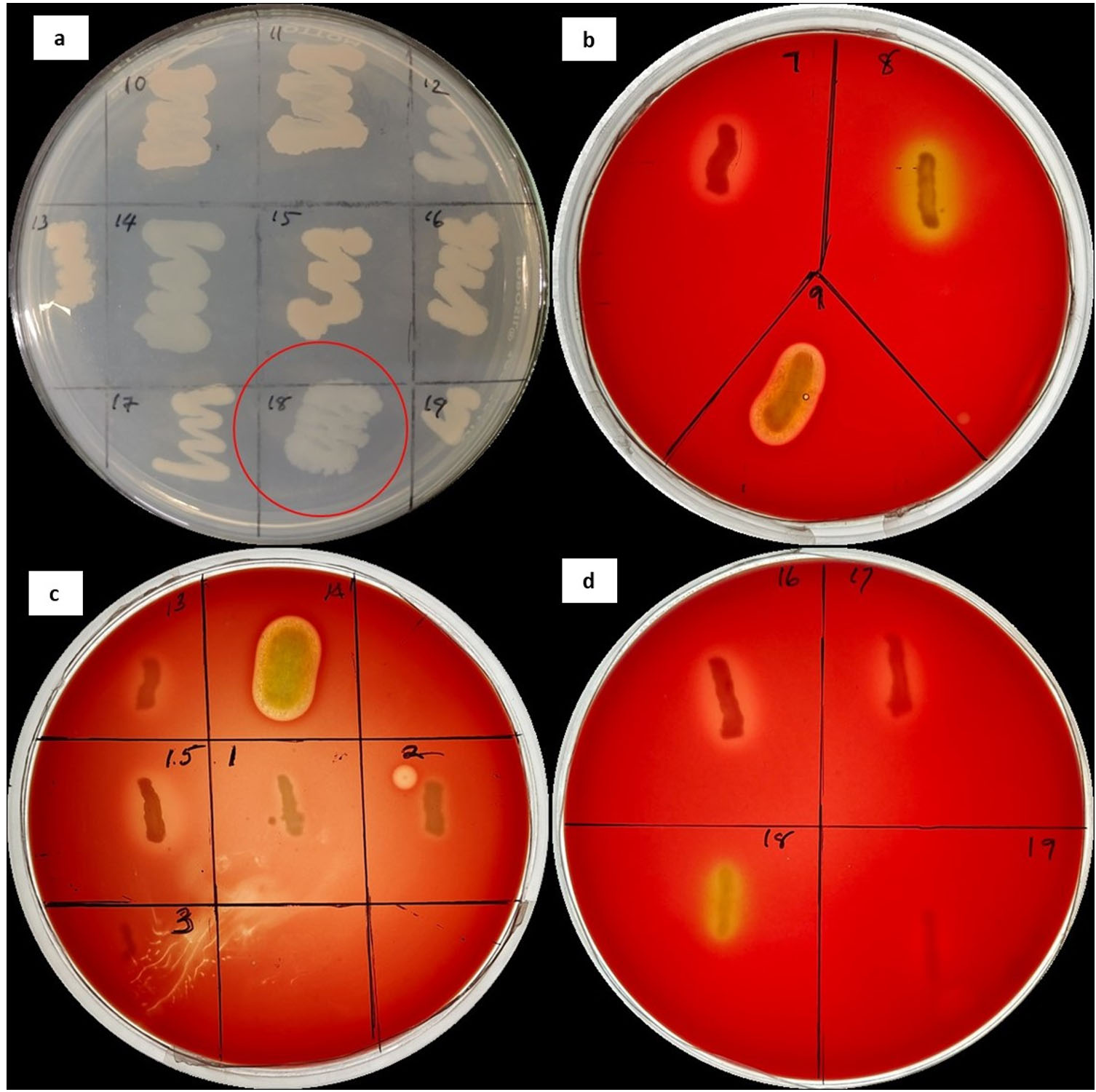
Figure 1. (a) Showing L. adecarboxylata (marked) along with bacterial colonies isolated from the gut of C. megacephala. (b), (c) and (d) Showing hemolytic activity of strains
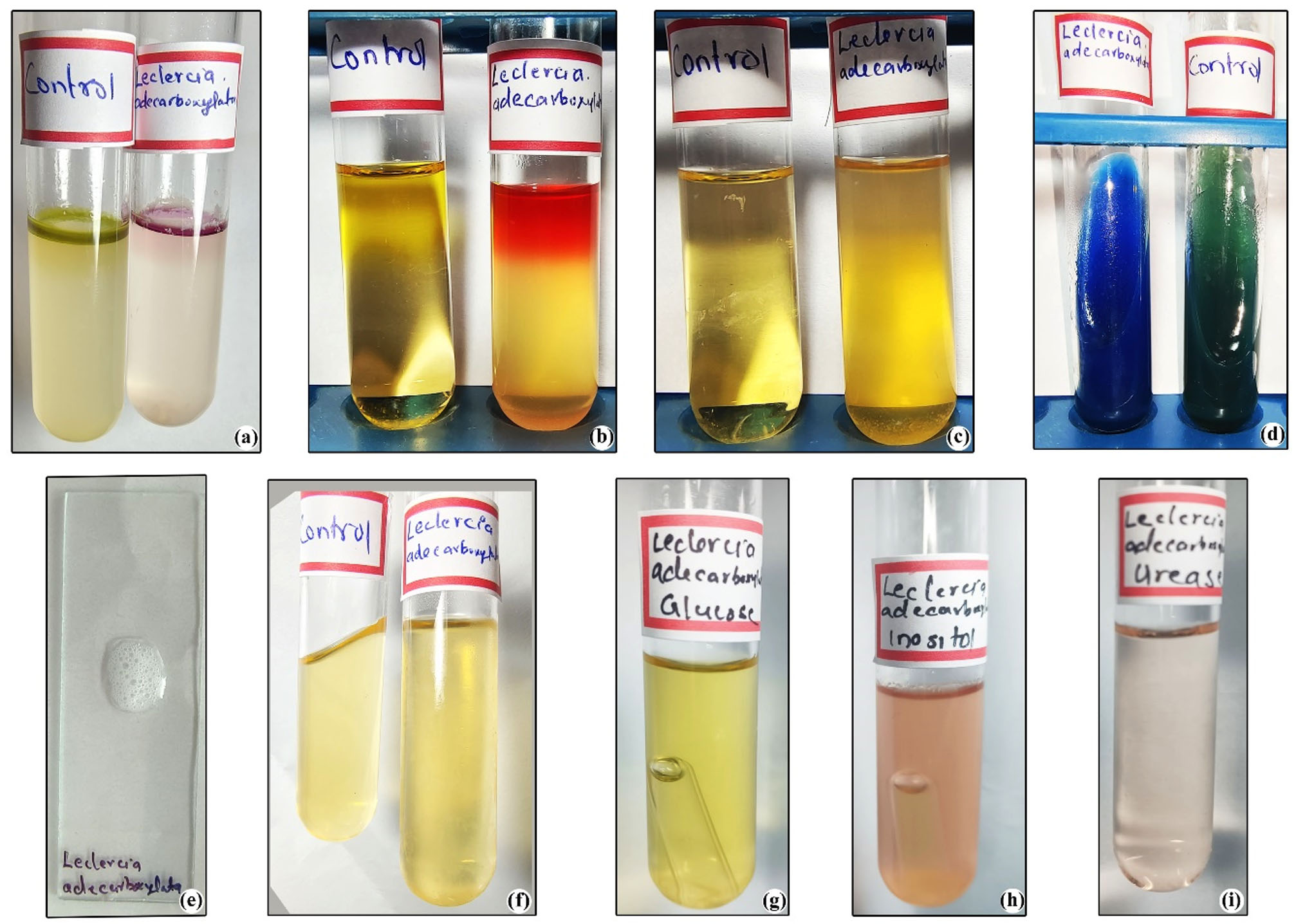
Figure 2. Biochemical Tests; (a) Indole Test, (b) MR Test, (c) Voges-Proskauer Test, (d) Citrate Test, (e) Catalase Test, (f) Gelatinase Test, (g) Glucose Acid Fermentation Test, (h) Inositol Test, (i) Urease Test
Molecular sequencing
Based on the 16S rRNA sequencing, the bacterial samples were identified as Pseudomonas aeruginosa, Enterococcus faecalis and Leclercia adecarboxylata. Bacterial sequences generated from the molecular sequencing were analyzed to check the quality. Sequence editing and assembling were done using BioEdit sequence alignment tool version.44 Aligned samples were then used to check the species similarity with the help of BLAST function of NCBI. The generated sequences were submitted to NCBI and accession numbers were generated as follows: PP381874, PP396147, PP396897, PP397013. The percentage of query coverage values ranged from 80-99%. The evolutionary history was constructed using Neighbor Joining method (NJ).45 Along with that, the evolutionary distances were calculated using Maximum Composite Likelihood method.46 In addition to molecular sequencing, phylogenetic relationship between species based on 16S rRNA gene were carried out to identify the sample species and the phylogenetic tree is represented in Figure 3.
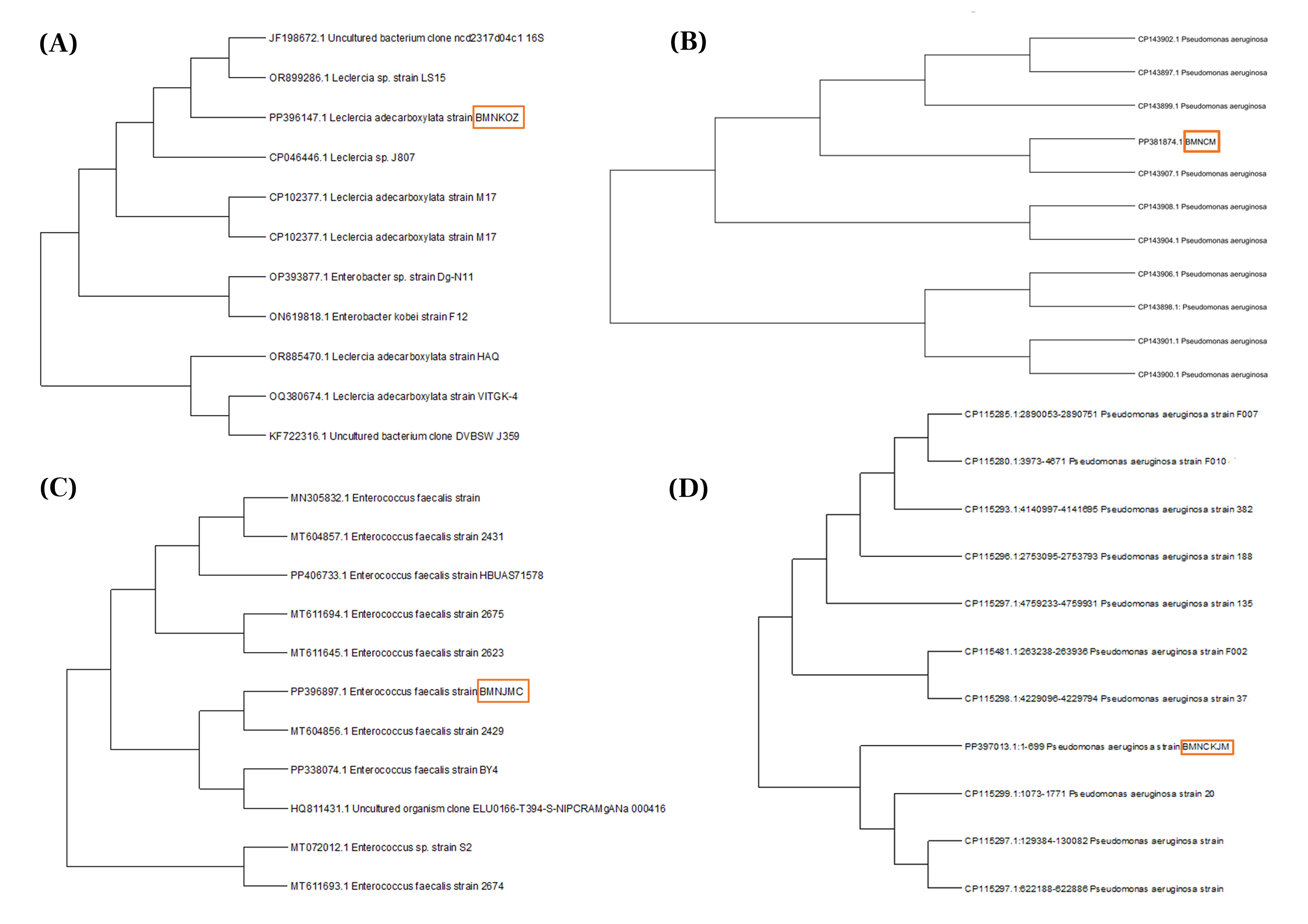
Figure 3. Phylogentic tree (a) L. adecarboxylata BMNKOZ (b) P. aeruginosa BMNCM, (c) E. faecalis BMNJMC (d) P. aeruginosa BMNCKJM
Antibacterial susceptibility
The zone of inhibition values is presented in Table 2. Ciprofloxacin was found to be the most effective antibiotics against all four strains of bacteria, followed by Streptomycin (Figure 4). However, Tetracycline showed the least effect against all four bacterial strains. While Amoxicillin and ampicillin had no effect on P. aeruginosa, Neomycin also had no effect on E. faecalis and also the strain showed some resistance to Streptomycin. In the case of L. adecarboxylata, all antibiotics showed considerable sensitivity making the strain not resistant towards the common antibiotics in use.
Table (2):
Zone of inhibition values (in mm) shown by antibiotics against bacterial strains
Pseudomonas aeruginosa (9)(mm) |
Pseudomonas aeruginosa (14)(mm) |
Enterococcus faecalis (8)(mm) |
Leclercia adecarboxylata (18)(mm) |
Sa |
MSb |
Rc |
|
|---|---|---|---|---|---|---|---|
Antibiotics |
|||||||
Amoxicillin (30) |
NE |
NE |
27 |
22 |
≥18 |
14-17 |
≤13 |
Ampicillin (10) |
NE |
NE |
25 |
21 |
≥17 |
14-16 |
≤13 |
Ciprofloxacin (1) |
35 |
32 |
20 |
30 |
≥21 |
16-20 |
≤15 |
Neomycin (10) |
14 |
17 |
NE |
16 |
≥20 |
15-20 |
≤14 |
Streptomycin (300) |
26 |
25 |
14 |
18 |
≥21 |
15-20 |
≤14 |
Tetracycline (10) |
8 |
8 |
2 |
19 |
≥19 |
15-18 |
≤14 |
Sa – Sensitive, MSb – Moderate sensitive, Rc– Resistant98 , NE- No Effect
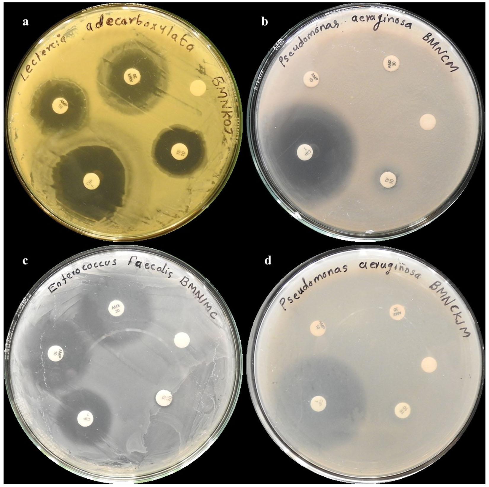
Figure 4. Antibiotic assay showing zone of inhibition by (a) L. adecarboxylata BMNKOZ, (b) P. aeruginosa BMNCM (c) E. faecalis BMNJMC (d) P. aeruginosa BMNCKJM
Flies are considered to be an important vector of pathogens due to their abundance and close association with people, animals and their waste.47 Poor hygiene in animal rearing areas attracts flies to breed and feed on waste, acting as vectors and increase pathogen transmission near human settlements.48 Calliphoridae are abundant in and around the farms and can travel up to 2-3 km making it to transfer the pathogens from one area to another.49 A study by Urban and Broce, suggested that flies can disseminate loads of bacteria directly to the raw meat which are given as food for dogs that results in intestinal infections and mortality.49 Basson et al. showed that species like C. albiceps and Chrysomya marginalis can disseminate Bacillus anthracis and the high abundance of blow flies feeding on disease affected carcass can lead to the continuation of anthrax infection to other animals due to frequent visit of blow flies with animals. Due to its capability of transfer, it can also take up antibiotic strains of bacteria from one region to other.50 To support this, studies have shown that flies like Lucilia sericata can harbor both antibiotic-resistant and sensitive strains of bacteria, such as Proteus mirabilis, within their gut for extended periods, potentially serving as reservoirs and vectors for transmission.51 Similarly, isolation of antibiotic resistant strains from Calliphoridae flies were done by several researchers which is represented in Table 3. Research by Deel suggests that new bacteria introduced into the environment could potentially integrate into the microbiome of flies and also there is a chance that while these flies colonize the human cadaver having pathogenic bacteria, it will get incorporated into fly microbiome and disseminate them thus acting as a carrier.52 The early and prominent colonization of carcasses by C. megacephala,6 necessitates studies on their vector potential due to inevitable contact with pathogenic environments. With respect to these findings, from our study we were able to isolate and identify P. aeruginosa, E. faecalis and L. adecarboxylata from the gut of C. megacephala. L. adecarboxylata and is reported for the first time from C. megacepahala.
Table (3):
Antibiotic resistant strains isolated from various Calliphoridae flies from different studies
| Calliphoridae sps | Strains isolated | Antibiotic Resistances | References |
|---|---|---|---|
| Green bottle flies | Pseudomonas putida | AMP, CEF, NAL | 99 |
| Pseudomonas spp. | CEF, NAL, AMPP | ||
| Enterococcus faecalis | KAN, CIP, NAL | ||
| Blow flies | E. coli | Amp, AML, CIP, IMI, E, P-G, TE | 100 |
| Klebsiella spp. | P-G, CHL | ||
| Salmonella spp. | AMP, IMI, P-G, TE | ||
| Shigella spp. | AMP, AML, IMI, TE, | ||
| Enterobacter spp. | P-G, TE | ||
| Staphylococcus spp. | AMP, AML, C, E, P-G, TE | ||
| Bacillus spp. | P-G, | ||
| Lucilia sericata | E. coli | AMP, STR, TET, CHL | 101 |
| Staphylococcus spp. | AMP, TET, | ||
| Lucilia sericata | S. aureus | TET, AMP, | 102 |
| E. coli | TET, AMP, CEP, COT | ||
| Chrysomya megacephala | Lemef17 K. ascorbate | AMP | 103 |
| Lucilia cuprina | Lemef105 E. coli (MDR) | AMP, ASB, CPM, CAZ, CRO, CIP, ETP, PPT |
Ampicillin (AMP), Amoxicillin (AML), Ampicillin-Sulbactam (ASB), Cefepime (CPM), Ceftazidime (CAZ), Cephalexin (CEP), Chloramphenicol (CHL), Ciprofloxacin (CIP), Cotrimoxazole (COT), Ceftriaxone (CRO), Ertapenem (ETP), Erythromycin (E), Imipenem (IMI), Penicillin-G (P), Piperacicllin-Tazobactam (PPT), Streptomycin (STR), Tetracycline (TE).
The first report of Vibrio parahaemolyticus in C. megacephala from Thailand has been reported highlighting the potential of C. megacephala as a significant mechanical vector for V. parahaemolyticus, a bacterium known for causing gastroenteritis from Calliphoridae family.53 The first report of L. adecarboxylata was found from Calliphora vicina collected from hospital premises along with strains of E. coli serotype E1525. The authors concluded that, bacterial strain E. coli serotype E1525 obtained from C. vicina pointed that these flies should be carefully monitored considering it as a mechanism of dispersing bacteria strains. The recommendation is that fly-proof measure should be employed in order to prevent the ingress of fly, thereby limit the introduction of bacteria from a pathogenic environment to non-pathogenic environment.23 However, till present no literature survey showed the presence of L. adecarboxylata from C. megacephala. They mostly cause human infections that are polymicrobial and usually occur in immunocompromised humans.20 The data and studies focusing on L. adecarboxylata were scanty which often results in the misdiagnosis of the pathogen as they share similar biochemical features with E. coli and causes diseases like sepsis, septic arthritis, diarrhoea, peritonitis,
gallbladder infections which are common diseases of Enterobacteriaceae.19,54 However to study the phenotypic characters, biochemical aspects of the bacterial strains were studied and considerable results were obtained. The L. adecarboxylata strain was found to be positive for tests like catalase, indole, MR, citrate, urease, and acid fermentation tests which come in concordance with the previous study. By considering these difficulties in identification we considered the molecular sequencing of L. adecarboxylata to confirm the species. Owing to the importance, recently several case reports have confirmed the problems associated with L. adecarboxylata. A medical study report showed the death of a 24-week-old premature neonate due to the nosocomial sepsis infection from L. adecarboxylata 55 The researcher further suggested that a thorough knowledge of L. adecarboxylata is necessary for the neonatologist to understand the nature of neonatal sepsis. In addition to this, a case report showed that a 48-year-old female was diagnosed with Peritonitis from L. adecarboxylata and the possible route of Leclercia infection was through water source or from farm animals.56 L. adecarboxylata also found to cause Necrotizing Soft Tissue Infection (NSTI) which can lead to serious health problems like damage to skin, muscle, soft tissues and even septic shock and consequent multiorgan failures.57,58 Also, a multidrug resistant L. adecarboxylata which causes respiratory distress in cow was reported from India. The authors suggested that the epidemiology and antimicrobial resistance of L. adecarboxylata should be studied with respect to different geographic locations around the world and more studies will bring the zoonotic significance of the species.59 While keeping the suggestion of Choudhary to conduct studies in different geographical locations, we selected the district of Kozhikode, Kerala, India. The district of Kozhikode in currently a hotspot of disease outbreaks like NIPAH 2018 and Shigella.60,61 Not only this particular district, every place should by carefully monitored to mitigate the chance of a new disease outbreak. To mitigate the disease, outbreak a better understanding of new strains along with their transmission pattern through vectors should be monitored. Since Calliphoridae flies are commonly associated with farm animals, the chance of Leclercia spp. to reach these animals and to humans cannot be neglected.55
Several bacteria secrete virulence factors (toxins), that allow them to disrupt the host defensive mechanisms and impair host functions. Bacterial hemolysins are exotoxins which attack blood cell membrane thereby disrupting its cell wall. This action could increase the severity of infections.62 Therefore, the hemolysis is one virulence factor that can determine the pathogenicity of bacteria strain. In our study L. adecarboxylata showed hemolysis which contradicts the studies done by Muratoglu and Anuradha where the specimen showed negative blood hemolysis.25,37 A possible explanation for this contrasting result was suggested by Snak, where the author pointed that this change might be due to the presence of hemolysin gene in the strain.63 Our strain showed alpha hemolysis which can interpreted in such a manner that they can act as pathogens and cause infection in humans and animals. Also, in the era of emerging antimicrobial resistance, a worldwide focus was given in terms to reduce the susceptibility of public to various resistant pathogens. Leclercia is beginning to exhibit antibiotic resistance, much like the majority of antibiotic-resistant bacteria. This indicates that they are a potentially harmful organism and should be treated with caution.64 Even though L. adecarboxylata is susceptible to many antibiotics, resistant strains been reported recently. The resistance of L. adecarboxylata isolated from pig farms to aminoglycosides, quinolones and trimethoprim-sulfamethoxazole was been reported by Yao et al.,65 The transferable drug resistance observed in L. adecarboxylata via R plasmids raises concerns, as these plasmids have the potential to be transferred to human pathogenic bacteria, thereby heightening the risk of drug-resistant bacterial development. This underscores the emergence of antibiotic resistance in L. adecarboxylata as an issue linked to environmental antibiotic exposure in food production.66 From our study L. adecarboxylata strain showed susceptibility to all antibiotics namely Amoxicillin, Ampicillin, Ciprofloxacin, Neomycon and Streptomycin. In our study, Ciprofloxacin showed the best result whereas neomycin showed the least effect. Regular monitoring of L. adecarboxylata for antibiotic resistance changes is crucial, warranting attention in future research endeavors. Furthermore, educating individuals involved in farming is imperative. Promoting educational initiatives that underscore the prevention of antibiotic misuse and the avoidance of multi-drug resistant bacteria emergence should also be prioritized.
Investigating the vector harboring capacities of flies is crucial for various biotechnological applications including biomedicine (treatment of diseases), agrobiotechnology (consume organic waste rapidly thereby help in disposal of meat and fish as well as manure) and insect-borne disease prevention.67-70 Moreover, Zaher et al., isolated bacteria species like Escherichia coli, Bacillus subtilis, Staphylococcus epidermidis, Pseudomonas aeruginosa, Staphylococcus aureus, Proteus mirabilis, and Staphylococcus hominis from Calliphoridae and other forensically important necrophagous insects.71 Our research isolated two strains of P. aeruginosa from the gut of C. megacephala, consistent with prior findings of Gaszv et al., where the author suggested P. aeruginosa prevalence in Lucilia spp., where both are important carrion feeders under Calliphoridae.4 P. aeruginosa is found to be a pathogen for causing infections such as nosocomial pneumonia, urinary tract infections, malignant external otitis, endophthalmitis, endocarditis, meningitis and septicemia.72 In our study, P. aeruginosa strains exhibited hemolytic activity, similar to findings from (Al-Saffar and Jarallah) and Macin et al., where the isolated P. aeruginosa from hospital environments showed hemolysis.73,74 Furthermore, our hemolytic strain showed antibiotic resistance to few antibiotics which was similar to the antibiotic resistant hemolytic strain isolated by (Macin et al.,74 Hemolysis is a crucial pathogenicity deciding factor in P. aeruginosa, observed consistently across different hosts and environments, as demonstrated by Hossain et al., in cattle samples.75 While taking the case of antibiotic resistance in P. aeruginosa, Sh AL-Salihi and Hameed, show resistance of P. aeruginosa to the antibiotic in the following order as ampicillin (100%), amoxicillin (97.3), neomycin (91.4%), ciprofloxacin (84%).76 In our study, P. aeruginosa strains show high resistant to amoxicillin, ampicillin and moderate resistant to neomycin. However, it shows contradicting result in the case of ciprofloxacin where we got high zone of inhibition values. The authors suggested that the reason for the development of resistance is due to the beta-lactamase production, the presence of strong barrier to diffusion at the outer membrane of bacteria and bacterial efflux of P. aeruginosa. Furthermore, our P. aeruginosa strain has no effect from amoxicillin and ampicillin which come in accordance with the study by Ahmed et al., where the researcher pointed that amoxicillin and ampicillin were ineffective against P. aeruginosa. These insights will help while administrating antibiotics against P. aeruginosa infection in humans and animals.77
In our current study, we also isolated E. faecalis from the gut of C. megacephala. Enterococcus species are gram-positive cocci which causes enterococcal bacteremia, urinary tract infections, meningitis, endocarditis which subsequently results in hospitalization and mortality. Out of this, E. faecalis is one common species that causes bacteremia.78-80 Our E. faecalis strain showed hemolytic activity which is consistent with the previous findings of Izumi et al.,81 Wang et al., detected the cylA gene in majority of E. faecalis strain (71%) potentially contributing to g-hemolytic activity. These studies aimed to evaluate the probiotic and safety aspects of E. faecalis, suggesting that strains lacking hemolytic activity and other virulence genes could be potential probiotic candidates.82 This was supported by Hashem et al., who investigated the virulence factors contributing to E. faecalis pathogenicity including hemolytic assay and found a significant correlation between hemolysis and the presence of Cyl gene which produce the Cytolysin toxin that increases the severity of infection.83 Although our study did not investigate gene-level correlations, the observed hemolysis activity in our samples may contribute to increased infection levels of E. faecalis, warranting further investigation. In our study, E. faecalis showed no resistance towards Ciprofloxacin and rather show sensitivity towards amoxicillin, ampicillin and streptomycin. However, bacteria strain has no effect by neomycin and tetracycline. This resistance towards tetracycline result was come in accordance with the study by Kim et al., where the isolated E. faecalis strain showed resistance towards tetracycline. The authors further supported the resistant phenomenon with gene level studies and showed that common genes responsible for the resistance were tet (M), tet (L).84 In addition to this, research by Xuan et al. showed that E. faecalis is resistant towards erythromycin (91.1%), tetracycline (100%), ciprofloxacin (66.1%), bacitracin (87.8%) and chloromycetin (41.1%). The resistance to ciprofloxacin contradicts with our strains result which showed moderate sensitivity. Xuan et al., pointed out that resistance of bacteria towards few antibiotics and absence of resistance to few antibiotics like nosipheptide and enramycin results can be used in policy making while suggesting the use of antibiotics in pig farms.85
Another important aspect to look into the vector potential of fly is the phenomenon of horizontal gene transfer among bacteria. Horizontal transmission of antibiotic resistance was found to occur in the crop of insects.86 Petridis et al. reported that the gastrointestinal tract of houseflies provides a suitable environment for the horizontal transfer of resistant genes and virulence genes.87 Additionally, the plasmid mediated horizontal resistant gene transfer were illustrated by Akhtar et al. in houseflies.88 Zurek and Ghosh reported that in the gut of housefly there is a frequent transfer of tetracycline resistance gene tet (M) between E. faecalis strains. So, inside the gut of insects, bacteria can actively share toxins and antibiotic genes along with proliferation of bacterial species.89 With respects to these studies, the same phenomenon in blow flies cannot be neglected as the feed and breed in pathogenic environments. Also, according to our knowledge, no studies has been reported to find out the horizontal gene transfer in C. megacephala and limited studies being reported about the pathogens associated with this fly. Additional importance of understanding the blow fly microbiome is that the information regarding blowfly associated microorganisms is found to be crucial in the development of PMI prediction models. Previous researches showed that soil and insects are two main factors associated with carrions. Hence, microbial communities associated with soil and insects also have significant role in the development of PMI models.90,91 Therefore, the current study of microbiome association with blowflies along with future metagenomic studies will help in increasing the accuracy of PMI by forensic scientists.92,93
Furthermore, while looking into the relevance, our current study also plays an influence in economic aspects. Due to the infestation of blow flies, into the sea food industry a large economic loss was found and it was shown by several research studies. Fish samples were taken from the Puthiyappa fishing harbor along the Kozhikode district’s coastline to check for the presence of Campylobacter spp. The results showed the presence of Campylobacter coli and Campylobacter jejuni, which can be a source of food-borne campylobacteriosis transmission.94 Similar to this, it was confirmed that there is antibiotic-resistant Listeria spp. present in fishes in Kerala’s fish harbors. This is concerning because Kerala is a major seafood exporter and the presence of these bacteria can cause regulatory alerts from importing nations thus causing huge economic loss.95 C. megacephala is found to be abundant in and around harbors where sea food processing takes place and they can spread the pathogens to nearby areas.96 Aak et al. reported an estimated loss of half a million euros from stock fish industry in Norway due to the infestation by Calliphoridae.97
In our current study, L. adecarboxylata is the bacteria was reported for the first time from C. megacephala. The strain showed hemolysis which contribute to their pathogenicity whereas the strain is susceptible to antibiotics. Since there is a potential that it will eventually develop antibiotic resistance, a thorough investigation into antibiotic pathogens pertaining to blow flies have to be conducted which will help in policy making and use of antibiotics in various animal and human related fields. Also, the results from the current study indicates that C. megacephalas has a vector potential in carrying pathogenic bacteria. To get a better understanding future studies focusing on in vivo experimental studies to determine vectoral capacity of flies, epidemiological aspects, and transmission high resolution sequence-based studies are to be carried out.
ACKNOWLEDGMENTS
The authors acknowledge CHRIST (Deemed to be University) for the facilities they provided for this work. Authors also acknowledge RGCB (Rajiv Gandhi Centre for Biotechnology), Thiruvananthapuram, for the sequencing support. Authors are also grateful to John Merston (Research scholar, CHRIST) for the support.
CONFLICT OF INTEREST
The authors declare that there is no conflict of interest.
AUTHORS’ CONTRIBUTION
BMN and MT conceptualized the study. BMN performed investigation. MT supervised the study. BMN wrote the manuscript. MT reviewed and edited the manuscript. Both authors read and approved the final manuscript for publication.
FUNDING
None.
DATA AVAILABILITY
All datasets generated or analyzed during this study are included in the manuscript.
ETHICS STATEMENT
Not applicable.
- Tomberlin JK, Crippen TL, Tarone AM, et al. A review of bacterial interactions with blow flies (Diptera: Calliphoridae) of medical, veterinary, and forensic importance. Ann Entomol Soc Am. 2017;110(1):19-36.
Crossref - Barreiro C, Albano H, Silva J, Teixeira P. Role of flies as vectors of foodborne pathogens in rural areas. ISRN Microbiol. 2013;2013:718780.
Crossref - Chaiwong T, Srivoramas T, Sueabsamran P, Sukontason K, Sanford MR, Sukontason KL. The blow fly, Chrysomya megacephala, and the house fly, Musca domestica, as mechanical vectors of pathogenic bacteria in Northeast Thailand. Trop Biomed. 2014;31(2):336-346.
- Gasz NE, Geary MJ, Doggett SL, Harvey ML. Bacterial association observations in Lucilia sericata and Lucilia cuprina organs through 16S rRNA gene sequencing. Appl Microbiol Biotechnol. 2021;105(3):1091-1106.
Crossref - Fouda A, Fouda MA, Aldally AG, Ismael SB, Awad MA. Isolation and identification of bacterial species associated with non-biting flies in Egypt. Egypt. Acad. J. Biolog. Sci. 2016;9(4):37-45.
Crossref - Badenhorst R, Villet MH. The uses of Chrysomya megacephala (Fabricius, 1794) (Diptera: Calliphoridae) in forensic entomology. Forensic Sci Res. 2018;3(1):2-15.
Crossref - Sukontason KL, Bunchoo M, Khantawa B, Piangjai S, Rongsriyam Y, Sukontason K. Comparison between Musca domestica and Chrysomya megacephala as carriers of bacteria in northern Thailand. Southeast Asian J Trop Med Public Health. 2007;38(1):38.
- Ferraz ACP, Proenca B, Gadelha BQ, et al. First record of human myiasis caused by association of the species Chrysomya megacephala (Diptera: Calliphoridae), Sarcophaga (Liopygia) Ruficornis (Diptera: Sarcophagidae), and Musca domestica (Diptera: Muscidae). J Med Entomol. 2010;47(3):487-490.
Crossref - Yang S, Liu Z. Pilot-scale biodegradation of swine manure via Chrysomya megacephala (Fabricius) for biodiesel production. Appl Energy. 2014;113:385-391.
Crossref - Inouye DW, Larson BMH, Ssymank A, Kevan PG. Flies and flowers III: Ecology of foraging and pollination. J Poll Ecol. 2015;16:115-133.
Crossref - Ramirez F, Davenport TL. Mango (Mangifera indica L.) pollination: A review Report on the occurrence of synanthropic derived. Scientia Horticulturae. 2016;203:158-168.
Crossref - Lancu L, Lulia RA, Victoria LP, et al. Microbiome pattern of Lucilia sericata (Meigen) (Diptera: Calliphoridae) and feeding substrate in the presence of the foodborne pathogen. Sci Rep. 2021;11(1):15296.
Crossref - Jiranantasak T, Benn JS, Metrailer MC, et al. Characterization of Bacillus anthracis replication and persistence on environmental substrates associated with wildlife anthrax outbreaks. Plos one. 2022;17(9):e0274645.
Crossref - Batz MB, Hoffmann S, Morris Jr JG. Ranking the disease burden of 14 pathogens in food sources in the United States using attribution data from outbreak investigations and expert elicitation. J Food Prot. 2012;75(7):1278-1291.
Crossref - European Centre for Disease Prevention and Control. Salmonellosis. ECDC. Annual Epidemiological Report for 2017. https://www.ecdc.europa.eu/. Accessed 10 April 2020.
- Junqueira ACM, Ratan A, Acerbi E, et al. The microbiomes of blowflies and houseflies as bacterial transmission reservoirs. Sci Rep. 2017;7(1):16324.
Crossref - Sawabe K, Hoshino K, Isawa H, et al. Detection and isolation of highly pathogenic H5N1 avian influenza A viruses from blow flies collected in the vicinity of an infected poultry farm in Kyoto, Japan, 2004. Am J Trop Med Hyg. 2006;75(2):327-332.
Crossref - Tamura K, Sakazaki R, Kosako Y, Yoshizaki E. Leclercia adecarboxylata gen. nov., comb. nov., formerly known as Escherichia adecarboxylata. Curr Microbiol. 1986;13:179-184.
Crossref - Hess B, Burchett A, Huntington MK. Leclercia adecarboxylata in an immunocompetent patient. J Med Microbiol. 2008;57(7):896-898.
Crossref - Zayet S, Lang S, Garnier P, et al. Leclercia adecarboxylata as emerging pathogen in human infections:Clinical features and antimicrobial susceptibility testing. Pathogens. 2021;10(11):1399.
Crossref - Bouamamaa L, Sorlozano A, Laglaoui A, Lebbadi M, Aarab A, Gutierrez J. Antibiotic resistance patterns of bacterial strains isolated from Periplaneta americana and Musca domestica in Tangier, Morocco. J Infect Dev Ctries. 2010;4(04):194-201.
Crossref - Muratoglu H, Demirbag Z, Sezen K. The first investigation of the diversity of bacteria associated with Leptinotarsa decemlineata (Coleoptera: Chrysomelidae). Biologia. 2011;66:288-293.
Crossref - Davies MP, Anderson M, Hilton AC. Microbiological analysis of non-biting flies collected from hospitals:novel pathogens and new vectors in the clinical environment. 2017;193-198.
- Rau J, Werner D, Beer M, Hoper D, Kampen H. The microbial RNA metagenome of Aedes albopictus (Diptera: Culicidae) from Germany. Parasitol Res. 2022;121(9):2587-2599.
Crossref - Anuradha M. Leclercia adecarboxylata isolation: Case reports and review. JCDR. 2014;8(12):DD03.
Crossref - Matsuura H, Sugiyama S. Sepsis and Leclercia adecarboxylata. QJM-Int J Med. 2018;111(10):733-734.
Crossref - Bali R, Sharma P, Gupta K, Nagrath S. Pharyngeal and peritonsillar abscess due to Leclercia adecarboxylata in an immunocompetant patient. J Infect Dev Ctries. 2013;7(1):46-50.
Crossref - Stone JP, Denis-Katz HS, Temple-Oberle C, Mercier P, Mizzau JB, Mitha AP. Leclercia adecarboxylata: the first reported infection of cerebrospinal fluid and a systematic review of the literature. J Neuroinfect Dis. 2015;6(3):21385207.
- Sirichoat A, Florez AB, Vazquez L, et al. Antibiotic susceptibility profiles of lactic acid bacteria from the human vagina and genetic basis of acquired resistances. Int J Mol Sci. 2020;21(7):2594.
Crossref - Dixit A, Kumar N, Kumar S, Trigun V. Antimicrobial resistance: Progress in the decade since emergence of New Delhi metallo-b-lactamase in India. IJCM. 2019;44(1):4-8.
Crossref - Marshall BM, Levy SB. Food animals and antimicrobials: impacts on human health. CMR. 2011;24(4):718-733.
Crossref - Gwenzi W, Chaukura N, Muisa-Zikali N, et al. Insects, rodents, and pets as reservoirs, vectors, and sentinels of antimicrobial resistance. Antibiotics. 2021;10(1):68.
Crossref - Senior WR. Diptera A, John S. Family Calliphoridae. The Fauna of British India, Including the Remainder of the Oriental Region. 1940;6:288.
- Zurek L, Schal C, Watson DW. Diversity and contribution of the intestinal bacterial community to the development of Musca domestica (Diptera: Muscidae) larvae. J Med Entomol. 2000;37(6):924-928.
Crossref - Gupta AK, Rastogi G, Nayduch D, Sawant SS, Bhonde RR, Shouche YS. Molecular phylogenetic profiling of gut associated bacteria in larvae and adults of flesh flies. Med Vet Entomol. 2014;28(4):345-354.
Crossref - Turista DDR, Puspitasari E. The growth of Staphylococcus aureus in the blood agar plate media of sheep blood and human blood groups A, B, AB, and O. J Teknol. 2019;8(1):1-7.
Crossref - Muratoglu H, Kati H, Demirbag Z, Sezen K. High insecticidal activity of Leclercia adecarboxylata isolated from Leptinotarsa decemlineata (Col.:Chrysomelidae). Afr J Biotechnol. 2009;8(24).
- Manero A, Blanch AR. Identification of Enterococcus spp. with a biochemical key. Appl Environ Microbiol. 1999;65(10):4425-4430.
Crossref - Su S, Lae K, Ngwe H. Isolation and Identification of Pseudomonas aeruginosa from the Clinical Soil. University of Yangon Res J. 2018;8:271-275
- Folmer O, Black M, Hoeh W, Lutz R, Vrijenhoek, R. DNA primers for amplification of mitochondrial cytochrome c oxidase subunit I from diverse metazoan invertebrates. Mol Mar Biol Biotechnol. 1994;3(5):294-299.
- Drummond AJ, Ashton B, Buxton S, et al. Geneious v5.1. 2010. http://www.geneious.com Acceseed date October 18, 2023
- Yao H, Liu J, Jiang X, Chen F, Lu X, Zhang J. Analysis of the clinical effect of combined drug susceptibility to guide medication for carbapenem-resistant Klebsiella pneumoniae patients based on the kirby-bauer disk diffusion method. Infect Drug Resist. 2021;14:79-87.
Crossref - Lopez-Siles M, Khan TM, Duncan SH, Harmsen HJM, Garcia-Gil LJ, Flint HJ. Cultured representatives of two major phylogroups of human colonic Faecalibacterium prausnitzii can utilize pectin, uronic acids, and host-derived substrates for growth. Appl Environ Microbiol. 2012;78(2):420-428.
Crossref - Saitou N, Nei M. The neighbor-joining method: A new method for reconstructing phylogenetic trees. Mol. Biol. Evol. 1987;4(4):406-425.
- Tamura K, Nei M, Kumar S. Prospects for inferring very large phylogenies by using the neighbor-joining method. Proc Natl Acad Sci U S A. 2004;101(30):11030-11035.
Crossref - Zhang J, Wang J, Chen L, et al. Housefly (Musca domestica) and blow fly (Protophormia terraenovae) as vectors of bacteria carrying colistin resistance genes. Appl Environ Microbiol. 2018;84(1):e01736-17.
Crossref - Anderson GS, Huitson NR. Myiasis in pet animals in British Columbia: The potential of forensic entomology for determining duration of possible neglect. Can Vet J. 2004;45(12):993-998.
- Fila M, Wozniakowski G. African swine fever virus-the possible role of flies and other insects in virus transmission. J Vet Res. 2020;64(1):1-7.
Crossref - Urban JE, Broce A. Flies and their bacterial loads in greyhound dog kennels in Kansas. Curr Microbiol. 1998;36:164-170.
Crossref - Basson L, Hassim A, Dekker A, et al. Blowflies as vectors of Bacillus anthracis in the Kruger National Park. Koedoe. 2018;60(1):1-6.
Crossref - Wei T, Miyanaga K, Tanji Y. Persistence of antibiotic-resistant and-sensitive Proteus mirabilis strains in the digestive tract of the housefly (Musca domestica) and green bottle flies (Calliphoridae). Appl Microbiol Biotechnol. 2014b;98(19):8357-8366.
Crossref - Deel HL, Montoya S, King K, et al. The microbiome of fly organs and fly-human microbial transfer during decomposition. Forensic Sci Int. 2022;340:111425.
Crossref - Chomchat C, Wongwigkarn J, Thongwat D, Sanit S, Bunchu N, Lamlertthon S. First report of isolation of Vibrio parahaemolyticus from Chrysomya megacephala (Diptera: Calliphoridae) in tak province, thailand. Southeast Asian J Trop Med Public Health. 2020;51(6):815-823.
- Stock I, Burak S, Wiedemann B. Natural antimicrobial susceptibility patterns and biochemical profiles of Leclercia adecarboxylata strains. Clin Microbiol Infect. 2004;10(8):724-733.
Crossref - Anaut MB, Montero JA, Abellas PG, de Uribe Viloria M, Zapata RMR. Fulminant Sepsis Caused by Leclercia adecarboxylata in a Premature Neonate. Pediatr Infect Dis J. 2022;41(5):e220-e222.
Crossref - Adapa, S, Konala VM, Nawaz F, et al. Peritonitis from Leclercia adecarboxylata: An emerging pathogen. Clin Case Rep. 2019;7(4):829-831.
Crossref - Lonneman MK, Devasahayam RJ, Phillips CJ. Leclercia adecarboxylata causing necrotising soft tissue infection in an immunocompetent adult. BMJ Case Rep. 2020;13(9):e235633.
Crossref - Suijker J, de Vries A, de Jong VM, Schepers T, Ponsen KJ, Halm JA. Health-related quality of life is decreased after necrotizing soft-tissue infections. J Surg Res. 2020;245:516-522.
Crossref - Choudhary M, Choudhary BK, Bhoyar S, et al. Isolation and characterization of multidrug-resistant Leclercia species from animal clinical case. Lett Appl Microbiol. 2018;66(1):44-48.
Crossref - Yadav PD, Sahay RR, Balakrishnan A, et al. Nipah virus outbreak in Kerala State, India amidst of COVID-19 pandemic. Front Public Health. 2022;10:818545.
Crossref - Loredana Popa G, Popa MI. Shigellosis outbreaks—an update. Rom Arch Microbiol Immunol. 2022;81(1):32.
Crossref - Vallet-Gely I, Novikov A, Augusto L, et al. Association of hemolytic activity of Pseudomonas entomophila, a versatile soil bacterium, with cyclic lipopeptide production. Appl Environ Microbiol. 2010;76(3):910-921.
Crossref - Snak A, Vendruscolo ECG, Santos MFD, Fiorini A, Mesa D. Genome sequencing and analysis of plant growth-promoting attributes from Leclercia adecarboxylata. Genet Mol Biol. 2021;44(1):e20200130.
Crossref - Keren Y, Keshet D, Eidelman M, Geffen Y, Raz-Pasteur A, Hussein K. Is Leclercia adecarboxylata a new and unfamiliar marine pathogen? J Clin Microbiol. 2014;52(5):1775-1776.
Crossref - Yao Q, Zeng Z, Hou J, et al. Dissemination of the rmtB gene carried on IncF and IncN plasmids among Enterobacteriaceae in a pig farm and its environment. J Antimicrob Chemother. 2011;66(11):2475-2479.
Crossref - Riazzo C, Lopez-Cerero L, Rojo-Martin MD, et al. First report of NDM-1-producing clinical isolate of Leclercia adecarboxylata in Spain. Diagn Microbiol Infect Dis. 2017;88(3):268-270.
Crossref - Choe S, Lee D, Park H, et al. Canine wound myiasis caused by Lucilia sericata (Diptera: Calliphoridae) in Korea. KJP. 2016;54(5):667-671.
Crossref - Xie S, Lan Y, Sun C, Shao Y. Insect microbial symbionts as a novel source for biotechnology. World J Microbiol Biotechnol. 2019;35(2):1-7.
Crossref - Yakovlev AY, Kruglikova AA, Chernysh SI. Calliphoridae flies in medical biotechnology. Entomol Rev. 2019;99(3):292-301.
Crossref - Ulanova RV, Tikhonova EN, Kravchenko IK. Bacteria associated with Lucilia sericata larvae reared on fish wastes. Entomologia Entomol Exp Appl. 2020;168(6-7):573-581.
Crossref - Zaher AH, Kabadaia MM, Hammad KM, Mekky AE, Salem SS. Forensic flies as carries of pathogenic bacteria associated with a pig carcass in Egypt. Al-Azhar Bulletin of Science. 2023;34(3):5.
Crossref - Reynolds D, Kollef M. The epidemiology and pathogenesis and treatment of Pseudomonas aeruginosa infections: an update. Drugs. 2021;81(18):2117-2131.
Crossref - Al-Saffar MF, Jarallah EM. Isolation and characterization of Pseudomonas aeruginosa from Babylon province. Biochem Cell Arch. 2019;19(1):20644.
- Macin S, Akarca M, Sener B, Akyon Y. Comparison of virulence factors and antibiotic resistance of Pseudomonas aeruginosa strains isolated from patients with and without cystic fibrosis. Rev Romana Med Lab. 2017;25(4):327-334.
Crossref - Hossain MG, Saha S, Rahman M, Singha JK, Mamun AA. Isolation, identification and antibiogram study of Pseudomonas aeruginosa from cattle in Bangladesh. J Vet Adv. 2013;3(7):180-185.
Crossref - Sh AL-Salihi S, H Hameed B. Antibiosis resistant of Pseudomonas aeruginosa isolated from different clinical specimens. Kirkuk J Sci. 2014;9(2):15-28.
Crossref - Ahmed Z, Khan SS, Khan M. In vitro trials of some antimicrobial combinations against Staphylococcus aureus and Pseudomonas aeruginosa. Saudi J Biol Sci. 2013;20(1):79-83.
Crossref - Chiang HY, Perencevich EN, Nair R, et al. Incidence and outcomes associated with infections caused by vancomycin-resistant enterococci in the United States: systematic literature review and meta-analysis. Infect Control Hosp Epidemiol. 2017;38(2):203-215.
Crossref - Uda A, Shigemura K, Kitagawa K, et al. Risk factors for the acquisition of Enterococcus faecium infection and mortality in patients with enterococcal bacteremia: A 5-year retrospective analysis in a tertiary care university hospital. Antibiotics. 2021;10(1):64.
Crossref - Seby R, Kim C, Khreis M, Khreis K. Enterococcus faecalis-induced infective endocarditis: An unusual source of infection and a rare clinical presentation. J Int Med Res. 2022;50(7):03000605221112019.
Crossref - Izumi E, Pires PD, De Marques EB, Suzart S. Hemagglutinating and hemolytic activities of Enterococcus faecalis strains isolated from different human clinical sources. Res Microbiol. 2005;156(4):583-587.
Crossref - Wang J, Da R, Tuo X, et al. Probiotic and safety properties screening of Enterococcus faecalis from healthy Chinese infants. Probiotics Antimicrob Proteins. 2020;12(3):1115-1125.
Crossref - Hashem YA, Abdelrahman KA, Aziz RK. Phenotype-genotype correlations and distribution of key virulence factors in Enterococcus faecalis isolated from patients with urinary tract infections. Infect Drug Resist. 2021;14:1713-1723.
Crossref - Kim YB, Seo KW, Jeon HY, Lim SK, Sung HW, Lee YJ. Molecular characterization of erythromycin and tetracycline-resistant Enterococcus faecalis isolated from retail chicken meats. Poult Sci. 2019;98(2):977-983.
Crossref - Xuan H, Yao X, Pan R, et al. Antimicrobial resistance in Enterococcus faecium and Enterococcus faecalis isolates of swine origin from eighteen provinces in China. J Vet Med Sci. 2021;83(12):1952-1958.
Crossref - Stoffolano JG. Fly foregut and transmission of microbes. Adv Insect Physiol. 2019;57:27-95.
Crossref - Petridis M, Bagdasarian M, Waldor MK, Walker E. Horizontal transfer of Shiga toxin and antibiotic resistance genes among Escherichia coli strains in house fly (Diptera: Muscidae) gut. J Med Entomol. 2014;43(2):288-295.
Crossref - Akhtar M, Hirt H, Zurek L. Horizontal transfer of the tetracycline resistance gene tetM mediated by pCF10 among Enterococcus faecalis in the house fly (Musca domestica L.) alimentary canal. Microb Ecol. 2009;58(3):509-518.
Crossref - Zurek L, Ghosh A. Insects represent a link between food animal farms and the urban environment for antibiotic resistance traits. Appl Environ Microbiol. 2014;80(12):3562-3567.
Crossref - Singh B, Crippen TL, Zheng L, et al. A metagenomic assessment of the bacteria associated with Lucilia sericata and Lucilia cuprina (Diptera: Calliphoridae). Appl Microbiol Biotechnol. 2015;99(2):869-883.
Crossref - Metcalf JL, Xu ZZ, Weiss S, et al. Microbial community assembly and metabolic function during mammalian corpse decomposition. Science. 2016;351(6269):158-162.
Crossref - Wohlfahrt D, Woolf MS, Singh B. A survey of bacteria associated with various life stages of primary colonizers: Lucilia sericata and Phormia regina. Sci Justice. 2020;60(2):173-179.
Crossref - Metcalf JL. Estimating the postmortem interval using microbes:Knowledge gaps and a path to technology adoption. Forensic Sci Int Genet. 2019;38:211-218.
Crossref - Deepa J, Sunil B, Latha C, Vrinda KM, Mini M, Aravindakshan TV. Prevalence of Campylobacter spp. in marine fishes, crustaceans and molluscs in Kozhikode district, Kerala. J Vet Med Anim Sci. 2022;53(1):32-38.
Crossref - Menon KV, Sunil B, Latha C. Prevalence and antibiotic resistance profile of Listeria spp. associated with seafoods from fish catchment areas in Kerala, India. Vet World. 2021;14(3):777-783.
Crossref - Ramaraj P, Selvakumar C, Ganesh A, Janarthanan S. Report on the occurrence of synanthropic derived form of Chrysomya megacephala (Diptera: Calliphoridae) from Royapuram fishing harbour, Chennai, Tamil Nadu, India. Biodivers Data J. 2014;(2):e111.
Crossref - Aak A, Birkemoe T, Mehl R. Blowfly (Diptera, Calliphoridae) damage on stockfish in northern Norway: pest species, damage assessment and the potential of mass trapping. J Pest Sci. 2010;83(3):329-337.
Crossref - CLSI. Performance Standards for Antimicrobial Susceptibility Testing. CLSI Supplement M100 27th edn, 1-15 (Clinical and Laboratory Standards Institute, 2017).
- Wei T, Ishida R, Miyanaga K, Tanji Y. Seasonal variations in bacterial communities and antibiotic-resistant strains associated with green bottle flies (Diptera: Calliphoridae). Appl Microbiol Biotechnol. 2014a;98(9):4197-4208.
Crossref - Akter T, Jahan S, Ahmed S, Sultana S, Begum S. Isolation of multi-drug resistant potential pathogenic bacteria from blow fly collected from different areas of Dhaka city. Bangladesh J Zool. 2021;49(2):205-214.
Crossref - Poudel A, Hathcock T, Butaye P, et al. Multidrug-resistant Escherichia coli, Klebsiella pneumoniae and Staphylococcus spp. in houseflies and blowflies from farms and their environmental settings. Int J Environ Res Public Health. 2019;16(19):3583.
Crossref - Haghi FM, Ahanjan M, Mohammadi KA, Eslamifar M. The first report of pathogenic bacteria isolated from blow flies Lucilia sericata (Diptera: Calliphoridae) in Sari city, North of Iran. J Entomol Zool Stud. 2018;6(2):20156-20161.
- Carramaschi IN, Lopes JCO, Leite JA, et al. Surveillance of antimicrobial resistant bacteria in flies (Diptera) in Rio de Janeiro city. Acta Tropica. 2021;220:105962.
Crossref
© The Author(s) 2024. Open Access. This article is distributed under the terms of the Creative Commons Attribution 4.0 International License which permits unrestricted use, sharing, distribution, and reproduction in any medium, provided you give appropriate credit to the original author(s) and the source, provide a link to the Creative Commons license, and indicate if changes were made.