


ISSN: 0973-7510
E-ISSN: 2581-690X
The Parthenium hysterophorus Linnaeus is one of the anti-inflammatory and antidiabetic ethnomedicine. Therefore the formulation of this plant as nanoparticles will be fruitful anti-inflammatory and antidiabetic as compared to conventional extract. In the current study, the aqueous kernel extract from Parthenium hysterophorus Linnaeus was subjected to synthesize iron oxide nanoparticles (IONPs) and explored their anti-inflammatory and anti-diabetic potentials. The results indicate that the aqueous kernel extract effectively produced IONPs, which were verified using standard analytical methods. UV-visible spectrophotometer analysis was used to check the formation of IONPs. The Fourier-transform infrared spectroscopy (FTIR) was used to check numerous functional groups from the valuable phytochemicals present in the extract. These functional groups play crucial roles as reducing, capping, and stabilizing agents during the synthesis of IONPs. Additionally, scanning electron microscopy (SEM) were utilized to investigate the surface characteristics of the nanoparticles. Notably, the IONPs fabricated from the extract demonstrated promising anti-inflammatory activity, inhibiting Human RBC by 79% and Heat Induced Hemolysis by 72%, as well as showing anti-diabetic potential with 60% inhibition of yeast glucose uptake and 72% inhibition of α-amylase activity, all at a concentration of 100 μg mL-1. These effects were partly comparable to standard drugs with anti-inflammatory activity of 85% inhibition of Human RBC and 78% inhibition of Heat Induced Hemolysis, and anti-diabetic activity of 67% inhibition of yeast glucose uptake and 78% inhibition of alpha amylase.
IONPs, Green Synthesis, Parthenium hysterophorus, Antidiabetic Assays, Anti-inflammatory Assays
Nanoscience is the fast emerging developing field, focuses on the manipulation of matter at the nanoscale, which is (10-9 m).1,2 At this scale, materials exhibit unique and enhanced properties compared to their bulk counterparts. Nanotechnology involves the production and utilization of nanoparticles, which are structures with dimensions in the range of a few nanometers to several hundred nanometers.3 The main objective of nanotechnology is to fabricate nanoparticles with controlled chemical compositions, sizes, and shapes, enabling them to be utilized for diverse applications.4-9 However, traditional methods of nanoparticle synthesis often require harsh conditions and hazardous chemicals, leading to environmental pollution and high production costs.10
To address these challenges, green synthesis has emerged as an eco-friendly and cost-effective approach to nanoparticle production.11 Biogenic synthesis involves using biological sources.12,13 The plant extract have varieties of secondary metabolites having biological activities which made this method therapeutically important and offers several advantages, including biocompatibility and less toxicity.14-18
Iron nanoparticles (FeNPs) have garnered significant interest due to their remarkable properties and potential applications in various fields.19 The nanosize of Fe NPs results in a high surface-to-volume ratio, providing more active sites for adsorption, catalysis, and biomedical interactions. Moreover, iron is biocompatible and abundant, making IONPs attractive for biomedical applications, including drug delivery and imaging.20
Parthenium hysterophorus L. a member of the Asteraceae (Compositae) family, is a widely distributed weed known for its diverse pharmacological activities.21,22 This plant contains numerous bioactive constituents, including a-methylene-g-lactone moiety, which has been associated with anti-inflammatory, antioxidant, antimicrobial, and anticancer properties.23,24 As such, Parthenium hysterophorus L. offers a promising source for green synthesis of IONPs with potential therapeutic applications.
In this study, we aimed to synthesize iron nanoparticles using the plant extract of Parthenium hysterophorus through a green synthesis approach. The obtained NPs were characterized for their size, shape, and stability. Subsequently, we investigated the in vitro antidiabetic and anti-inflammatory activities of these green-synthesized IONPs.
Collection of plant and extraction
Parthenium hysterophorus L. were collected from the local region of Peshawar Pakistan. They were washed and cleaned with deionized water and dried in shadow for 15 days. Crushed and grounded and, added to 10 mL of sterile deionized water, and heated for an hour at a moderate temperature. Whatman No. 1 filter paper was then used to filter the extract. By using a conventional sterilized filtration procedure, the filtrate stored for further use.
Synthesis of IONPs
The plant extract and iron salt were combined in a 6:1 ratio for IONPs synthesis. 5 mL of 0.001 M aqueous FeCl3 solution and 5 mL of plant extract were combined in a beaker while being constantly stirred at 50°C to 60°C. Within a respect to time, transformation in color change from light green to black color was obtained which indicate the successful synthesis of nanoparticles.
Instrumental characterizations UV-Vis spectra analysis
To comprehensively assess the size and morphology of the synthesized Iron oxide NPs, various characterization techniques were employed. UV spectroscopy was conducted for absorbance spectrum of the IONPs, utilizing a UV-Vis spectrophotometer. FTIR using the KBr pellet method was employed for functional groups present in the plant extract and their potential role in stabilizing and reducing the Iron oxide NPs. SEM was utilized to investigate the morphology and texture of the NPs. By employing these techniques, a comprehensive understanding of the synthesized NPs’ characteristics and properties was achieved.
In vitro Studies
Glucose uptake by Yeast cells assay
Yeast cells are chosen as a model for diabetes because of their natural attraction to glucose uptake. The glucose returns to the bloodstream when the insulin is unable to connect with cells. Consequently, the same technique was applied in this test.25 The amount of glucose uptake was determined spectrophotometrically. Baker’s yeast available in market and was washed by repeated centrifugation via distilled water. Centrifugation was maintained until a transparent supernatant fluid became visible. Then, in distilled water 10% (v/v) colloidal suspension from the pellet was obtained. Various concentrations of were supplemented with 1 mL of solution possessing glucose (5 mM). They were all incubated at 37°C for ten minutes. The reaction began when yeast suspension was added. They underwent vortexing and an additional hour of incubation at 37°C. The sample tubes were spun in a centrifuge for 5 minutes at 3800 rpm, after which the amount of remaining glucose in the precipitate was determined. Using the formula, the percent rise up in glucose uptake in yeast cells was calculated. Each test was run three times, and the average of the three results was calculated.
α-amylase inhibition assay
This assay aimed to assess the potential of samples as a-amylase inhibitor. Inhibiting a-amylase in the pancreas is crucial for managing postprandial hyperglycemia. The well-known 3,5-dinitrosalicylic acid (DNSA) method was used for a-amylase inhibition assays.26 The sample was dissolved in dimethyl sulfoxide and buffers of various concentrations (Na2HPO4, NaCl at pH 6.9). a-amylase solution was mixed with the NPs and incubated for 10 minutes at 30°C. Starch solution was added to each test tube and incubated for 3 minutes. Then, DNSA reagent was added and boiled for 10 minutes. After cooling down and dilution, absorbance was measured using a UV-visible spectrophotometer at 540 nm. Blank solutions were prepared to determine 100% enzyme activity. Acarbose served as a positive control. All experiments were performed in triplicate to ensure reproducibility.
Anti-inflammatory activities
For assessing the anti-inflammatory activities in vitro, HRBC assay was performed.27 Chemicals used included fresh human blood, PBS (pH 7.4), NaCl, deionized water, and diclofenac sodium. The principle of the assay relied on stabilizing or inhibiting lysosomal membranes, as inflammation often involves the release of lysosomal enzymes. Since the HRBC membrane’s composition is similar to lysosomes, its stability was evaluated as an indicator of anti-inflammatory potential. Fresh blood was taken from and treated with EDTA as an anticoagulant. After centrifugation and washing, a 10% HRBC suspension was prepared. The reaction mixtures were set up for control, standard, and test samples with varying concentrations of diclofenac sodium or test sample. Incubation at 37°C for 30 minutes was followed by centrifugation, and the supernatants were examined using a spectrophotometer at 560 nm. The percentage inhibition of hemoglobin denaturation was calculated using a specific formula. All tests were conducted in triplicate, and the results provide insights into the anti-inflammatory activity of the test samples.
Synthesis of Iron Oxide nanoparticles
Iron Oxide Nanoparticles was synthesized using Parthenium hysterophorus Linnaeus aqueous extract. The process involved the reduction of Fe+3 ions present in a FeCl3 solution by mixing it with the plant extract in a 6:1 ratio. Notably, the addition of the plant extract to the aqueous FeCl3 solution immediately induced a striking color change from light green to black. Additionally, there was a noticeable decrease in the pH of the solution following the mixing process. It is worth mentioning that pure ferric chloride typically exhibits a bright yellowish color when dissolved in distilled water; however, the observed change in color and pH after combining it with the plant extract served as strong evidence of iron nanoparticle formation. The reduction of Fe+3 ions into Fe nanoparticles was a crucial step in the synthesis process, and this transformation was effectively achieved using the Parthenium hysterophorus Linnaeus extract as a natural reducing and stabilizing agent. The obtained iron nanoparticles exhibited the characteristic black color, confirming their successful formation.
Characterizations
UV-Vis spectroscopy
UV spectroscopy is an essential tool for the characterization of nanoparticles, providing valuable insights into their electronic and optical properties. In this study, UV spectroscopy was employed to analyze the synthesized IONPs. The UV spectra obtained from the Iron oxide NPs revealed a distinct peak at 300 nm, indicative of the nanoparticles’ lambda maximum (lmax) as shown in Figure 1. The appearance of the lmax at 300 nm is a significant confirmation of the successful synthesis of Iron oxide NPs. The characteristic peak at this wavelength suggests the presence of specific electronic transitions within the nanoparticles, which is a typical feature of Iron oxide NPs. This observation aligns with previous studies that have reported lmax values around 300 nm for Iron oxide NPs.28,29
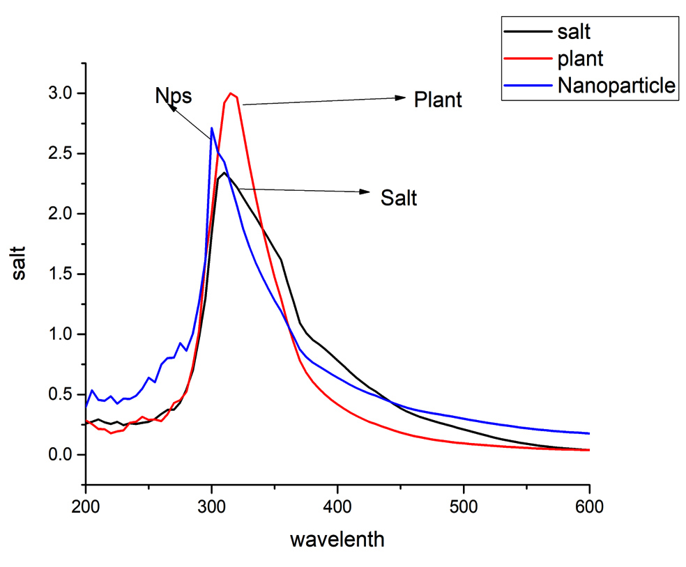
Figure 1. UV-Vis spectra of the synthesized Iron oxide NPs
FTIR spectroscopy
FTIR spectroscopy is a fundamental technique used to investigate functional groups present in a given sample based on their unique vibrational frequencies. In this study, both Parthenium Extract and Iron Oxide Nanoparticles were investigated using FTIR spectroscopy to gain insights into the different functional groups present in them. The FTIR spectrum of Parthenium Extract exhibited distinctive peaks at various wavenumbers, indicating the presence of specific functional groups. A broad peak at 3387 cm-1 was attributed to the stretching vibrations of C-H bonds, suggesting the presence of hydrocarbon groups. Peaks at 2918.30 cm-1 and 2843.86 cm-1 were associated with C-H bonds in alkanes, while peaks at 1714.72 cm-1 and 1633.71 cm-1 indicated the presence of carbonyl groups. The bands observed at 1338.60 cm-1, 1255.66 cm-1, and 1242.16 cm-1 were related to the stretching vibrations of C-O bonds and C-N bonds. Moreover, the band at 989.48 cm-1 confirmed the presence of C=C bonds, and the peaks at 719.45 cm-1 and 642.30 cm-1 suggested the presence of C-H bonds in alkenes with bonding vibrations, along with C-H bonds in alkene having SP2 hybridization at 567.07 cm-1. In the case of Iron Oxide Nanoparticles, the FTIR spectrum exhibited distinct peaks at different wavenumbers, indicative of specific functional groups. The presence of OH stretching and NH2 stretching was confirmed by the bands observed at 3311.78 cm-1, 3261.63 cm-1, and 3242.34 cm-1. Additionally, the bands at 1635.64 cm-1 and 1627.92 cm-1 suggested the presence of amide and amine linkages. The bands at 675.09 cm-1 and 623.01 cm-1 indicated the presence of para-disubstituted aromatic groups or C-H stretching in alkyne. The FTIR spectra of the plant extract and IONPs is given in Figure 2.
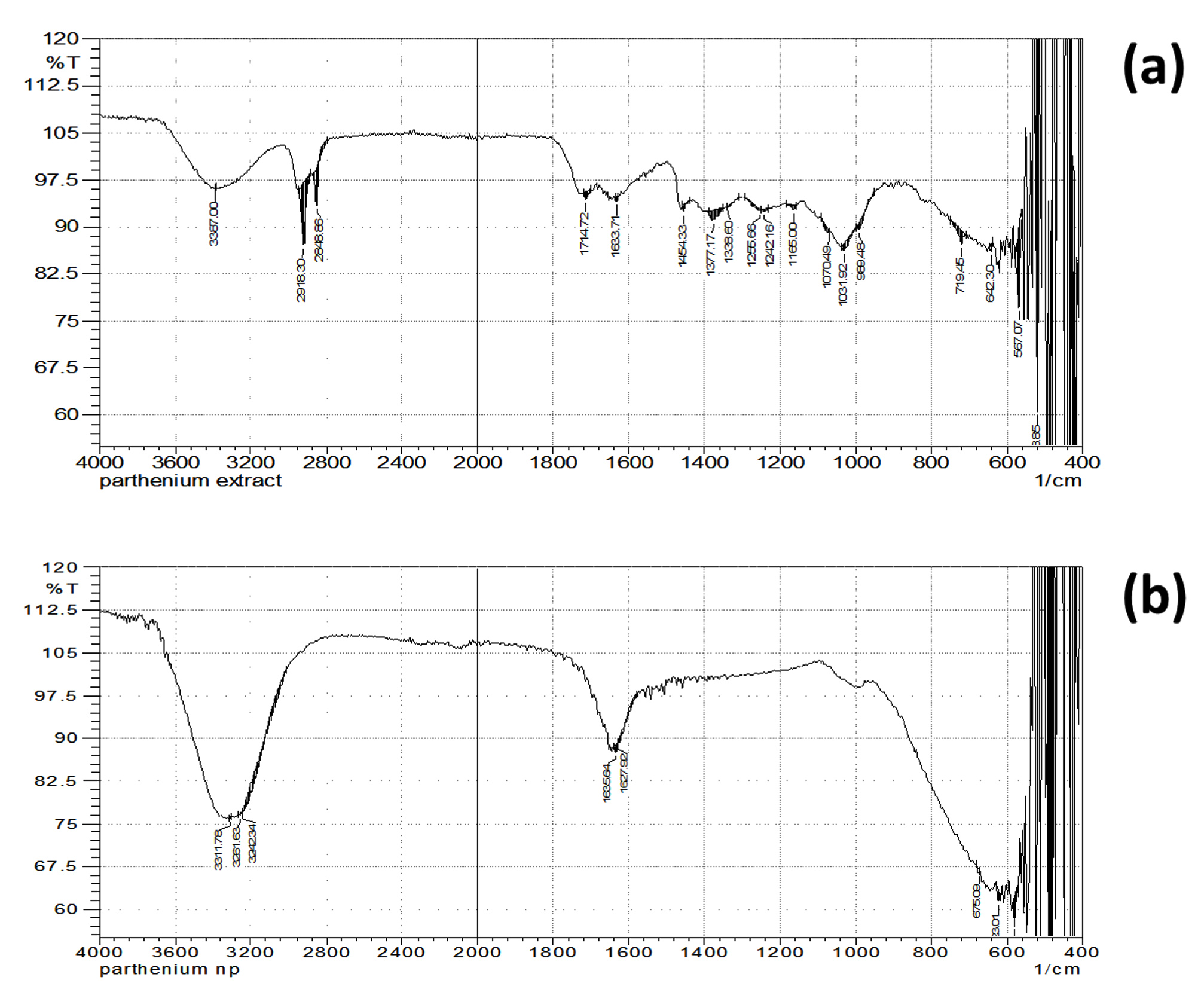
Figure 2. FTIR spectra of Parthenium hysterophorus Linnaeus (a) and Iron oxide NPs (b)
SEM
SEM is an effective method for examining the size and surface morphology of synthesized NPs. It offers crucial insights into the physical characteristics of NPs and offers useful information about the texture and overall structure of NPs. In this study, we used SEM to investigate the properties of the created NPs. We were able to analyze the surface features of the NPs in great detail because the SEM images were taken at various resolutions. The NPs’ uniform and spherical shape, which was revealed by the SEM analysis, suggested that the synthesis process had been successful. Additionally, the SEM images were used to estimate the size of the NPs. According to the analysis, the NPs had an average size of about 0.255 m (micrometres). It’s important to note that applications for nanotechnology frequently use these micro-scale sizes. The measured NP sizes are 255 nm, 18 nm, and 727 nm. The SEM images are given in the inset of Figure 3.

Figure 3. SEM images of the synthesized NPs at low resolution (a) and high resolution (b).
In-vitro screening
Anti-diabetic assays
Glucose uptake by Yeast cells
A dose-dependent increment in glucose uptake was observed in the Parthenium hysterophorus Linnaeus Iron oxide NPs-exposed yeast cells, according to the results of the glucose uptake assay. Various concentrations of NPs (10-100 mg) were incubated. Parthenium hysterophorus Linnaeus nanoparticles has increased the uptake from 8-60% in the yeast cell at 10 and 100 mg concentration respectively. Increase in uptake seems dose dependent. The reference drug, Metronidazole, also significantly influences glucose uptake by the yeast cells (Figure 4a).
α-amylase Inhibition
The a-amylase enzyme was significantly inhibited by Parthenium hysterophorus Linnaeus nanoparticles in the α-amylase inhibition assay. Various concentrations of Parthenium hysterophorus Linnaeus nanoparticles were used that are 10, 20, 40, 80 and 100 µg which showed the inhibition of 21.11, 29.65, 47.12, 58.98 and 72.09% respectively. As referenced and standard drug, diclofenac sodium was employed as 10, 20, 40, 80, and 100 µg/mL which revealed 27.09, 38.11, 46.76, 66.69 and 78.98% of inhibition, respectively. The lowest inhibition rate of Parthenium hysterophorus Linnaeus nanoparticles at 10 µg which was 21.76. While the highest was at 100µg that was 78% and that of Acarbose was at 10 µg 27% and 100 µg/mL 78.98 (Figure 4b).
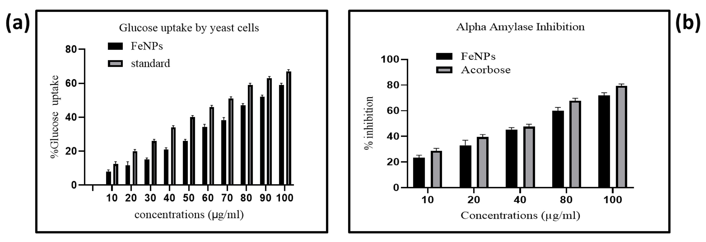
Figure 4. Antidiabetic assays, Glucose uptake by yeast cell (a) and Alpha amylase inhibition (b)
Anti-inflammatory assays
HRBC-membrane stabilizing assay
The HRBC-membrane stabilizing assay revealed the dose-dependent inhibition of HRBC membrane destabilization by Parthenium hysterophorus Linnaeus nanoparticles. Various concentrations of Parthenium hysterophorus Linnaeus nanoparticles were used that are 10, 20, 40, 60, 80 and 100 µg which exhibit 15.87, 31.43, 43.22, 57.11, 66.33 and 79.02% inhibition respectively. As a standard group, the diclofenac sodium was used as 10, 20, 40, 60, 80, and 100 µg/mL which revealed 26.66, 44.23, 56.76, 70.03, 80.95, and 85.71% of inhibition, respectively. The lowest and highest inhibition rate of Parthenium hysterophorus Linnaeus nanoparticles was at 10 µg and 100 µg was 15% and 79%, respectively (Figure 5a).
Effect on heat-induced hemolysis
The Parthenium hysterophorus Linnaeus nanoparticles’ capacity to prevent RBC membrane lysis under high temperatures was demonstrated by the heat-induced hemolysis assay. Various concentrations of Extract were used at 10, 20, 40, 60, 80 and 100 µg/mL which showed 14.96, 25.70, 43.16, 55.05, 66.41, and 78.82% inhibition, respectively. The diclofenac sodium as standard was used at 10, 20, 40, 60, 80 and 100 µg/mL which showed 32.66, 64.23, 77.38, 78.57, 80.95, and 85.71% of inhibition, respectively. The lowest rate inhibition of sample was at 10 µg/mL which was 14.96%. The highest level of inhibition was observed at a concentration of 100 µg/mL, resulting in a 78.82% inhibition rate (Figure 5b).
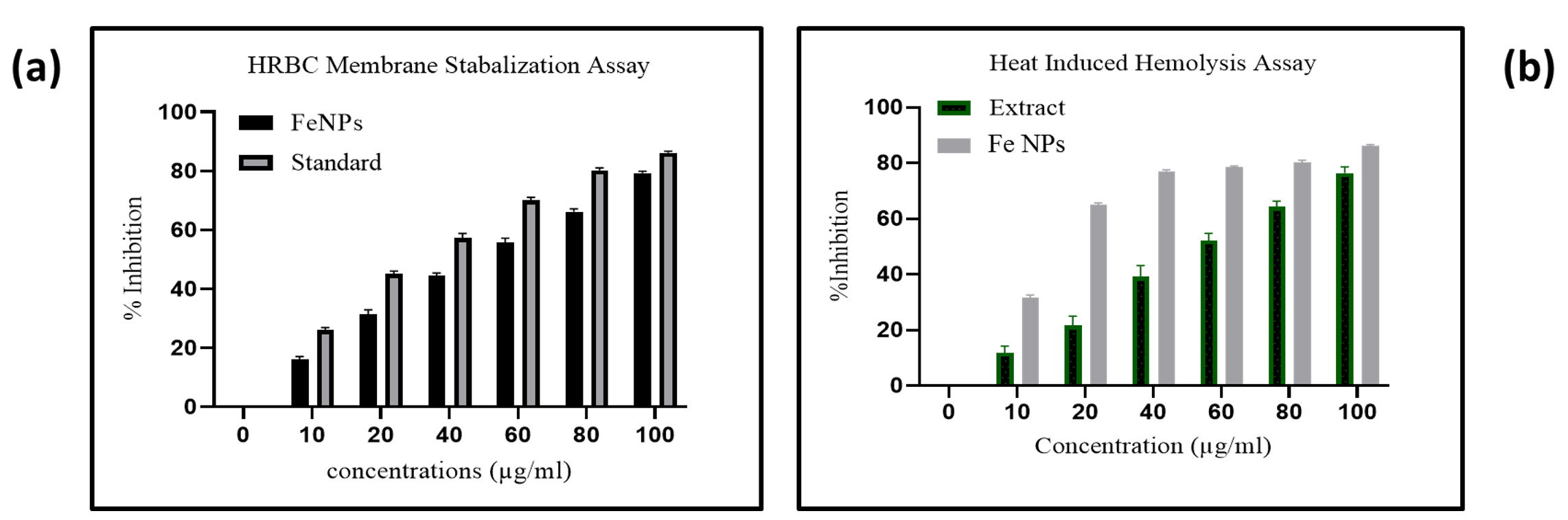
Figure 5. Anti-inflammatory studies by HRBC membrane stabilization (a) and Heat induce Hemolysis assays (b)
Nanoparticle synthesis, a cornerstone of modern materials science, which involves the controlled creation of nanoscale particles with unique characteristics. One intriguing avenue within this field is the biogenic synthesis of nanoparticles by employing plant extracts as reducing and stabilizing agents. This approach offers an ecofriendly and sustainable alternative to conventional chemical methods which often required hazards chemicals. A notable example is the synthesis of Iron Oxide NPs utilizing plant extracts, in this case, Parthenium hysterophorus Linnaeus extract. In this process, the phytochemicals inside the plant extract work as reducing agents, facilitating the formation of NPs by the reduction of metal ions. This “green” approach not only provides a cost-effective means of nanoparticle production but also harnesses the inherent properties of plant compounds which is responsible for their pharmacological activities. In this study, the successful synthesis of IONPs using Parthenium hysterophorus Linnaeus extract was supported by comprehensive characterization techniques. The characterization of these nanoparticles via UV-Vis spectroscopy, FTIR spectroscopy, and SEM provided insights into their properties and elemental composition. The in vitro screening assays demonstrated significant anti-diabetic and anti-inflammatory activities, suggesting promising applications in various biomedical fields. The UV-Vis spectroscopy analysis revealed a distinct peak at 300 nm in the spectra of Iron oxide NPs, showing the successful synthesis of IONPs. This peak aligns with previous studies, validating the consistency of the synthesis method and the presence of characteristic electronic transitions in Iron oxide NPs.28,30 The FTIR spectroscopy investigation of both Parthenium Extract and IONPs unveiled significant functional groups, validating interactions between the extract’s components and iron ions. These interactions likely play a crucial role in NPs synthesis by stabilizing and reducing the metal ions, contributing to their successful formation. The observed changes in the intensities and shifts in various peaks between the Parthenium Extract and IONPs. FTIR spectra indicate the involvement of functional groups of the plant extract responsible for the synthesis of IONPs. These changes in the spectra suggest possible interactions between the plant extract’s functional groups and the iron ions during the nanoparticle synthesis process, leading to the successful reduction and stabilization of IONPs. The SEM images of the synthesized NPs provided a clear depiction of their uniform and spherical shape. The analysis at various resolutions demonstrated the nanoparticles’ consistency in size and shape, reinforcing the effectiveness of the synthesis technique. The nanometer-scale size of the NPs is particularly noteworthy, as it aligns well with the requirements for numerous applications in nanotechnology, catalysis, and biomedical research. The in vitro screening assays revealed the promising anti-diabetic potential of the Iron oxide NPs. The dose-dependent increase in glucose uptake by yeast cells suggests that the nanoparticles may enhance cellular glucose uptake and utilization. This effect could be attributed to bioactive compounds in the nanoparticles that stimulate glucose transport systems. The similarity in trends between Iron oxide NPs and the standard drug Metronidazole underscores the potential of these nanoparticles for further exploration in anti-diabetic research. The anti-inflammatory assays demonstrated the ability of Iron oxide NPs to inhibit HRBC membrane destabilization and heat-induced hemolysis in a dose-dependent manner. This finding indicates the nanoparticles’ potential to stabilize cell membranes and protect against inflammation-induced damage. The similarity in inhibition trends between the nanoparticles and the standard drug diclofenac sodium strengthens the case for Parthenium hysterophorus Linnaeus nanoparticles as potential natural anti-inflammatory agents. These findings open avenues for further research and exploration of Iron oxide NPs for diverse applications in biomedicine, catalysis, and environmental remediation.
The aqueous kernel extract effectively synthesized IONPs, confirmed through standard analytical techniques. These IONPs demonstrated promising anti-inflammatory and anti-diabetic activities at a concentration of 100 μg mL-1, comparable to standard drugs. The findings suggest that the aqueous kernel extract holds promise as a potential pharmaceutical agent, warranting further in vivo studies to explore its practical applications in the medical field.
ACKNOWLEDGMENTS
The authors would like to thank the Department of Chemistry, University of Swabi, Pakistan for providing research facilities.
CONFLICT OF INTEREST
The authors declare that there is no conflict of interest.
AUTHORS’ CONTRIBUTION
AR conceptualized and supervised the study. R, MI, MRI, HAH, YSA, OB, AR, ZA MU and NM performed experimental work. R, MI, MRI, HAH, YSA, OB, AR, MU and NM performed biological activities and spectroscopic analysis. ZA performed data interpretation. ZA wrote and edited the manuscript. All authors read and approved the final manuscript for publication.
FUNDING
None.
DATA AVAILABILITY
All datasets generated or analyzed during this study are included in the manuscript.
ETHICS STATEMENT
Not applicable.
- Mohanraj V, Chen Y. Nanoparticles-a review. Trop J Pharm Res. 2006;5(1):561-573.
Crossref - Sharma R, Prajapati PK. Nanotechnology in medicine: Leads from Ayurveda. J Pharm Bioallied Sci. 2016;8(1):80-81.
Crossref - Bhattacharya D, Gupta RK. Nanotechnology and potential of microorganisms. Crit Rev Biotechnol. 2005;25(4):199-204.
Crossref - Schmid G. Nanoparticles: from theory to application: John Wiley & Sons. 2011.
Crossref - Ahmad Z, Shah SA, Khattak I, et al. Melia azedarach impregnated Co and Ni zero-valent metal nanoparticles for organic pollutants degradation: Validation of experiments through statistical analysis. J Mater Sci: Mater Electron. 2020;31(19):16938-16950.
Crossref - Khan N, Shahida B, Khan SA, et al. Anchoring Zero-Valent Cu and Ni Nanoparticles on Carboxymethyl Cellulose-Polystyrene-Block Polyisoprene-Block Polystyrene Composite Films for Nitrophenol Reduction and Dyes Degradation. J Polym Environ. 2022;31(1):1-13.
Crossref - Riaz M, Khan N, Khan SA, et al. Enhanced catalytic reduction/degradation of organic pollutants and antimicrobial activity with metallic nanoparticles immobilized on copolymer modified with NaY zeolite films. J Mol Liq. 2022;359:119246.
Crossref - Shah SA, Ahmad Z, Khan SA, et al. Biomass impregnated zero-valent Ag and Cu supported-catalyst: Evaluation in the reduction of nitrophenol and discoloration of dyes in aqueous medium. J Organomet Chem. 2021;938:121756.
Crossref - Islam MR, Naz H, Hamad H. Pharmaceutical Upgrade and Natural Perspective on Mucilage and Gum Applications in Pharmacology and Nanomedicine. Phytopharmacol Res J. 2023;2(2):18-21.
- Amstad E, Textor M, Reimhult E. Stabilization and functionalization of iron oxide nanoparticles for biomedical applications. Nanoscale. 2011;3(7):2819-2843.
Crossref - Hussain I, Singh NB, Singh A, Singh H, Singh SC. Green synthesis of nanoparticles and its potential application. Biotechnol Lett. 2016;38:545-560.
Crossref - Tariq M, Ahmad Z, Shah SA, Gul Z, Khan SA. Phytochemical Analysis and Antibacterial Activity of Nicotiana tabacum and Nicotiana rustica. RADS Journal of Biological Research & Applied Sciences. 2021;12(1):60-65.
Crossref - Ahmad Z, Rauf A, Zhang H, Ibrahim M, Muhammad N, Al-Awthan YS, Bahattab O S. Green synthesis and multifaceted characterization of iron oxide nanoparticles derived from Senna bicapsularis for enhanced in vitro and in vivo biological investigation. Green Process Synth, 2024; 13(1):20240001
- Mohanpuria P, Rana NK, Yadav SK. Biosynthesis of nanoparticles: technological concepts and future applications. J Nanoparticle Res. 2008;10:507-517.
Crossref - Rahman H, Rauf A, Khan SA, et al. Green Synthesis of Silver Nanoparticles Using Rhazya stricta Decne Extracts and Their Anti-Microbial and Anti-Oxidant Activities. Crystals. 2023;13(3):398.
Crossref - Abrar H, Niaz M, Ashfaq S, et al. Qualitative phytochemicals profile of five different crude solvents of Galium asperifolium Wall. Phytopharmacol Res J. 2023;2(2):26-37.
- Ahmad Z, Muhammad B, Khan N. Pharmacokinetics of Natural Compounds: Unlocking the Therapeutic Potential. Phytopharmacol Res J. 2023;2(2):22-25.
- Shabir T, Hussain M, Ishfaq S, et al. Antimicrobial and Cytotoxic Potential of Anemone tetrasepala Royle. Phytopharmacol Res J. 2023;2(1):41-48.
- Rukhsar M, Ahmad Z, Rauf A, Zeb H, Ur-Rehman M, Hemeg HA. An Overview of Iron Oxide (Fe3O4) Nanoparticles: From Synthetic Strategies, Characterization to Antibacterial and Anticancer Applications. Crystals. 2022;12(12):1809.
Crossref - Aljohny BO, Ahmad Z, Shah SA, Anwar Y, Khan SA. Cellulose acetate composite films fabricated with zero-valent iron nanoparticles and its use in the degradation of persistent organic pollutants. Appl Organomet Chem. 2020;34(11):e5892.
Crossref - Adkins S, Shabbir A. Biology, ecology and management of the invasive parthenium weed (Parthenium hysterophorus L.). Pest Manag Sci. 2014;70(7):1023-1029.
Crossref - Dhileepan K, McFadyen RC. Parthenium hysterophorus L.-parthenium. Biological Control of Weeds in Australia. 2012:448-462.
- Nath R. Parthenium hysterophorus L.-a review. Agric Rev. 1988;9(4):171-179.
- Kaur L, Malhi DS, Cooper R, et al. Comprehensive review on ethnobotanical uses, phytochemistry, biological potential and toxicology of Parthenium hysterophorus L.: A journey from noxious weed to a therapeutic medicinal plant. J Ethnopharmacol. 2021;281:114525.
Crossref - Thida M, Chan KN, Ei SL, Khai AA. In vitro antidiabetic activities of Myanmar medicinal plants: Cassia siamea Lam. and Butea monosperma Roxb. Indian Journal of Natural Products and Resources (IJNPR)[Formerly Natural Product Radiance (NPR)]. 2023;13(4):483-490.
- Patwekar M, Patwekar F, Mezni A, et al. Assessment of antioxidative and alpha-amylase potential of polyherbal extract. Evid Based Complement Alter Med. 2022;2022:7153526.
Crossref - Anwar S, Raut R, Kanwal B, Yahiya EA, Kumar V. In Vitro Investigation of Anti-inflammatory and Antioxidant Activities of Curcuma Longa Rhizome Methanol Extract. Int J Creat Res Thoughts. 2022.
- Irshad R, Tahir K, Li B, Ahmad A, Siddiqui AR, Nazir S. Antibacterial activity of biochemically capped iron oxide nanoparticles: A view towards green chemistry. J Photochem Photobiol B Biol. 2017;170:241-246.
Crossref - Kiwumulo HF, Muwonge H, Ibingira C, Lubwama M, Kirabira JB, Ssekitoleko RT. Green synthesis and characterization of iron-oxide nanoparticles using Moringa oleifera: a potential protocol for use in low and middle income countries. BMC Res Notes. 2022;15(1):149.
Crossref - Niraimathee VA, Subha V, Ravindran RSE, Renganathan S. Green synthesis of iron oxide nanoparticles from Mimosa pudica root extract. Int J Environ Sustain Dev. 2016;15(3):227-240.
Crossref
© The Author(s) 2024. Open Access. This article is distributed under the terms of the Creative Commons Attribution 4.0 International License which permits unrestricted use, sharing, distribution, and reproduction in any medium, provided you give appropriate credit to the original author(s) and the source, provide a link to the Creative Commons license, and indicate if changes were made.