


ISSN: 0973-7510
E-ISSN: 2581-690X
 , Manoj Kumar, Shanmugasundaram Nagarajan, Harshad V. Murugkar, Pushpendra K. Namdeo, Deepak K. Pankaj, Sonam Agarwal, Aniket Sanyal and Chakradhar Tosh
 Highly pathogenic avian influenza (HPAI) H5N1 virus poses a significant threat to the global poultry sector. The report highlights the genetic and antigenic features of a clade 2.3.4.4b HPAI H5N1 virus isolated from a free-range layer duck in the Kuttanad delta of Kerala, India, in 2022. The virus is similar to an H5N1 virus detected in Russia in October 2021 and antigenically distinct from clade 2.3.2, showing a 16-fold reduction in hemagglutination inhibition titer and 23.95% amino acid differences at the HA antigenic sites. The duck virus shares genetic similarities with H5N1 viruses found in Eurasian wild birds, suggesting a role for these birds in its introduction. The study underscores the importance of strengthening avian influenza surveillance to monitor virus evolution and spread.
Highly Pathogenic Avian Influenza Virus, H5N1 Subtype, Clade 2.3.4.4b, Free-Range Duck, Phylogenetic Analysis, Antigenic Analysis, India
HPAI viruses pose a serious threat to India’s poultry sector, with more than 500 combined H5N1 and H5N8 outbreaks reported since their first detection in 2006.1-3 Multiple genetic clades of HPAI viruses have emerged over time, including the initial identification of the clade 2.2 H5N1 virus in domestic poultry and sporadic cases in crows.4,5 In early 2011, the ancestor of the current clade 2.3.2.1a emerged and spread among poultry, with occasional human cases.6,7 The H5N1 clade 2.3.2.1c was detected in poultry during 2014-15, followed by H5N8 clade 2.3.4.4b in wild birds and poultry starting in 2016.8,9 In 2020, a new reassortant H5N1 clade 2.3.4.4b caused significant morbidity and mortality in both wild birds and poultry worldwide.10 Although ducks are known to be silent carriers of HPAI,11 data on HPAI infection in free-range ducks remain limited. Here, we report the genetic and antigenic analysis of a clade 2.3.4.4b H5N1 virus isolated from a free-range layer duck in India.
Clinical sample processing and animal ethics
We received five free-range layer duck carcasses (12 weeks old) from a single farm in Alappuzha district (Village: Cheruthana, Block: Haripad; Latitude: 9.34139; Longitude: 76.4424) in Kerala State, India, on November 3, 2022. The region lies within the Kuttanad delta region on the west coast of Kerala, where rice is cultivated below sea level.12 It is home to the Vembanad-Kol wetland (Ramsar site), where many migratory waterfowl from the Central Asian Flyway visit during winter migration (October-March). The birds had a history of corneal opacities, torticollis, and unusual mortality. An autopsy was performed, and organs were collected from all birds. Approximately 100 mg of pooled organ sample was triturated in 900 µL of 1X PBS (pH 7.2) using 2.2 mm bead and tissue homogenizer iRUPT24P (Neuation). The homogenizer runs at 3000 rpm for 10 cycles, with a run time and pause time of 15 seconds each and an agitation speed of 5 m/s. The tissue triturate was then centrifuged at 3000 rpm for 5 minutes. Animal experimentation and sample processing were carried out in the animal and laboratory wings, respectively, of the BLS III containment laboratory in accordance with the Animal Ethics Committee of the ICAR-National Institute of High Security Animal Diseases, Bhopal (M.P.), under approval No. 131/IAEC/NIHSAD/22.
RNA extraction and real-time RT-PCR
Viral RNA was extracted from the supernatant of the tissue triturate using the QIAamp Viral RNA Mini Kit (Qiagen, Germany) according to the manufacturer’s protocol. Influenza A virus typing and HA and NA subtyping were performed using the GoTaq® Probe 1-Step RT-qPCR System (Promega, USA).5,13,14
Virus isolation
Isolation of the virus was performed in both 9 day-old specific pathogen-free (SPF) chicken eggs and 12 day-old duck eggs as described earlier.15 The supernatant of the tissue triturate was used as an inoculum for virus propagation in eggs. A total of 800 µL of inoculum was prepared by treating the supernatant with 2X antibiotics (Antimycotic mix and Gentamicin). The eggs were then inoculated with 100 µL of inoculum each via the allantoic cavity route. The eggs were kept in a humid incubator at 37 °C till the death of the embryo. All the eggs were screened every 12 hours, and the dead eggs were chilled at 4 °C. The infected allantoic fluid was harvested aseptically and stored in 1 mL aliquots at -80 °C until further use. Hemagglutination inhibition (HI) assay with H5-subtype-specific sera was used to confirm the viral subtype.15
Intravenous pathogenic index
Intravenous pathogenicity index (IVPI) of the virus (harvested from chicken eggs) was estimated using 6 week-old SPF chickens as per the WOAH guidelines.15
RNA extraction, sequencing and analysis
The RNA was extracted from infected allantoic fluid collected from duck eggs using the QIAamp® Viral RNA Mini Kit (Qiagen, Germany) according to the manufacturer’s protocol. The cDNA library was prepared separately using Protoscript® First Strand cDNA synthesis Kit (New England Biolabs, USA) with random hexamer and U12 primer,16 which were mixed after preparation. The complete genome sequencing, using Sanger’s chemistry, has been determined as described previously.17 The sequences have been deposited in GenBank under accession numbers PP340455-PP340462. Representative avian influenza virus sequences were taken from public database/ GenBank for comparison. The nucleotide sequence alignment was performed using BioEdit (ver 7.7.1),18 and phylogenetic relationships were estimated using MEGA (ver 7.0.26).19 Neighbor-joining trees were constructed for all 8 genes [polymerase basic (PB) 2, PB1, polymerase acidic (PA), HA, nucleoprotein (NP), NA, matrix (M), and non-structural (NS)] by using Tamura-Nei model of nucleotide substitution available in MEGA software. Trees were statistically evaluated using the bootstrap method (with 1000 resampling datasets).
Polyclonal sera and antigenic analysis
Polyclonal antisera were raised in chickens as described previously.20 The H5N1 virus [A/duck/India/11CA04/2022 (dk/11CA04/22)] harvested from the chicken eggs used in the antigenic analysis was inactivated by β-propiolactone at a final concentration of 0.1%. Three SPF chickens were inoculated with the inactivated virus along with the adjuvant. The first inoculation used Freund’s complete adjuvant, followed by a booster dose on the 14th day, with Freund’s incomplete adjuvant. The serum was collected on 21st day, aliquoted, and stored at -40 °C until further use. Other serum and viruses, A/Duck/India/04CA05/2024(H5N1) and A/Chicken/India/03CL488/2011(H5N1) used in this study were obtained from the repository of ICAR-NIHSAD, Bhopal. Antigenic analysis was performed using the hemagglutination inhibition assay.15
Out of the five samples received, 4 were positive for influenza A (H5N1 subtype) by real-time RT-PCR and virus isolation. One representative isolate, A/duck/India/11CA04/2022 (dk/11CA04/22), was selected for further study. The complete genome of dk/11CA04/22 was amplified, and the virus was characterized as HPAI because it harbors multiple basic amino acids at the HA cleavage site (PLKEKRRKR/G), which was confirmed by an IVPI index of 2.96 (out of 3.0) in chickens.15 The duck isolate had conserved amino acid residues at HA positions Q222 and G224 (H5 numbering), indicating a preference for avian-like receptors.21 The PB2 mammalian virulence (E627K) and host-adaptation (D701N) factors were absent in the isolate. The amino acid markers in M2 (V27 and S31) and M1 (I43V) indicated sensitivity to amantadine and increased virulence of the duck H5N1 virus in mice, chickens, and ducks, respectively.
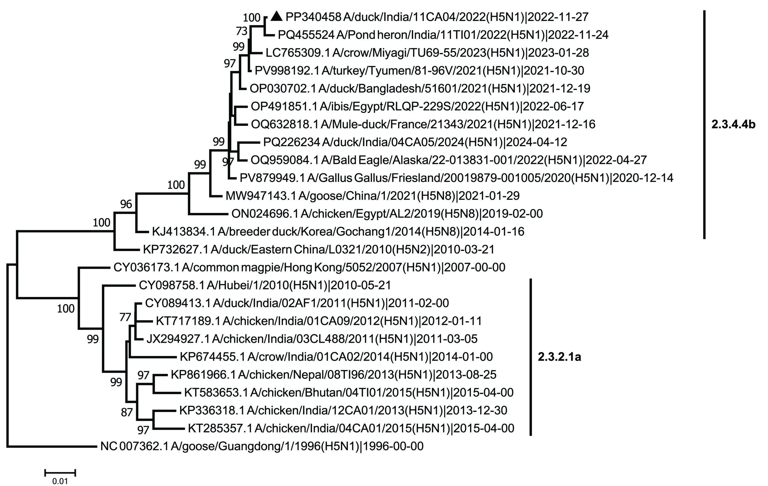
The dk/11CA04/22 revealed high nucleotide sequence identities (99.5%-100%) with a free-flying bird virus (A/pond heron/India/11TI01/2022 (H5N1)) isolated in India, and a turkey virus (A/turkey/Tyumen/81-96V/2021(H5N1) (99.1%-99.9%) isolated in Russia across all 8 genes, indicating an epidemiological link between the outbreaks. Furthermore, dk/11CA04/22 had over 98.0% homology across all genes, except PB2, with H5N1 viruses detected during 2021-2022 in Asia, Africa, Europe and North America (data not shown). In the PB2 gene, the Indian isolates, along with H5N1 viruses from China, Japan and Russia, showed a close genetic relationship (98.6% homology) with a low pathogenic avian influenza (LPAI) virus from Mongolia (A/duck/Mongolia/826/2019(H4N6), indicating reassortment. Phylogenetic analysis placed dk/11CA04/22 within clade 2.3.4.4b (Figure 1A). Within this clade, dk/11CA04/22 grouped with recent H5N1 viruses isolated in Asia, Europe, and North America. Since the emergence of the novel H5N1 clade 2.3.4.4b virus in late 2020, it has been predominantly detected in poultry and wild birds in Europe and has spread globally.22 By October 2021, it was detected in the Russia (A/turkey/Tyumen/81-96V/2021(H5N1). It is likely to have been spread to India via winter bird migration along the Central Asian flyway. Barman and co-workers identified the H5N1 clade 2.3.4.4b virus in Bangladesh in 2021.23 The Indian virus shares seven genes with the Bangladesh virus (A/duck/Bangladesh/51601/2021(H5N1), with only the PB2 gene differing (Figure 1A, B, C, and D [NP gene; representative of other internal genes]). The PB2 gene contributed by LPAI viruses of A/duck/Bangladesh/58751/2023(H6N2)-like and A/duck/Mongolia/826/2019(H4N6)-like, respectively, with Bangladesh and Indian viruses (Figure 1C). A similar reassortant genotype (G10) virus was identified in China, with only the PB2 gene differing from that of the ancestral genotype (G1) virus that first emerged in Europe.22
(a)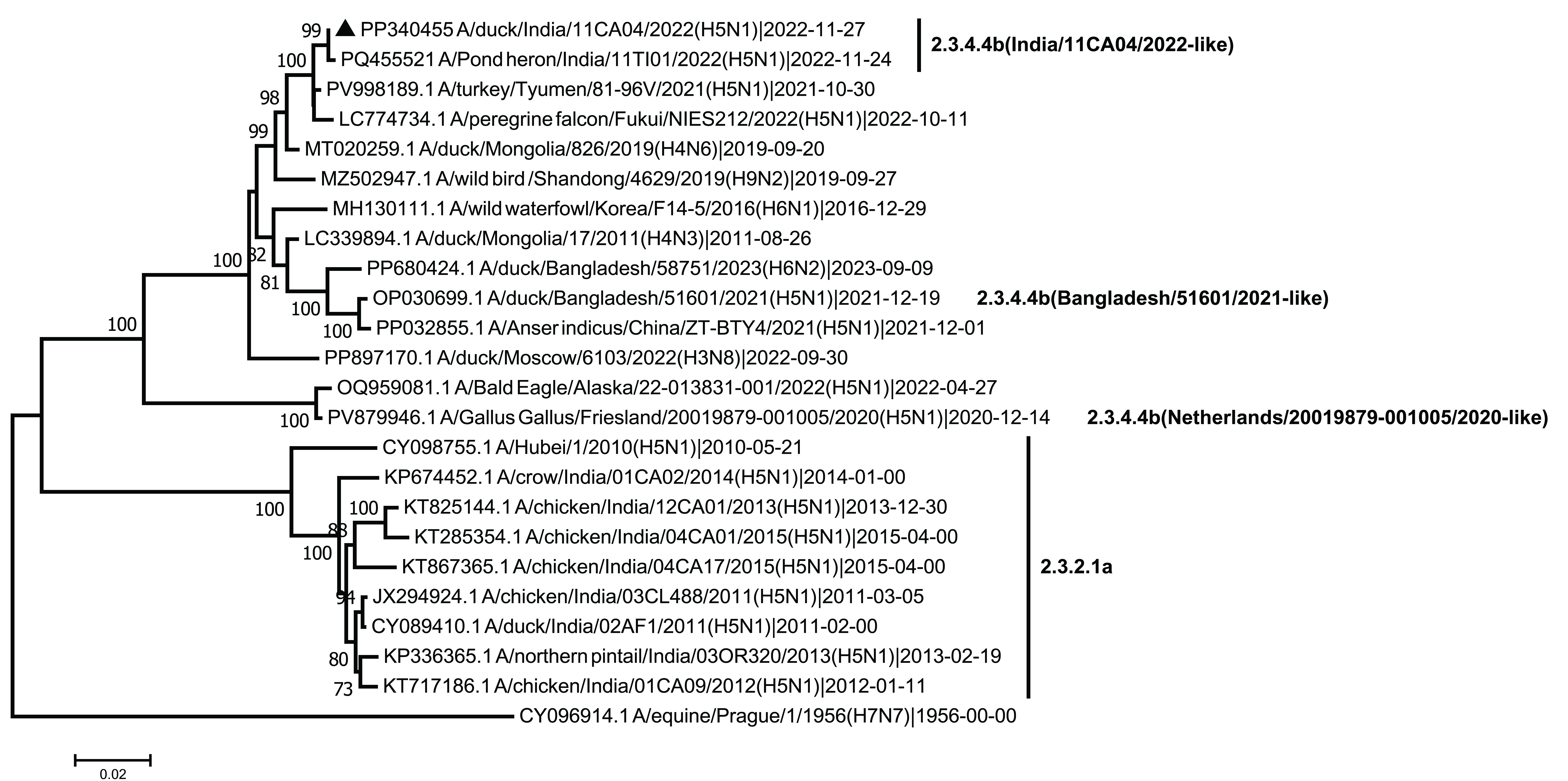 (b)
(b)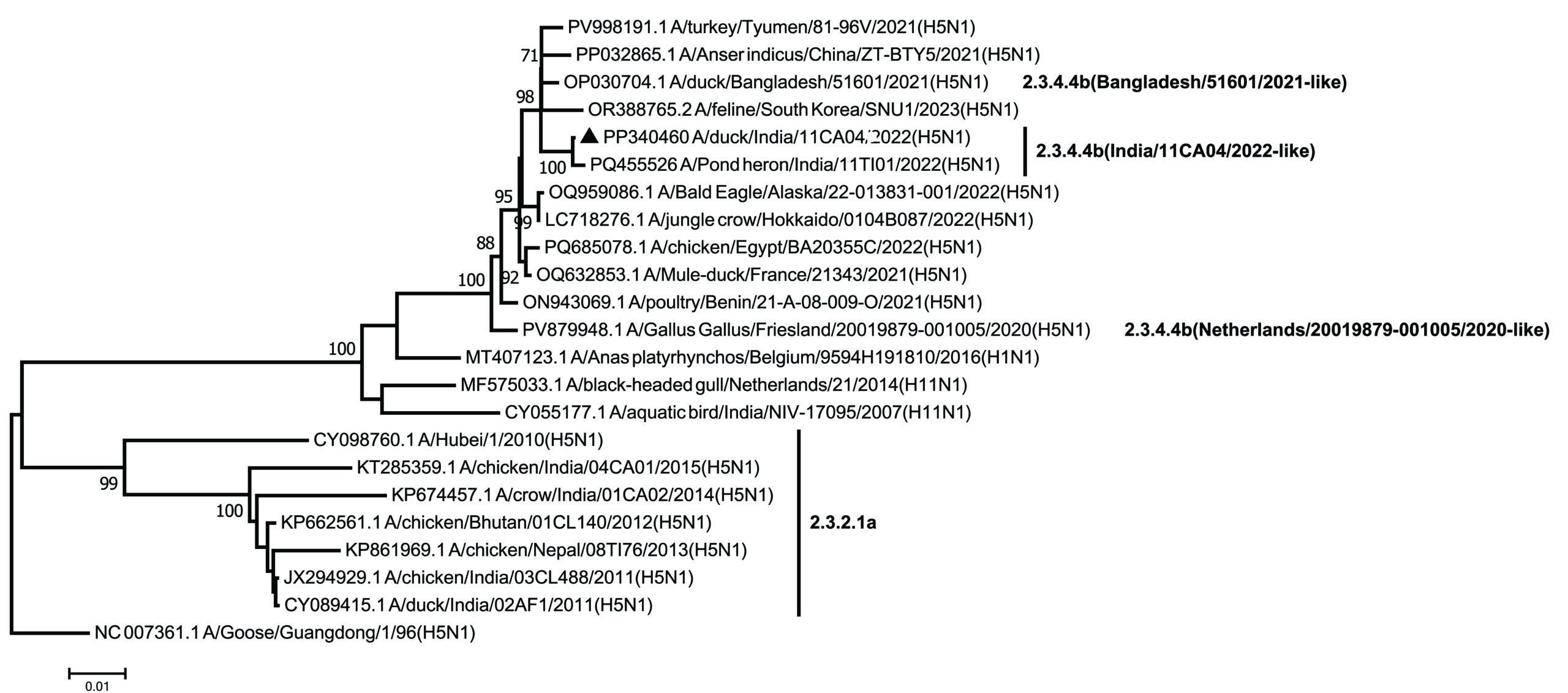 (c)
(c)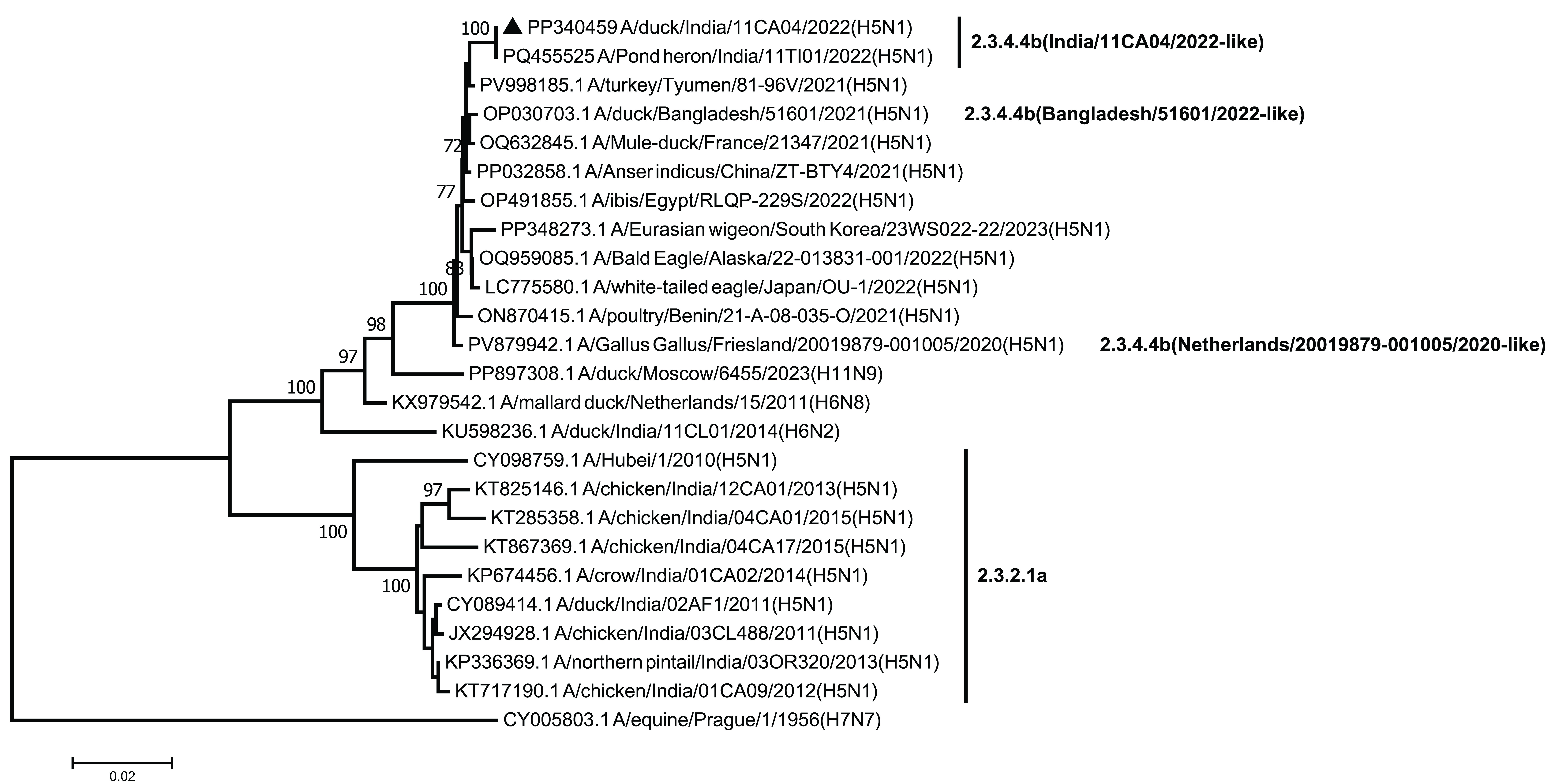
(d)
Figure 1. Neighbor-joining phylogenetic trees of (a) Hemagglutinin (nucleotide positions 29-1735), (b) PB2 (nucleotide positions 28-2307), (c) NA (nucleotide positions 21-1433), and (d) NP (nucleotide positions 46-1542) genes of H5N1 and other viruses. Bootstrap values (³70%, out of 1,000 replicates) are indicated near the nodes. The virus sequenced in this study is highlighted by a solid triangle. The scale bar indicates the number of nucleotide substitutions per site. Clade designations are shown to the right. The HA and NA trees was rooted to A/goose/Guangdong/1/1996(H5N1) and the PB2 and NP trees to A/equine/Prague/1/1956(H7N7)
Antigenic analysis using chicken antisera in an HI assay was performed to assess cross-reactivity between clade 2.3.4.4b and clade 2.3.2 (the ancestor of clade 2.3.2.1a) H5N1 viruses. Chicken antisera raised against dk/11CA04/22 did not react with the clade 2.3.2 virus A/chicken/India/03CL488/2011(H5N1); however, antisera against the clade 2.3.2 virus reacted with clade 2.3.4.4b viruses, with titers 16- to 32-fold lower, indicating antigenic divergence (Table). Similar inter-clade antigenic diversity among H5 subtypes has been reported in China.24 Although both clade 2.3.4.4b viruses are antigenically similar, they have HA substitutions (23.95-25%) that have caused antigenic drift compared to the clade 2.3.2 virus. In addition to H5N1 HPAI, an H9N2 LPAI virus is circulating among poultry in India, with sporadic transmission to humans.25,26
Table. Cross-reactivity of hemagglutination inhibition (HI) antibody titers and amino acid substitutions at the antigenic sites of H5N1 viruses
| Virus isolates | Clade | HI titer of antiserum against antigensa | Amino acid differences at the antigenic site (out of a total 96 amino acids)b | ||
|---|---|---|---|---|---|
| dk/11CA04/22 | ck/03CL488/11 | dk/11CA04/22 | ck/03CL488/11 | ||
| dk/11CA04/22 | 2.3.4.4b | 64 | 16 | 0 (0%) | 23 (23.95%) |
| dk/04CA05/24 | 2.3.4.4b | 64 | 8 | 5 (5.2%) | 24 (25%) |
| ck/03CL488/11 | 2.3.2 | <2 | 256 | 23 (23.95%) | 0 (0%) |
aTiters are expressed as the reciprocal of the highest dilution of serum that completely inhibits hemagglutination.
Boldface indicates homologous HI titers
bAmino acid position and antigenic sites27
Abbreviations: dk/11CA04/22, A/duck/India/11CA04/2022 (H5N1); ck/03CL488/11, A/chicken/India/03CL488/2011(H5N1); dk/04CA05/24, A/duck/India/04CA05/2024 (H5N1)
This study details the antigenic and genetic characteristics of an H5N1 clade 2.3.4.4b HPAI virus isolated from a free-range layer duck. The findings suggest that wild migratory birds likely introduced the H5N1 virus to India through interactions with free-range ducks in wetlands. The H5N1 clade 2.3.4.4b virus is antigenically distinct from the clade 2.3.2 virus, a distinction that should inform future control strategies. In addition to H5N1, an H9N2 LPAI virus is circulating among poultry and ducks in India and occasionally infects humans. The ongoing co-circulation of H5N1 and H9N2 viruses increases the risk of reassortment, potentially producing novel influenza virus(es) with pandemic potential. Therefore, the study highlights the need to strengthen surveillance to track the evolution and spread of these viruses.
ACKNOWLEDGMENTS
The authors thank ICAR, New Delhi, and the Director, ICAR-NIHSAD, Bhopal, for providing the facilities needed to carry out this work. The first author acknowledges the Director, ICAR-IVRI, Bareilly, India, for funding the study through a scholarship. The authors also thank the Director of Animal Husbandry, Kerala, for sharing the clinical samples used in this study.
CONFLICT OF INTEREST
The authors declare that there is no conflict of interest.
AUTHORS’ CONTRIBUTION
SN, HVM, AS and CT conceived and designed the study. KG, PKN, DKP, SA and MK performed the experiment and sequencing. CT, MK and SN performed data and phylogenetic analysis and wrote the manuscript. All authors reviewed, revised and approved the final manuscript for publication.
FUNDING
This study was supported by the CDDL fund of the DAHD, Ministry of Fisheries, Animal Husbandry and Dairying, Government of India.
DATA AVAILABILITY
Molecular sequence data generated in this study have been deposited in GenBank under the accession numbers PP340455-PP340462.
ETHICS STATEMENT
This study was approved by the Institutional Animal Ethics Committee (Approval No. 131/IAEC/NIHSAD/22).
- Pattnaik B, Pateriya AK, Khandia R, et al. Phylogenetic analysis revealed genetic similarity of the H5N1 avian influenza viruses isolated from HPAI outbreaks in chickens in Maharashtra, India with those isolated from swan in Italy and Iran in 2006. Curr Sci. 2006;91(1):77-81.
- Ministry of Fisheries, Animal Husbandry & Dairying. Government and Poultry Industry Collaborate to Prevent Bird Flu Outbreaks. PIB Delhi. 2025. https://www.pib.gov.in/PressReleasePage.aspx?PRID=2119198®=3&lang=2. Accessed February 9, 2026.
- WOAH. WAHIS: World Animal Health Information System. 2026. https://wahis.woah.org/#/dashboards/country-or-disease-dashboard (Accessed on 14 May 2026)
- Tosh C, Murugkar HV, Nagarajan S, et al. Outbreak of avian influenza virus H5N1 in India. Vet Rec. 2007;161(8):279-279.
Crossref - Nagarajan S, Tosh C, Murugkar HV, et al. Isolation and molecular characterization of a H5N1 virus isolated from a Jungle crow (Corvus macrohynchos) in India. Virus Genes. 2010;41(1):30-36.
Crossref - Nagarajan S, Tosh C, Smith DK, et al. Avian Influenza (H5N1) Virus of Clade 2.3.2 in Domestic Poultry in India. PLoS ONE. 2012;7(2):e31844.
Crossref - Potdar V, Brijwal M, Lodha R, et al. Identification of Human Case of Avian Influenza A(H5N1) Infection, India. Emerg Infect Dis. 2022;28(6).
Crossref - Tosh C, Nagarajan S, Kumar M, et al. Multiple introductions of a reassortant H5N1 avian influenza virus of clade 2.3.2.1c with PB2 gene of H9N2 subtype into Indian poultry. Infect Genet Evol. 2016;43:173-178.
Crossref - Nagarajan S, Kumar M, Murugkar HV, et al. Novel Reassortant Highly Pathogenic Avian Influenza (H5N8) Virus in Zoos, India. Emerg Infect Dis. 2017;23(4):717-719.
Crossref - Chrzastek K, Lieber CM, Plemper RK. H5N1 Clade 2.3.4.4b: Evolution, Global Spread, and Host Range Expansion. Pathogens. 2025;14(9):929.
Crossref - Hulse-Post DJ, Sturm-Ramirez KM, Humberd J, et al. Role of domestic ducks in the propagation and biological evolution of highly pathogenic H5N1 influenza viruses in Asia. Proc Natl Acad Sci USA. 2005;102(30):10682-10687.
Crossref - FAO. Kuttanad Below Sea Level Farming System. Family Farming Knowledge Platform. 2012. https://www.fao.org/family-farming/detail/en/c/283069/. Accessed May 12, 2026.
- Spackman E, Senne DA, Myers TJ, et al. Development of a Real-Time Reverse Transcriptase PCR Assay for Type A Influenza Virus and the Avian H5 and H7 Hemagglutinin Subtypes. J Clin Microbiol. 2002;40(9):3256-3260.
Crossref - Fereidouni SR, Harder TC, Gaidet N, et al. Saving resources: Avian influenza surveillance using pooled swab samples and reduced reaction volumes in real-time RT-PCR. J Virol Methods. 2012;186(1-2):119-125.
Crossref - WOAH. Chapter 3.3.4: Avian Influenza (Including Infection with High Pathogenicity Avian Influenza Viruses). Manual of Diagnostic Tests and Vaccines for Terrestrial Animals, thirteenth edition 2024. 2021. https://www.woah.org/fileadmin/Home/fr/Health_standards/tahm/3.03.04_AI.pdf. Accessed February 9, 2026.
- Hoffmann E, Stech J, Guan Y, Webster RG, Perez DR. Universal primer set for the full-length amplification of all influenza A viruses. Arch Virol. 2001;146(12):2275-2289.
Crossref - Tosh C, Nagarajan S, Murugkar HV, et al. Phylogenetic evidence of multiple introduction of H5N1 virus in Malda district of West Bengal, India in 2008. Vet Microbiol. 2011;148(2-4):132-139.
Crossref - Hall T. BioEdit: a user-friendly biological sequence alignment editor and analysis program for Windows 95/98/NT. Nucleic Acids Symposium Series. 1999;41:95-98.
- Kumar S, Stecher G, Tamura K. MEGA7: Molecular Evolutionary Genetics Analysis Version 7.0 for Bigger Datasets. Mol Biol Evol. 2016;33(7):1870-1874.
Crossref - Bhat S, Bhatia S, Pillai AS, et al. Genetic and antigenic characterization of H5N1 viruses of clade 2.3.2.1 isolated in India. Microb Pathog. 2015;88:87-93.
Crossref - Suttie A, Deng YM, Greenhill AR, Dussart P, Horwood PF, Karlsson EA. Inventory of molecular markers affecting biological characteristics of avian influenza A viruses. Virus Genes. 2019;55(6):739-768.
Crossref - Cui P, Shi J, Wang C, et al. Global dissemination of H5N1 influenza viruses bearing the clade 2.3.4.4b HA gene and biologic analysis of the ones detected in China. Emerg Microbes Infect. 2022;11(1):1693-1704.
Crossref - Barman S, Turner JCM, Kamrul Hasan M, et al. Emergence of a new genotype of clade 2.3.4.4b H5N1 highly pathogenic avian influenza A viruses in Bangladesh. Emerg Microbes Infect. 2023;12(2):e2252510.
Crossref - Tian J, Bai X, Li M, et al. Highly Pathogenic Avian Influenza Virus (H5N1) Clade 2.3.4.4b Introduced by Wild Birds, China, 2021. Emerg Infect Dis. 2023;29(7):1367-1375.
Crossref - Tare DS, Pawar SD, Keng SS, et al. The evolution, characterization and phylogeography of avian influenza H9N2 viruses from India. Virology. 2023;579:9-28.
Crossref - WHO (2024) Disease Outbreak News; Avian Influenza A (H9N2) in India. Available at: https://www.who.int/emergencies/disease-outbreak-news/item/2024-DON523
- Luczo JM, Spackman E. Epitopes in the HA and NA of H5 and H7 avian influenza viruses that are important for antigenic drift. FEMS Microbiol Rev. 2024;48(3):fuae014.
Crossref
© The Author(s) 2026. Open Access. This article is distributed under the terms of the Creative Commons Attribution 4.0 International License which permits unrestricted use, sharing, distribution, and reproduction in any medium, provided you give appropriate credit to the original author(s) and the source, provide a link to the Creative Commons license, and indicate if changes were made.