


ISSN: 0973-7510
E-ISSN: 2581-690X
 , Sasinapa Thodsarat1, Prarisa Phadankaew1, Piyatida Pimvichai1 and Rattikan Yutthasin2
This study aimed to identify the causal agent of cassava leaf blight in Thailand and to evaluate the biocontrol potential of Bacillus spp. isolated from the digestive tract of small millipedes. Symptomatic cassava leaves were collected from Maha Sarakham Province, Thailand, and a total of 32 bacterial isolates were obtained and classified into seven morphotypes. Pathogenicity tests revealed that isolates Mnbr-2 and Mnbr-22 induced typical leaf blight symptoms, with Mnbr-2 producing the largest lesion diameter (28.30 ± 3.50 mm) and therefore selected for further study. Based on morphological, biochemical, and 16S rRNA gene analyses, isolate Mnbr-2 was identified as a member of the genus Pantoea and was closely related to Pantoea stewartii, showing 99.75% sequence similarity. These findings provide evidence that Pantoea-associated bacteria are involved in cassava leaf blight in Thailand. The antagonistic activity of 28 Bacillus isolates against P. stewartii Mnbr-2 was evaluated using the agar well diffusion method. Several isolates, including Bacillus velezensis, B. cereus, B. safensis, and B. subtilis, exhibited strong inhibitory activity, with inhibition zones ranging from 25.00 ± 1.50 to 30.00 ± 5.00 mm. Cell-free culture filtrates of selected isolates also suppressed pathogen growth, showing inhibition comparable to that of streptomycin (100 ppm). Detached leaf assays demonstrated that B. cereus N5_TCR and B. velezensis N8_TCR achieved the highest disease suppression under both preventive and curative treatments, with inhibition efficiencies ranging from 91.35%-94.41%. These findings indicate that the selected Bacillus isolates possess strong antagonistic activity and may serve as promising candidates for the development of sustainable and environmentally friendly biocontrol strategies against cassava leaf blight caused by Pantoea.
Bacillus spp., Biological Control, Cassava Leaf Blight, Pantoea stewartii, Small Millipede
Cassava (Manihot esculenta Crantz) is one of the most important tropical root crops cultivated worldwide for food, animal feed, and industrial starch production due to its high carbohydrate content, drought tolerance, and adaptability to marginal soils. Thailand is among the leading cassava-producing countries, where the crop plays a crucial role in agricultural income and export-oriented starch industries. Despite its agronomic advantages, cassava productivity is significantly constrained by various diseases affecting leaves, stems, and storage roots. Among these, foliar diseases are particularly important as they reduce photosynthetic efficiency, plant vigor, and yield.1 Cassava bacterial diseases, especially cassava bacterial blight (CBB) caused by Xanthomonas phaseoli pv. manihotis, are among the most destructive, leading to angular water-soaked lesions, blight, wilting, and severe yield losses under favorable conditions.2 In addition to Xanthomonas, other bacterial genera such as Pseudomonas and Pantoea have been associated with leaf blight-like symptoms in cassava. These bacteria typically invade plant tissues through natural openings or wounds and produce a range of virulence factors, including extracellular enzymes, toxins, and biofilms, which contribute to host colonization and disease progression.3,4
Among them, Pantoea stewartii is an economically important phytopathogen best known as the causal agent of Stewart’s wilt in maize. This Gram-negative bacterium, belonging to the family Erwiniaceae, is transmitted by insect vectors such as flea beetles (Chaetocnema spp.) and can persist epiphytically on plant surfaces. Its pathogenicity is strongly associated with the production of exopolysaccharides (EPS), particularly stewartan, which facilitate biofilm formation, xylem occlusion, and systemic infection within host plants.5 Although extensively studied in maize, the occurrence and role of P. stewartii in cassava remain poorly understood, particularly in Thailand. Recent reports have indicated a broader host range of this pathogen, including its association with bronzing disease in jackfruit (Artocarpus heterophyllus) in Vietnam,6 highlighting its ecological adaptability and potential threat to diverse crops. Accurate identification of bacterial pathogens associated with cassava leaf blight is therefore essential, as symptoms caused by different bacterial species are often indistinguishable, whereas effective disease management strategies depend on pathogen identity and epidemiology. Confirmation of the causal agent through pathogenicity testing in accordance with Koch’s postulates, together with molecular characterization, is critical for establishing disease etiology.
Biological control has emerged as a promising and environmentally sustainable approach for managing plant bacterial diseases. The excessive use of chemical bactericides has led to increased production costs, environmental contamination, and the development of resistant pathogen populations.7,8 Species of Bacillus are widely recognized as effective biocontrol agents due to their ability to produce a broad spectrum of antimicrobial metabolites, particularly lipopeptides such as surfactin, iturin, and fengycin, which disrupt pathogen cell membranes. In addition, the production of extracellular enzymes and stress-resistant endospores enhances their persistence and efficacy under field conditions.9-11 Several species, including B. subtilis, B. velezensis, and B. cereus, have demonstrated strong antagonistic activity against a wide range of phytopathogens through mechanisms such as antibiosis, competition, and induction of systemic resistance.12,13 The digestive tract of small millipedes represents a unique and underexplored microbial niche, harboring diverse bacteria adapted to decomposition-rich environments.14 Such arthropod-associated microbiota have gained increasing attention as promising sources of novel bioactive compounds, including those with antimicrobial properties.15 However, the potential application of Bacillus spp. isolated from millipede digestive systems for controlling cassava bacterial diseases remains largely unexplored. This study is the first to report P. stewartii as a causal agent of cassava leaf blight in Thailand and highlights the digestive tract of millipedes as a novel reservoir of biocontrol bacteria. Therefore, this study aimed (i) to isolate and identify the bacterial pathogen associated with cassava leaf blight symptoms, (ii) to confirm its pathogenicity and molecular identity, and (iii) to evaluate the antagonistic efficacy of Bacillus spp. isolated from the digestive tract of small millipedes against the identified pathogen under laboratory and detached-leaf conditions.
Bacillus spp. isolates
A total of 28 Bacillus isolates were used in this study, comprising seven species: Bacillus altitudinis (2 isolates: N7_TCR and N6_LTND), Bacillus cereus (8 isolates: N6_TCR, N2_TCR, N12_TCR, N8_TCR, N2_LTND, N11_TCR, N5_TCR, and N9_TCR), Bacillus subtilis (2 isolates: N3_LCP and N2_LCP, Bacillus nitratireducens (2 isolates: N3_LTNC and N1_TCWR), Bacillus safensis (2 isolates: N3_LTND and N1_TCR), Bacillus toyonensis (6 isolates: N2_TCWR, N1_LTNC, N7_TCWR, N5_TCWR, N3_TCDLP, and N1_TCDLP), and Bacillus velezensis (6 isolates: N2_LTNC, N4_LDLP, N2_LDLP, N4_TCWR), N5_LTND, and N4_LTND). All isolates were kindly provided by the Mycology and Bioproduct Laboratory, Department of Biology, Faculty of Science, Mahasarakham University, Thailand. The bacterial cultures had been preserved in 20% glycerol stock and were revived by streaking onto nutrient agar (NA) plates to obtain pure colonies. Pure cultures were subsequently maintained on NA slants for further experiments.
Isolation of the causal agent of cassava leaf blight
Symptomatic cassava leaves showing leaf blight symptoms were collected from a cassava cultivation area in Ban Fang, Non Rasi sub-district, Borabue District, Maha Sarakham Province, Thailand (15.912998°N, 103.135021°E). Leaf tissues were excised from the margin between diseased and healthy areas into small sections (approximately 0.5 × 0.5 cm). The tissue pieces were surface sterilized in 5% Clorox solution for 1 min, followed by rinsing twice with sterile distilled water for 1 min each with gentle shaking to completely remove residual disinfectant from the leaf surface. The sterilized leaf tissues were then macerated thoroughly with a small amount of sterile distilled water. The resulting suspension was streaked onto NA plates and yeast extract peptone glucose agar (YPGA) plates and incubated at 28 ± 2 °C for 24-72 hrs. Single colonies were selected and purified to obtain pure bacterial isolates, which were subsequently maintained on NA slants and stored at 4 °C for further experiments.
Pathogenicity test
Healthy cassava leaves (cultivar Kasetsart 72) free from visible disease symptoms were used for pathogenicity testing. Leaf surfaces were disinfected with 70% ethanol and allowed to air dry. The petioles were wrapped with sterile moist cotton and covered with aluminum foil to maintain leaf freshness during the assay. Small wounds were created on the leaf surface using a sterile needle. Bacterial isolates suspected to be the causal agents of cassava leaf blight were cultured for 24-48 hrs and prepared as cell suspensions at a concentration of 106 cells/ml. The bacterial suspension was inoculated into the wounded leaf tissues using a sterile syringe without a needle by gently pressing the suspension onto the abaxial side of the leaf until a water-soaked area became visible beneath the epidermis. After inoculation, the leaves were maintained under high humidity at 25-30 °C for 48 hrs and then transferred to room temperature for 5 days. Disease symptoms were observed daily and recorded throughout the experimental period.
Identification of the causal bacterial pathogen
Morphological characterization
The bacterial isolate selected as the causal agent of cassava leaf blight was streaked onto NA plates and incubated at room temperature for 24 hrs. Colony morphology was examined based on colony size, color, surface texture, margin, and elevation. Gram staining was performed, and bacterial cells were observed under a light microscope at 1,000× magnification to determine Gram reaction, cell shape, and cell size.
Biochemical characterization
Biochemical characteristics of the bacterial pathogen were determined using standard microbiological assays, including catalase, oxidase, starch hydrolysis, citrate utilization, urease, and gelatin hydrolysis tests. For the catalase test, a fresh bacterial colony was transferred onto a clean glass slide, and one drop of 3% hydrogen peroxide was added. Immediate bubble formation was recorded as a positive reaction, indicating catalase production. For the oxidase test, 1% tetramethyl-p-phenylenediamine dihydrochloride prepared in sterile 0.85% saline solution was applied to sterile filter paper. A fresh bacterial colony was streaked onto the reagent-soaked paper, and development of a purple to dark blue color within 10 sec was considered positive for cytochrome oxidase activity. Starch hydrolysis was evaluated by streaking the isolate onto starch agar plates followed by incubation at 37 °C for 24 hrs. After incubation, the plates were flooded with Lugol’s iodine solution. A clear zone surrounding the colony against a blue-black background indicated positive starch hydrolysis due to amylase production, whereas absence of a clear zone indicated a negative result.16 For citrate utilization, the isolate was streaked on Simmons citrate agar slants and incubated for 48 hrs. A color change of the medium from green to blue indicated positive citrate utilization. Urease activity was determined using urea medium supplemented with phenol red as a pH indicator. Development of a red to pink coloration indicated ammonia production resulting from urea hydrolysis and was recorded as a positive reaction. Gelatin hydrolysis was assessed using nutrient broth supplemented with 12% (w/v) gelatin by stab inoculation. Cultures were incubated at 35-37 °C for 3 hrs. After incubation, tubes were refrigerated for 30 min before interpretation. Liquefaction of gelatin at low temperature was considered a positive result, indicating gelatinase activity.
Molecular identification
The selected bacterial pathogen was cultured on NA plates, and submitted to Macrogen Inc. (Seoul, South Korea) for genomic DNA extraction, PCR amplification of 16S rRNA gene using universal primers 27F (5′-AGA GTT TGA TCM TGG CTC AG-3′) and 1492R (5′-TAC GGY TAC CTT GTT ACG ACT T-3′), and DNA sequencing. The obtained nucleotide sequence was compared with sequences available in the GenBank database of the National Center for Biotechnology Information using BLAST analysis to determine bacterial identity. Phylogenetic relationships between the obtained isolate and reference strains were analyzed using 16S rRNA sequence data, and a phylogenetic tree was constructed using MEGA11.17
Evaluation of antagonistic activity of Bacillus spp. against the causal pathogen using the agar well diffusion method
The cassava leaf blight pathogen isolate selected based on its highest pathogenicity, Mnbr-2, was used as the test pathogen. The isolate was cultured on NA plates for 24 hrs, and a bacterial suspension was prepared in sterile distilled water and adjusted to approximately 1 × 10⁸ CFU/ ml using the 0.5 McFarland standard. The suspension was uniformly spread onto NA plates using the swab plate technique and allowed to dry. Twenty-eight Bacillus isolates were cultured separately on NA plates for 24 hrs. Bacterial suspensions were then prepared in sterile distilled water and adjusted to approximately 1 × 10⁸ CFU/ml using the 0.5 McFarland standard. Wells of 6 mm diameter were made in the center of each pathogen-inoculated NA plate using a sterile cork borer, and 20 µl of each Bacillus suspension was added into each well. Sterile distilled water and streptomycin (100 ppm) were used as negative and positive controls, respectively, following the method of Sutthisa et al.18 Plates were incubated at 37 °C for 24 hrs, and the diameter of the inhibition zone was measured. Each treatment was performed in triplicate.
Evaluation of cell-free filtrates of Bacillus spp. against the cassava leaf blight pathogen by agar well diffusion
Selected Bacillus isolates showing strong antagonistic activity in the preliminary screening were cultured in nutrient broth (NB) for 48-72 hrs. The bacterial cultures were adjusted to an optical density at 600 nm (OD600) of 0.2 using a spectrophotometer, corresponding to approximately 1 × 10⁸ CFU/ml. Cultures were centrifuged at 6,000 rpm for 15 min, and the supernatants were collected and filtered through a 0.22 µm membrane filter to obtain cell-free filtrates. The bacterial isolate Mnbr-2 was cultured in nutrient broth (NB) for 24 hrs and the culture was adjusted to an optical density at 600 nm (OD600) of 0.2 using a spectrophotometer, corresponding to approximately 1 × 10⁸ CFU/ml. The pathogen suspension was swabbed uniformly onto NA plates and allowed to dry for 3-5 min. Wells of 6 mm diameter were made at the center of the agar plate using a sterile cork borer, and 20 µl of each cell-free filtrate was added into each well. Plates were incubated at 37 °C for 48 hrs, and inhibition zones were measured in millimeters. Each treatment was conducted in triplicate and compared with the control treatments.
Evaluation of Bacillus spp. efficacy against cassava leaf blight using the detached leaf technique
Healthy cassava leaves (cultivar Kasetsart 72) without visible disease symptoms were surface disinfected with 70% ethanol and air-dried. Petioles were wrapped with sterile moist cotton and covered with aluminum foil to maintain leaf freshness. Small wounds were created on the leaf surface using a sterile needle. Test bacterial isolates were cultured in NB at 28 °C for 24-48 hrs under shaking conditions at 120 rpm. The bacterial culture was adjusted to an optical density at 600 nm (OD600) of 0.2 using a spectrophotometer to obtain a cell density of approximately 1 × 10⁸ CFU/ml. Two inoculation methods were used: Treatment 1 (curative method): 50 µl of the pathogen suspension was applied onto the wounded leaf surface, followed 24 hrs later by 50 µl of the test Bacillus suspension. Treatment 2 (preventive method): 50 µl of the test Bacillus suspension was first applied onto the wounded leaf surface, followed 24 hrs later by 50 µl of the pathogen suspension.19 Each treatment consisted of five replicates. Sterile distilled water was used as the negative control, while streptomycin (100 ppm) served as the positive control. Inoculated leaves were maintained in moist plastic boxes at room temperature. Disease severity was evaluated 7 days after inoculation by measuring lesion diameter, and inhibition percentage was calculated using the following equation:
Inhibition percentage = [Lc – Lt / Lc] × 100
where Lc represents the mean lesion diameter in the control treatment and Lt represents the mean lesion diameter in the treated leaves.
Statistical analysis
All experiments were independently repeated twice using a completely randomized design (CRD) with three to five replicates per treatment in each independent experiment. Data were expressed as mean ± standard deviation (SD). Statistical analysis was performed using one-way analysis of variance (ANOVA), followed the least significant difference (LSD) test at a significance level of P < 0.05. All statistical analyses were performed using IBM SPSS Statistics Version 30 (licensed by Mahasarakham University).
Collection and isolation of cassava leaf blight pathogen
Cassava leaves exhibiting typical leaf blight symptoms were collected from fields in Ban Fang, Non Rasi Subdistrict, Borabue District, Maha Sarakham Province, Thailand. The observed symptoms included water-soaked angular lesions with yellow halos, brown necrotic streaks along the midrib, and progressive blight across the leaf lamina. Four symptomatic cassava plants (cultivar Kasetsart 72, 6 months old) were randomly selected for pathogen isolation. A total of 32 bacterial isolates were obtained from infected tissues and distributed among the four sampled plants as follows: Plant 1 (15 isolates; Mnbr-2 to Mnbr-16), Plant 2 (8 isolates; Mnbr-1, Mnbr-17 to Mnbr-23), Plant 3 (2 isolates; Mnbr-24 and Mnbr-25), and Plant 4 (7 isolates; Mnbr-26 to Mnbr-32). All isolates were subsequently subjected to morphological, biochemical, and molecular characterization. The recovery of 32 bacterial isolates from symptomatic tissues indicates that cassava leaf blight in the study area is associated with a diverse bacterial community rather than a single pathogen. The observed symptoms are consistent with those reported for bacterial diseases in cassava, particularly infections caused by Xanthomonas spp. and related phytopathogens.2 However, the presence of multiple morphologically distinct isolates suggests the coexistence of primary pathogens and secondary colonizers within infected leaf tissues, highlighting the complexity of microbial interactions during disease development.20,21
Morphological characterization
The 32 bacterial isolates were classified into seven morphotypes based on colony morphology and Gram-staining characteristics (Figure 1). Group 1 consisted of small, round, cream-colored, convex, shiny colonies with Gram-positive, short rod-shaped cells (1 isolate: Mnbr-1). Group 2 included small, round, cream-white colonies with a yellow tinge, convex and shiny, with Gram-negative slender rod-shaped cells (3 isolates: Mnbr-6, Mnbr-15, Mnbr-22). Group 3 comprised small, round, opaque white, convex, smooth colonies with Gram-positive coccoid cells (4 isolates: Mnbr-7, Mnbr-29, Mnbr-30, Mnbr-31). Group 4 showed large, round, opaque white colonies with a dry convex surface that appeared mucoid when touched, consisting of Gram-positive slender rod-shaped cells (1 isolate: Mnbr-32). Group 5 included small, round, translucent white, convex, shiny colonies with Gram-positive short rod-shaped cells (1 isolate: Mnbr-21). Group 6 was characterized by small, round, dark yellow, convex, mucoid colonies with Gram-negative short rod-shaped cells (11 isolates: Mnbr-2, Mnbr-3, Mnbr-4, Mnbr-5, Mnbr-13, Mnbr-18, Mnbr-19, Mnbr-20, Mnbr-24, Mnbr-27, Mnbr-28). Similarly, Group 7 consisted of small, round, pale yellow, convex, mucoid colonies with Gram-negative short rod-shaped cells (11 isolates: Mnbr-8, Mnbr-9, Mnbr-10, Mnbr-11, Mnbr-12, Mnbr-14, Mnbr-16, Mnbr-17, Mnbr-23, Mnbr-25, Mnbr-26). These two groups represented the majority of isolates (22 out of 32). The predominance of yellow-pigmented, mucoid, Gram-negative rod-shaped bacteria (Groups 6 and 7), indicated that these isolates share morphological characteristics commonly found among several plant-associated Gram-negative bacterial genera.22 However, these phenotypic traits alone are insufficient for accurate genus identification. Therefore, molecular identification was performed to confirm the identity of the selected pathogenic isolate.
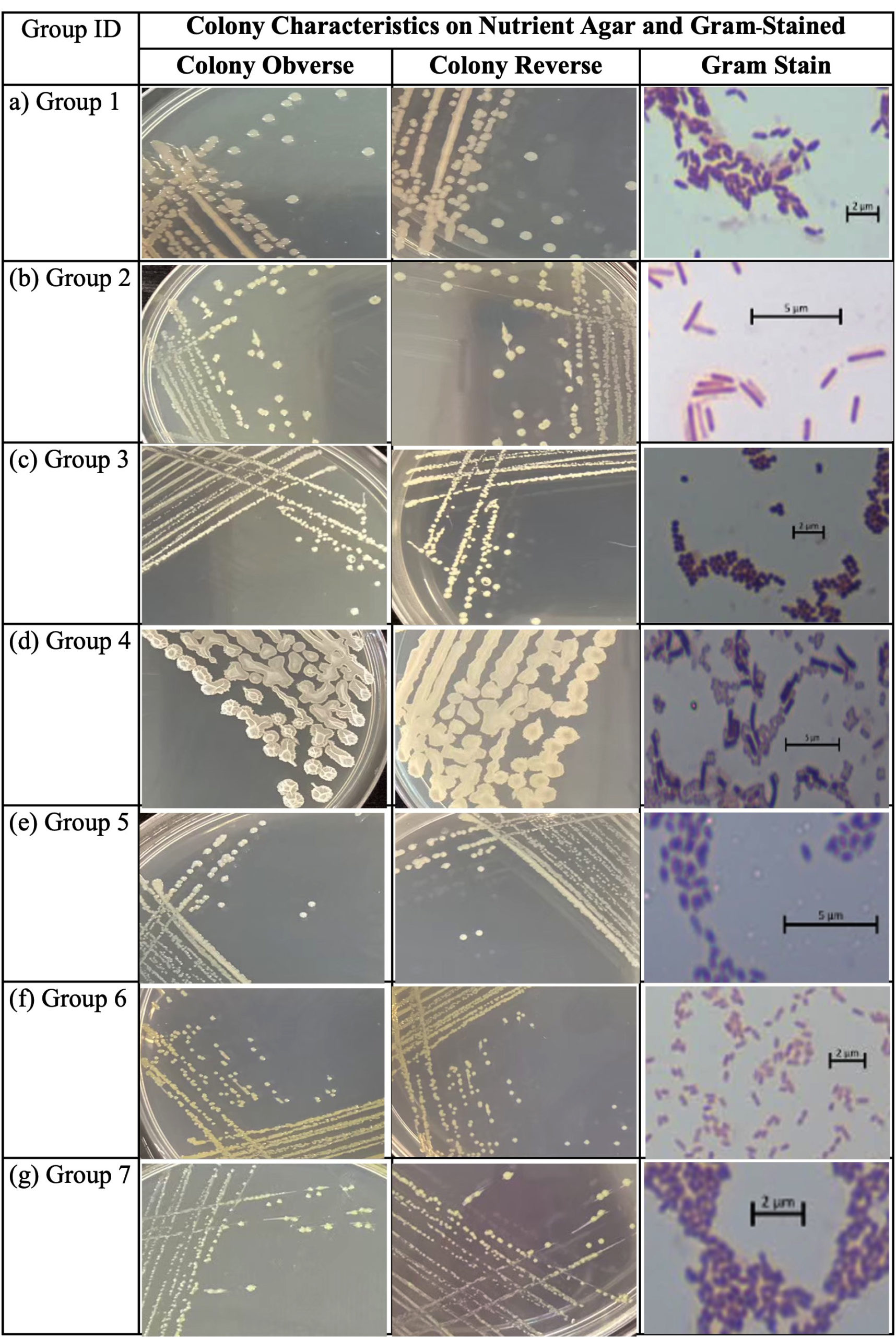
Figure 1. Colony characteristics on the obverse and reverse sides of nutrient agar plates, and cell morphology including Gram staining of bacterial isolates obtained from cassava leaves exhibiting leaf blight symptoms: (a) Group 1; (b) Group 2; (c) Group 3; (d) Group 4; (e) Group 5; (f) Group 6; (g) Group 7. Scale bars represent 2-5 µm
In contrast, the remaining morphotypes, particularly Gram-positive cocci and rods (Groups 1, 3, 4, and 5), are likely to represent non-pathogenic or opportunistic bacteria colonizing damaged plant tissues, which commonly occur during disease progression and tissue degradation.20,21 The presence of Gram-negative but non-mucoid isolates (Group 2) may also indicate the involvement of other genera, such as Pseudomonas, which are frequently associated with plant surfaces and diseased tissues and may function as epiphytes or opportunistic colonizers.23 Colony characteristics, especially pigmentation and mucoid texture, are often associated with virulence traits in plant-associated bacteria. In many Gram-negative phytopathogens, exopolysaccharide (EPS) production plays a crucial role in biofilm formation, environmental stress tolerance, and host colonization.24-26 For example, P. stewartii produces stewartan, an exopolysaccharide (EPS) involved in xylem occlusion and systemic infection.5 Similarly, EPS production is a key virulence factor in many plant pathogenic bacteria, contributing to biofilm formation, environmental persistence, and host colonization.27 Therefore, the high proportion of mucoid, yellow-pigmented isolates observed in this study may suggest the presence of potentially virulent strains contributing to disease development. However, morphological characterization alone is insufficient for accurate bacterial identification due to overlapping phenotypic traits among taxa and the strong influence of environmental conditions on colony morphology.28 Therefore, further characterization using biochemical assays and molecular techniques, particularly 16S rRNA gene sequencing, is necessary to confirm the taxonomic identity and pathogenic role of the isolates. Overall, these results demonstrate the diversity of bacterial populations associated with cassava leaf blight and indicate that morphological characteristics alone are insufficient for accurate bacterial identification. To evaluate pathogenicity, 15 representative isolates were selected from the seven morphotypes based on their colony morphology and Gram staining characteristics. This selection ensured that each morphotype was represented in the pathogenicity assay while minimizing redundancy among isolates with similar phenotypic characteristics. Pathogenicity testing and subsequent molecular analyses were then performed to identify the causal pathogen.
Pathogenicity of bacterial isolates on cassava leaves
The pathogenicity test was performed using 15 representative bacterial isolates selected from the seven morphotypes identified during morphological characterization. All isolates induced disease symptoms on detached cassava leaves (cv. Kasetsart 72). No symptoms were observed in the negative control treated with sterile distilled water, confirming that lesion development was solely attributed to bacterial infection (Table 1). At 3 days post-inoculation (dpi), all isolates produced small lesions ranging from 6.80 ± 2.14 mm (Mnbr-9) to 11.36 ± 1.56 mm (Mnbr-29). Statistical analysis revealed no significant differences among most isolates (P < 0.05), although Mnbr-29 exhibited the largest lesion size at this stage. This suggests that early infection ability was relatively similar among isolates. By 5 dpi, lesion diameters increased markedly, indicating progressive disease development. Among the tested isolates, Mnbr-2 caused significantly larger lesions (28.30 ± 3.50 mm) than other isolates (P < 0.05), followed by Mnbr-22 and Mnbr-29 (Figure 2). The remaining isolates produced moderate lesion sizes ranging from 10.44-16.50 mm. These findings indicate variability in virulence among isolates, with Mnbr-2 exhibiting the highest aggressiveness. The increase in lesion size over time reflects active colonization and tissue maceration, which are typical characteristics of bacterial plant pathogens. Similar patterns of symptom progression have been reported in cassava bacterial diseases, where initial water-soaked lesions expand into necrotic areas due to enzymatic degradation of plant tissues.29,30
Table 1. Pathogenicity of bacterial isolates obtained from symptomatic Cassava leaves
| Isolate | Lesion diameter (mm) | |
|---|---|---|
| 3 days post-inoculation | 5 days post-inoculation | |
| Mnbr-1 | 8.07 ± 3.72ab | 15.66 ± 7.09b |
| Mnbr-2 | 7.52 ± 3.99ab | 28.30 ± 3.50a |
| Mnbr-3 | 7.60 ± 0.56ab | 11.42 ± 4.13b |
| Mnbr-6 | 9.50 ± 2.79ab | 12.88 ± 3.32b |
| Mnbr-9 | 6.80 ± 2.14b | 10.44 ± 3.06b |
| Mnbr-14 | 7.38 ± 1.61ab | 13.44 ± 1.64b |
| Mnbr-15 | 8.40 ± 2.76ab | 15.08 ± 4.75b |
| Mnbr-16 | 7.52 ± 2.40ab | 11.46 ± 2.85b |
| Mnbr-18 | 7.38 ± 1.35ab | 14.24 ± 4.61b |
| Mnbr-19 | 8.96 ± 1.34ab | 16.50 ± 4.13b |
| Mnbr-20 | 7.50 ± 0.52ab | 11.42 ± 1.85b |
| Mnbr-21 | 8.26 ± 3.81ab | 15.68 ± 5.30b |
| Mnbr-22 | 10.82 ± 1.76ab | 18.78 ± 4.00ab |
| Mnbr-29 | 11.36 ± 1.56a | 17.92 ± 4.39b |
| Mnbr-32 | 9.22 ± 2.80ab | 12.48 ± 2.49b |
| Control | 0.00 ± 0.00c | 0.00 ± 0.00c |
Values are presented as mean ± standard deviation (SD). Means followed by the same letter in the same column are not significantly different at P < 0.05
.
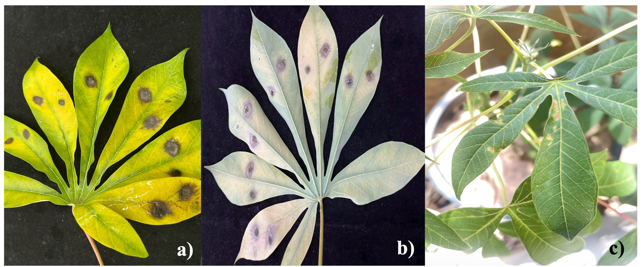
Figure 2. Pathogenicity assay of bacterial isolate Mnbr-2 isolated from diseased cassava leaves: (a) adaxial leaf surface showing symptom development; (b) abaxial leaf surface showing symptom development; (c) disease symptoms on cassava seedlings at 14 days after inoculation
The pathogenicity assays demonstrated that all tested isolates induced leaf blight symptoms on detached cassava leaves, indicating that these isolates possess pathogenic potential. However, since pathogen re-isolation and confirmation were not conducted, the results should be interpreted as evidence of symptom induction rather than complete fulfillment of Koch’s postulates. The absence of symptoms in the control further validates the experimental design and excludes abiotic factors. Variation in lesion size among isolates suggests differences in virulence, which may be associated with the production of pathogenicity factors such as cell wall-degrading enzymes, extracellular polysaccharides, and toxins. Highly virulent isolates, such as Mnbr-2, may possess enhanced abilities to invade host tissues and suppress plant defense responses. Similar variability in aggressiveness among bacterial isolates has been widely reported in plant pathogenic bacteria, including Xanthomonas spp., the causal agents of cassava bacterial blight (CBB), where high genetic diversity and variability among strains are associated with differences in virulence and disease severity.31 The rapid expansion of lesions between 3 and 5 dpi indicates that the pathogens were able to establish infection efficiently and proliferate within host tissues. This progression is consistent with previous studies showing that bacterial pathogens colonize intercellular spaces and vascular tissues, often forming biofilms that facilitate systemic infection and severe tissue damage.32 Detached leaf assays, as employed in this study, provide a reliable and rapid method for screening pathogenicity and comparing virulence among bacterial isolates. However, environmental conditions in detached leaf systems may differ from those in natural field conditions. To confirm pathogenicity, a bacterial cell suspension of isolate Mnbr-2 was sprayed onto cassava seedlings, which subsequently developed disease symptoms on the leaves 14 days after inoculation (Figure 2). Overall, these findings provide evidence that several bacterial isolates associated with cassava leaf blight possess pathogenic potential and highlight the importance of identifying highly virulent isolates for disease management strategies.
Identification of the causal agent of cassava leaf blight
The most virulent isolate (Mnbr-2), selected based on the pathogenicity assay, exhibited small, circular, convex, dark yellow colonies and Gram-negative short rod-shaped cells, consistent with the morphological characteristics previously described for Morphotype 6. Therefore, only biochemical and molecular characterization were further performed for species identification.
Biochemical characterization
Biochemical analysis showed that isolate Mnbr-2 was positive for catalase activity, motility, acetoin production (Voges–Proskauer test), and citrate utilization. In contrast, the isolate was negative for urea hydrolysis, indole production, starch hydrolysis, casein hydrolysis, methyl red test, oxidase activity, and nitrate reduction (Table 2). The biochemical profile of Mnbr-2 was consistent with that of Pantoea stewartii, particularly in its positive VP reaction, citrate utilization, and negative oxidase and nitrate reduction tests, which distinguish it from related genera such as Pseudomonas and Xanthomonas.
Table 2. Biochemical characteristics of the cassava leaf blight bacterium Mnbr-2 compared with reference strains
Test |
Mnbr-2 |
Pantoea stewartii |
Pseudomonas spp. |
Xanthomonas spp. |
|---|---|---|---|---|
Cell shape |
Short rod |
Short rod |
Short rod |
Short rod |
Gram stain |
Negative |
Negative |
Negative |
Negative |
Catalase |
+ |
+ |
+ |
+ |
Motility |
+ |
+ |
+ |
+ |
Acetone production (VP) |
+ |
+ |
– |
+ |
Methyl red (MR) |
– |
– |
– |
– |
Citrate |
+ |
+ |
– |
– |
Urea |
– |
– |
– |
– |
Indole |
– |
– |
– |
– |
Starch hydrolysis |
– |
– |
– |
– |
Casein hydrolysis |
– |
– |
– |
– |
Oxidase |
– |
– |
– |
– |
Nitrate reduction |
– |
– |
– |
– |
+ = positive; – = negative; Reference: Bergey’s Manual of Systematic Bacteriology.33
Molecular characterization
Molecular identification was performed by sequencing the 16S rRNA gene using universal primers 27F and 1492R, yielding an approximately 1,500 bp fragment. Comparative analysis against the NCBI database showed that isolate Mnbr-2 shared 99.75% 16S rRNA gene sequence similarity with Pantoea stewartii (Figure 3), indicating that the isolate belongs to the genus Pantoea and is closely related to P. stewartii. The integration of morphological, biochemical, and 16S rRNA gene analyses indicated that isolate Mnbr-2 belongs to the genus Pantoea and is closely related to P. stewartii, a known plant pathogenic bacterium. Morphologically, the yellow-pigmented, convex colonies observed on nutrient agar are characteristic of Pantoea spp., which often produce carotenoid pigments contributing to their distinctive coloration. The Gram-negative, rod-shaped cell structure further supports its classification within the family Enterobacteriaceae (now Erwiniaceae). Biochemical profiling provided additional evidence supporting this identification. The positive Voges–Proskauer reaction and citrate utilization, combined with negative oxidase and nitrate reduction tests, are key diagnostic features distinguishing P. stewartii from other Gram-negative plant pathogenic bacteria such as Pseudomonas spp. and Xanthomonas spp. These findings are consistent with recent taxonomic and phenotypic descriptions of Pantoea spp., which exhibit diverse morphological and physiological characteristics associated with plant environments.34 Molecular identification based on 16S rRNA gene sequencing further supported the taxonomic placement of Mnbr-2 within the genus Pantoea. Although the 16S rRNA gene is widely used for bacterial identification at the genus level, it has limited discriminatory power for distinguishing closely related species within the genus Pantoea. Therefore, additional housekeeping genes or multilocus sequence analysis (MLSA), such as gyrB, rpoB, recA, atpD, infB, would provide higher taxonomic resolution.3,35 P. stewartii is well known as the causal agent of Stewart’s wilt in maize, but members of the genus Pantoea have also been reported as opportunistic pathogens on a wide range of plant hosts.
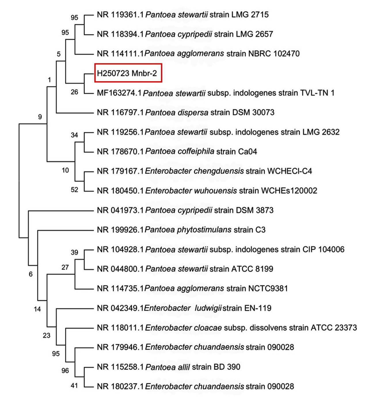
Figure 3. Neighbor-joining phylogenetic tree based on 16S rRNA gene sequences showing the phylogenetic relationship of isolate Mnbr-2 (this study; highlighted by the red box) with reference strains of Pantoea spp. and related taxa retrieved from the NCBI GenBank database. GenBank accession numbers are shown before each strain name. Bootstrap values (>50%) based on 1,000 replications are indicated at the branch nodes
Their pathogenicity is often associated with the production of exopolysaccharides and other virulence factors that facilitate host colonization, biofilm formation, and symptom development, thereby enhancing bacterial persistence and disease progression.11,27,32 In this study, the strong pathogenicity of Mnbr-2 observed in detached leaf assays supports its association with cassava leaf blight. The ability of this isolate to induce severe lesions highlights its potential impact on cassava production and underscores the importance of developing effective management strategies. Overall, the combined morphological, biochemical, and 16S rRNA gene analyses indicate that isolate Mnbr-2 belongs to the genus Pantoea and is closely related to P. stewartii. However, additional housekeeping genes or multilocus sequence analysis (MLSA) will be required to unequivocally confirm its species-level identity.
In vitro antagonistic activity of Bacillus spp. against cassava leaf blight pathogen by agar well diffusion
The antagonistic activity of 28 Bacillus isolates against the cassava leaf blight pathogen was evaluated using the agar well diffusion method (Table 3). All isolates exhibited varying degrees of inhibitory activity, as indicated by the size of inhibition zones. At 1 day after incubation, inhibition zones ranged from 0.00 ± 0.00 mm (B. toyonensis N7_TCWR) to 26.00 ± 1.00 mm (B. cereus N8_TCR), with the latter showing the strongest early antagonistic effect. At 2 days, inhibition zones increased for most isolates, indicating sustained or enhanced antimicrobial activity. The largest inhibition zones were observed in B. subtilis N3_LCP (29.00 ± 3.00 mm) and B. safensis N3_LTND (29.00 ± 3.50 mm), followed by B. cereus N8_TCR (28.25 ± 1.25 mm). Several other isolates, including B. subtilis N2_LCP and B. cereusN5_TCR, also demonstrated strong antagonistic activity, with inhibition zones exceeding 25 mm. In contrast, some isolates exhibited moderate to low activity, such as B. altitudinis N7_TCR and B. toyonensis N3_TCDLP, which produced inhibition zones below 15 mm at 2 days. Interestingly, B. toyonensis N7_TCWR showed no inhibition at 1 day but developed a moderate inhibition zone (18.75 ± 0.25 mm) at 2 days, suggesting delayed antimicrobial activity. Overall, these results demonstrate that several Bacillus isolates, particularly B. subtilis, B. safensis, and B. cereus, possess strong in vitro antagonistic activity against the cassava leaf blight pathogen.
Table 3. In vitro antagonistic activity of Bacillus spp. against cassava leaf blight pathogen using the agar well diffusion method
| Isolate | Inhibition zone (mm) | |
|---|---|---|
| 1 day | 2 day | |
| Bacillus altitudinis N6_LTND | 7.25 ± 0.75fg | 16.25 ± 0.75fghij |
| Bacillus altitudinis N7_TCR | 6.75 ± 0.25g | 13.50 ± 1.00jk |
| Bacillus cereus N2_LTND | 9.00 ± 1.50cdefg | 16.75 ± 0.75fghij |
| Bacillus cereus N2_TCR | 9.00 ± 2.00cdefg | 15.75 ± 0.75fghij |
| Bacillus cereus N5_TCR | 8.50 ± 0.00cdefg | 25.00 ± 5.00abc |
| Bacillus cereus N6_TCR | 10.75 ± 0.75cdefg | 18.75 ± 0.25defghij |
| Bacillus cereus N8_TCR | 26.00 ± 1.00a | 28.25 ± 1.25abc |
| Bacillus cereus N9_TCR | 7.25 ± 0.75fg | 14.75 ± 0.25hijk |
| Bacillus cereus N11_TCR | 6.75 ± 0.25g | 14.25 ± 0.25ijk |
| Bacillus cereus N12_TCR | 10.00 ± 0.00cdefg | 17.75 ± 6.25fghij |
| Bacillus nitratireducens N1_TCWR | 11.00 ± 3.50cdefg | 19.75 ± 0.25cdefgh |
| Bacillus nitratireducens N3_LTNC | 12.75 ± 0.25cd | 19.25 ± 0.75defgi |
| Bacillus safensis N1_TCR | 9.75 ± 1.25cdefg | 20.75 ± 3.25cdef |
| Bacillus safensis N3_LTND | 8.75 ± 1.75cdefg | 29.00 ± 3.50a |
| Bacillus subtilis N2_LCP | 11.75 ± 0.25cdef | 25.00 ± 1.50abc |
| Bacillus subtilis N3_LCP | 11.00 ± 1.00cdefg | 29.00 ± 3.00a |
| Bacillus toyonensis N1_LTNC | 8.00 ± 0.00efg | 14.75 ± 1.25hijk |
| Bacillus toyonensis N1_TCDLP | 9.75 ± 2.25cdefg | 18.50 ± 2.50defghij |
| Bacillus toyonensis N2_TCWR | 7.75 ± 0.25efg | 18.00 ± 0.00efghij |
| Bacillus toyonensis N3_TCDLP | 7.25 ± 0.25fg | 10.00 ± 0.00k |
| Bacillus toyonensis N5_TCWR | 9.50 ± 0.50cdefg | 18.50 ± 0.00defghij |
| Bacillus toyonensis N7_TCWR | 0.00 ± 0.00h | 18.75 ± 0.25defghij |
| Bacillus velezensis N2_LDLP | 9.08 ± 2.90cdefg | 20.25 ± 2.25cdefg |
| Bacillus velezensis N2_LTNC | 8.50 ± 0.50cdefg | 20.50 ± 2.00cdefg |
| Bacillus velezensis N4_LDLP | 10.50 ± 4.50cdefg | 20.75 ± 0.50cdef |
| Bacillus velezensis N5_LTND | 17.50 ± 0.00b | 23.50 ± 0.50bcd |
| Bacillus velezensis N4_LTND | 23.00 ± 6.50a | 30.00 ± 5.00a |
| Bacillus velezensis N4_TCWR | 12.00 ± 2.50cde | 27.25 ± 3.25ab |
Values are expressed as mean ± standard deviation (SD). Means followed by the same letter in the column are not significantly different at P < 0.05
The isolation of antagonistic Bacillus spp. from the digestive tract of small millipedes is of particular interest because the gut of soil-dwelling arthropods represents a unique ecological niche that supports diverse microbial communities and selectively enriches microorganisms adapted to survive under highly competitive conditions.36 These microorganisms may contribute to host nutrition, degradation of organic matter, and protection against invading microorganisms through the production of antimicrobial compounds. Such ecological interactions may contribute to the antagonistic activity observed among the Bacillus isolates in the present study. Previous studies have also reported that insect-associated Bacillus spp. produce a wide range of bioactive metabolites and represent promising sources of biological control agents for agricultural applications.37,38 Therefore, the digestive tract of small millipedes represents a promising yet underexplored source of beneficial microorganisms for sustainable plant disease management.
The present study highlights the strong antagonistic potential of Bacillus spp. against the cassava leaf blight pathogen, supporting their role as promising biocontrol agents. The observed inhibition zones in the agar well diffusion assay indicate the production of diffusible antimicrobial metabolites. Among the tested isolates, B. subtilis and B. safensis exhibited the highest inhibitory activity, which is consistent with previous reports identifying these species as prolific producers of bioactive secondary metabolites, including lipopeptides such as surfactin, iturin, and fengycin. These compounds are known to disrupt pathogen cell membranes, destabilize membrane integrity, and interfere with essential cellular processes, ultimately inhibiting bacterial growth.39 The strong activity observed in B. cereus isolates also aligns with earlier studies demonstrating their ability to produce antimicrobial compounds and enzymes that degrade pathogen cell walls. However, the variability in inhibition among B. cereus isolates suggests strain-specific differences in metabolite production and regulatory mechanisms. The increase in inhibition zones from day 1 to day 2 indicates that antimicrobial compound production is time-dependent, likely associated with bacterial growth phase and quorum sensing regulation. Delayed inhibition, as observed in B. toyonensis N7_TCWR, may reflect the time-dependent synthesis and accumulation of antimicrobial metabolites, whose production and activity vary among Bacillus species and often require sufficient concentration to exert inhibitory effects.11,40 Differences in antagonistic activity among isolates may be attributed to multiple mechanisms, including (i) production of antibiotics, (ii) secretion of hydrolytic enzymes such as chitinases and proteases, and (iii) competition for nutrients and space. In addition, some Bacillus spp. can induce systemic resistance in plants, further enhancing their effectiveness under in vivo conditions.12 Although agar well diffusion is a useful preliminary screening method, it primarily reflects the activity of diffusible compounds under in vitro conditions. Therefore, further studies under greenhouse and field conditions are necessary to confirm the biocontrol efficacy of the most promising isolates, particularly B. subtilis N3_LCP and B. safensis N3_LTND. Overall, this study demonstrates the potential of selected Bacillus isolates as effective biological control agents against cassava leaf blight, providing a foundation for the development of environmentally friendly disease management strategies.
In vitro antagonistic activity of selected Bacillus isolates and their cell-free supernatants against cassava leaf blight pathogen
The antagonistic activity of eight selected Bacillus isolates against the cassava leaf blight pathogen was evaluated using the agar well diffusion method, comparing whole bacterial cells and cell-free supernatants (Table 4). For whole-cell treatments, all isolates exhibited inhibitory activity, with inhibition zones ranging from 9.50 ± 1.50 mm (B. velezensis N4_LTND) to 21.25 ± 3.75 mm (B. velezensis N4_TCWR). Among the tested isolates, B. velezensis N4_TCWR showed the strongest antagonistic activity, followed by B. subtilis N2_LCP (18.25 ± 0.25 mm) and B. cereus N5_TCR (17.00 ± 1.75 mm). However, all bacterial treatments showed lower inhibition compared to the positive control, streptomycin (100 ppm), which produced inhibition zones of 24.50 ± 0.05 mm.
Table 4. In vitro antagonistic activity of Bacillus isolates against the cassava leaf blight pathogen, comparing whole cells and cell-free supernatants using the agar well diffusion method
| Isolate | Inhibition zone (mm) | |
|---|---|---|
| Whole cells (mm) | Cell-free supernatant (mm) | |
| B. velezensis N5_LTND | 10.50 ± 2.50c | 8.75 ± 0.25b |
| B. velezensis N4_LTND | 9.50 ± 1.50c | 8.75 ± 1.50b |
| B. cereus N5_TCR | 17.00 ± 1.75abc | 10.50 ± 0.50b |
| B. cereus N8_TCR | 16.25 ± 0.00abc | 10.75 ± 0.25b |
| B. safensis N3_LTND | 12.25 ± 0.25bc | 8.75 ± 0.25b |
| B. subtilis N2_LCP | 18.25 ± 0.25abc | 13.00 ± 0.00b |
| B. subtilis N3_LCP | 14.50 ± 0.00bc | 17.25 ± 0.25ab |
| B. velezensis N4_TCWR | 21.25 ± 3.75ab | 15.50 ± 3.00ab |
| Streptomycin 100 ppm | 24.50 ± 0.05a | 24.75 ± 0.25a |
Values are mean ± standard deviation (SD). Means followed by the same letter in a column are not significantly different at P < 0.05
In the case of cell-free supernatants, all isolates also demonstrated inhibitory effects, confirming the production of extracellular antimicrobial compounds. The inhibition zones ranged from 8.75-17.25 mm. The highest activity was observed in B. subtilis N3_LCP (17.25 ± 0.25 mm), followed by B. velezensis N4_TCWR (15.50 ± 3.00 mm) and B. subtilis N2_LCP (13.00 ± 0.00 mm). Other isolates, including B. cereus and B. safensis, exhibited relatively lower inhibition (8.75-10.75 mm). The positive control again showed the highest inhibition (24.75 ± 0.25 mm). Interestingly, some isolates such as B. subtilis N3_LCP exhibited stronger inhibition in the cell-free supernatant than in whole-cell assays, whereas others showed reduced activity, indicating differences in the mode of antagonism.
The present study demonstrates that selected Bacillus isolates possess strong antagonistic activity against the cassava leaf blight pathogen, mediated through both direct bacterial interaction and the production of extracellular antimicrobial metabolites. The higher inhibition observed in whole-cell treatments, particularly in B. velezensis N4_TCWR, suggests that multiple mechanisms may be involved, including competition for nutrients and space, as well as continuous production of antimicrobial compounds during bacterial growth. In contrast, the activity observed in cell-free supernatants confirms that diffusible metabolites play a significant role in pathogen suppression. Notably, B. subtilis N3_LCP exhibited stronger inhibition in the cell-free supernatant than in the whole-cell assay, indicating that its antagonistic activity is largely mediated by secreted bioactive compounds. This is consistent with previous studies reporting that B. subtilis produces a wide range of lipopeptides (e.g., surfactin, iturin, and fengycin) with strong antimicrobial activity against plant pathogens.9,41 The comparatively lower inhibition zones observed in some isolates, particularly in their supernatants, may reflect lower production or slower accumulation of antimicrobial metabolites. Additionally, differences between whole-cell and supernatant activity highlight the importance of bacterial viability and dynamic interactions in antagonism. The superior performance of B. velezensis isolates aligns with previous findings that this species is a highly effective biocontrol agent due to its ability to produce diverse secondary metabolites and form stable root-associated populations. Similarly, B. cereus and B. subtilis produce a range of extracellular enzymes, such as proteases and cellulases, that contribute to pathogen inhibition by degrading structural components of microbial cells and interfering with pathogen integrity.13 Although streptomycin exhibited the highest inhibition, the relatively strong activity of Bacillus isolates highlights their potential as environmentally friendly alternatives to chemical control. Unlike antibiotics, Bacillus-based biocontrol agents offer sustainable disease management with reduced risk of resistance development and environmental contamination. Overall, the findings indicate that both whole-cell activity and extracellular metabolites contribute to the antagonistic effects of Bacillus spp. The isolates B. velezensis N4_TCWR and B. subtilis N3_LCP are particularly promising candidates for further development as biocontrol agents. Future studies should focus on metabolite characterization, gene expression analysis, and validation under greenhouse and field conditions.
Efficacy of Bacillus spp. against cassava leaf blight on detached leaves
The detached leaf assay revealed that all tested Bacillus treatments significantly reduced disease severity of cassava leaf blight under both curative and preventive conditions (P < 0.05) (Table 5). In the curative treatment (pathogen inoculated prior to bacterial application), disease inhibition increased over time for most treatments. At 3 days after inoculation, B. cereus N8_TCR and streptomycin completely suppressed disease development (100% inhibition), while other treatments such as B. cereus N5_TCR, B. subtilis N2_LCP, and the cell-free supernatant of B. subtilis N3_LCP achieved high levels of suppression (81%-86%). By day 7, B. cereus N8_TCR maintained strong inhibition (94.11%), comparable to streptomycin (95.23%), whereas other isolates showed moderate to high inhibition ranging from 59.04%-91.35%. In the preventive treatment (bacterial application prior to pathogen inoculation), several isolates provided substantial disease prevention. At 3 days, B. cereus N5_TCR and streptomycin achieved complete protection (100%), while B. velezensis N4_TCWR and B. cereus N8_TCR also showed high inhibition (>80%). At 7 days, B. velezensis N4_TCWR and the cell-free supernatants of B. subtilis N3_LCP and B. velezensis N4_TCWR maintained consistent disease suppression (approximately 75%-79%), whereas B. subtilis N2_LCP showed lower protective efficacy (57.15%). Overall, both whole-cell and cell-free treatments significantly reduced disease severity, with several Bacillus isolates demonstrating efficacy comparable to the chemical control.
Table 5. Efficacy of Bacillus isolates in controlling Cassava leaf blight on detached leave
| Treatment | Disease inhibition (%) | |||
|---|---|---|---|---|
| 3 dpi | 5 dpi | 7 dpi | ||
| Curative (Pathogen first) | B. velezensis N4_TCWR | 41.17 ± 23.72c | 65.94 ± 15.97a | 59.04 ± 15.22ef |
| B. cereus N5_TCR | 82.12 ± 19.52ab | 89.54 ± 10.30a | 91.35 ± 9.87abc | |
| B. subtilis N2_LCP | 81.17 ± 20.93ab | 76.72 ± 14.96a | 74.80 ± 9.00bcdef | |
| B. cereus N8_TCR | 100.0 ± 0.00a | 98.97 ± 2.29a | 94.11 ± 3.12abc | |
| Cell-free B. subtilis N3_LCP | 85.64 ± 16.73a | 90.05 ± 10.09a | 85.51 ± 7.88abcd | |
| Cell-free B. velezensis N4_TCWR | 47.53 ± 12.60bc | 68.41 ± 9.85a | 71.08 ± 6.87cdef | |
| Streptomycin | 100.0 ± 0.00a | 98.46 ± 3.44a | 95.23 ± 7.24ab | |
| Protective (Bacillus first) | B. velezensis N4_TCWR | 84.95 ± 9.03a | 68.69 ± 17.85a | 79.67 ± 11.57abcdef |
| B. cereus N5_TCR | 100.0 ± 0.00a | 95.41 ± 6.76a | 82.09 ± 15.39abcde | |
| B. subtilis N2_LCP | 46.90 ± 4.53bc | 30.03 ± 7.65b | 57.15 ± 4.62f | |
| B. cereus N8_TCR | 81.15 ± 7.73ab | 76.63 ± 10.74a | 66.52 ± 10.65def | |
| Cell-free B. subtilis N3_LCP | 76.19 ± 4.04abc | 75.96 ± 6.95a | 78.69 ± 8.35abcdef | |
| Cell-free B. velezensis N4_TCWR | 79.02 ± 10.03ab | 77.55 ± 10.45a | 75.15 ± 7.90abcdef | |
| Streptomycin | 100.0 ± 0.00a | 100.0 ± 0.00a | 98.88 ± 1.60a | |
Values are mean ± standard deviation (SD). Means followed by the same letter within a column are not significantly different at P < 0.05. dpi = days post-inoculation
The results clearly demonstrate that selected Bacillus isolates possess strong curative and preventive efficacy against cassava leaf blight under detached leaf conditions. The ability to suppress disease both before and after pathogen infection highlights their potential as versatile biocontrol agents. In the curative treatment, the high efficacy of B. cereus N8_TCR suggests strong antibacterial activity against the established pathogen. This may be attributed to the rapid production of antimicrobial compounds and extracellular enzymes capable of inhibiting pathogen growth and degrading infected plant tissues. Similar curative effects have been reported for Bacillus spp., which produce a wide range of antibiotics and lytic enzymes that directly suppress phytopathogens and disrupt their structural integrity.11,42,43 In the preventive treatment, the strong performance of B. cereus N5_TCR and B. velezensis N4_TCWR indicates their ability to prevent pathogen establishment. This may involve early colonization of leaf surfaces, competition for nutrients and infection sites, and the production of antimicrobial metabolites that inhibit pathogen entry. In addition, Bacillus spp. are well known to induce systemic resistance in plants, priming host defense responses against subsequent pathogen attack.12 The comparable performance of cell-free supernatants, particularly from B. subtilis N3_LCP, further confirms that extracellular metabolites play a key role in disease suppression. These metabolites, including lipopeptides such as surfactin, iturin, and fengycin, have been widely reported to disrupt pathogen cell membranes and inhibit growth.9 The effectiveness of supernatants also suggests potential for developing metabolite-based bioproducts. Interestingly, differences between curative and protective efficacy among isolates indicate variation in their modes of action. Some isolates, such as B. subtilis N2_LCP, performed better under curative conditions than preventive ones, suggesting stronger direct antagonism than colonization ability. Conversely, isolates like B. velezensis N4_TCWR showed relatively stable performance under both conditions, indicating multiple mechanisms of action. Although streptomycin provided the highest and most consistent disease control, the performance of several Bacillus isolates was comparable, highlighting their potential as sustainable alternatives to chemical bactericides. Importantly, biological control agents reduce the risks associated with antibiotic resistance and environmental contamination. It should be noted that detached leaf assays provide controlled conditions that may not fully represent field environments. Therefore, further validation under greenhouse and field conditions is necessary to confirm the practical efficacy of the most promising isolates, particularly B. cereus N8_TCR, B. cereus N5_TCR, and B. velezensisN4_TCWR.
Cassava leaf blight is a significant disease affecting cassava production in Thailand. In this study, the most virulent isolate as (Mnbr-2) was identified as a member of the genus Pantoea and was closely related to P. stewartii based on morphological, biochemical, and 16S rRNA gene analyses, showing 99.75% sequence similarity. Pathogenicity tests confirmed that Mnbr-2 is highly virulent to cassava leaves. Among the 28 Bacillus isolates tested, several strains, including B. subtilis, B. cereus, and B. velezensis, exhibited strong in vitro antagonistic activity against the cassava leaf blight pathogen, both as whole cells and through cell-free supernatants, indicating the production of effective extracellular antimicrobial compounds. Detached leaf assays demonstrated that these Bacillus isolates significantly reduced disease severity under both curative (pathogen inoculated before Bacillus) and preventive (Bacillus inoculated before pathogen) treatments. Certain isolates, such as B. cereus N8_TCR, B. cereus N5_TCR, and B. subtilis N2_LCP, showed comparable efficacy to streptomycin, suggesting their potential as biological control agents for sustainable management of cassava leaf blight.
ACKNOWLEDGMENTS
This research project was financially supported by Mahasarakham University. The authors are grateful to Prof. Motoyuki Sumida for English language editing.
CONFLICT OF INTEREST
The authors declare that there is no conflict of interest.
AUTHORS’ CONTRIBUTION
All authors listed have made a substantial, direct and intellectual contribution to the work, and approved it for publication.
FUNDING
This research project was financially supported by Mahasarakham University.
DATA AVAILABILITY
All datasets generated or analyzed during this study are included in the manuscript.
ETHICS STATEMENT
Not applicable.
- Borku AW, Tora TT, Masha M. Cassava in focus: A comprehensive literature review, its production, processing landscape, and multi-dimensional benefits to society. Food Chem Adv2025;7:100945.
Crossref - Zarate-Chaves CA, Moufid Y, Lopez CE, Bernal A, Szurek B, Yanez JM. First report of Cassava bacterial blight caused by Xanthomonas phaseoli pv. manihotis in the Amazonian forest of Ecuador. Plant Dis. 2024;108(6):1879.
Crossref - Ryan RP, Vorholter FJ, Potnis N, et al. Pathogenomics of Xanthomonas: Understanding bacterium–plant interactions. Nat Rev Microbiol.2011;9(5):344-355.
Crossref - Zhao A, Sun J, Liu Y. Understanding bacterial biofilms: From definition to treatment strategies. Front Cell Infect Microbiol. 2023;13:1137947.
Crossref - Roper MC. Pantoea stewartii subsp. stewartii: lessons learned from a xylem-dwelling pathogen of sweet corn. Mol Plant Pathol. 2011;12(7):628-637.
Crossref - Ha VTN, Hoang LK, Huyen PK. Pantoea stewartiisubsp. stewartii, the causative agent of Thai jackfruit’s bronzing disease and its possible host range in Vietnam. J Plant Prot Res. 2024;64(2):149-157.
Crossref - Compant S, Duffy B, Nowak J, Clement C, Barka EA. Use of plant growth-promoting bacteria for biocontrol of plant diseases:Principles, mechanisms, and future prospects. Appl Environ Microbiol.2005;71(9):4951-4959.
Crossref - Sundin GW, Wang N. Antibiotic resistance in plant-pathogenic bacteria. Ann Rev Phytopathol.2018;56:161-180.
Crossref - Ongena M, Jacques P. Bacillus lipopeptides:Versatile weapons for plant disease biocontrol. Trends Microbiol. 2008;16(3):115-125.
Crossref - Fira D, Dimkic I, Beric T, Lozo J, Stankovic S. Biological control of plant pathogens by Bacillus species. J Biotechnol. 2018;285:44-55.
Crossref - Tran C, Cock IE, Chen X, Feng Y. Antimicrobial Bacillus:Metabolites and their mode of action. Antibiotics.2022;11(1):88.
Crossref - Kloepper JW, Ryu CM, Zhang S. Induced systemic resistance and promotion of plant growth by Bacillus spp. Phytopathology. 2004;94(1):1259-1266.
Crossref - Rabbee MF, Ali MS, Choi J, Hwang BS Jeong, SC, Baek K-h. Bacillus velezensis:A valuable member of bioactive molecules within plant microbiomes. Molecules 2019;24(6):1046.
Crossref - Koubova A, Lorenc F, Horvathova T, Chrooakova A, Sustr V. Millipede gut-derived microbes as a potential source of cellulolytic enzymes. World J Microbiol Biotechnol.2023;39(7):1-12.
Crossref - Engel P, Moran NA. The gut microbiota of insects – diversity in structure and function. FEMS Microbiol Rev.2013;37(5):699-735.
Crossref - Ashmawy NA, Shoeib AA, Youssef HFB, Mahmoud SM. Pathological, biochemical and molecular characterization of the seed borne bacteria Pantoea spp., Xanthomonas spp. and Pseudomonas spp. from solanaceous plants in Egypt. J Microbiol Biotechnol Food Sci. 2020;10(20):289-295.
Crossref - Sutthisa W. Comparison of the antagonistic potential of the entomopathogenic bacterium Serratia nematodiphila GCSR38 with other effective microorganisms for the control of rice bacterial leaf blight. J Pure Appl Microbiol. 2022;16(1):557-566.
Crossref - Sutthisa W, Kaewpipat D, Wongkhomchang W, Manphae A. Development of alginate-based Bacillus subtilis biopellets for tomato bacterial wilt control. Trends Sci. 2025;22(9):10332.
Crossref - Sutthisa W, Nachai P, Yutthasin R. Development of Entomopathogenic Bacterium Serratia nematodiphila GCSR38 Formulation for the Control of Mango Anthracnose Disease. J Pure Appl Microbiol. 2025;19(2):1410-1418.
Crossref - Trivedi P, Leach JE, Tringe SG, Sa T, Singh BK. Plant–microbiome interactions: From community assembly to plant health. Nat Rev Microbiol. 2020;18(11):607-621.
Crossref - Ge J, Li D, Ding J, Xiao X, Liang Y. Microbial coexistence in the rhizosphere and the promotion of plant stress resistance: A review. Environ Res. 2023;222:115298.
Crossref - Walterson AM, Stavrinides J. Pantoea:Insights into a highly versatile and diverse genus within the Enterobacteriaceae. FEMS Microbiol Rev.2015;39(6):968-984.
Crossref - Xin XF, Kvitko B, He SY. Pseudomonas syringae:what it takes to be a pathogen. Nat Rev Microbiol. 2018;16(5):316-328.
Crossref - Denny TP. Involvement of bacterial polysaccharides in plant pathogenesis. Ann Rev Phytopathol. 1995;33:173-97.
Crossref - Kaur N, Dey P. Bacterial exopolysaccharides as emerging bioactive macromolecules:from fundamentals to applications. Res Microbiol. 2023;174(4):104024.
Crossref - Dong J, Chi Z, Lu S, et al. Bacterial exopolysaccharides: Characteristics and antioxidant mechanism. Int J Biol Macromol. 2025;289:138849.
Crossref - Vailleau F, Genin S. Ralstonia solanacearum:An arsenal of virulence strategies and prospects for resistance. Ann Rev Phytopathol.2023;5:61:25-47.
Crossref - Hafi LA, Alocilja EC. Morphometric characterization of bacteria associated with bacteremia. Encyclopedia. 2025;5(3):130.
Crossref - Lozano JC. Cassava bacterial blight: A manageable disease. Plant Dis.1986;70(12):1089-1093.
Crossref - Zárate-Chaves CA, Gómez de la Cruz D, Verdier V, López CE, Bernal A, Szurek B. Cassava diseases caused by Xanthomonas phaseoli pv. manihotis and Xanthomonas cassavae. Mol Plant Pathol. 2021; 22(12): 1520-1537.
Crossref - Veley KM, Elliott K, Jensen G, et al. Improving Cassava bacterial blight resistance by editing the epigenome. Nat Commun. 2023;14(1):85
Crossref - Carezzano ME, Paletti Rovey MF, Cappellari LdR, et al. Biofilm-forming ability of phytopathogenic bacteria:A review of its involvement in plant stress. Plants. 2023;12(11):2207.
Crossref - Šindelářová M. Schaad NW, Jones JB, Chun W, eds. Laboratory Guide for Identification of Plant Pathogenic Bacteria. Biol Plant. 2001;44(4):546.
Crossref - Tambong JT. Taxogenomics and systematics of the genus Pantoea. Front Microbiol. 2019;10:2463.
Crossref - Crosby KC, Rojas M, Sharma P, et al. Genomic delineation and description of species and within-species lineages in the genus Pantoea. Front Microbiol. 2023;14:1254999.
Crossref - Byzov BA, Kurakov AV, Tretyakova EB, et al. Principles of the digestion of microorganisms in the gut of soil millipedes:specificity and possible mechanisms. Appl Soil Ecol. 1998;9(1-3):145-151.
Crossref - Konig H. Bacillus species in the intestine of termites and other soil invertebrates. J Appl Microbiol. 2006; 101(3):620-627.
Crossref - Caulier S, Nannan C, Gillis A, Licciardi F, Bragard C, Mahillon J. Overview of the antimicrobial compounds produced by members of the Bacillus subtilis group. Front Microbiol. 2019;10:302.
Crossref - Ghosh S, Basu S, Anbarasu A, Ramaiah S. A comprehensive review of antimicrobial agents against clinically important bacterial pathogens:Prospects for phytochemicals. Phytother Res. 2025;39(1):138-161.
Crossref - Da Silva ML, De Freitas BC, Costa MD, De Souza Carneiro JE, De Queiroz MV. Potential of endophytic Bacillus toyonensis BAC3151 for phytopathogen control and growth promotion in common bean. J Plant Dis Prot. 2026;133(2).
Crossref - Chowdhury SP, Hartmann A, Gao X, Borriss R. Biocontrol mechanism by root-associated Bacillus amyloliquefaciensFZB42—a review. Front Microbiol. 2015;6:780.
Crossref - Rabbee MF, Baek KH. Antimicrobial activities of lipopeptides and polyketides of Bacillus velezensis for agricultural applications. Molecules. 2020; 25(21):4973.
Crossref - Ajuna HB, Lim HI, Moon JH, et al. The prospect of hydrolytic enzymes from Bacillus species in the biological control of pests and diseases in forest and fruit tree production. Int J Mol Sci. 2023;24(23):16889.
Crossref
© The Author(s) 2026. Open Access. This article is distributed under the terms of the Creative Commons Attribution 4.0 International License which permits unrestricted use, sharing, distribution, and reproduction in any medium, provided you give appropriate credit to the original author(s) and the source, provide a link to the Creative Commons license, and indicate if changes were made.