


ISSN: 0973-7510
E-ISSN: 2581-690X
In blood donations, chlorhexidine gluconate (CHG) and isopropyl alcohol (IPA) are routinely used for skin disinfection. Despite this measure, contamination of platelet concentrates with biofilm-forming Staphylococcus epidermidis from donor skin remains a concern in transfusion medicine. Essential oils (EOs) not only possess antibacterial abilities but have possible synergistic effects with traditional disinfectants. This study evaluated the anti-biofilm activity of EOs from four Saudi Arabian plants (Clitoria ternatea, Aerva lanata, Lavandula dentata, and Plumbago zeylanica) as potential synergistic enhancers for skin disinfection. EO composition was evaluated by gas chromatography-mass spectrometry (GC-MS), and safe EO concentrations were determined using the rabbit skin irritation test. In anti-biofilm activity assays, S. epidermidis biofilms were treated with EOs alone or in conjunction with CHG or CHG-IPA and log-reduction of bacteria was calculated. Synergistic effects were evaluated through the coefficient of drug interaction. EO concentrations up to 20% were found to be non-irritating. Most EOs exhibited some anti-biofilm activity; P. zeylanica EO exerted the strongest effect, and also displayed suggestive of synergistic action with CHG and improved CHG-IPA performance. GC-MS identified plumbagin (79%) as the main oxygenated terpenoid component in P. zeylanica EO, and plumbagin individual activity demonstrated to be almost equivalent to the activity of P. zeylanica. The effectiveness of CHG-IPA against biofilms was greatly improved by combination with P. zeylanica EO or plumbagin. These results add to the evidence that natural compounds could enhance skin disinfection agents and help lower the risk of bacterial contamination in blood products.
Biofilm, Staphylococcus epidermidis, Platelets, Skin Disinfection, Essential Oil
At present, the main transfusion-associated infection risk from bacterial contamination of platelet concentrates (PCs).1 PCs are made in an additive, pH-neutral solution containing around 25 g/L glucose, and are maintained under continuous agitation at 22-28 °C.2,3 Blood centers in Canada, the United States, Germany, China, and New Zealand have shown the most frequently-identified aerobic contaminant of PCs to be coagulase-negative Staphylococcus epidermidis.4-7 A normal human commensal that prefers moist niches such as the antecubital area, S. epidermidis is most likely introduced by the venipuncture during blood product collection.2,8,9 A number of precautions have been implemented to help PC contamination, including screening donors for illness symptoms, skin disinfection prior to venipuncture, diversion of the initial 30-40 mL drawn, and screening PCs for bacteria.2,10 Despite these measures, bacterial contamination and missed detections continue to occur, and therefore the risk of potentially fatal S. epidermidis-associated transfusion reactions.11,12
As an effective, broad-spectrum cationic disinfectant, chlorhexidine has become widely adopted by blood centers for donor skin disinfection over the past decade,13,14 particularly in the form of the one-step ChloraPrep kits, which combine 2% chlorhexidine gluconate (CHG) with 70% isopropyl alcohol (IPA). Used in tandem, these disinfectants reduce the rate of PC contamination, but do not fully eliminate it.2,13,15 Notably, we previously demonstrated that where planktonic S. epidermidis cells are vulnerable to CHG-IPA, cell aggregates encased in a self-produced matrix, termed biofilms, are markedly more resistant.16
Plant essential oils (EOs) have been widely demonstrated to possess antimicrobial activity, especially those with oxygenated terpenoid components, such as alcohols and phenolic oxygenated terpenoid.17-19 Some EOs are also reported to have synergistic action with CHG, enhancing its penetration into skin and its antibacterial activity.8,20,21 Among plants native to Saudi Arabia, Clitoria ternatea (butterfly pea), Aerva lanata (mountain knotgrass), Lavandula dentata (French lavender), and Plumbago zeylanica (Ceylon leadwort) are notable medicinal plants known to possess antibacterial and/or anti-biofilm properties.22-25 In this study, EOs were extracted from these four plants and investigated for their antimicrobial efficacy and their synergy with CHG for potential enhancement of skin disinfection practices to improve the purity and safety of PCs. We hypothesized that EOs with elevated concentrations of oxygenated terpenoids would synergistically enhance the anti-biofilm activity of CHG-based disinfectants.
Bacterial strain
Staphylococcus epidermidis strain ATCC 35984 was used in anti-biofilm activity assays on account of its documented ability to form strong biofilms. Bacteria were stored at -80 °C in brain heart infusion broth enriched with 15% glycerol until required.26
Ethical approval statement
This study was approved by the Ethics Review Committee (ERC), College of Medicine, Imam Mohammad Ibn Saud Islamic University, Riyadh, Saudi Arabia (Approval No. 23R/442/07, dated 12 November 2024).
EO extraction and analysis
Four plants native to Saudi Arabia (specifically Jazan Province) were selected for EO extraction: Clitoria ternatea, Aerva lanata, Lavandula dentata, and Plumbago zeylanica. EOs were extracted by hydro-distillation from aerial parts,27 and their respective constituents were identified by means of gas chromatography-mass spectrometry (GC-MS) at King Saud University’s Chemistry College Mass Spectrometry Facility. This analysis utilized an Agilent 7820A gas chromatograph with a Stabilwax column (Restek; 30 m, 0.25 mm film thickness, and 0.25 mm internal diameter) and a 5975 series mass selective detector. One microliter of diluted oil (1 μL oil in 40 μL ethyl acetate) was used as input. Helium carrier gas was delivered at a steady rate of 1.8 mL/min. The initial oven temperature of 40 °C was ramped up to 220 °C at 10 °C/min, then held at that temperature for two minutes. For the first three minutes of each run, the GC-MS detector was deactivated to avoid solvent signals. EO components were identified by comparison to the Wiley 275 database.27
Reagents
Stock solutions of EOs, plumbagin (99%, Fisher Scientific), and disinfectants were formulated in 8% Tween-80 (Sigma-Aldrich). EOs were diluted to concentrations of 10%-50%, plumbagin from 4.5%-9%, CHG (Sigma-Aldrich) to 2%, and IPA (Fisher Scientific) to 70%. The neutralizing solution containing 0.5% lecithin (Fisher Scientific) and 4% Tween-20 (Fisher Scientific) in distilled water was prepared and validated according to the American Society for Testing and Materials protocol (ASTM E1054-08).28,29
Rabbit skin test
Skin irritation tests were conducted in accordance with the US Environmental Protection Agency guideline (OPPTS; 870.2500).30 Each oil was evaluated on three rabbits, each weighing roughly 1-2 kg. On the first day, the rabbits received anesthesia (1 mL/kg of 66% urethane), and a 3 × 7 cm region on their dorsal surface was shaved and sanitized. On the subsequent day, two circular areas within the sanitized surface were designated: one to receive the non-irritant control (10 μL of Tween-80 solution) and the other the test oil (10 μL). Irritation was evaluated at 30 sec, 10 min, 1 hr, and 4 hrs after application.
Anti-biofilm activity assays
Anti-biofilm activity was determined according to the procedure of Alabdullatif et al. with some modification.19,27 Briefly, S. epidermidis ATCC 35984 was streaked on trypticase soy agar plates (Watin Biolife, KSA) and incubated at 37 °C overnight. The next day, one to three colonies were inoculated into 3 mL of Mueller-Hinton broth (Watin Biolife, KSA) and the suspension was incubated at 37 °C with agitation at 260 rpm for
17-18 hrs. The resultant cultures were adjusted to an optical density of 0.1 at 600nm, which is equivalent to ~1.0 x 10⁷ cells/mL. For biofilm production, 0.1 mL of adjusted culture was introduced into each well of a 96-well plate (Corning, Inc.) and incubated at 37 °C for 24 hours. Planktonic cells were then removed, and each well was carefully washed three times with 0.1 mL of 0.9% saline at pH 7.4.
EOs were evaluated for their effects against S. epidermidis biofilms either alone, with CHG, or with CHG-IPA. Concentrations of 10% and 20% were tested for each EO, and 4.5% and 9% for plumbagin. To mimic traditional skin-disinfection methods, the biofilms were subjected to 0.1 mL of treatment for one minute, followed by 0.2 mL of the neutralizing solution for five minutes. After removal of the neutralizing solution, each well was carefully washed three times with 0.2 mL of 0.9% saline. Finally, in accordance with our prior protocol, 0.2 mL of fresh 0.9% saline was dispensed into each well and biofilm cells were extracted from the plate through 30 minutes of sonication at 60 Hz with an ultrasonic cleaner (Model 5510; Branson Ultrasonics Corp.). The obtained solutions were serially diluted with 0.9% saline, plated on trypticase soy agar, incubated overnight at 37 °C, and colonies counted the next morning.
Determination of drug interaction
Drug-drug interactions between each EO and CHG were assessed in terms of the coefficient of drug interaction (CDI), calculated with the formula CDI = AB / (A x B), where AB denotes the ratio of bacterial counts (CFU/mL) in the combination treatment (EO + CHG) to counts in the control treatment, and A (EO) and B (CHG) the respective counts themselves. Interactions were classified based on the CDI value as antagonistic (CDI > 1), additive (CD = 1), or synergistic (CDI < 1).31,32 An antagonistic interaction occurs when the effect of a combination treatment is less than the cumulative effects of the individual treatments, whereas in a synergistic interaction, the combination is more effective than the sum of the individual treatments.31,33
Statistical analysis
Viable cell counts were log10 transformed in order to determine the logarithmic difference in viable cells between treated and untreated (control) cultures (i.e. log reduction). Multiple treatment comparisons to detect differences were performed in the Statistical Analysis System (SAS Institute, Inc.) and used mixed-model analysis, with the results considered significant for P-values below 0.05. A minimum of three independent biological experiments were performed for each treatment, each with two technical replicates.
Irritation of rabbit skin by EOs
A positive (+) reaction was determined to be present if irritation was observed at any assessment time point, and a negative (-) reaction if no irritation was detected after four hours of exposure. The control solution induced no signs of irritation at any time point. EOs tested at 20% concentration also produced no evident signs of irritation (Table 1). However, skin irritation was observed for all EOs at higher concentrations; specifically, C. ternatea and P. zeylanica induced irritation at 25%, A. lanata at 30%, and L. dentata at 40%. As a result, the anti-biofilm assays were conducted with EO concentrations of 10% and 20%, well below the levels shown to produce significant irritation.
Table (1): Rabbit skin test results for the four EOs
| Plant | EOs concentrations | |||||||
|---|---|---|---|---|---|---|---|---|
| 50% | 40% | 30% | 25% | 20% | 15% | 12.50% | 10% | |
| C. ternatea | +++ | +++ | +– | -+- | — | — | — | — |
| A. lanata | +++ | -++ | –+ | — | — | — | — | — |
| L. dentata | +++ | +– | — | — | — | — | — | — |
| P. zeylanica | +++ | +++ | –+ | –+ | — | — | — | — |
+, Irritation; −, no irritation (n = 3)
Anti-biofilm activity of EOs
EOs were further evaluated for their ability to reduce preformed bacterial biofilms upon 60 seconds of exposure. Neither distilled water nor the control solution, 8% Tween-80, produced any substantial change in viable bacterial counts; moreover, the combination of control solution with either CHG or CHG-IPA did not affect the anti-biofilm properties of the latter (data not shown). The tested EOs demonstrated varying degrees of anti-biofilm activity. The P. zeylanica EO exhibited the most potent effect, with significant log reductions of 0.54 ± 0.15 CFU/mL at 10% concentration (P = 0.01) and 1.21 ± 0.10 CFU/mL at 20% concentration (P < 0.01). The L. dentata EO also showed considerable activity, with log reductions of 0.65 ± 0.26 CFU/mL at 10% concentration (P = 0.03) and 1.24 ± 0.11 CFU/mL at 20% concentration (P = 0.04). Meanwhile, C. ternatea and A. lanata EOs displayed lesser, non-significant anti-biofilm effects. Specifically, the C. ternatea EO achieved log reductions of 0.19 ± 0.10 CFU/mL at 10% and 0.29 ± 0.10 CFU/mL at 20% (P = 0.09 and P = 0.27, respectively), while the A. lanata EO showed minimal anti-biofilm activity (P = 0.23 and P = 0.07 at 10% and 20%, respectively).
Interaction of EOs with CHG
Compared to the 8% Tween-80 control solution, CHG by itself significantly lowered bacterial concentration (P < 0.0001), achieving an estimated 2 log reduction (Figure 1A). Similarly, compared to each EO alone, combined treatment with CHG produced a significant increase in anti-biofilm activity (P < 0.0001). The CDI was calculated to determine the nature of these interactions. Interestingly, the results for P. zeylanica EO were suggestive of a synergistic interaction with CHG at the tested levels, as evidenced by CDI values of 0.56 at 10% and 0.78 at 20% concentration. However, it should be noted that these interactions may be concentration-dependent and specific to the ratios tested in this study. The other EOs exhibited antagonistic effects when combined with CHG (C. ternatea, CDI of 1.15 and 1.2; A. lanata, CDI of 1.01 and 1.2; L. dentata, CDI of 2.04 and 1.78) (Table 2).
Table (2): Efficacy of EOs in reducing S. epidermidis (CFU/mL) when administered alone or in combination with 2% CHG
| EO concen., % | EOs CFU/mL Log reduction ± SD* | P-value † | EO plus CHG CFU/mL Log reduction ± SD* | P-value ‡ | CDI | Result |
|---|---|---|---|---|---|---|
| C. ternatea | ||||||
| Control, 0% | 0.00 ± 0.11 | 1.60 ± 0.19 | ||||
| 10% | 0.19 ± 0.10 | 0.09 | 1.72 ± 0.24 | 0.53 | 1.15 | Anta |
| 20% | 0.29 ± 0.10 | 0.27 | 1.80 ± 0.39 | 0.77 | 1.2 | Anta |
| A. lanata | ||||||
| Control, 0% | 0.00 ± 0.18 | 1.18 ± 0.16 | ||||
| 10% | -0.19 ± 0.12 | 0.23 | 0.99 ± 0.07 | 0.18 | 1.01 | Anta |
| 20% | 0.25 ± 0.24 | 0.07 | 1.31 ± 0.16 | 0.06 | 1.32 | Anta |
| L. dentata | ||||||
| Control, 0% | 0.00 ± 0.14 | 1.31 ± 0.08 | ||||
| 10% | 0.65 ± 0.26 | 0.03 | 1.65 ± 0.26 | 0.14 | 2.04 | Anta |
| 20% | 1.24 ± 0.11 | 0.04 | 2.30 ± 0.10 | 0.04 | 1.78 | Anta |
| P. zeylanica | ||||||
| Control, 0% | 0.00 ± 0.15 | 1.67 ± 0.20 | ||||
| 10% | 0.54 ± 0.15 | 0.01 | 2.46 ± 0.51 | 0.01 | 0.56 | Syn |
| 20% | 1.21 ± 0.10 | <0.01 | 2.99 ± 0.08 | <0.01 | 0.78 | Syn |
* Mean log10 reduction (CFU/mL) ±SD; n = 3. † The P-value is shown for each EO concentration compared with controls.
‡ The P-value is shown for each EO plus CHG compared with CHG alone.
SD = standard deviation; Anta = antagonistic; Syn = synergistic
P. zeylanica EO is abundant in oxygenated terpenoids, mostly as plumbagin
The determination of EO constituents through GC-MS analysis revealed P. zeylanica to harbor the highest percentage of oxygenated terpenoids (76%). Second was L. dentata, with 56% oxygenated terpenoids, followed by A. lanata at 34%. The lowest oxygenated terpenoid content was observed for C. ternatea, at 27% (Table 3).
In the P. zeylanica EO, the most abundant oxygenated terpenoid compound was plumbagin, which constituted 79% of the oil. In L. dentata, 1,8-cineole predominated at 63%. In A. lanata, lupeol was the dominant oxygenated terpenoid at 38%, and in C. ternatea, the leading compound was stigmasterol at 41%.
Table (3): Oxygenated terpenoids percentage and most abundant EO oxygenated terpenoid
Plant |
Total percentage of oxygenated terpenoids, % |
Most abundant oxygenated terpenoid compound (%) |
|---|---|---|
C. ternatea |
27 |
stigmasterol (41) |
A. lanata |
34 |
lupeol (38) |
L. dentata |
56 |
1,8-cineole (63) |
P. zeylanica |
76 |
plumbagin (79) |
Anti-biofilm activity of P. zeylanica EO and plumbagin with CHG-IPA
The combination of CHG and IPA substantially reduced bacterial concentrations, achieving a 3-log decrease, significantly greater than when using CHG alone (P < 0.0001) (Figure 1B). Likewise, addition of CHG-IPA to either EOs or plumbagin resulted in a significant enhancement of bacterial reduction (P < 0.0001), with decreases reaching up to 5.5 logs. Both P. zeylanica and plumbagin exhibited synergistic interactions with CHG and the most significant enhancements of anti-biofilm activity (P < 0.05). However, no significant difference was observed for P. zeylanica and plumbagin when used in conjunction with CHG versus CHG-IPA (P > 0.05) (Figure 1A and 1B).
While the anti-biofilm activity of P. zeylanica closely mirrored that of pure plumbagin, it is important to note that the EO’s total efficacy may not be exclusively restricted to this dominant compound. The presence of other minor oxygenated terpenoids and trace constituents could contribute to the overall potency or stability of the EO’s antimicrobial action.
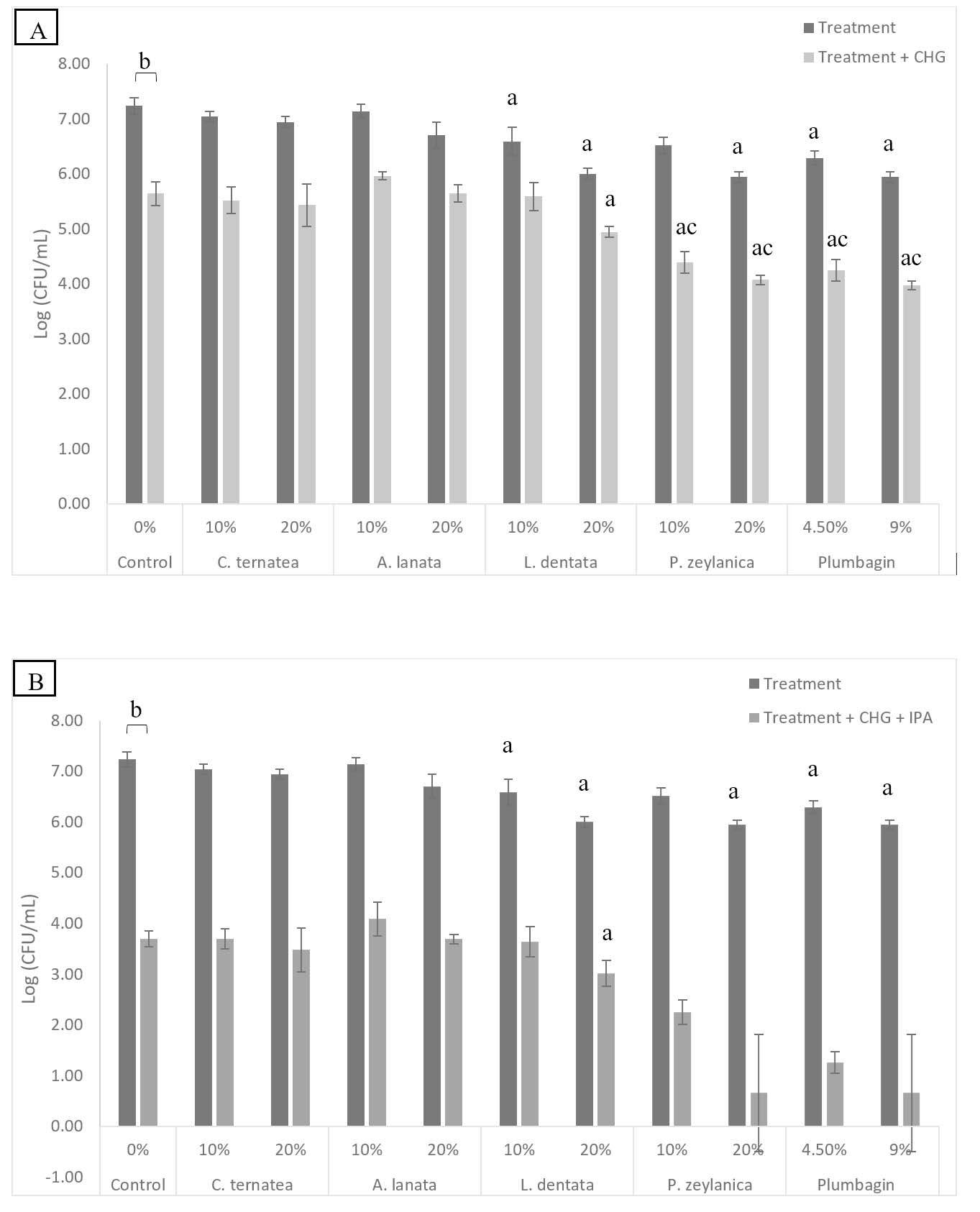
Figure 1. (A) Shows the anti-biofilm activity of 10% and 20% EOs, as well as 4.5% and 9% plumbagin, was tested alone and in combination with 2% CHG. (B) Shows the anti-biofilm activity of 10% and 20% EOs, as well as 4.5% and 9% plumbagin, was tested alone and in combination with CHG-IPA. EOs and plumbagin experiments were conducted in three independent replicates (n = 3). (a) represents P < 0.05, and (b) represents P < 0.0001 were used in the mixed-model analysis that was conducted for multiple comparisons. As determined by the coefficient of drug interactions, (c) represents that synergy was observed when combined with CHG. Treatments = Control or EO; Control = 8% Tween 80
The safety of blood products is a universal priority of blood centers. Bacterial contamination of PCs nonetheless remains a persistent challenge. Such contamination is frequently associated with skin flora such as S. epidermidis.2,10 Many bacteria, including S. epidermidis, live in biofilms: populations of microorganisms surrounded by a matrix. S. epidermidis biofilms have been observed between squamous epithelial cells, as deep as five cells (10-20 µm below the surface).34,35 CHG and IPA are often used in combination as a skin disinfectant; interestingly, Taha and colleagues found that while this combination can decrease the number of viable S. epidermidis biofilms, it does not totally eradicate them.16 Karpanen and his coworkers further reported that even after as much as 30 minutes of contact, 2% CHG penetrates less than 300 µm into the skin; however, bacteria can live at up to 1500 µm deep in hair follicles.35,36 These findings suggest a possible explanation for the recurrent bacterial contamination of PCs and blood products in spite of skin disinfection procedures.
Plant EOs are of considerable interest for their disinfecting properties and potential to enhance transdermal medication delivery.22-25,37 In addition, many EOs are known to have anti-biofilm properties; for example, eucalyptus oil can reduce viable biofilms of S. aureus, Escherichia coli, and S. epidermidis, and can even boost the effectiveness of chemical disinfectants such as CHG against biofilms.21,38 However, eucalyptus oil is also reported to cause skin irritation at concentrations as low as 1%, limiting its practical application.39 Identifying EOs that possess antibacterial activity alongside minimal skin irritation is critical for establishing better disinfectant methods.
This study examined EOs extracted from four plants native to Saudi Arabia, C. ternatea, A. lanata, L. dentata, and P. zeylanica, for their potential as anti-biofilm disinfectant agents. Initial assessments in a rabbit model demonstrated concentrations of up to 20% to be generally non-irritating, aligning with the “generally regarded as safe” classification of EOs and their oxygenated terpenoids by the US Food and Drug Administration.40 This low irritancy profile is very important for their possible use in disinfecting skin, especially in the context of sensitive operations like blood donation.19
Our findings align with existing research and the broad recognition of natural compounds, EOs, and plant extracts as having potent antimicrobial activities.22-25 In particular, the significant anti-biofilm activity of P. zeylanica EO observed in this work is consistent with reports that other EOs, such as eucalyptus oil, can reduce viable S. epidermidis biofilms and enhance the activity of chemical disinfectants.8,20,21 Likewise, our observation that higher oxygenated terpenoid content corresponds to greater activity against S. epidermidis biofilms corroborates previous studies.17-19,41 Finally, we also identified plumbagin as the predominant oxygenated terpenoid in P. zeylanica and observed it to have a synergistic anti-biofilm effect when combined with CHG, confirming a prior report.42 Taken together, these results highlight the potential of P. zeylanica and its active constituent plumbagin as valuable adjuncts to disinfection protocols aimed at minimizing bacterial contamination in blood products.
The complex chemical profile of EOs suggests that their biological activity often results from a delicate balance between major and minor constituents. In this study, the antagonistic interactions observed with C. ternatea, A. lanata, and L. dentata when combined with CHG highlight how minor components may interfere with or modulate the disinfectant’s efficacy. Conversely, the synergy observed in P. zeylanica may be facilitated by minor compounds acting in concert with plumbagin to enhance membrane permeability or inhibit biofilm matrix integrity, a phenomenon requiring further investigation.
While the findings of this study are promising, several limitations must be acknowledged. First, the in vitro nature of the anti-biofilm assays may not fully reflect the complexities of the human physiological environment. Similarly, the rabbit skin irritation model, while useful for initial safety screening, cannot be directly extrapolated to human skin responses. This is due to significant interspecies differences in skin structure, permeability, and immune response; consequently, human safety cannot be assumed based on these animal results alone. Most notably, this study relied exclusively on a single reference strain, S. epidermidis ATCC 35984. Although this is a robust model for studying biofilm-forming bacteria, it does not represent the clinical heterogeneity or the varied antimicrobial resistance profiles found in diverse clinical isolates. Consequently, these results should not be broadly extrapolated to all clinical settings. Future research should incorporate a diverse panel of clinically isolated strains, including multidrug-resistant isolates, to enhance the translational relevance and confirm the broad-spectrum efficacy of these EO-disinfectant combinations. Furthermore, while the CDI values indicate a positive interaction between P. zeylanica or plumbagin with CHG, these findings are based on single-concentration pairings rather than extensive dose response or ratio-dependent validation. Consequently, the observed effects should be viewed as suggestive of synergy within the specific parameters of this assay, and further research is required to determine the optimal ratios and concentration-dependent thresholds of these interactions.
Future studies should interrogate the synergistic interaction of P. zeylanica or plumbagin with CHG, utilizing checkerboard assays and dose-response modeling to fully characterize the concentration-dependent nature of this interaction across a broader range of ratios. In addition, it should be determined whether P. zeylanica EO or plumbagin might enhance the transdermal penetration of CHG-IPA in a blood donation scenario, as was previously reported for linalool, the main component of Lavandula multifida EO.19,43 Finally, rigorous in vivo studies in appropriate animal models must be conducted to validate the current in vitro findings, optimize formulations, and evaluate their safety and long-term effectiveness in the clinical setting. Given that CHG can cause skin reactions, further research on the allergenic potential of its combinations is warranted.
This study successfully identified P. zeylanica EO and its principal constituent, plumbagin, as highly promising and non-irritating agents that synergistically boost the anti-biofilm activity of CHG. This synergy provides a novel approach to overcome the failure of traditional disinfectants to eradicate deep resistant S. epidermidis biofilms and offering a viable pathway to significantly reduce bacterial contamination in platelet concentrates. Future research should focus on elucidating the molecular mechanisms of this powerful plumbagin-CHG synergy and determining if plumbagin enhances the transdermal penetration of CHG-IPA in vivo.
ACKNOWLEDGMENTS
The author would like to thank Ms. Bayan Abalkhail for data analysis and article comments as well as Imam Muhammad Ibn Saud University (IMSIU) for constant support. The author also appreciates the Chemistry College Mass Spectrometry Facility of King Saud University for gas chromatography-mass spectrometry (GC-MS).
FUNDING
None.
DATA AVAILABILITY
The datasets generated and/or analysed during the current study are available from the corresponding author on reasonable request.
ETHICS STATEMENT
This study was approved by the Ethics Review Committee (ERC), College of Medicine, Imam Mohammad Ibn Saud Islamic University, Riyadh, Saudi Arabia (Approval No. 23R/442/07, dated 12 November 2024).
- Corash L. Bacterial contamination of platelet components: potential solutions to prevent transfusion-related sepsis. Expert Rev Hematol. 2011;4(5):509-525.
Crossref - Ramirez-Arcos S, Goldman M. Bacterial contamination. In Popovsky MA, ed. Transfusion Reactions. 4th ed. American Association of Blood Banks (AABB) Press. 2012:153-189.
- Canadian Blood Services. Circular of information for the use of human blood components. 2015. https://www.blood.ca/sites/default/files/Pooled_Platelets_LR_CPD_Apheresis
_Platelets_0.pdf. Accessed June 24, 2025. - Jacobs MR, Smith D, Heaton WA, Zantek ND, Good CE, PGD Study Group. Detection of bacterial contamination in pre storage culture-negative apheresis platelets on day of issue with the Pan Genera Detection test. Transfusion. 2001;51(12):2573-2582.
Crossref - Muller B, Walther-Wenke G, Kalus M, et al. Routine bacterial screening of platelet concentrates by flow cytometry and its impact on product safety and supply. Vox Sang. 2015;108(3):209-218.
Crossref - Zhu L, Xu J, Yang X, et al. Detection of bacterial contamination of apheresis platelets in a Chinese blood center. Transfus Med. 2009;19(6):357-362.
Crossref - Dickson M, Dinesh D. Bacterial contamination of platelet concentrates produced in New Zealand. N Z Med J. 2013;126(1376):12-21.
- Karpanen TJ, Conway BR, Worthington T, Hilton AC, Elliott TSJ, Lambert PA. Enhanced chlorhexidine skin penetration with eucalyptus oil. BMC Infect Dis. 2010;10:278.
Crossref - Grice EA, Segre JA. The skin microbiome. Nat Rev Microbiol. 2011;9(4):244-253.
Crossref - de Korte D, Marcelis J. Platelet concentrates: reducing the risk of transfusion-transmitted bacterial infections. Int J Clin Transfus Med. 2014;2:29-37.
Crossref - Kou Y, Pagotto F, Hannach B, Ramirez-Arcos S. Fatal false-negative transfusion infection involving a buffy coat platelet pool contaminated with biofilm-positive Staphylococcus epidermidis: a case report. Transfusion. 2015;55(10):2384-2389.
Crossref - US Food and Drug Administration. Fatalities reported to FDA following blood collection and transfusion: annual summary for FY 2015; 2015. Accessed June 25, 2025. https://www.notifylibrary.org/sites/default/files/FDA%20Fatality%20Report-2015.pdf
- Weinstein RA, Milstone AM, Passaretti CL, Perl TM. Chlorhexidine: expanding the armamentarium for infection control and prevention. Clin Infect Dis. 2008;46(2):274-281.
Crossref - Mangram AJ, Horan TC, Pearson ML, Silver LC, Jarvis WR. Guideline for Prevention of Surgical Site Infection, 1999. Infection Control & Hospital Epidemiology. 1999;20(4):247-280.
Crossref - Benjamin RJ, Dy B, Warren R, Lischka M, Eder AF. Skin disinfection with a single-step 2% chlorhexidine swab is more effective than a two-step povidone-iodine method in preventing bacterial contamination of apheresis platelets. Transfusion. 2011;51(3):531-538.
Crossref - Taha M, Kalab M, Yi QL, et al. Biofilm-forming skin microflora bacteria are resistant to the bactericidal action of disinfectants used during blood donation. Transfusion. 2014;54(11):2974-2982.
Crossref - Khwaza V, Aderibigbe BA. Antibacterial Activity of Selected Essential Oil Components and Their Derivatives: A Review. Antibiotics. 2025; 14(1):68.
Crossref - Aboulwafa MM, Mostafa NM, Youssef FS, Eldahshan OA, Singab ANB. Lavandula dentata leaves as potential natural antibiofilm agents against Pseudomonas aeruginosa. Scientific Reports. 2025;15(1):8540.
Crossref - Alabdullatif M, Boujezza I, Mekni M, et al. Enhancing blood donor skin disinfection using natural oils. Transfusion. 2017;57(12):2920-2927.
Crossref - Casey AL, Karpanen TJ, Conway BR, et al. Enhanced chlorhexidine skin penetration with 1,8-cineole. BMC Infect Dis. 2017;17(1):350.
Crossref - Hendry ER, Worthington T, Conway BR, Lambert PA. Antimicrobial efficacy of eucalyptus oil and 1,8-cineole alone and in combination with chlorhexidine digluconate against microorganisms grown in planktonic and biofilm cultures. J Antimicrob Chemother. 2009;64(6):1219-1225.
Crossref - Sathiaseelan A, Song KP, Tan HS, Choo WS. Antibiofilm activity of Clitoria ternatea flowers anthocyanin fraction against biofilm-forming oral bacteria. FEMS Microbiol Lett. 2025;372.
Crossref - Qamer S, Siddiqui N, Shafqat R. Antibacterial activity of methanolic crude extracts from Aerva lanata (L.) A.L. Juss. ex Schultes against Staphylococcus aureus and Escherichia coli using in vitro bladder model for catheter-associated urinary tract infection. Ann Phytomed. 2023;12(2):630-637.
Crossref - Rocha CT, de Lima PMN, Pereira TC, et al. Antibacterial and antibiofilm effect of Lavandula dentata L. essential oil as endodontic irrigate against standard and clinical strains of Enterococcus spp. Appl Sci. 2024;15(10):5534.
Crossref - Qais FA, Ahmad I, Husain FM, et al. Interference of quorum sensing regulated bacterial virulence factors and biofilms by Plumbago zeylanica extract. Microscopy Research and Technique. 2021;84(12):3150-3160.
Crossref - Fernández-Calderón MC, Fernández-Babiano I, Navarro-Pérez ML, Pazos-Pacheco C, Calvo-Cano A. Biofilm formation and role of other pathogenic factors in the virulence of Staphylococcus epidermidis clinical isolates. Front Cell Infect Microbiol. 2025;15:1630341.
Crossref - Alabdullatif M, Atreya CD, Ramirez-Arcos S. Antimicrobial peptides: an effective approach to prevent bacterial biofilm formation in platelet concentrates. Transfusion. 2018;58(8):2013-2021.
Crossref - Kampf G, Shaffer M, Hunte C. Insufficient neutralization in testing a chlorhexidine-containing ethanol-based hand rub can result in a false positive efficacy assessment. BMC Infect Dis. 2005;5(1):48.
Crossref - ASTM International. Standard Test Methods for Evaluation of Inactivators of Antimicrobial Agents. ASTM International. 2008:E1054-08.
- US Environmental Protection Agency. Health Effects Test Guidelines: OPPTS 870.2500 Acute Dermal Irritation. EPA.1998.
- Chou TC, Talalay P. Quantitative analysis of dose-effect relationships: the combined effects of multiple drugs or enzyme inhibitors. Adv Enzyme Regul. 1984;22:27-55.
Crossref - Zhao Y, Gao JL, Ji JW, et al. Cytotoxicity enhancement in MDA-MB-231 cells by the combination treatment of tetrahydropalmatine and berberine derived from Corydalis yanhusuo W. T. Wang. J Intercult Ethnopharmacol. 2014;3(2):68-72.
Crossref - Yap PSX, Yiap BC, Ping HC, Lim SHE. Essential oils, a new horizon in combating bacterial antibiotic resistance. Open Microbiol J. 2014;8(1):6-14.
Crossref - Mishra R, Panda AK, De Mandal S, Shakeel M, Bisht SS, Khan J. Natural anti-biofilm agents: strategies to control biofilm-forming pathogens. Front Microbiol. 2020;11:566325.
Crossref - Berrow M, Starr NJ, Scurr DJ, de Cogan F. Enhanced topical antimicrobial delivery for improved skin antisepsis. J Pharm Sci. 2026;115(1):104258.
Crossref - Duffy HR, Ashton NN, Stulce P, et al. Skin-dwelling bacteria survive preoperative skin preparation in reconstruction surgery. J Clin Med. 2025;14(10):3417.
Crossref - Herman A, Herman AP. Essential oils and their constituents as skin penetration enhancer for transdermal drug delivery: a review. J Pharm Pharmacol. 2015;67(4):473-485.
Crossref - Karpanen TJ, Worthington T, Hendry ER, Conway BR, Lambert PA. Antimicrobial efficacy of chlorhexidine digluconate alone and in combination with eucalyptus oil, tea tree oil and thymol against planktonic and biofilm cultures of Staphylococcus epidermidis. J Antimicrob Chemother. 2008;62(5):1031-1036.
Crossref - Vilaplana J, Romaguera C. Allergic contact dermatitis due to eucalyptol in an anti-inflammatory cream. Contact Dermatitis. 2000;43(2):118.
- Vaddi HK, Ho PC, Chan SY. Terpenes in propylene glycol as skin-penetration enhancers: permeation and partition of haloperidol, Fourier transform infrared spectroscopy, and differential scanning calorimetry. J Pharm Sci. 2002;91(7):1639-1651.
Crossref - Bassole IH, Juliani HR. Essential oils in combination and their antimicrobial properties. Molecules. 2012;17(4):3989-4006.
Crossref - Liu H, Chen H, Ma Z, et al. Plumbagin enhances antimicrobial and anti-biofilm capacities of chlorhexidine against clinical Klebsiella pneumoniae while reducing resistance mutations. Microbiol Spectr. 2024;12(5):e00896-24.
Crossref - Jager W, Buchbauer G, Jirovetz L, et al. Percutaneous absorption of lavender oil from a massage oil. J Soc Cosmet Chem. 1992;43(1):49-54.
© The Author(s) 2026. Open Access. This article is distributed under the terms of the Creative Commons Attribution 4.0 International License which permits unrestricted use, sharing, distribution, and reproduction in any medium, provided you give appropriate credit to the original author(s) and the source, provide a link to the Creative Commons license, and indicate if changes were made.