


ISSN: 0973-7510
E-ISSN: 2581-690X
Escherichia coli is a key indicator of faecal contamination and a reservoir of antimicrobial resistance genes (ARGs) in aquatic environments. Monitoring its environmental prevalence and resistance patterns in water sources is essential for public health and environmental safety. In this study, a total of 300 water samples were collected from diverse sources. E. coli were isolated from 32% samples, with the highest prevalence in river water (72.72%) and the lowest in tap water (10%). All isolates were positive for the yaiO gene. Complete (100%) resistance was observed against ampicillin-sulbactam and polymyxin-B, while high resistance was displayed by isolates against Tigecycline (94.6%), ciprofloxacin (94.4%), colistin (94.4%), and amikacin (94.4%). Comparatively, higher sensitivity was noted to azithromycin (46%) and meropenem (41.1%). PCR analysis detected tetA/tetB genes in 37.5% and dfrA in 20.83% of isolates, with significant variation in gene distribution across water sources. The study revealed a substantial prevalence of multidrug-resistant E. coli in surface and groundwater sources, posing a potential risk to human and animal health. The presence of ARGs highlights the aquatic environment as a reservoir for resistance determinants, underscoring the need for regular water quality surveillance, improved sanitation, and prudent antibiotic stewardship within a One Health framework.
Escherichia coli, Water Contamination, Antimicrobial Resistance, Multidrug-resistance, One Health
Antimicrobial resistance (AMR) constitutes a significant global health challenge, impacting human, animal, and environmental health. Among bacterial species, Escherichia coli plays a pivotal role due to its capacity to acquire and disseminate resistance to multiple antibiotic classes, including β-lactams, fluoroquinolones, aminoglycosides, and tetracyclines.1 Although typically a commensal organism within the intestinal microbiota, pathogenic strains of E. coli are capable of causing urinary tract, gastrointestinal, and systemic infections.2,3 Furthermore, E. coli serves as a sentinel indicator of fecal contamination in aquatic environments.
The waterborne transmission of E. coli poses a substantial public health risk, particularly in low-resource settings where inadequate sanitation and limited access to safe drinking water facilitate the spread of resistant bacteria. Contaminated water not only acts as a conduit for human exposure but also serves as a reservoir for resistance determinants that can be horizontally transferred to other bacterial pathogens.4,5 Environmental contamination is further exacerbated by anthropogenic activities, including agricultural runoff, wastewater discharge, and inadequate sanitation infrastructure, all of which introduce antibiotics and resistant bacteria into surface and groundwater.6,7 Such water systems function as ecological hotspots for the selection, maintenance, and dissemination of antimicrobial resistance.
The mechanisms by which E. coli acquires resistance are diverse and frequently involve horizontal gene transfer mediated by plasmids, integrons, and transposons.8 These mobile genetic elements facilitate rapid adaptation and the spread of resistance genes across bacterial communities. The selective pressure exerted by widespread antibiotic use in healthcare and agriculture, coupled with the persistence of antibiotics in aquatic ecosystems, accelerates the emergence of resistant strains.9,10 Consequently, monitoring E. coli in environmental waters provides critical insights into both fecal pollution and the broader dynamics of AMR transmission within the community.
Advancements in molecular techniques have enhanced the detection of resistance genes in environmental isolates, thereby improving the understanding of AMR dissemination and informing surveillance strategies.11 Given the increasing role of aquatic ecosystems in the AMR burden, this study aimed to isolate and characterize E. coli from different water sources, determine their antimicrobial resistance profiles, and detect selected resistance genes. The findings are anticipated to support environmental AMR surveillance and guide water quality management and promote responsible stewardship efforts.
Sampling of water
A total of 300 water samples were collected from different locations across Uttar Pradesh, India. Samples of 10-15 ml were collected in sterile centrifuge tubes and transported to the Veterinary Microbiology laboratory under cold-chain condition for microbiological analysis. The sampling locations were selected across different localities, representing seven types of water sources: tap water, streams, ponds, rivers, handpumps, dams and wells. These sites were identified after several preliminary visits to various communities. Details of samples collected are given in Table 1.
Table (1): Details of water samples collected in the present study
No. |
Source of water sampling |
No. of samples collected |
|---|---|---|
1. |
Tap water |
40 |
2. |
Stream |
42 |
3. |
Pond |
93 |
4. |
River |
22 |
5. |
Handpump |
32 |
6. |
Dam |
17 |
7. |
Well |
54 |
Total |
300 |
Standard bacterial culture
Standard strain of E. coli ATCC 25922 procured from HiMedia (India) was used as a reference strain for the present study.
Bacterial Culture
Pre-enrichment of water samples
Water samples were mixed with pre-enrichment broth, Buffered Peptone water (BPW, 2% w/v) (HiMedia, India), at a 1:10 dilution (1 ml of sample + 9 ml of BPW) to provide a suitable environment to help stressed or low-population bacteria recover and multiply before being transferred to a more selective medium. The mixture in tubes were incubated in a shaking incubator for 12-18 hrs at 37 °C for pre-enrichment.
Isolation and characterization of E. coli
A loopful of each pre-enriched water sample was streaked onto MacConkey Lactose Agar (MLA, HiMedia, India) and incubated aerobically at 37 °C for 18-24 hrs to encourage the selective growth of Gram-negative bacteria like E. coli. MLA is a selective and differential medium which selectively inhibit the growth of Gram-positive bacteria and supports the growth of Gram-negative lactose fermenting bacteria only. Pink colonies on MLA were further sub-cultured on Eosin Methylene Blue (EMB) agar for the development of greenish-black colonies with a characteristic metallic sheen, typical feature of E. coli.12 Morphological, cultural, and bio-chmemical tests were carried out for confirmation of E. coli isolates, such as Gram staining (shape, size, arrangement of colonies), catalase, oxidase, IMViC (Indole, Methyl red, Voges Proskauer, Citrate utilization test), Nitrate reduction, Urease, and sugar fermentation (glucose, lactose, sucrose, maltose) tests. Isolates showing Gram-negative rods, positive for catalase, indole, and methyl red, and negative for oxidase, Voges-Proskauer, citrate, and urease were identified as pure colonies of E. coli and preserved on nutrient agar slants at 4 °C for future analysis.
DNA extraction (snap chill method)
The snap-chilling method was used to extract bacterial DNA from E. coli.13 Isolates were incubated in 5 ml Brain Heart Infusion (BHI) broth at 37 °C overnight with constant shaking. 1 ml of the broth culture was centrifuged at 8000 rpm for 10 min at 4 °C. The bacterial pallet was washed three times with sterile normal saline solution (0.85% NaCl) and resuspended in 300 µl nuclease-free sterile distilled water. The bacterial suspension was boiled for 5-10 mins in a boiling water bath followed by immediate chilling for 10 min and then lysate was centrifuged at 5000 rpm for 5 min. The supernatant containing crude DNA was collected and stored at -20 °C. The purity and concentration of DNA were checked using a Nanodrop and gel electrophoresis.
PCR confirmation of E. coli
All the isolates of E. coli, identified by both cultural and biochemical characterization, were confirmed by PCR targeting the yaiO gene, a highly conserved and species-specific molecular maker for E. coli14 and the primer sequences used were F: TGATTTCCGTGCGTCTGAATG and R: ATGCTGCCGTAGCGTGTTTC. Agarose gel electrophoresis (1.5% agarose in 1X TAE buffer) was used to evaluate the amplified PCR products. It was run at 80 V/cm for 45 min and stained with ethidium bromide (0.5 µg/ml). PCR product of each sample (5-10 µl) was loaded in individual wells of gel alongside a positive control. The amplicon size was determined by a 100 bp DNA ladder as a molecular weight marker. The products were visualized under a UV transilluminator and documented using a gel documentation system (AlphaImager).
Antibiotic sensitivity test
All the pure culture isolates of E.coli were subjected to in vitro antibiotic sensitivity testing by disc diffusion method15 against 22 commonly used antibiotics for E. coli (Gentamycin, Ampicillin sulbactam, Doxycycline, Azithromycin, Ofloxacin, Amikacin, Nitrofurantoin, Tetracycline, Trimethoprim, Sulphafurazole, Aztreonam, Co-trimoxazole, Fosfomycin, Enrofloxacin, Polymyxin B, Colistin, Tigecycline, Levofloxacin, Ciprofloxacin, Meropenem, Cefepime, Imipenem, and Amoxycillin clavulanic acid) following CLSI guidelines. E. coli ATCC 25922 culture served as quality control strain. Fresh colonies of E. coli were suspended in sterile normal saline (0.85% NaCl) and adjusted to a turbidity equivalent to 0.5 McFarland standard (approximately 1.5 × 10⁸ CFU/mL). A sterile cotton swab was used to uniformly inoculate the suspension onto Mueller-Hinton agar (MHA) plates, ensuring complete coverage of the agar surface. After drying the plates for 3-5 minutes at room temperature, antibiotic-impregnated disks (HiMedia, India) were placed on the agar surface using sterile forceps and incubated at 37 °C for 18-24 hrs. Results were recorded after 18-24 hrs incubation and interpreted as sensitive, intermediate, or resistant according to CLSI standards.16
Multiplex PCR for detection of tetA and tetB gene
All the E. coli isolates were screened by multiplex PCR assay for tetA and tetB genes17 with suitable modifications. PCR was carried out in a 0.2 ml thin wall PCR tubes with final volume of 25 µl reaction mixture with PCR cycling conditions listed in Table 2. Agarose gel electrophoresis (1% agarose in 1X TAE buffer) was used to evaluate the amplified PCR products. The gel was run at 80 V/cm for 45 min and stained with ethidium bromide (0.5 µg/ml). PCR product of each sample (5-10 µl) was loaded in individual wells of gel alongside a positive control. The amplicon size was determined by a 1 kb DNA ladder as a molecular weight marker. The products were visualized under a UV transilluminator and documented using a gel documentation system (AlphaImager).
PCR for detection of dfrA gene
For the dfrA gene detection, conventional PCR was carried out in all E. coli isolates17 with PCR cycling condition listed in Table 2. Rest of the procedure was same as used for tetA and tetB detection and the amplicon size was determined by a 100 bp DNA ladder as the molecular weight marker.
Table (2): PCR cycling conditions for detection of tetA, tetB and dfrA gene of E. coli
Target gene |
Primer sequence |
PCR product size |
PCR conditions |
PCR reaction volume (25 µl) |
|---|---|---|---|---|
tetA tetB |
F: 5`-GGTTCACTCGAACGACGTCA-3` R: 5`-CTGTCCGACAAGTTGCATGA -3` F: 5`-CCTCAGCTTCTCAACGCGTG -3` R: 5` -GCACCTTGCTGATGACTCTT -3` |
577 bp
634 bp |
1. First cycle (95 °C for 3 min), 2. Subsequent 35 cycles of Denaturation (94 °C for 1 min), Annealing (56° C for 1 min), & Extension (72°C for 1 min) 3. Finalextension(72 °C for 10 min) |
12.5 µl of PCR master mix (1X, Thermo fisher) 10 picomole/µl of each primers forward and reverse, 4.5 µl DNA template |
dfrA |
F: 5`-GGAGTGCCAAAGGTGAACAGC -3` R: 5 -GAGGCGAAGTCTTGGGTAAAAAC -3` |
367 bp |
1. First cycle (95 °C for 3 min), 2. Subsequent 35 cycles of Denaturation (94 °C for 1 min), Annealing (45° C for 1.5 min), & Extension (72°C for 1 min) 3. Final extension (72 °C for 10 min) |
12.5 µl of PCR master mix (1X, Thermo fisher) 10 picomole/µl of each primers forward and reverse, 4.5 µl DNA template |
Statistical analysis
The data on the prevalence of E. coli across water sources, distribution of resistance genes (tetA/tetB, dfrA), and antibiotic susceptibility profiles were analyzed using the Chi-square (χ²) test of independence. Fisher’s exact test was applied where expected frequency were £5. Statistical analysis was performed using SPSS version 26 (IBM, USA). A P-value <0.05 was considered statistically significant.
Isolation and Identification of E. coli
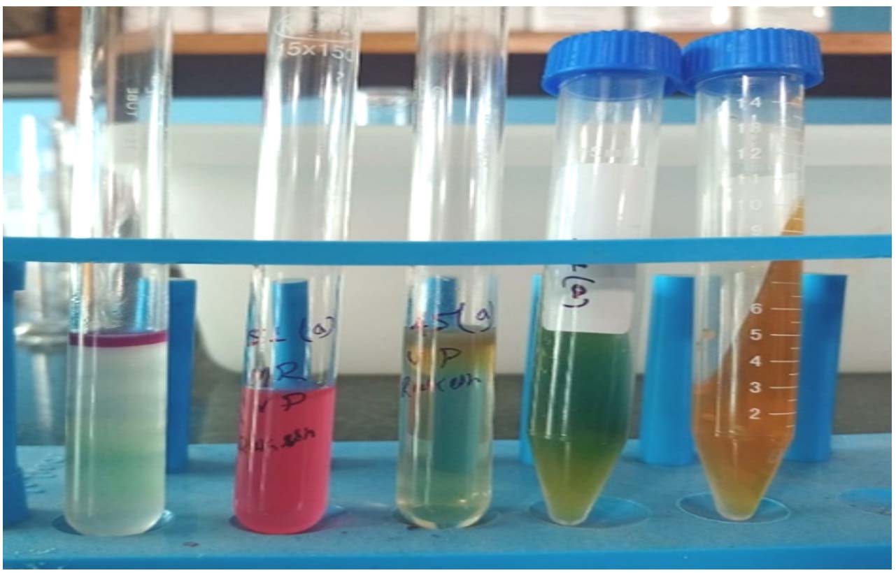
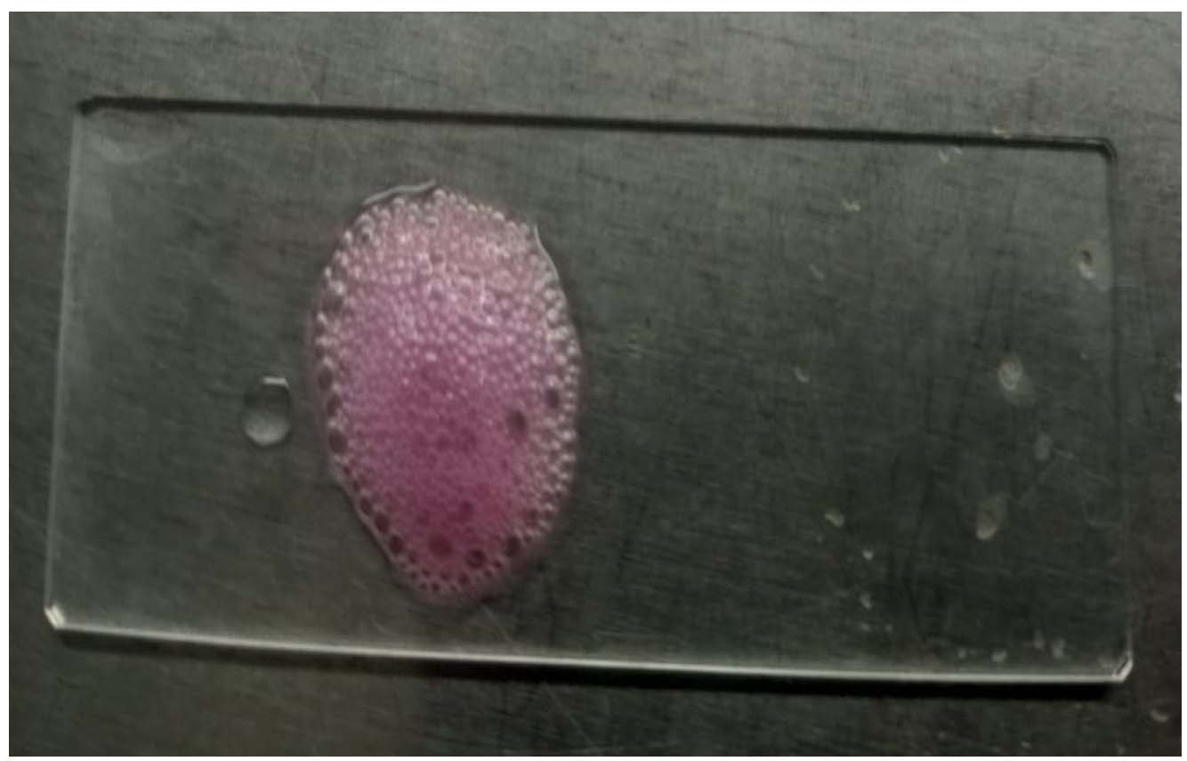
Morphological characterization using Gram staining revealed Gram-negative rod-shaped bacteria in scattered arrangement (Figure 1). Cultural characterization of isolates revealed pink lactose-fermenting colonies on MLA and a greenish metallic sheen on EMB agar (Figures 2a, 2b). TSI (Triple Sugar Iron) agar slant inoculation showed fermentation of all the three sugars; glucose, lactose, and sucrose with the production of gas turning the media to yellow colour completely. Biochemical characterization revealed the isolates positive for catalase, Indole, Methyl red, and nitrate reduction, whereas they tested negative for oxidase, Voges-Proskauer, citrate utilization, and Urease (Figures 3a, 3b). These morphological and biochemical characteristics confirmed a total of 96 isolates as E. coli recovered from 300 water samples.
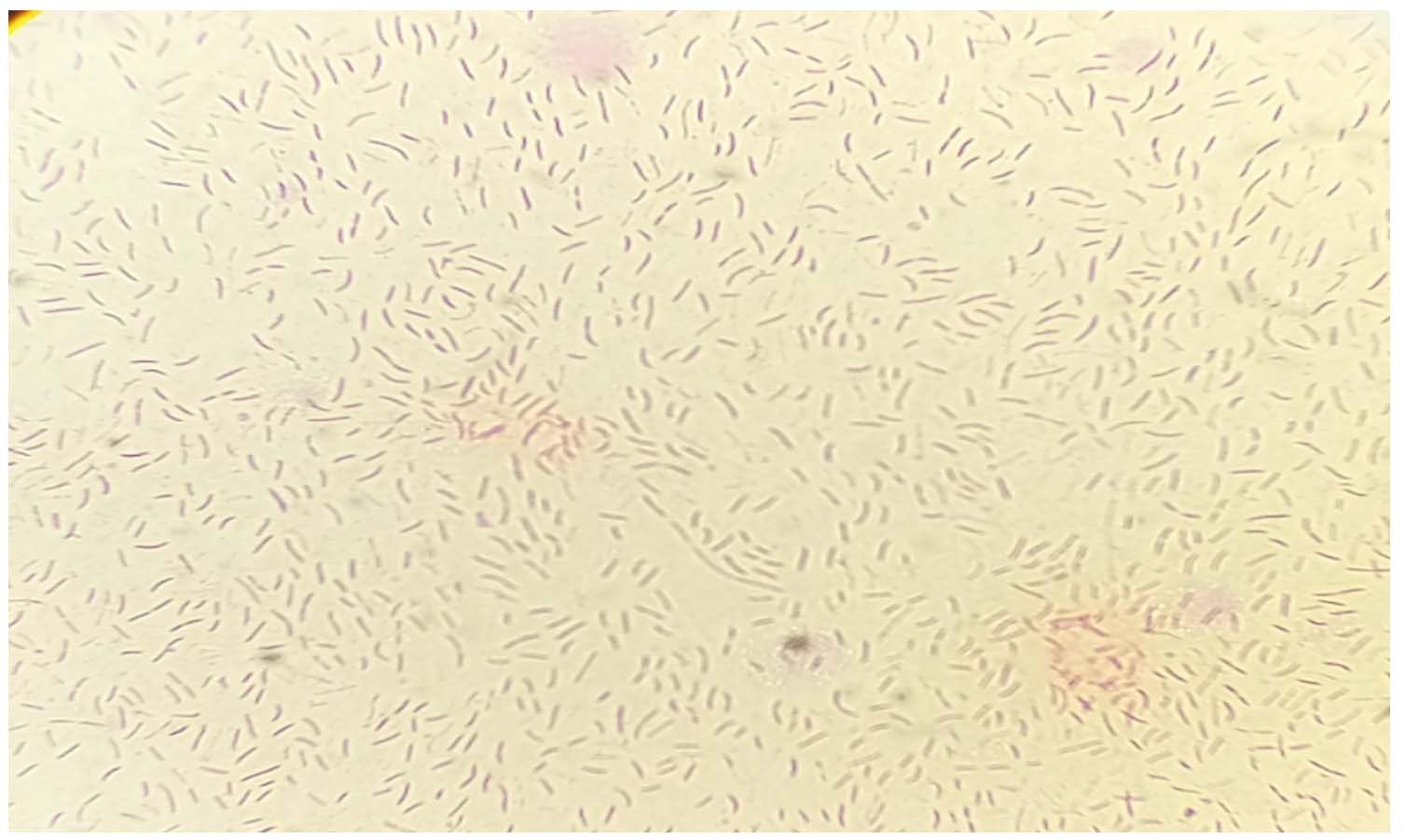
Figure 1. Gram staining of E. coli exhibiting pink coloured, rod shaped bacteria in scattered arrangement
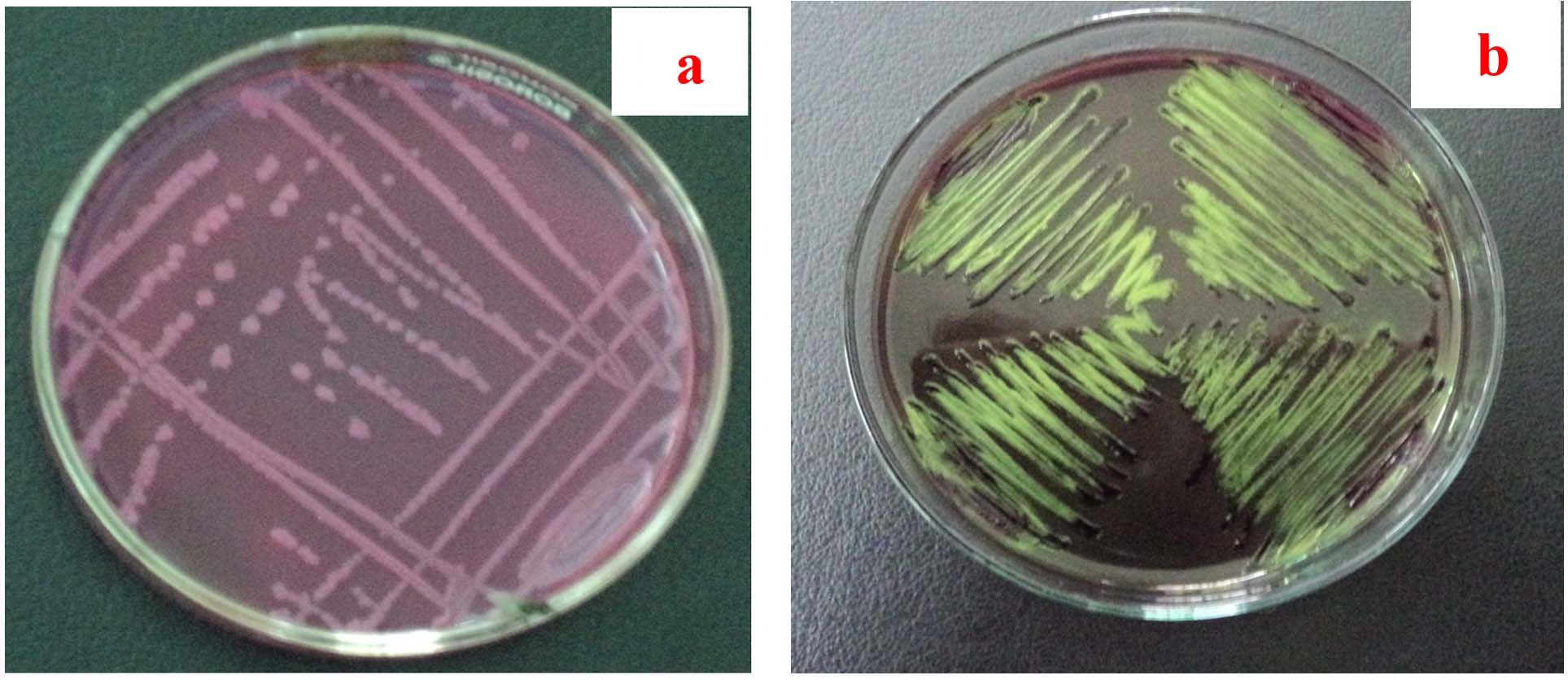
Figure 2. (a) E. coli exhibiting pink colored lactose fermenting colony on MLA; (b) E. coli exhibiting greenish metallic sheen colony on EMB agar
(a)
 (b)
(b)
Figure 3. (a) IMViC test of E. coli with indole positive, MR positive, VP negative and Citrate utilization test negative results. TSI slant exhibiting all 3 sugars fermentation; (b) Catalase test positive result for E. coli
Prevalence of E. coli in water samples
A total of 96 (32%) E. coli isolates were recovered from 300 water samples collected from different sources (Table 3). The prevalence varied across water sources: the highest was in river water (72.7%), followed by ponds (43.0%), and canals (35.7%), while the lowest was observed in hand pump (12.5%) and tap water (10%) samples. The results indicate higher contamination levels in surface water compared to treated or groundwater sources.
Table (3): Percentage prevalence of E. coli isolates in water samples collected from different sources
No. |
Source of water sampling |
No. of samples collected |
No. of E. coli isolated |
Prevalence (%) |
|---|---|---|---|---|
1 |
Tap water |
40 |
04 |
10.0 |
2 |
Stream |
42 |
15 |
35.71 |
3 |
Pond |
93 |
40 |
43.01 |
4 |
River |
22 |
16 |
72.72 |
5 |
Hand pump |
32 |
04 |
12.5 |
6 |
Dam |
17 |
05 |
29.41 |
7 |
Well |
54 |
12 |
22.22 |
Total |
300 |
96 |
32 |
Molecular confirmation of E. coli by detection of yaiO gene
All the 96 isolates of E. coli identified through cultural and biochemical characterization were further confirmed as E. coli (96/96 = 100%) by PCR targeting yaiO gene, which is a species-specific marker gene for identification of E. coli (Figure 4).
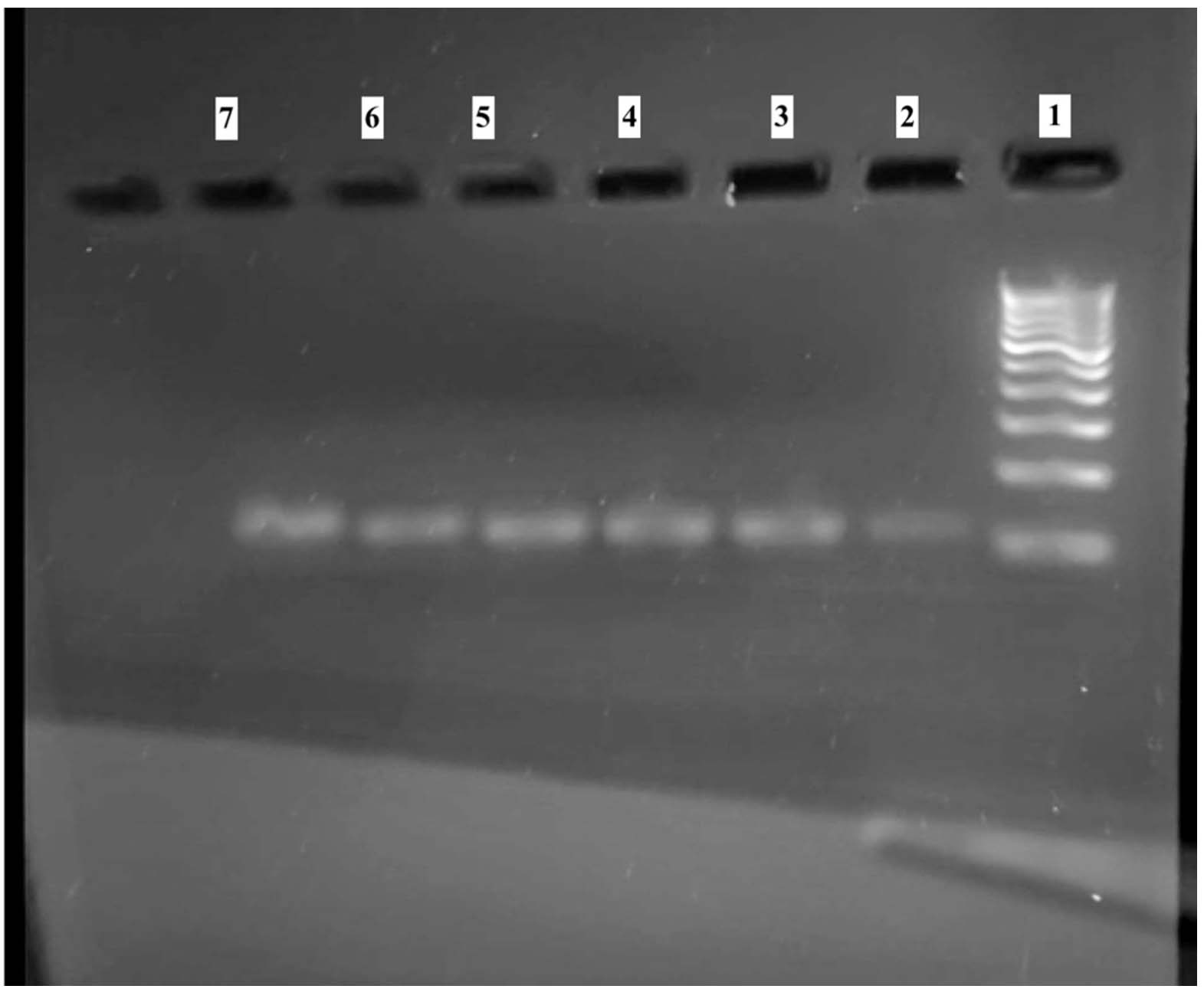
Figure 4. PCR amplification of yaiO gene
Lane 1: 100 bp DNA ladder
Lane 2-7: yaiO positive sample (amplicon size 115 bp)
Antibiotics sensitivity test
Antibiotic sensitivity testing of all the 96 E. coli isolates using Kirby-Bauer method against 22 commonly used antibiotics revealed varying resistance patterns (Table 4). The highest sensitivity was exhibited to Azithromycin (46%), followed by Meropenem (41.1%), Co-trimoxazole (36.8%), Sulphafurazole (36.3%), Imipenem (30.4%), Gentamycin (28.57%), Ofloxacin (28.5%), Cefepime (21%), Levofloxacin (20%), Fosfomycin (17.3%), Aztreonam (11.1%), Enrofloxacin (5.5%), Tigecycline (5.4%) and Doxycycline (1.3%). On the other hand, complete resistance (100%) was exhibited against Ampicillin sulbactam and Polymixin-B, followed by Tigecycline (94.6%), Ciprofloxacin (94.4%), Colistin (94.4%), Enrofloxacin (94.4%), Amikacin (94.4%), Azteronam (88.8%), Amoxycillin clavulinic (80%), Nitrofurantoin (70.5%), Azithromycin (68.4%), Fosfomycin (60.8%), Co-trimoxazole (52.63%), Levofloxacin (50%), Trimethoprim (50%), Cefepime (47.8%), Ofloxacin (47.6%), Imipenem (43.4%), Gentamycin (42.8%), Sulphafurazole (36.36%), Meropenem (35.2%), and Doxycycline (33.3%) (Figure 5). These results display widespread multidrug-resistance (MDR), with more than 80% showing resistance to three or more antibiotics.
Table (4): Result of antibiotic sensitivity testing by disc diffusion method
No. |
Name of antibiotics |
Disc concen. |
Sensitive |
Intermediate |
Resistance |
|---|---|---|---|---|---|
1. |
Ampicillin Sulbactam (A/S) |
10/10 mcg |
– |
– |
100% |
2. |
Polymyxin-B (PB) |
300 units |
– |
– |
100% |
3. |
Tigecycline (TGC) |
15 mcg |
5.4% |
– |
94.6% |
4. |
Amikacin (AK) |
30 mcg |
– |
5.5% |
94.4% |
5. |
Enrofloxacin (EX) |
10 mcg |
5.5% |
– |
94.4% |
6. |
Ciprofloxacin (CIP) |
5 mcg |
5.5% |
– |
94.4% |
7. |
Aztreonam (AT) |
30 mcg |
11.11% |
– |
88.8% |
8. |
Amoxycillin Clavulanic acid (AMC) |
30 mcg |
– |
20% |
80% |
9. |
Nitrofurantoin (NIT) |
2 mcg |
– |
29.4% |
70.5% |
10. |
Azithromycin (AZM) |
15 mcg |
46% |
– |
68.4% |
11. |
Fosfomycin (FO) |
50 mcg |
17.3% |
21.7% |
60.8% |
12. |
Co-trimoxazole (COT) |
25 mcg |
36.8% |
10.5% |
52.63% |
13. |
Tetracycline (TR) |
30 mcg |
16% |
33.3% |
50% |
14. |
Levofloxacin (LE) |
5 mcg |
20% |
30% |
50% |
15. |
Trimethoprim (TE) |
5 mcg |
– |
50% |
50% |
16. |
Cefepime (CPM) |
50 mcg |
21% |
31.2% |
47.8% |
17. |
Ofloxacin (OF) |
5 mcg |
28.5% |
23.8% |
47.6% |
18. |
Imipenem (IPM) |
10 mcg |
30.4% |
26.2% |
43.4% |
19. |
Gentamycin (GEN) |
10 mcg |
28.57% |
28.57% |
42.8% |
20. |
Sulphafurazole (SF) |
300 mcg |
36.3% |
27.2% |
36.36% |
21. |
Meropenem (MRP) |
10 mcg |
41.1% |
23.7% |
35.2% |
22. |
Doxycycline (DO) |
30 mcg |
1.3% |
58.33% |
33.3% |
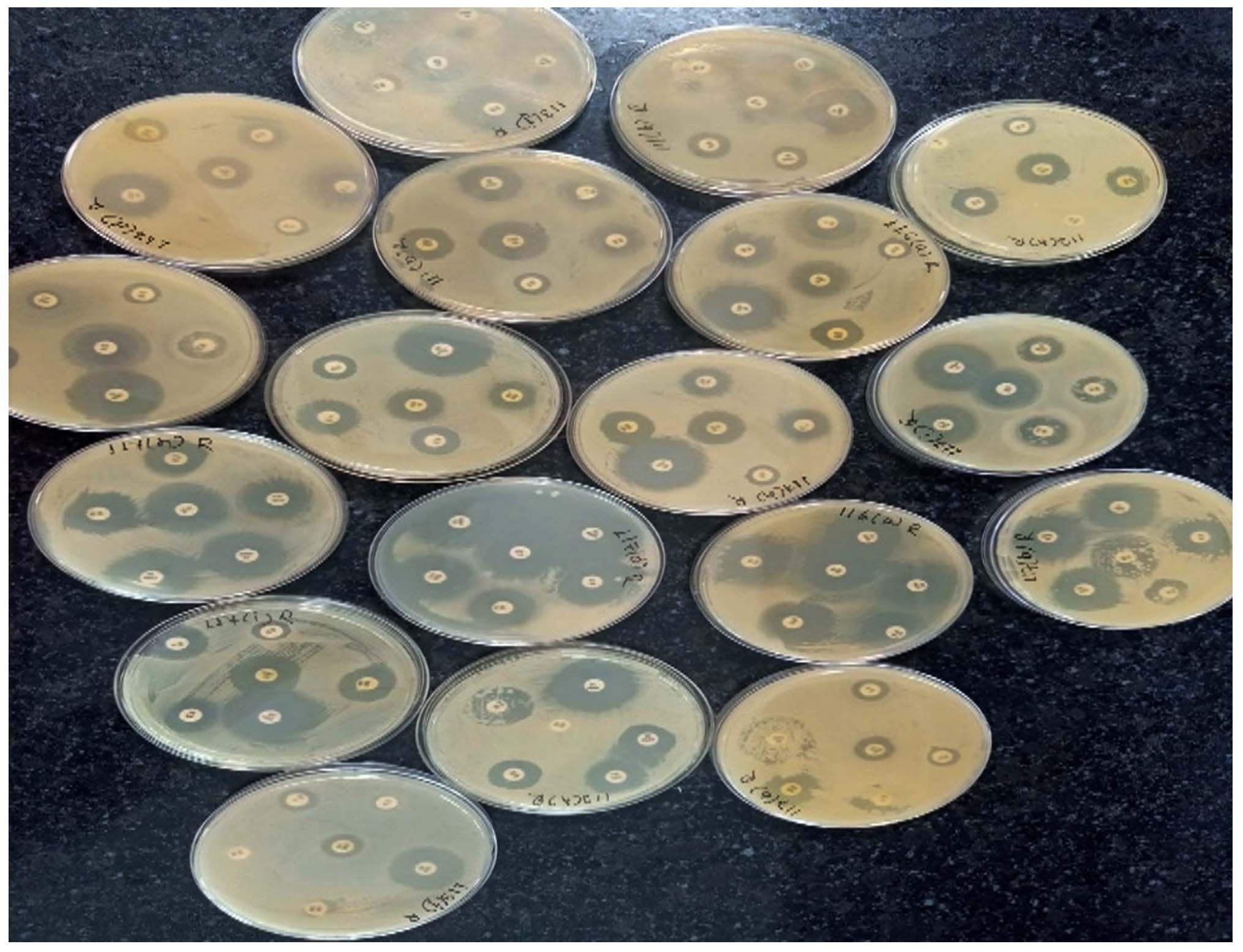
Figure 5. MHA plates showing different sensitivity and resistance pattern in ABST test result
Multidrug-resistance profile of E. coli isolates from different sources
A total of 13 E. coli isolates exhibited diverse multidrug-resistance (MDR) profiles, with resistance ranging from 3-7 antimicrobial classes (Table 5). The majority of isolates showed resistance to ≥5 antibiotic classes, indicating a high burden of MDR. Resistance was most frequently observed against ampicillin–sulbactam, ciprofloxacin, amikacin, polymyxin B, colistin, and tigecycline, while comparatively fewer isolates exhibited resistance to tetracycline, co-trimoxazole, sulphafurazole, and nitrofurantoin. Isolates from river and pond sources demonstrated higher resistance complexity, with some exhibiting resistance to up to 7 antibiotic classes, highlighting substantial variability and environmental influence on resistance patterns.
Table (5): Multidrug-resistance profiles of E. coli isolates from water sources
Isolate ID |
Source |
No. of Antibiotic Classes Resistant |
Resistant Antibiotics |
|---|---|---|---|
EC-01 |
Tap water |
5 |
A/S, CIP, AK, TR, TGC, PB |
EC-02 |
Stream |
5 |
A/S, CIP, AK, TE, COT |
EC-03 |
Stream |
6 |
A/S, CIP, AK, PB, COL, TGC |
EC-04 |
Stream |
3 |
TR, COT, SF |
EC-05 |
Pond |
4 |
CIP, LE, OF, EX |
EC-06 |
Pond |
5 |
CIP, OF, LE, AK, TE |
EC-07 |
Pond |
5 |
CIP, OF, LE, EX |
EC-08 |
River |
6 |
A/S, PB, COL, CIP, AK, AMC |
EC-09 |
River |
7 |
A/S, CIP, AK, PB, COL, EX, TGC |
EC-10 |
River |
4 |
TE, COT, SF, NIT |
EC-11 |
River |
3 |
CIP, LE, OF |
EC-12 |
Dam |
5 |
A/S, COL, CIP, TR |
EC-13 |
Well |
5 |
A/S, AK, PB, COL, TGC |
A/S: Ampicillin Sulbactam, CIP: Ciprofloxacin, AK: Amikacin, TR: Tetracycline, PB: Polymyxin-B, TE: Trimethoprim, COT: Co-trimoxazole, COL: Colistin, TGC: Tigecycline, SF: Sulphafurazole, LE: Levofloxacin, OF: Ofloxacin, EX: Enrofloxacin, AMC: Amoxycillin clavulanic acid, NIT: Nitrofurantoin
Detection of antibiotic resistance genes
Detection of tetA and tetB genes
Multiplex PCR screening of 96 E. coli isolates, using tetA and tetB specific primers, revealed that 37.5% isolates (36/96) were positive for tetA and tetB genes (Table 6, Figure 6) and all of these isolates were phenotypically resistant to tetracycline. These genes encode tetracycline efflux pumps, which confer resistance by actively pumping tetracyclines out of the bacterial cell.
Table (6): Multiplex PCR result for detection of tetA, tetB, and dfrA genes in E. coli
Source of sample collection |
No. of samples collected |
No. of E. coli isolated |
No. of isolates positive for tetA gene |
No. of isolates positive for tetB gene |
No. of isolates positive for tetA & tetB gene |
No. of E. coli isolate positive for dfrA gene |
|---|---|---|---|---|---|---|
Tap water |
40 |
04 |
01 |
0 |
0 |
0 |
Canal |
42 |
15 |
04 |
02 |
01 |
03 |
Pond |
93 |
40 |
09 |
05 |
3 |
10 |
River |
22 |
16 |
03 |
02 |
01 |
04 |
Hand pump |
32 |
04 |
01 |
0 |
0 |
0 |
Dam |
17 |
05 |
01 |
01 |
0 |
01 |
Well |
54 |
12 |
04 |
03 |
02 |
02 |
Total |
300 |
96 |
23 |
13 |
07 |
20 |
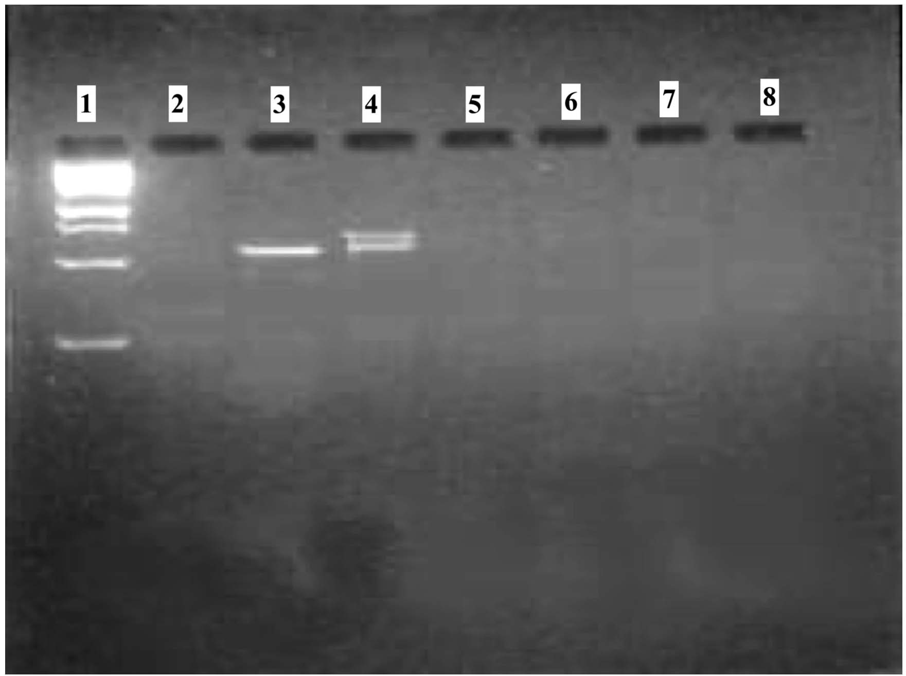
Figure 6. Multiplex PCR of tetA and tetB gene detection
Lane 1:1 kb DNA ladder 1st band-250 bp, 2nd band-500 bp and so on
Lane 3: tetA positive sample with 577 bp PCR product size
Lane 4: tetA (577 bp) and tetB (634 bp) positive sample
Detection of dfrA gene
The dfrA gene encodes a modified dihydrofolate reductase, which is insensitive to trimethoprim inhibition, and thus confers resistance against trimethoprim, a co-trimoxazole component. Conventional PCR revealed that 20.83% (20/96) of the isolates were positive for the dfrA gene (Table 6, Figure 7).
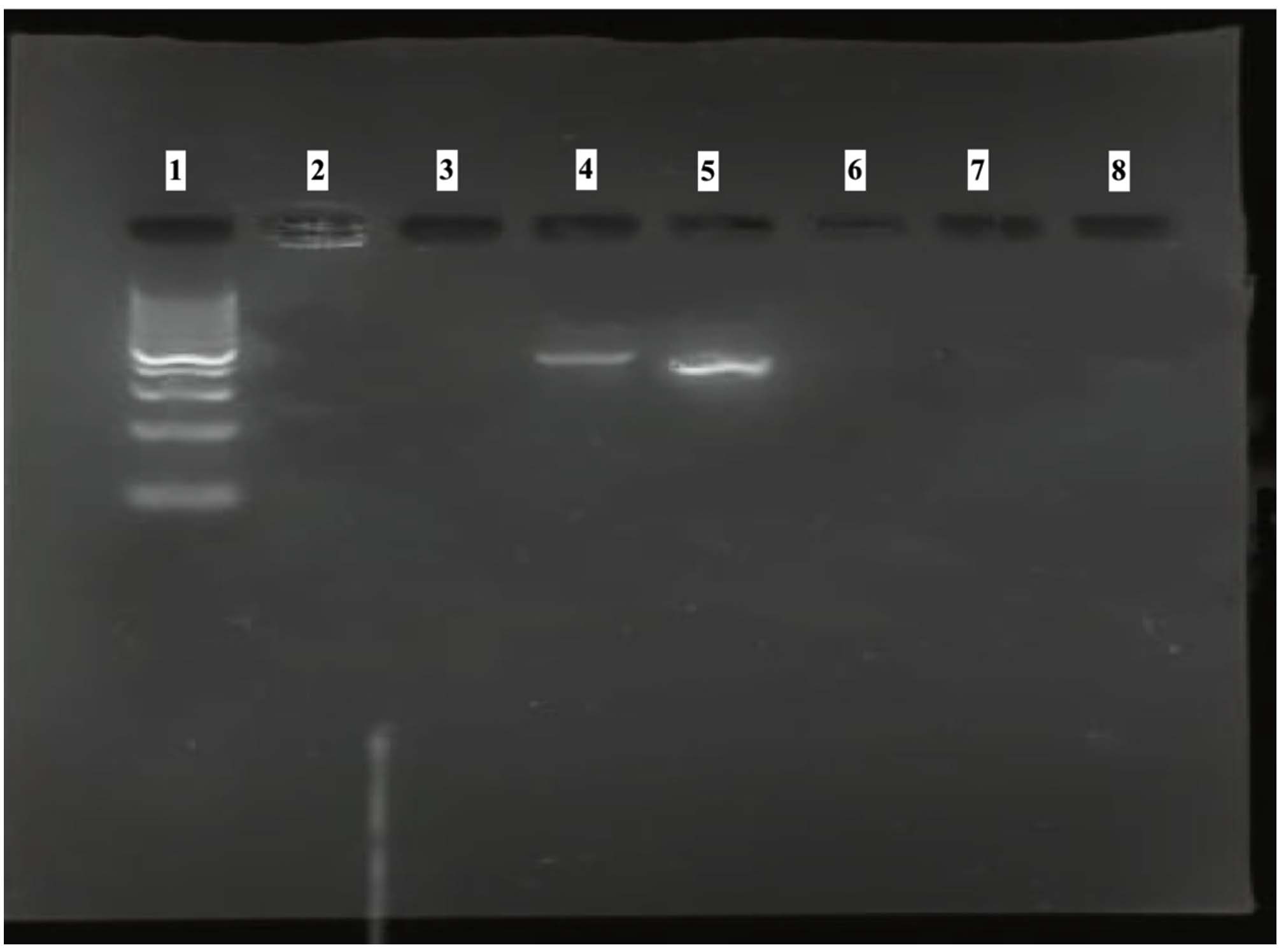
Figure 7. PCR amplification of dfrA gene
Lane 1:100 bp DNA ladder
Lane 4-5: dfrA gene positive sample (amplicon size 367 bp)
The present study investigated the identification, prevalence, and antimicrobial resistance (AMR) profiles of Escherichia coli isolated from different water sources in Uttar Pradesh, India. The isolates displayed morphological characteristics typical to E. coli and culturally they produced pink lactose-fermenting colonies on MLA, with a characteristic metallic-green sheen on EMB agar which confirmed their identity as E. coli. Identity of the isolates was further validated by biochemical characterization, whereby all the isolates were positive for catalase, indole, methyl red, and nitrate reduction tests; and negative for oxidase, Voges-Proskauer, citrate utilization, and urease tests. The complete agreement between the cultural and biochemical results suggests that the conventional approach remains a reliable and cost-effective method for preliminary screening of E. coli from environmental samples.18,19 Molecular confirmation targeting yaiO gene, a species specific molecular marker for E. coli, further validated all 96 isolates as E. coli. This gene is a reliable marker for species confirmation and complements traditional culture methods, reducing false negatives and improving diagnostic accuracy.20 These findings are consistent with Anbazhagan et al.21 and Molina et al.,20 who demonstrated that yaiO-based PCR offers high specificity and sensitivity for E. coli identification. Integrating molecular methods strengthens environmental surveillance by identifying viable but non-culturable cells that conventional techniques often miss.22
The detection of E. coli in environmental water sources is a well-recognized indicator of fecal contamination and poses significant risks for the transmission of waterborne diseases. In the present study, E. coli was isolated from 32% (96/300) of water samples collected from diverse aquatic sources. The highest prevalence was recorded in river water (72.7%), followed by pond water (43.0%), and canal water (35.71%). On the other hand, comparatively lower prevalence was observed in hand pump (12.5%) and tap water (10%). The high prevalence in rivers and ponds may be attributed to direct discharge of untreated domestic sewage, animal waste runoff, and agricultural effluents.23 Though at different rates, the prevalence of E. coli in different water sources in India is a consistent observation, such as 28.7% in Ayodhya,24 9.6% in Himachal Pradesh,25 and 56.5% in Punjab.26 Similarly, the prevalence rate of 25.2% was reported in Nepal27 and 56% in Bangladesh28 with whom India shares the river waters. In contrast, the relatively low prevalence in tap and hand-pump water reflects the effect of chlorination and filtration practices that limit bacterial survival. However, even the presence of E. coli in these treated sources indicates possible post-treatment contamination or leakages in water pipelines. The groundwater contamination may also be by percolation or inadequate protection as well.
The antibiotic susceptibility testing of 96 E. coli isolates revealed a diverse and concerning resistance pattern, indicating widespread multidrug-resistance (MDR) among isolates obtained from water sources in Uttar Pradesh. Multidrug-resistance (MDR) has been defined as acquired non-susceptibility to at least one agent in three or more antimicrobial classes.29 The results demonstrated complete resistance (100%) to ampicillin-sulbactam and polymyxin B, along with very high resistance (³94%) to tigecycline, ciprofloxacin, colistin, enrofloxacin, and amikacin. Moderate resistance was observed to imipenem (43.4%), gentamicin (42.8%), and co-trimoxazole (52.6%), while azithromycin (46%), meropenem (41.1%), and co-trimoxazole (36.8%) were among the few antibiotics still exhibiting some efficacy. These findings signify the alarming presence of MDR E. coli in environmental waters, which could act as both reservoir and channel for resistance genes transferable to human and animal pathogens. The high resistance to β-lactams, polymyxins, and fluoroquinolones indicates extensive antibiotic selection pressure, likely arising from the misuse of these drugs in human medicine, livestock production, and agricultural applications.30,31 The 100% resistance to ampicillin-sulbactam is consistent with reports by Avatsingh et al.32 and Johnson et al.,33 who documented complete loss of β-lactam efficacy in E. coli isolated from wastewater and river samples. Similarly, colistin resistance (94.4%) is particularly concerning since colistin is a last-resort antibiotic for carbapenem-resistant Enterobacteriaceae. The high colistin resistance in environmental isolates may be attributed to the dissemination of mcr (mobilized colistin resistance) genes.34 In this study, the resistance to third-generation cephalosporins such as cefepime and to fluoroquinolones exceeded the levels previously reported from India and other low and middle-income countries,25 suggesting dissemination of ESBL-producing strains and plasmid-mediated quinolone resistance mechanisms.35
The moderate resistance to carbapenems (imipenem 43.4%; meropenem 35.2%) suggests the potential emergence of carbapenemase-producing E. coli strains even in natural water systems. This incomplete resistance is consistent with earlier reports that carbapenems still retain efficacy against many ESBL-producing E. coli.36 This trend mirrors findings by Akiba et al.,37 who observed similar resistance levels among E. coli isolates from Indian rivers receiving untreated hospital effluents. However, the presence of carbapenem resistance in environmental isolates underscores the role of aquatic environments in the persistence and dissemination of highly resistant pathogens. Interestingly, despite overall resistance trends, azithromycin (46%) retained partial effectiveness. Azithromycin’s residual sensitivity might be due to its relatively lower use in environmental contexts compared to β-lactams or fluoroquinolones. A critical finding in this study was that 83.3% of isolates were multidrug-resistant (MDR), which is equivalent to that reported from Panjab (86.5%),26 but exceeding the average of 43.6% in low and middle-income countries.38 The high MDR prevalence suggests strong environmental selection pressures driven by antibiotic misuse in humans, livestock, and aquaculture, as well as persistence of resistance genes in sediments and biofilms.39-41 Mobile genetic elements such as plasmids, integrons, and transposons likely facilitated resistance gene dissemination among aquatic bacteria.42
Multiplex PCR analysis revealed that 37.5% of the E. coli isolates carried tetracycline resistance genes (tetA and tetB). These genes encode efflux pump proteins that actively expel tetracycline molecules from bacterial cells, preventing inhibition of protein synthesis. The presence of tetA and tetB genes in environmental E. coli isolates suggests that tetracycline residues are present in the aquatic environment, likely originating from livestock farms and aquaculture effluents where tetracyclines are extensively used as growth promoters and prophylactics. The presence of tetracycline resistance genes in environmental E. coli has been documented in other studies as well.43-45 Similar trend in the detection rates of these resistance genes have been reported by Daghrir et al.46 from wastewater and soil in North India (35%-45%) and Rathinavelu et al.47 and Karkman et al.48 from river and pond water (30%-40%). On the other hand, lower prevalence (7%) of these resistance genes has been reported in E. coli isolates from the River Yamuna32 and far higher prevalence (70.0%) has been reported from drinking water in China.49 The consistent reports of these findings across different studies reinforces the notion that tet genes are highly stable and easily transmissible in environmental microbiota. The findings reflect the environmental dissemination of ARGs, likely driven by selective pressure from antibiotic use in human, veterinary, and agricultural settings.50,51 Similarly, the dfrA gene was detected in 20.83% of the isolates, conferring resistance to trimethoprim, a component of co-trimoxazole. This detection rate is consistent with earlier reports which revealed the prevalence rate of dfrA gene in the range of 18%-25% from environmental and clinical sources.52-55 The gene encodes an altered dihydrofolate reductase enzyme that is insensitive to inhibition by trimethoprim, thereby maintaining folate synthesis and bacterial growth. In this study, relatively lower prevalence of dfrA compared to tetA/B can be ascribed to the fact that trimethoprim is clinically less used compared to tetracyclines. However, the presence of dfrA in waterborne isolates is epidemiologically significant because of the fact that this gene is frequently located on plasmids or class 1 integrons, which facilitate horizontal transfer among bacterial species. The detection of resistance genes at higher frequencies in isolates form river and pond water reinforces the fact that stagnant and open water bodies serve as hotspots for the persistence and propagation of resistant strains. Environmental persistence of antibiotic resistance genes has been shown to correlate strongly with nutrient load, biofilm formation, and organic pollution.56 The considerable level of concurrence between phenotypic resistance and the presence of tet and dfrA genes in several isolates in this study indicates that genetic determinants are the primary contributors to the observed MDR phenotype. However, the presence of phenotypically resistant isolates without resistant gene detection suggests the involvement of other mechanisms such as mutations in target sites, reduced membrane permeability, or efflux pump overexpression other than tet and dfrA.57
This study establishes the widespread distribution of E. coli along with an alarming rise of antimicrobial resistance among the environmental isolates from the aquatic ecosystems of Uttar Pradesh posing a serious public health and environmental safety concern. The presence of MDR E. coli harboring tetA, tetB, and dfrA genes and their persistence in the environment increases the risk of genetic exchange with pathogenic bacteria, potentially leading to recalcitrant infections under both medical and veterinary settings. Hence, this study underscores the need for regular surveillance of antimicrobial residues and resistance genes in aquatic systems, rational antibiotic stewardship in agriculture and clinical sectors, and wastewater treatment and sanitation.
ACKNOWLEDGMENTS
None.
CONFLICT OF INTEREST
The authors declare that there is no conflict of interest.
AUTHORS’ CONTRIBUTION
JB conceptualized the study and collected resources. JB, RRM and NAM performed validation. RRA supervised the study. JB, RK, ESR, RRM and VK applied methodology. RK, ESR and VK performed investigation and data curation. NAM performed formal analysis and wrote the original draft. RRM and JB wrote, reviewed and revised the manuscript. All authors read and approved the final manuscript for publication.
FUNDING
None.
DATA AVAILABILITY
The datasets generated and/or analysed during the current study are available from the corresponding author on reasonable request.
ETHICS STATEMENT
This article does not contain any studies on human participants or animals performed by any of the authors.
- Poirel L, Madec JY, Lupo A, et al. Antimicrobial resistance in Escherichia coli. Microbiol Spectr. 2018;6(4):ARBA-0026-2017.
Crossref - Odonkor ST, Ampofo JK. Escherichia coli as an indicator of bacteriological quality of water: an overview. Microbiol Res. 2013;4(1):e2.
Crossref - Donkor ES, Odoom A, Osman AH, Darkwah S, Kotey FCN. A systematic review on antimicrobial resistance in Ghana from a One Health perspective. Antibiotics. 2024;13(7):662.
Crossref - Kummerer K. Antibiotics in the aquatic environment – A review – Part I. Chemosphere. 2009;75(4):417-434.
Crossref - Wang W, Weng Y, Luo T, Wang Q, Yang G, Jin Y. Antimicrobial and the resistances in the environment: ecological and health risks, influencing factors, and mitigation strategies. Toxics. 2023;11(2):185.
Crossref - Baquero F, Alvarez-Ortega C, Martinez JL. Ecology and evolution of antibiotic resistance. Environ Microbiol Rep. 2009;1(6):469-476.
Crossref - Laxminarayan R, Matsoso P, Pant S, et al. Access to effective antimicrobials: a worldwide challenge. Lancet. 2016;387(10014):168-175.
Crossref - Galindo-Méndez M. Antimicrobial Resistance in Escherichia coli. In: Rodrigo L, ed. E. Coli Infections – Importance of Early Diagnosis and Efficient Treatment. IntechOpen; 2020.
Crossref - Martinez JL. Environmental pollution by antibiotics and by antibiotic resistance determinants. Environ Pollut. 2009;157(11):2893-2902.
Crossref - Puvaca N, de Llanos Frutos R. Antimicrobial resistance in Escherichia coli strains isolated from humans and pet animals. Antibiotics. 2021;10(1):69.
Crossref - Anjum MF, Zankari E, Hasman H. Molecular methods for detection of antimicrobial resistance. Microbiol Spectr. 2017;5(6):ARBA-0011-2017.
Crossref - Hadiujjaman M, Rahman MM, Ahasan MD, Banu MA, Khatun MM, Islam MA. Isolation and identification of Escherichia coli from apparently healthy chicken of selected areas of Bangladesh. Int J Nat Soc Sci. 2016;3(3):15-23.
- Begum J, Dutta TK, Chandra R, et al. Molecular and phenotypic characterization of shiga toxigenic Escherichia coli (STEC) and enteropathogenic E. coli (EPEC) from piglets and infants associated with diarrhoea in Mizoram, India. Afr J Biotechnol. 2014;13(13):1452-1461
- Ahmadi S, Keshavarzi F. Determining contamination by Escherichia coli in water resources through yaiO gene molecular analysis. J Res Med Dent Sci. 2018;6(1):392-396.
Crossref - Bauer AW, Kirby WMM, Sherris JC, Turck M. Antibiotic susceptibility testing by a standardized single disk method. Am J Clin Pathol. 1966;45(4):493-496.
- Clinical and Laboratory Standards Institute. Performance standards for antimicrobial susceptibility testing. CLSI supplement M100. 33rd ed. 2023
- Momtaz H, Rahimi E, Moshkelani S. Molecular detection of antimicrobial resistance genes in E. coli isolated from slaughtered commercial chickens in Iran. Vet Med. 2012;57(4):193-197.
Crossref - Quinn PJ, Markey BK, Leonard FC, Hartigan P, Fanning S, FitzPatrick ES. Veterinary Microbiology and Microbial Disease (Second Edition). Wiley-Blackwell; 2016
- Singh AK, Das S, Kumar S, et al. Distribution of Antibiotic-Resistant Enterobacteriaceae Pathogens in Potable Spring Water of Eastern Indian Himalayas: Emphasis on Virulence Gene and Antibiotic Resistance Genes in Escherichia coli. Front Microbiol. 2020;11:581072.
Crossref - Molina F, Lopez-Acedo E, Tabla R, Roa I, Gomez A, Rebollo JE. Detection and Quantification Methods for Viable but Non-culturable (VBNC) Cells in Process Wash Water of Fresh-Cut Produce: Industrial Validation. BMC Biotechnol. 2015;15(1):48.
Crossref - Anbazhagan D, Mui WS, Mansor M, Yan GO, Yusof MY, Sekaran SD. Development of conventional and real-time multiplex PCR assays for the detection of nosocomial pathogens. Braz J Microbiol. 2011;42(2):448-458.
Crossref - Truchado P, Gil MI, Larrosa M, Allende A. Detection and quantification methods for viable but non-culturable (VBNC) cells in process wash water. Front Microbiol. 2020;11:673.
Crossref - Hanna N, Purohit M, Diwan V, et al. Monitoring of water quality, antibiotic residues, and antibiotic-resistant Escherichia coli in the Kshipra River in India over a 3-year period. Int J Environ Res Public Health. 2020;17(21):7706.
Crossref - Shekhar C, Joshi N, Singh A. Prevalence of multidrug-resistant Escherichia coli in drinking water in and around Ayodhya (U.P.), India. Indian J Vet Sci Biotechnol. 2023;19(1):112-115.
Crossref - Thakur SD, Panda AK, Diwan V, Kumar P, Singh M, Sharma DK. Microbial assessment of drinking water sources in Himachal Pradesh, India. Indian J Public Health. 2024;68(4):572-574.
Crossref - Gautam B, Dara SN, Sachan RSK. A study on Escherichia coli contamination in drinking water sources in Punjab, India. J Water Health. 2025;23(2):155-165.
Crossref - Uprety S, Dangol B, Nakarmi P, et al. Assessment of microbial risks by characterization of Escherichia coli presence to analyze the public health risks from poor water quality in Nepal. Int J Hyg Environ Health. 2020;226:113484.
Crossref - Ferdous J, Rashid RB, Sultana R, et al. Is it human or animal? The origin of pathogenic Escherichia coli in the drinking water of a low-income urban community in Bangladesh. Trop Med Infect Dis. 2021;6(4):181.
Crossref - Magiorakos AP, Srinivasan A, Carey RB, et al. Multidrug-resistant, extensively drug-resistant and pandrug-resistant bacteria: an international expert proposal for interim standard definitions for acquired resistance. Clin Microbiol Infect. 2012;18(3):268-281.
Crossref - Kousar A, Jabbar Z, Nisa ZU, et al. Novel approaches to overcome antibiotic resistance: Phage therapy, nanoparticles, and natural antimicrobials. Lett Anim Biol. 2025;5(2):50-60.
Crossref - Rafee AR, Abd Alfatlawi MA, Asad A, et al. Zoonotic Escherichia coli infections: Pathogenic variants, mechanism of disease, transmission routes, and foodborne outbreaks. Lett Anim Biol. 2025;5(2):107-117.
Crossref - Avatsingh AU, Sharma S, Kour S, et al. Prevalence of antibiotic-resistant Gram-negative bacteria with ESBL phenotypes in polluted irrigation wastewater from Indian agro-ecosystems. Front Microbiol. 2023;14:1227132.
Crossref - Johnson A, Ginn O, Bivins A, et al. Extended-spectrum β-lactamase-positive Escherichia coli in urban aquatic environments in Kanpur, India. J Water Health. 2020;18(5):849-854.
Crossref - Liu YY, Wang Y, Walsh TR, et al. Emergence of plasmid-mediated colistin resistance mechanism MCR-1 in animals and human beings in China: a microbiological and molecular biological study. Lancet Infect Dis. 2016;16(2):161-168.
Crossref - Zurfluh K, Abgottspon H, Hächler H, Nüesch-Inderbinen M, Stephan R. Quinolone Resistance Mechanisms among Extended-Spectrum Beta-Lactamase (ESBL) Producing Escherichia coli Isolated from Rivers and Lakes in Switzerland. PLoS ONE. 2014;9(4):e95864.
Crossref - Shin H, Kim Y, Han D, Hur H. Emergence of High Level Carbapenem and Extensively Drug Resistant Escherichia coli ST746 Producing NDM-5 in Influent of Wastewater Treatment Plant, Seoul, South Korea. Front Microbiol. 2021;12:645411.
Crossref - Akiba M, Sekizuka T, Yamashita A, et al. Distribution and relationships of antimicrobial resistance determinants among extended-spectrum-cephalosporin-resistant or carbapenem-resistant Escherichia coli isolates from rivers and sewage treatment plants in India. Antimicrob Agents Chemother. 2016;60(5):2973–2980.
Crossref - Desye B, Mawugatie T, Asmare L, et al. Antimicrobial resistance profile of Escherichia coli in drinking water from one health perspective in low and middle income countries. Front Public Health. 2024;12:1440908.
Crossref - Pandey S, Doo H, Keum GB, et al. Antibiotic resistance in livestock, environment and humans: One Health perspective. J Anim Sci Technol. 2024;66(2):266-278.
Crossref - Grenni P. Antimicrobial Resistance in Rivers: A Review of the Genes Detected and New Challenges. Environ Toxicol Chem. 2022;41(3):687-714.
Crossref - Zhao Y, Yang QE, Zhou X, et al. Antibiotic resistome in the livestock and aquaculture industries: Status and solutions. Crit Rev Environ Sci Technol. 2020;51(19):2159-2196.
Crossref - Partridge SR, Kwong SM, Firth N, Jensen SO. Mobile genetic elements in antimicrobial resistance. Clin Microbiol Rev. 2018;31(4):e00088-17.
Crossref - Perewari DO, Otokunefor K, Agbagwa OE. Tetracycline-Resistant Genes in Escherichia coli from Clinical and Nonclinical Sources in Rivers State, Nigeria. Int J Microbiol. 2022;2022:9192424.
Crossref - Cianelli C. Prevalence Rates of tetB, tetA, and tetM Tetracycline Resistance Genes in Environmental Escherichia coli Isolates From Local Waterways. [Honors project]. Bridgewater, VA: Bridgewater College; 2025. https://digitalcommons.bridgewater.edu/honors_projects/832
- Martinez-Vazquez AV, Vazquez-Villanueva J, Leyva-Zapata LM, Barrios-Garcia H, Rivera G, Bocanegra-Garcia V. Multidrug Resistance of Escherichia coli Strains Isolated From Bovine Feces and Carcasses in Northeast Mexico. Front Vet Sci. 2021;8:643802.
Crossref - Daghrir R, Drogui P. Tetracycline antibiotics in the environment: a review. Environ Chem Lett. 2013;11:209–227.
Crossref - Rathinavelu S, Uluseker C, Sonkar V, Thatikonda S, Nambi IM, Kreft JU. Mapping the scarcity of data on antibiotics in natural and engineered water environments across India. Front Antibiot. 2024;3:1337261.
Crossref - Karkman A, Thuy T DO, Walsh F, Virta MPJ. Antibiotic-Resistance genes in waste water. Trends Microbiol. 2017;26(3):220-228.
Crossref - Chen Z, Yu D, He S, et al. Prevalence of Antibiotic-Resistant Escherichia coli in Drinking Water Sources in Hangzhou City. Front Microbiol. 2017;8:1133.
Crossref - Taneja N, Sharma M. Antimicrobial resistance in the environment: the Indian scenario. Indian J Med Res. 2019;149(2):119-128.
Crossref - Xu C, Kong L, Liao Y, et al. Mini-Review: Antibiotic-Resistant Escherichia coli from Farm Animal-Associated Sources. Antibiotics. 2022;11(11):1535.
Crossref - Kumar G, Balakrishna K, Mukhopadhyay C, Eshwara VK. Comparison of integron mediated antimicrobial resistance in clinical isolates of Escherichia coli from urinary and bacteremic sources. BMC Microbiol. 2024;24(1):102.
Crossref - Kumar G, Balakrishna K, Mukhopadhyay C, Eshwara VK. Characterization and comparative analysis of antimicrobial resistance in Escherichia coli from hospital and municipal wastewater treatment plants. J Water Health. 2024;22(12):2276-2288.
Crossref - Muller H, Sib E, Gajdiss M, et al. Dissemination of multi-resistant Gram-negative bacteria into German wastewater and surface waters. FEMS Microbiol Ecol. 2018;94(5):fiy057.
Crossref - Bajaj P, Singh NS, Virdi JS. Distribution and molecular characterization of CTX-M and AmpC β-lactamases in Escherichia coli from an Indian urban aquatic environment. Sci Total Environ. 2015;505:350–356.
Crossref - Berendonk TU, Manaia CM, Merlin C, et al. Tackling antibiotic resistance: environmental framework. Nat Rev Microbiol. 2015;13(5):310-317.
Crossref - Ali A, Ambrose S, Hussain D, et al. Antimicrobial resistance and antimicrobial activity of plant-based antimicrobial peptides against bacteria. Lett Anim Biol. 2024:4(2):19-27.
Crossref
© The Author(s) 2026. Open Access. This article is distributed under the terms of the Creative Commons Attribution 4.0 International License which permits unrestricted use, sharing, distribution, and reproduction in any medium, provided you give appropriate credit to the original author(s) and the source, provide a link to the Creative Commons license, and indicate if changes were made.