


ISSN: 0973-7510
E-ISSN: 2581-690X
Chitosan, a positively charged polymer obtained by treating chitin with hot alkali to remove acetyl groups, has extensive biological applications due to its non-toxicity, biocompatibility, and natural biodegradability. In this study, chitosan nanoparticles were prepared and encapsulated with bacteriocin isolated from Lactococcus lactis subsp. lactis, resulting in chitosan-bacteriocin conjugate nanoparticles produced using the ionic gelation method. The antibacterial activity of this formulation was evaluated as a potential food preservative against Bacillus cereus, Escherichia coli, Salmonella typhimurium, and Staphylococcus aureus. The effectiveness of the conjugate nanoparticles was compared to that of crude chitosan, chitosan nanoparticles, and free bacteriocin. The agar diffusion method was employed to assess the in vitro dissolution of the substance and to investigate the effects of temperature and pH on its stability. The results indicated that the release of chitosan nanoparticles conjugated with bacteriocin in vitro was controlled at around 60% within 24 hours, demonstrating a cumulative and sustained effect. This release control was significantly higher compared to the free bacteriocin, which achieved a 90% release. Among the formulations tested, chitosan nanoparticles conjugated with bacteriocin (CSNps-B) demonstrated the most potent antibacterial activity. CSNps-B achieved inhibition zones ranging from 30.32 to 32.45 mm against Gram-positive bacteria and from 30.22 to 36.26 mm against Gram-negative bacteria. The superior performance of CSNps-B was statistically significant, with a p-value of less than 0.05 compared to the other formulations. In contrast, chitosan alone showed inhibition zones of 6.7 to 8.45 mm, chitosan nanoparticles (CSNps) exhibited zones of 10.12 to 12.6 mm, and free bacteriocin (free-B) ranged from 13.38 to 15.67 mm. The enhanced antibacterial effectiveness of CSNps-B underscores its potential as a superior antimicrobial agent compared to the other formulations evaluated.
Antimicrobial Agents, Drug Delivery Systems, Antibacterial Activity, Gram-positive Bacteria, Gram-negative Bacteria, Biopolymers, Nanoparticle Technology, Food Preservatives
Chitosan, a natural biopolymer derived from chitin, is renowned for its versatile applications and biocompatibility. Chitin, the second most abundant polysaccharide found in the exoskeletons of crustaceans and insects, is converted into chitosan through a process of deacetylation.1 This process produces a compound with notable attributes, including biodegradability, non-toxicity, and excellent film-forming properties. As a result, chitosan has attracted considerable attention across various fields such as biomedical engineering, pharmaceuticals, agriculture, and food technology. Its unique capacity to form hydrogels, nanoparticles, and films, combined with its inherent antimicrobial activity and potential for drug delivery, makes chitosan a key material in the development of innovative solutions for health and industrial applications.2
Chemically, chitosan is a linear polysaccharide primarily composed of β-(1→4)-linked D-glucosamine units, which are partially deacetylated derivatives of chitin. Its chemical structure features repeating units of (C₆H₁₁NO₄)n, where “n” denotes the degree of polymerization, which varies depending on the source and preparation method.3 This biopolymer generally consists of a combination of glucosamine (deacetylated units) and N-acetylglucosamine (acetylated units), with the ratio between these units determining the degree of deacetylation (DD). The DD is a crucial factor affecting chitosan’s solubility and functional properties. The free amino groups in the glucosamine units confer a positive charge to chitosan under acidic conditions, allowing it to interact effectively with negatively charged surfaces and molecules.4 This distinctive chemical composition underlies chitosan’s solubility in acidic solutions, its capacity to form films and gels, and its broad range of applications across fields such as biomedicine, agriculture, and environmental science.5
In addition, Chitosan and its nanoparticle derivatives exhibit notable antimicrobial activities, making them highly effective for combating microbial diseases.6 The antimicrobial activity of chitosan is primarily attributed to its polycationic nature, which enables it to interact with the negatively charged membranes of bacterial cells, leading to increased membrane porosity, the release of intracellular contents, and ultimately, cell death.7 This interaction disrupts the metabolic processes of bacteria, fungi, and some viruses, making chitosan effective against a wide range of pathogens. Additionally, chitosan nanoparticles amplify these antimicrobial properties due to their larger surface area and enhanced bioavailability.8 These nanoparticles can penetrate microbial cells more efficiently, delivering higher concentrations of chitosan to targeted sites. Furthermore, the small size and high surface-to-volume ratio of chitosan nanoparticles facilitate improved interaction with microbial membranes, thereby enhancing the disruption of cellular functions.9
Similarly, Lactic acid bacteria (LAB) are widely used in food preservation due to their ability to produce metabolites with strong antibacterial properties. These metabolites effectively inhibit the growth of harmful and pathogenic bacteria in food products. Among these metabolites, bacteriocins are particularly significant. Bacteriocins are ribosomally synthesized antimicrobial peptides secreted by LAB to inhibit the growth of both closely related and unrelated bacterial species.10 These peptides disrupt the cell membranes of target bacteria, leading to cell lysis and death. Additionally, LAB produces other antimicrobial compounds such as acids, hydrogen peroxide (H₂O₂), and diacetyl, which create an inhospitable environment for harmful microorganisms by lowering the pH and generating oxidative stress. The combined action of these metabolites not only extends the shelf life of food items but also enhances food safety by reducing the risk of contamination by pathogens. Consequently, incorporating LAB into food preservation strategies harnesses their natural antagonistic activity, offering a sustainable and effective method to maintain both the quality and safety of food.11
Despite their potential, the direct use of bacteriocins as food preservatives has been slow to gain widespread adoption, largely due to limitations such as sensitivity to storage conditions, temperature fluctuations, and production processes. To overcome these challenges, bacteriocin-nanoconjugates have emerged as a promising solution. Nanoparticles, with dimensions ranging from 1 to 100 nm, offer unique properties that significantly enhance the effectiveness of bacteriocins. Their small size and increased surface area compared to bulk materials improve the protection and delivery of bacteriocins’ antibacterial effects.12
Building on this, nanoencapsulation, the process of embedding bacteriocins within nanoparticles has been shown to enhance the antimicrobial activity of these peptides. Nanoparticles are capable of effectively diffusing and crossing biological cell membrane barriers, which improves the delivery of bacteriocins to their target sites.13
In light of these advancements, this study aims to evaluate the performance of bacteriocins encapsulated in chitosan nanoparticles under various environmental conditions and to assess their potential effectiveness against harmful bacteria. The findings could lead to improved methods for utilizing bacteriocins in diverse applications, including food safety and medical treatments.
Lyophilized strains of pathogenic bacteria were procured from various culture collections, as detailed in Table 1. In our earlier investigation, we produced bacteriocin using the Levilactobacillus brevis strain ABRIINW-k.
Table (1):
Catalogue of foodborne pathogens and their corresponding culture types
No. |
Pathogens |
Culture type |
|---|---|---|
1 |
Salmonella typhimurium |
MTCC 3224 |
2 |
Bacillus cereus |
MTCC 1272 |
3 |
Staphylococcus aureus |
MTCC 740 |
4 |
Escherichia coli |
MTCC 1310 |
Preparation
The ionic gelation process was used to produce chitosan nanoparticles (CSNps) as described by Hoang et al.14 The experiment began by dissolving 0.2 grams of chitosan (purchased from Sigma-Aldrich, USA, with a deacetylation degree of 93%) in 100 milliliters of 1% acetic acid to create a uniform solution. Sodium tripolyphosphate (TPP), also from Sigma-Aldrich, USA, was then added gradually to the solution while stirring continuously. Chitosan and TPP were added drop by drop and stirred for 3 hours to ensure sufficient interaction and ionic cross-linking, leading to the formation of nanoparticles. Following the reaction period, the product was centrifuged at 10,000 RPM for 10 minutes. This centrifugation step facilitated the separation of the chitosan nanoparticles, forming a solid mass at the bottom of the centrifuge tube. The solid mass, consisting of the CSNps, was then available for further use or examination.
Analysis of drug release in vitro
The in vitro release of bacteriocin was determined using the dialysis bag method, as described by Bohrey et al.,15 with some modifications. A total of 5 milliliters of release medium, specifically 0.1 M phosphate-buffered saline (PBS) with a pH of 7.4, was prepared. This medium contained either 50 milligrams of chitosan nanoparticles conjugated with bacteriocin (CSNps-B) or free bacteriocin (free-B). The release medium was placed into a dialysis bag, which was then immersed in a beaker containing 100 milliliters of PBS. The beaker was positioned on a magnetic stirrer set to rotate at 100 revolutions per minute (rpm), and the temperature was carefully maintained at 37 ± 1°C to simulate physiological conditions.
To monitor bacteriocin release, 2 milliliter samples were periodically withdrawn from the beaker at various time intervals, including 1, 2, and 3 hours, and continuing up to 24 hours. After each sample was taken, an equal volume of fresh PBS was added to the beaker to maintain a constant volume. The collected samples were then analyzed for bacteriocin release percentage using an ultraviolet-visible spectrophotometer at a wavelength of 291 nm. This method provided an accurate profile of bacteriocin release over the designated period.
Formulation preparation
In a sterile environment, three-fold tubes with dimensions of 12 × 75 mm were utilized to divide the formulations into four distinct groups: chitosan nanoparticles (CSNps), crude chitosan, free bacteriocin (free-B), and chitosan nanoparticles conjugated with bacteriocin (CSNps-B). The quantification of each group was conducted precisely, and the formulations were then combined with 0.25% acetic acid to achieve a final concentration of 100 µg/mL, following the procedure outlined by Abdeltawab et al.16 This standardized preparation ensured that each formulation possessed the correct concentration required for subsequent trials or analyses.
Antimicrobial activity
The antibacterial activities of different formulations against foodborne bacteria were evaluated using the agar well diffusion technique. This procedure involved introducing bacterial cultures onto Mueller Hinton Agar plates to create a uniform bacterial layer. Sterile cork borers were then used to create wells by removing plugs of agar from the inoculated plates. Each well was subsequently filled with 100 µl of one of the distinct formulations: free bacteriocin (free-B), chitosan nanoparticles (CSNps), crude chitosan, or chitosan nanoparticles conjugated with bacteriocin (CSNps-B). The plates were incubated at 37°C for 24 hours, allowing bacterial growth and interaction with the formulations. Following incubation, the zones of inhibition, where bacterial growth was halted, were measured in millimeters to assess the antibacterial effectiveness of each formulation. Measurements were taken three times to ensure accuracy and consistency, following the methodology described by Balouiri et al.17
Stability pH
The impact of pH on the stability of the formulated groups was evaluated following the method outlined by Guirguis et al.18 In this procedure, 5 mL of each formulation was placed into tubes, and the pH levels were adjusted between 2 and 12 by adding sterile lactic acid (1% w/v) or sodium hydroxide (1 M) at room temperature (22°C) for 2 hours. After the incubation period, the pH of each formulation was readjusted to a neutral pH of 7. The antimicrobial efficacy of the pH-adjusted concentrates was then assessed using the agar well diffusion technique, as described by Mostafa et al.19 This involved measuring the zones of inhibition around the wells containing the treated samples on inoculated agar plates, thereby determining the effectiveness of each formulation in killing bacteria at various pH levels.
Statics
Statistical analysis was conducted on the data obtained from the duplicated experiments to assess the significance of differences among various formulations and treatments. IBM-SPSS software, version 20 (USA), was used for data entry and processing. The analysis utilized two primary statistical methods: One-way ANOVA (Analysis of Variance) and the paired-samples T-test.
The efficacy of chitosan nanoparticles (CSNps) as a food preservative was evaluated by investigating the release of bacteriocin from both CSNps and free bacteriocin (free-B) in vitro. As shown in Figure 1, transmission electron microscopy (TEM) images illustrate the morphology and size distribution of the nanoparticles. The left panel shows chitosan nanoparticles (CSNps) alone, with particle sizes around 10.10 nm, 12.56 nm, and 20.56 nm. In contrast, the right panel displays chitosan nanoparticles conjugated with bacteriocin (CSNps-B), showing a larger particle size range, with some particles measuring approximately 50.10 nm and 90.12 nm. This difference in size suggests the successful conjugation of bacteriocin onto the chitosan nanoparticles, potentially enhancing their functionality as a food preservative.
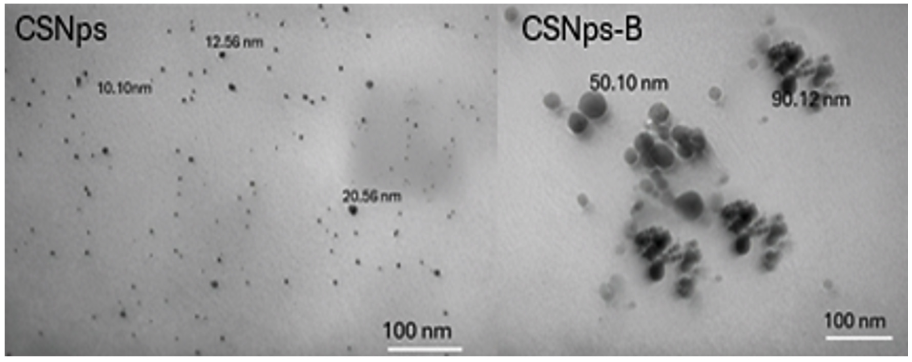
Figure 1. TEM images of chitosan nanoparticles and chitosan nanoparticles conjugated with bacteriocin
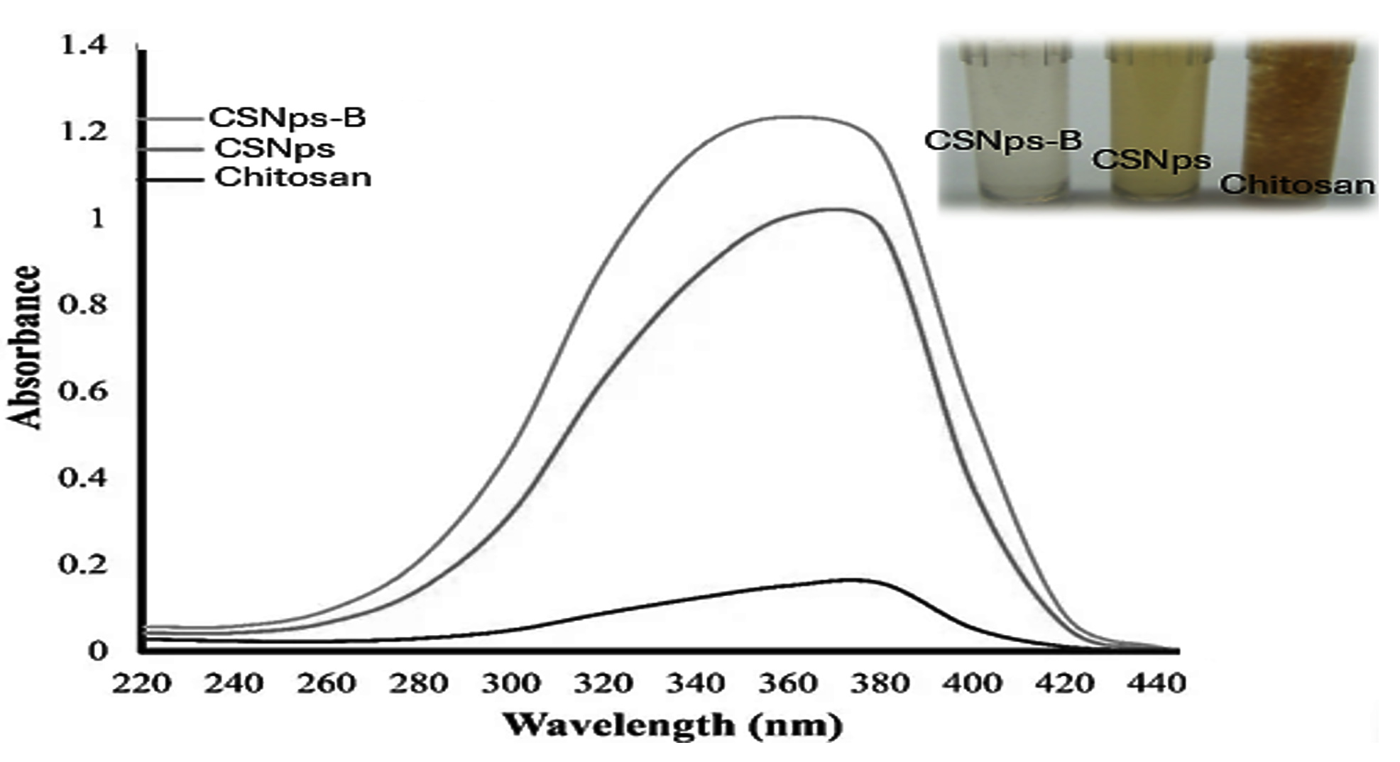
Figure 2. UV-visible absorbance spectra of chitosan, chitosan nanoparticles (CSNps), and chitosan nanoparticles conjugated with bacteriocin (CSNps-B) nanocomposites
Additionally, Figure 2 presents the UV-visible absorbance spectra of chitosan, chitosan nanoparticles (CSNps), and chitosan nanoparticles conjugated with bacteriocin (CSNps-B) nanocomposites. The spectra illustrate a significant increase in absorbance for CSNps-B compared to CSNps and chitosan, indicating effective bacteriocin loading and enhanced interaction with light at specific wavelengths, which may correlate with its antimicrobial efficacy.
This investigation involved examining the release patterns of bacteriocin from CSNps loaded with bacteriocin, as well as from its separate components-chitosan, CSNps alone, and free bacteriocin (free-B)-against food-borne pathogenic microbes. As illustrated in Figure 3, the comparative analysis of in vitro drug release profiles shows distinct release patterns between free bacteriocin and chitosan nanoparticles conjugated with bacteriocin (CSNps-B). This comparison highlights the controlled and sustained release of bacteriocin from the CSNps-B formulation, potentially enhancing its antimicrobial efficacy against food-borne pathogens.
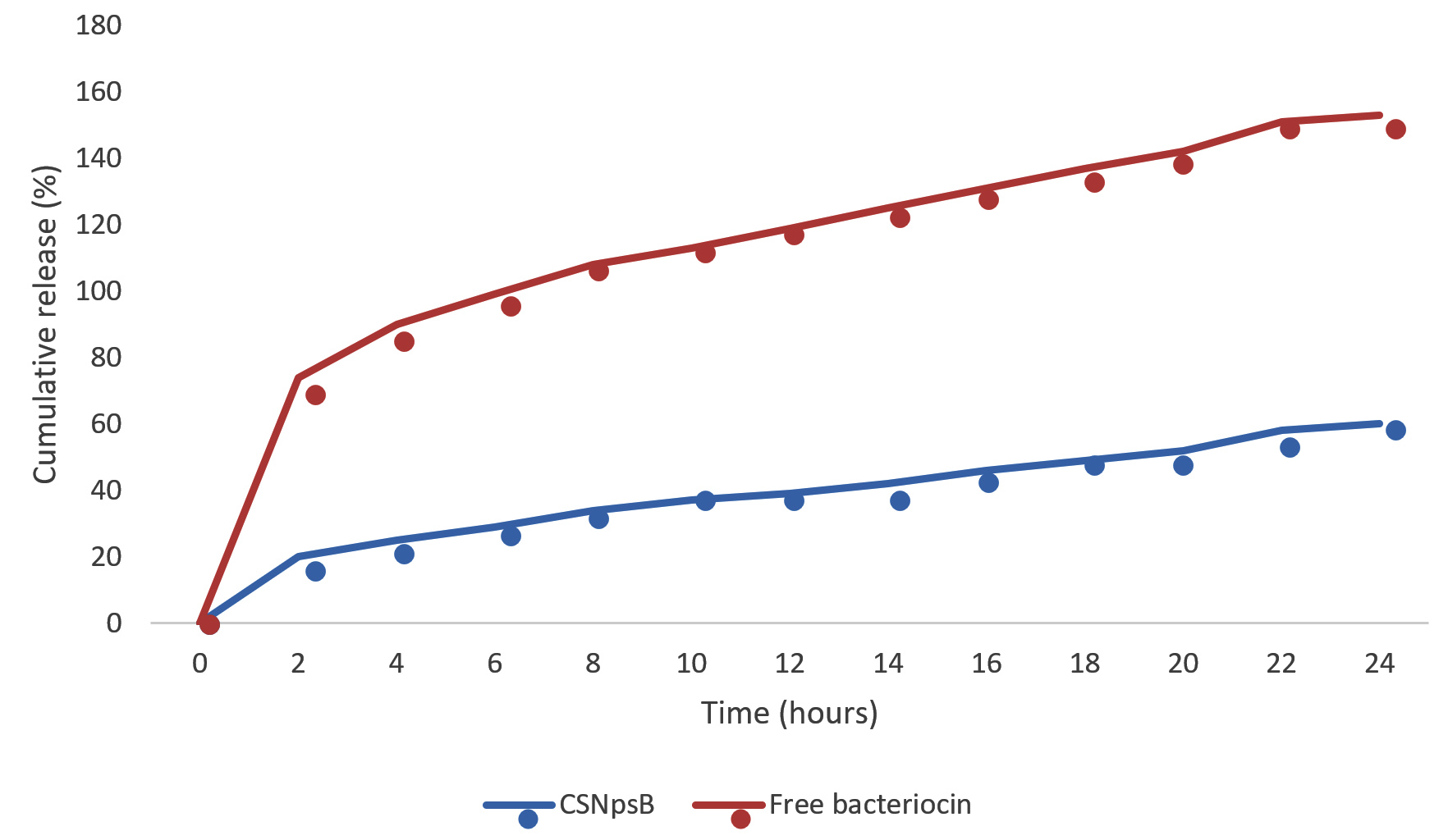
Figure 3. Comparative analysis of in vitro drug release of free bacteriocin and chitosan nanoparticles conjugated with bacteriocin (CSNps-B)
In this context, the controlled release observed with CSNps-B in our study aligns with findings from several recent works. For instance, Narayanan et al.20 demonstrated that encapsulation of bacteriocins in chitosan nanoparticles led to a significant reduction in the initial burst release, which is crucial for maintaining consistent antimicrobial activity over time. Their work showed that by modulating the chitosan-to-bacteriocin ratio and cross-linking conditions, the release profile could be finely tuned to extend the bacteriocin’s efficacy in food preservation applications.
In contrast, the initial burst release observed with free-B, where 65% of the bacteriocin was released within the first 4 hours, is a phenomenon commonly seen in systems without encapsulation. This rapid release can be beneficial for applications requiring immediate antimicrobial action but may also lead to a quick depletion of active bacteriocin, potentially reducing long-term effectiveness. Chen et al.21 echoed this observation, reporting that free bacteriocin formulations often exhibit a burst release due to the lack of a barrier controlling the diffusion of the active compound. They also noted that such rapid release could result in suboptimal antimicrobial activity over extended periods, making it less suitable for applications where prolonged activity is desired.
In contrast to the burst release observed with free-B, the prolonged release observed with CSNps-B, where only 25% of the bacteriocin was released within the first 4 hours and 60% after 24 hours, suggests a more sustained delivery, which is crucial for food preservation and other applications requiring long-term antimicrobial protection. A study by Niazmand et al.22 highlighted that such controlled release systems can significantly enhance the shelf life of perishable food products by maintaining a consistent level of antimicrobial agents. This controlled release mechanism, facilitated by the nanoparticle matrix, ensures that bacteriocin is gradually released, reducing the risk of microbial resistance and improving overall food safety.
Furthermore, the paired-sample t-test results, indicating significant differences in release patterns between CSNps-B and free-B with a p-value <0.05, further confirm the efficacy of chitosan nanoparticles in regulating the release of bacteriocin. These findings are consistent with recent studies, such as the work by Ali et al.,23 which reported similar statistical significance when comparing the release profiles of free and nanoparticle-encapsulated antimicrobial peptides. Their work emphasized that controlled release not only prolongs the antimicrobial effect but also reduces the frequency of required applications, making it a cost-effective solution for industrial use.
Therefore, the controlled release pattern of CSNps-B could have significant implications for food preservation, as it provides more consistent delivery of bacteriocin over time. This could help prevent spoilage and extend the shelf life of food products without the need for repeated applications. The potential to fine-tune release profiles by modifying the nanoparticle formulation offers a versatile tool for various food preservation needs, from fresh produce to processed foods.
Moreover, the superior antibacterial activity of CSNps-B, with inhibition zones ranging from 30.32 to 36.26 mm, indicates that encapsulating bacteriocin in chitosan nanoparticles significantly enhances its efficacy against both Gram-positive and Gram-negative bacteria as shown in Figure 4. This enhanced activity is supported by recent studies. For instance, Mohanty et al.24 found that bacteriocin-loaded chitosan nanoparticles exhibited significantly higher antibacterial activity compared to free bacteriocin, particularly against resistant strains of E. coli and S. typhimurium. The nanoparticles’ ability to disrupt bacterial cell membranes more effectively than free bacteriocin or chitosan alone was highlighted as a key factor in this enhanced activity.
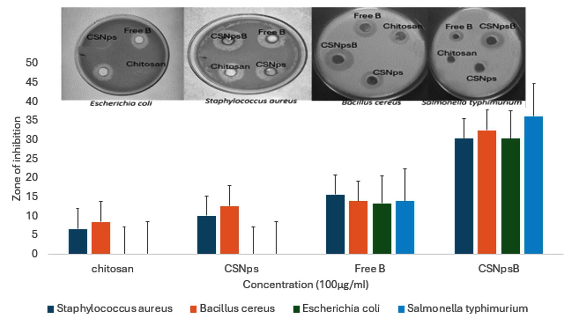
Figure 4. Antimicrobial activity of prepared Formulations
The mechanism behind the enhanced antibacterial activity of CSNps-B can be attributed to several factors. First, the nanoparticles’ small size allows them to penetrate bacterial cell walls more easily, increasing the local concentration of bacteriocin at the site of action. Second, the chitosan matrix itself possesses inherent antimicrobial properties, which may act synergistically with the bacteriocin to enhance overall effectiveness. Recent research by Rashki et al.25 demonstrated that the positive charge of chitosan nanoparticles facilitates strong electrostatic interactions with the negatively charged bacterial cell membranes, leading to increased permeability and subsequent cell death.
This enhanced effectiveness of CSNps-B is further highlighted by a comparative analysis of the four formulations in your study CSNps-B, CSNps, chitosan, and free-B reveals the distinct advantages of using CSNps-B. Chitosan alone, with inhibition zones ranging from 6.7 to 8.45 mm, exhibited minimal antibacterial activity, particularly against Gram-negative bacteria as shown in Table 2. This finding is consistent with recent studies, such as the one by Yan et al.,26 which reported that chitosan’s antibacterial efficacy is limited against Gram-negative bacteria due to the outer membrane’s protective barrier, which impedes chitosan’s penetration.
Table (2):
Summarization of activity of different formulations at different pH
| Formulations | Bacillus cereus ZOI (mm) | Staphylococcus aureus ZOI (mm) | Escherichia coli ZOI (mm) | Salmonella typhimurium ZOI (mm) | ||||||||
|---|---|---|---|---|---|---|---|---|---|---|---|---|
| pH | pH | pH | pH | |||||||||
| 2 | 4 | 12 | 2 | 4 | 12 | 2 | 4 | 12 | 2 | 4 | 12 | |
| Chitosan | 5.12 | 8.45 | 5.32 | 3.2 | 6.7 | 2.5 | 0 | 0 | 0 | 0 | 0 | 0 |
| CSNp | 3.5 | 12.6 | 5.12 | 5.6 | 10.12 | 6.10 | 0 | 0 | 0 | 0 | 0 | 0 |
| Free B | 8.12 | 13.88 | 10.10 | 10.23 | 15.67 | 8.21 | 9.34 | 13.80 | 10.12 | 8.19 | 13.76 | 10.1 |
| CSNpsB | 9.13 | 32.45 | 9.23 | 12.56 | 30.32 | 23.1 | 9.89 | 30.36 | 10.23 | 12.4 | 36.22 | 10.2 |
In contrast, CSNps, with inhibition zones ranging from 10.12 to 12.6 mm, demonstrated moderate antibacterial activity, likely due to the nanoparticles’ ability to interact with bacterial cell membranes. However, the absence of bacteriocin limits their effectiveness compared to CSNps-B. The results align with those of Kaur et al.,27 who observed that chitosan nanoparticles alone could disrupt bacterial membranes, but their antibacterial activity was significantly enhanced when combined with other antimicrobial agents.
However, Free bacteriocin (free-B) showed better antibacterial activity than chitosan and CSNps, with inhibition zones ranging from 13.38 to 15.67 mm. However, the encapsulation of bacteriocin in chitosan nanoparticles (CSNps-B) resulted in even greater efficacy, highlighting the importance of controlled release and sustained activity provided by the nanoparticle matrix. This finding is supported by Sharma et al.,28 who reported that encapsulating bacteriocins in nanoparticles not only enhances their stability but also prolongs their antimicrobial activity, making them more effective against a wider range of pathogens.
The potent antibacterial activity of CSNps-B, particularly against resistant strains of food-borne pathogens like E. coli and S. typhimurium, underscores its potential as an effective food preservative. The ability to achieve such strong inhibition with a single application suggests that CSNps-B could significantly reduce the risk of foodborne illnesses, especially in high-risk products like raw meats and dairy. Recent studies by Fadiji et al.29 have shown that the application of antimicrobial nanoparticles in food packaging can significantly extend the shelf life of perishable products while maintaining safety and quality.
Furthermore, the study examined the antibacterial activity of different formulations-chitosan, chitosan nanoparticles (CSNp), free bacteriocin (Free-B), and bacteriocin-loaded chitosan nanoparticles (CSNps-B)-against Bacillus cereus, Staphylococcus aureus, Escherichia coli, and Salmonella typhimurium across three pH levels (2, 4, and 12). The results revealed that the antibacterial efficacy of all formulations was significantly enhanced at pH 4, with CSNps-B demonstrating the highest zones of inhibition (ZOI) across all tested bacteria. For Bacillus cereus, the ZOI reached 32.45 mm at pH 4 with CSNps-B, compared to 13.88 mm with Free-B, 12.6 mm with CSNp, and 8.45 mm with chitosan. Similarly, Staphylococcus aureus showed a ZOI of 30.32 mm with CSNps-B at pH 4, while Free-B, CSNp, and chitosan achieved lower inhibition zones of 15.67 mm, 10.12 mm, and 6.7 mm, respectively. Both Escherichia coli and Salmonella typhimurium were inhibited most effectively by CSNps-B at pH 4, with ZOIs of 30.36 mm and 36.22 mm, respectively. In contrast, chitosan and CSNp exhibited no antibacterial activity against these two bacteria at any pH. These findings indicate that pH 4 is optimal for maximizing the antibacterial properties of bacteriocin-loaded chitosan nanoparticles, which emerged as the most potent formulation in the study.
Consequently, the study’s results demonstrated statistically significant differences in antibacterial activity among the various formulations at different pH levels. Notably, the zones of inhibition (ZOI) observed with bacteriocin-loaded chitosan nanoparticles (CSNps-B) at pH 4 were significantly larger compared to those achieved with free bacteriocin, chitosan nanoparticles (CSNp), and chitosan alone. These differences were statistically significant (p < 0.05), indicating that the enhanced antibacterial efficacy of CSNps-B at pH 4 is not due to random variation but reflects a true improvement in antibacterial activity. This statistical significance further supports the conclusion that pH 4 is optimal for maximizing the efficacy of bacteriocin-loaded chitosan nanoparticles, making CSNps-B the most potent formulation in the study.
The findings from this study highlight the crucial role of pH in modulating the antibacterial efficacy of different formulations, particularly bacteriocin-loaded chitosan nanoparticles (CSNps-B). The observed superiority of CSNps-B at pH 4 across all tested bacteria aligns with recent studies that emphasize the importance of environmental pH in influencing the activity of chitosan-based systems. At lower pH levels, the amino groups in chitosan are protonated, leading to increased solubility and enhanced interaction with bacterial cell membranes, which likely explains the heightened antibacterial activity observed.
The significant zones of inhibition (ZOI) recorded for CSNps-B, particularly against Bacillus cereus (32.45 mm), Staphylococcus aureus (30.32 mm), Escherichia coli (30.36 mm), and Salmonella typhimurium (36.22 mm) at pH 4, indicate the synergistic effect of combining chitosan nanoparticles with bacteriocin. This synergy is supported by recent research that suggests the nanoscale size of CSNps enhances the stability and controlled release of bacteriocins, further improving their antimicrobial effectiveness.30 In comparison, the lack of activity observed with chitosan and CSNp alone against E. coli and Salmonella typhimurium at all pH levels underscores the critical role of bacteriocin in these formulations.
Moreover, the findings are consistent with recent advancements in antimicrobial delivery systems, where the combination of natural antimicrobials like bacteriocins with nanocarriers such as chitosan nanoparticles has been shown to provide prolonged and targeted antibacterial effects.31 The pronounced activity of CSNps-B at pH 4 could also be attributed to optimal conditions for both chitosan protonation and bacteriocin stability, as suggested by recent studies on the physicochemical properties of these systems.32
In conclusion, the incorporation of bacteriocins into chitosan nanoparticles (CSNps) to form CSNps-B has significantly enhanced the antibacterial effectiveness of bacteriocins as food preservatives. This formulation not only prolongs the freshness of food items but also maintains their quality attributes. CSNps-B demonstrated exceptional antibacterial activity across a broad range of temperatures and pH levels, making it versatile for various food preservation applications. Specifically, CSNps-B showed superior antibacterial effects against Gram-positive pathogenic bacteria, particularly under acidic conditions, compared to Gram-negative bacteria. This indicates that CSNps-B is highly effective in inhibiting the growth of food-borne bacteria, making it a promising candidate for use as a bio-preservative in food products. Its ability to perform well under different environmental conditions underscores its potential for widespread application in enhancing food safety and extending shelf life.
ACKNOWLEDGMENTS
None.
CONFLICT OF INTEREST
The authors declare that there is no conflict of interest.
AUTHORS’ CONTRIBUTION
All authors listed have made a substantial, direct and intellectual contribution to the work, and approved it for publication.
FUNDING
None.
DATA AVAILABILITY
All datasets generated or analyzed during this study are included in the manuscript.
ETHICS STATEMENT
This article does not contain any studies on human participants or animals performed by any of the authors.
- Iber BT, Kasan NA, Torsabo D, Omuwa JW. A review of various sources of chitin and chitosan in nature. J Renew Mater. 2022;10(4):1097-1123.
Crossref - Wang HM, Yuan TQ, Song GY, Sun RC. Advanced and versatile lignin-derived biodegradable composite film materials toward a sustainable world. Green Chem. 2021;23(11):3790-3817.
Crossref - Pratt DY. Sorption of arsenic species using chitosan-based biopolymer sorbent materials (Doctoral dissertation, University of Saskatchewan). 2011.
- Bakshi PS, Selvakumar D, Kadirvelu K, Kumar NS. Chitosan as an environment-friendly biomaterial-a review on recent modifications and applications. Int J Biol Macromol. 2020;150:1072-1083.
Crossref - Chadha U, Bhardwaj P, Selvaraj SK, et al. Retracted: Advances in chitosan biopolymer composite materials: from bioengineering, wastewater treatment to agricultural applications. Mater Res Express. 2022;9(5):052002.
Crossref - Confederat LG, Tuchilus CG, Dragan M, Sha’at M, Dragostin OM. Preparation and antimicrobial activity of chitosan and its derivatives: A concise review. Molecules. 2021;26(12):3694.
Crossref - Tachaboonyakiat W. Antimicrobial applications of chitosan. Chitosan Based Biomaterials. 2017;2:245-274.
Crossref - Wang Y, Xu J, Yu C, et al. Prevention of bacterial biofilm formation on orthodontic brackets by non-crosslinked chitosan coating. International Journal of Biological Macromolecules, 2023;251:126283.
- Akashpriya C, Gopishankar T, Praveen N, Vasantha VL. Impact of chitosan and chitosan-based nanoparticles on genetic transformation: an overview. Role of Chitosan and Chitosan-Based Nanomaterials in Plant Sciences. 2022:387-400.
Crossref - Ibrahim SA, Ayivi RD, Zimmerman T, et al. Lactic acid bacteria as antimicrobial agents: Food safety and microbial food spoilage prevention. Foods. 2021;10(12):3131.
Crossref - Mathews G. Food and Dairy Microbiology. Scientific e-Resources. 2018.
- Sidhu PK, Nehra K. Bacteriocin-nanoconjugates as emerging compounds for enhancing antimicrobial activity of bacteriocins. J King Saud Univ Sci. 2019;31(4):758-767.
Crossref - Namasivayam AK, Pukonen M, Goshulak D, et al. Treatment intensity and childhood apraxia of speech. Int J Lang Commun Disord. 2015;50(4):529-546.
Crossref - Hoang NH, Thanh TL, Sangpueak R, et al. Chitosan nanoparticles-based ionic gelation method: a promising candidate for plant disease management. Polymers. 2022;14(4):662.
Crossref - Bohrey S, Chourasiya V, Pandey A. Polymeric nanoparticles containing diazepam: preparation, optimization, characterization, in-vitro drug release and release kinetic study. Nano Converg. 2016;3(1):3.
Crossref - Abdeltawab H, Mohamed YARI. Mobile energy storage sizing and allocation for multi-services in power distribution systems. IEEE Access. 2019;7:176613-176623.
Crossref - Balouiri M, Sadiki M, Ibnsouda SK. Methods for in vitro evaluating antimicrobial activity: A review. J Pharm Anal. 2016;6(2):71-79.
Crossref - Guirguis E. Enhancing the antibacterial effect of bacteriocin from Lactococcus lactis subsp. Lactis using chitosan nanoparticles. J Microbiol Biotechnol Food Sci. 2021;11(3):e3777-e3777.
Crossref - Mostafa AMS, Gould-Williams JS, Bottomley P. High-performance human resource practices and employee outcomes: the mediating role of public service motivation. Public Adm Rev. 2015;75(5):747-757.
Crossref - Narayanan KB, Bhaskar R, Han SS. Bacteriophages: Natural antimicrobial bioadditives for food preservation in active packaging. Int J Biol Macromol. 2024;133945.
Crossref - Chen X, Ma W, Hu W, et al. Progress in release-activated food packaging films. Packaging Technology and Science. 2023;36(10):889-902.
Crossref - Niazmand R, Razavizadeh BM, Sabbagh F. Simulating release model and antimicrobial efficiency of LDPE film carrying ferula asafetida leaf and gum extracts. Polymer Bulletin. 2022;79(2):1151-1174.
Crossref - Ali MM, Shoukri RA, Yousry C. Thin film hydration versus modified spraying technique to fabricate intranasal spanlastic nanovesicles for rasagiline mesylate brain delivery: Characterization, statistical optimization, and in vivo pharmacokinetic evaluation. Drug Deliv Transl Res. 2023;13(4):1153-1168.
Crossref - Mohanty D, Suar M, Panda SK. Nanotechnological interventions in bacteriocin formulations-advances, and scope for challenging food spoilage bacteria and drug-resistant foodborne pathogens. Crit Rev Food Sci Nutr. 2023:1-18.
Crossref - Rashki S, Asgarpour K, Tarrahimofrad H, et al. Chitosanbased nanoparticles against bacterial infections. Carbohydr Polym. 2021;251:117108.
Crossref - Yan D, Li Y, Liu Y, Li N, Zhang X, Yan C. Antimicrobial properties of chitosan and chitosan derivatives in the treatment of enteric infections. Molecules,. 2021;26(23):7136.
Crossref - Kaur M, Cohen Y, Poverenov E, Eltzov E. Binding selectivity of N-alkylaminated modified chitosan nanoparticles produce a synergistic antibacterial effect against gram-negative strains. React Funct Polym. 2023;186:105567.
Crossref - Sharma P, Yadav M. Enhancing antibacterial properties of bacteriocins using combination therapy. J Appl Biol Biotechnol. 2023;11(1):232-243.
- Fadiji T, Rashvand M, Daramola MO, Iwarere SA. A review on antimicrobial packaging for extending the shelf life of food. Processes. 2023;11(2):590.
Crossref - Derakhshan-Sefidi M, Bakhshi B, Rasekhi A. Thiolated chitosan nanoparticles encapsulated nisin and selenium: antimicrobial/antibiofilm/anti-attachment/immunomodulatory multi-functional agent. BMC Microbiol. 2024;24(1):257.
Crossref - Singh BP, Rohit, Manju KM, et al. Nano-conjugated food-derived antimicrobial peptides as natural biopreservatives: a review of technology and applications. Antibiotics. 2023;12(2):244.
Crossref - Mirbagheri VS, Alishahi A, Ahmadian G, Petroudi SHH, Ojagh SM, Romanazzi G. Toward understanding the antibacterial mechanism of chitosan: Experimental approach and in silico analysis. Food Hydrocolloids. 2024;147(Part A):109382.
Crossref
© The Author(s) 2024. Open Access. This article is distributed under the terms of the Creative Commons Attribution 4.0 International License which permits unrestricted use, sharing, distribution, and reproduction in any medium, provided you give appropriate credit to the original author(s) and the source, provide a link to the Creative Commons license, and indicate if changes were made.