

ISSN: 0973-7510
E-ISSN: 2581-690X
This study reports the effect of different levels of wheat/cassava composite flour on liver architecture of wistar rat. The cassava variety- TMS 98/419 and Twenty (20) male wistar rats were used for the study. The ages of the wistar rats ranged between 10-12 weeks and weighed 90-100 g. After one (1) week period of adaptation, the rats were divided randomly into five groups (A, B, C, D and E) of four (4) rats per group. Group A served as the control while groups B, C, D and E were the experimental groups. The blood glucose levels and body weight of the animals were monitored. The result showed that among the different rat groups, their body weights increased from week 2 of the study while their blood glucose levels significantly decreased (P<0.05) with increase in the quantity of cassava flour. The histo-pathological analysis showed that from 20 % cassava flour substitution, significant distortion of the liver architecture of the wistar rats were observed resulting in intra-hepatic haemorrhage as well as necrosis. The result revealed that 10 % cassava flour substitution is adequate as this has no effect on the liver architecture of the wistar rat.
Wistar rat, wheat flour, cassava flour, composite bread, and liver architecture.
Bread is one of the most widely consumed food products in the world and bread making technology is probably one of the oldest technologies known1. It is an important staple food for many countries. The product is basically made of hard wheat flour, yeast, fat, sugar, salt and water2. It is a cereal product that is naturally low in protein and nutritionally not a balanced diet because it is low in lysine, an essential amino acid3.
Due to the high cost, geographical scarcity and high demand for wheat flour, efforts are being directed toward the provision of alternative source of flour, for bread making. Hofmann et al.4 reported that composite bread can be made by substituting 5, 10, 15, 20 and 30% plantain flour for wheat flour. According to Idowu et al5, the possibility of using starchy staples for bread making depends on the physical and chemical properties of the product. In the light of this, cocoyam, cassava, taro and other tuber crops have been found to be alternative sources of major raw materials for bread making6.
Cassava (Manihot esculenta Crantz) is a perennial crop, it grows well in the tropical poor soil and can withstand drought. Nigeria is ranked highest in the production of cassava7. In spite of its high cyanide content, cassava products with encouraging international market, efforts are being made to achieve the quality standards especially if processed to reduce its cyanide content8. According to Giami et al6, up to 20 % substitution of cassava flour had no adverse sensory and organoleptic effects on bread. Because of this, wheat-cassava composite bread is gaining general acceptance.
Nigeria and most developing countries are largest importer of American red winter wheat9. This implies that these countries are totally dependent on foreign country for their bread production. Therefore, the use of wheat-cassava flour for production of baked goods would help to lower the dependency of developing nations on imported wheat. Although cassava is known to be high in cyanide, new and improved cassava varieties have been found to be in low cyanide levels. Thus, the purpose of this study is to determine the effect of wheat-cassava composite bread on the liver architecture of wistar rat.
Source of Materials
Cassava variety (Tropical Manihot Species (TMS) 98/419) was purchased from Ebonyi State Agricultural Development Programme (EBADEP) Onuebonyi, Ebonyi State. The wheat flour, margarine, sugar, concentrated milk, salt, and dried yeast were purchased from Abakpa Main Market, Abakaliki, Ebonyi State, Nigeria.
Preparation of Cassava Flour
The cassava variety TMS 98/419 was washed with tap water and peeled using stainless steel knife and sliced into chips. The sliced cassava chips were soaked in tap water for 5 hours. The soaked cassava chips were sun-dried until the moisture content was reduced to less than 15 %. The dried cassava chips were milled into flour using attrition milling machine. After milling, the flour was sieved using sieve mesh size 20, to achieve uniform particle size. The cassava flour was used together with wheat flour for the bread production.
Bread Baking Procedure
The dough was prepared using straight dough method. The dough was prepared with 100 % wheat flour, 90/10, 80/20, 70/30 and 60/40 % wheat-cassava flour substitutions. The bread baked with 100 % wheat flour was used as control. Six hundred grams (600g) of the dough was baked at 230 oC for 20 minutes using gas oven. Baking was done in duplicates.
Experimental Animals
Twenty (20) male Wistar rats (Rattus norvegius) were purchased from the Animal House of the Department of Anatomy, Ebonyi State University, Abakaliki, Nigeria. The ages of the rats ranged between 10-12 weeks and they weighed between 90-100 g. They were housed in iron cages containing hard wood chips for bedding in controlled animal house (25±2ºC). The animals were allowed to acclimatize in the animal house of the department of Biochemistry, Ebonyi State University, Abakaliki, for one (1) week and were fed with standard livestock pellets (Guinea Feed Nigeria Limited).
Experimental Design
After acclimatization, the rats were divided randomly into five groups (A, B, C, D and E) with four rats each. The rats were marked with a permanent marker for easy identification. Group A served as the control while groups B, C, D and E were the experimental groups. The rats were fed with bread baked with different compositions of composite flours. Group B animals received 10 % concentration of cassava flour and 90 % of wheat flour; Group C animals received 20 % concentration of cassava flour and 80 % of wheat flour; Group D animals received 30 % concentration of cassava flour and 70 % of wheat flour while Group E animals received 40 % concentration of cassava flour and 60 % of wheat flour. At the end of each week, one animal from each compartment was fasted overnight. The animal was sacrificed and dissected, the liver was removed, weighed and frozen (-20oC).
Bread Moisture Content Determination
The method of AOAC10 was used in determining the moisture content of the bread. This was done to determine the shelf stability of the bread. The determination was done in triplicates.
Statistical analysis
The data obtained was analyzed using SPSS 16.0. One-Way Analysis of Variance (ANOVA) and mean separation of the values were carried out using Duncan test at 5 % significant level.
Effect of Cassava Floor Substitution on the moisture content of the bread
Results are means of three independent determinations. Mean values having different superscripts within the same column are significantly different (pÂ0.05).
Table (1):
Moisture Contents of Wheat-Cassava Composite Bread.
Sample |
MC (%) |
|---|---|
100 % wheat (Control) |
12.47±0.06d |
10 % Cassava flour |
12.55±0.05d |
20 % Cassava flour |
13.79±0.10c |
30 % Cassava flour |
14.32±0.10b |
40 % Cassava flour |
15.10±0.06a |
Results are means of three independent determinations. Mean values having different superscripts within the columns are significantly different (p<0.05) while mean values with same superscripts within column are the same.
Results of the Histopathological Effects of the Diets on the Rats Figures 1-15 shows the results of the histopathological effects of the diets on the animals.
The moisture contents of wheat-cassava composite bread ranged from 12.47 to 15.10 %. Moisture contents increased significantly as the substitution of cassava flour increased with the highest moisture content (15.10 %) obtained for bread sample containing 40 % cassava flour while the least moisture content (12.47 %) was obtained for bread control sample containing only wheat flour. This could be due to the fact that cassava flour is fairly high in moisture11. Results showed that there were significant differences (pÂ0.05) in all the moisture contents except for bread control sample and bread containing 10 % cassava flour, which showed no statistical difference. The moisture content values obtained for this study is lower than 30.10-34.03 % reported by Masamba and Jinazali12 for bread made from wheat- fermented and unfermented cassava flour blends. However, similar results have been reported by Eddy et al13 for wheat-cassava composite bread. At the baking temperature (which is normally greater than 100°C) the moisture content of the raw samples must have been greatly reduced. However, different food materials have different capacities for absorbing/retaining moisture which may exist as occluded or absorbed water. As a result, it can be deduced that even at the high baking temperature, some moisture are still present in the samples.
Table (2):
Effect of the Diets on the Weight Gain of the Wistar Rats.
|
Weight (g) |
|||
|---|---|---|---|
| Sample | Initial Weight (g) |
Final weight (g) |
Weeks |
| Control 100% wheat | |||
| Rat 1 | 84.20±0.10f | 41.00±1.00n | week 1 |
| Rat 2 | 73.10±0.10j | 86.40±0.10f | week 2 |
| Rat 3 | 75.17±0.59i | 145.40±0.10b | week 3 |
| 10 % cassava | |||
| Rat 1 | 76.00±1.00i | 48.20±0.10o | week 1 |
| Rat 2 | 83.10±0.10e | 94.20±0.10k | week 2 |
| Rat 3 | 80.30±0.10h | 93.20±0.10i | week 3 |
| 20 % cassava | |||
| Rat 1 | 61.20±1.00n | 43.28±0.08l | week 1 |
| Rat 2 | 45.100±1.00m | 66.40±0.10j | week 2 |
| Rat 3 | 57.10±1.00c | 85.17±0.10h | week 3 |
| 30 % cassava | |||
| Rat 1 | 127.50±0.10b | 96.40±0.10e | week 1 |
| Rat 2 | 97.20±0.10d | 109.30±0.10c | week 2 |
| Rat 3 | 134.4±0.10a | 156.20±0.10a | week 3 |
| 40 % cassava | |||
| Rat 1 | 65.10±0.10l | 47.60±0.10g | week 1 |
| Rat 2 | 83.00±1.00g | 103.40±0.10d | week 2 |
| Rat 3 | 42.30±0.10k | 69.40±0.10m | week 3 |
The result presented in Table 2 shows the weight of wistar rat fed with wheat-cassava bread. It was observed that among the different rat groups, the wistar rat weight decreased after one week but increased on the second and third week of being fed with the composite bread. The initial reduction in the wistar rats’ weights in the first week could be as a result of the wistar rats’ initial rejection of the feed as the feed is different from the wisrar rats’ normal feed. The increased body weight can be attributed to abnormality in nutrient absorption by the liver which results in fat accumulation. Fat accumulation is more frequently observed in the liver, since this is the main organ involved in lipid metabolism. Lipid content in hepatocytes is regulated by the activities of cellular enzymes that catalyze lipid uptake, synthesis, oxidation and externalization from the cell. When the fat amount that enters into hepatocytes exceeds the capacity for their oxidation or externalization, hepatic steatosis settles14.
Figure 1 shows the micrograph of the liver’s control in first week. The result showed normal hepatocyte (NH) with normal cytoplasm. The normality observed showed that the diet had no lethal effect on the liver of the rats. This result agrees perfectly with the result obtained by Orisakwe et al. 15.
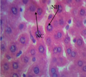
Fig. 1. The photomicrograph section of the liver’s control in first week, stained with H/E (X 600) shows normal hepatocyte (NH) with normal cytoplasm. The overall architecture of liver is normal.
Figures 2 –5 showed the micrograph of the liver tissue of experimental animals fed with varying compositions of the composite bread after a period of 1 week. Figures 4 and 5 showed the photomicrograph of the liver of wistar rat fed with 40 percent cassava and 60 percent wheat composite bread showing moderate fatty change (MFC) and granulated cytoplasm (GC). Thus, the cytoplasm of this liver is affected. The disintegration of cytoplamsmic membrane might likely lead to disruption in the filtration and concentration of urine, it may also affect the fluid and electrolytic balance of the rat as well as regulation of total body homeostasis. Disruption of cellular membranes has been reported as one of the consequences of ingestion of oxidized lipids16, 17. This is as a result of peroxidation of membrane phospholipid due to free radical attack18. This leads to the formation of lipid peroxides. The toxicity of this compound is usually evidenced by an elevation of serum marker enzymes, such as aspartate aminotransferase (AST), alanine aminotransferase (ALT) and gamma glutamyl transferase (GGT) as reported by Palanivel et al19. The changes mostly include hepatocellular necrosis or apoptosis, fatty accumulation, inflammatory cells infiltration and other histological manifestations20. Figure 6 shows the results of the micrograph of the liver tissue of control animal after two weeks. The results revealed normal hepatocyte (NH) with normal cytoplasm (NC). This result indicates the absence of lethal constituents in the meal fed to the control animal.
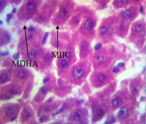
Fig. 2. The photomicrography section of the liver’s 10% cassava flour and 90% wheat flour in first week, stained with H/E (X 600) shows mild distortion of hepatic architecture (MDHA) with mild infiltrate of inflammatory cell (MIIC).
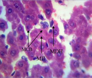
Fig. 3. The photomicrography section of the liver’s 20% cassava flour and 80% wheat flour in first week, stained with H/E (X 600) shows moderate distortion of hepatic architecture (MDHA), with moderate infiltrate of inflammatory cell (MIIC) and mild congestion of the blood vessel
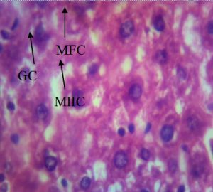
Fig. 4. The photomicrography section of the liver’s 30% cassava flour and 70% wheat flour in first week, stained with H/E (X 600) shows moderate fatty change (MFC), with moderate infiltrate of inflammatory cell (MIIC) and granulated cytoplasm (GC).
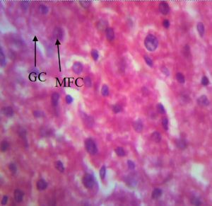 Fig. 5. The photomicrography section of the liver’s 40% cassava flour and 60% wheat flour in first week, stained with H/E (X 600) shows moderate fatty change (MFC) and granulated cytoplasm (GC). Thus the cytoplasm of this liver is affected.
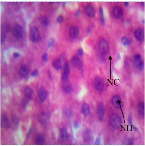
Fig. 6. The photomicrography section of the liver’s control in second week, stained with H/E (X 600) shows normal hepatocyte (NH) with normal cytoplasm (NC). The overall architecture of liver is normal.
Figures 7 – 10 shown above are the micrographs of the liver tissue of experimental animals fed with varying compositions of the composite bread after a period of 2 weeks. Figure 7 is the result obtained after feeding the rat with 10 percent cassava and 90 percent wheat composite bread showing mild fatty change (MFC) with mild infiltrate of inflammatory cell (MIIC) and moderate distortion of hepatic architecture (MDHA). Figure 8 showed the photomicrograph of the liver of wistar rat fed with 20 percent cassava and 80 percent wheat composite bread. The result shows moderate fatty change (MFC) with mild infiltrate of inflammatory cell (MIIC). Also, Figure 9 is the photomicrograph of the liver of wistar rat fed with 30 percent cassava and 70 percent wheat composite bread showing clumping of the hepatic cell (CH) with congestion of hepatic vessel. Figure10 showed the photomicrograph of the liver of wistar rat fed with 40 percent cassava and 60 percent wheat composite bread shows hypertrophic hepatocyte (HH) with moderate fatty change (MFC). It has been reported that when cassava is eaten, most of the ingested cyanide is converted into thiocyanate, a reaction catalysed by the enzyme Rhodanase, which uses up part of the pool of S-containing essential amino acids methionine and cysteine/cystine21. Consumption of cassava products containing large amounts of cyanide can cause acute intoxication, with symptoms of dizziness, headache, nausea, vomiting, stomach pains, diarrhoea and sometimes death22. Other health disorders include hyperthyroidism, tropical ataxic neuropathy, and konzo23. Thus, the emphasis should be on using cassava that is well processed to avoid cyanide poisoning in the cassava composite bread.
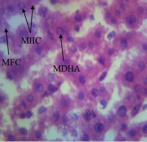
Fig. 7. The photomicrography section of the liver’s 10% cassava flour and 90% wheat flour in second week, stained with H/E (X 600) shows mild fatty change (MFC) with mild infiltrate of inflammatory cell (MIIC) and moderate distortion of hepatic architecture (MDHA).
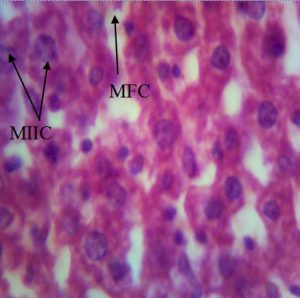
Fig. 8. The photomicrography section of the liver’s 20% cassava flour and 80% wheat flour in second week, stained with H/E (X 600) shows moderate fatty change (MFC) with mild infiltrate of inflammatory cell (MIIC).
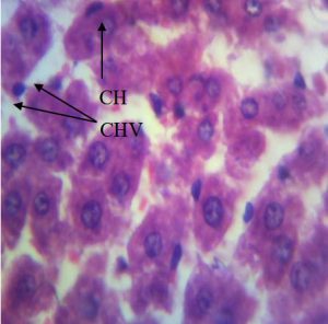
Fig. 9. The photomicrography section of the liver’s 30% cassava flour and 70% wheat flour in second week, stained with H/E (X 600) shows clumping of the hepatic cell (CH) with congestion of hepatic vessel.
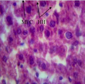
Fig. 10. The photomicrography section of the liver’s 40% cassava flour and 60% wheat flour in second week, stained with H/E (X 600) shows hypertrophic hepatocyte (HH) with moderate fatty change (MFC).
Figure 11 shows the micrograph of the liver’s control in third week. The result obtained showed that although there was normality in the hepatocytes as the weeks increased a mild separation (MS) occurred on the liver. This result is supported by Longman-Adman,24 who also observed that the progression in the duration of storage causes organ dysfunction.
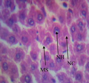
Fig. 11. The photomicrography section of the liver’s control in third week, stained with H/E (X 600) shows normal hepatocyte (NH) with normal cytoplasm (NC).
Figures 12 – 15 shown above are the micrographs of the liver tissue of experimental animals fed with varying compositions of the composite bread after a period of 3 weeks. Figure 12 is the result obtained after feeding the rat with 10 percent cassava and 90 percent wheat composite bread showing mild fatty change (MFC) with mild separation of hepatic tissue (MSHT) and congestion of the hepatic tissue (CHT). Figure13 showed the photomicrograph of the liver of wistar rat fed with 20 percent cassava and 80 percent wheat composite bread. The result shows mild fatty change (MFC) with change in hepatic vessel (CHV) and granulated cytoplasm (GC). Figure14 is the photomicrograph of the liver of wistar rat fed with 30 percent cassava and 70 percent wheat composite bread showing mild infiltration of inflammatory cells (MIIC) with intra-hepatic haemorrhage (IHH). Figure 16 showed the photomicrograph of the liver of wistar rat fed with 40 percent cassava and 60 percent wheat composite bread shows hepatic necrosis (HN). Hepatic necrosis is caused by fat accumulation in the liver, and this condition may be caused by the presence of toxic substances25. Animal models exposed to hydrogen cyanide demonstrated extensive CNS toxicity, including dyspnea and vomiting, with vascular and cellular CNS lesions identified post-mortem [26]. The toxic manifestations strongly suggest that high dose of cyanide in diet in association with low protein intake could precipitate hepatic dysfunction.
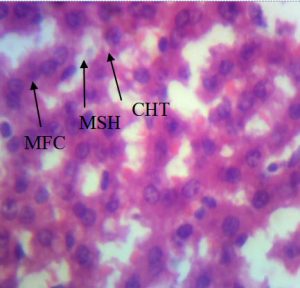
Fig.12. The photomicrography section of the liver’s10% cassava flour and 90% wheat flour in third week, stained with H/E (X 600) shows mild fatty change (MFC) with mild separation of hepatic tissue (MSHT) and congestion of the hepatic tissue (CHT).
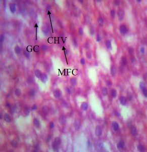
Fig. 13. The photomicrography section of the liver’s 20% cassava flour and 80% wheat flour in third week, stained with H/E (X 600) shows mild fatty change (MFC) with change in hepatic vessel (CHV) and granulated cytoplasm (GC).
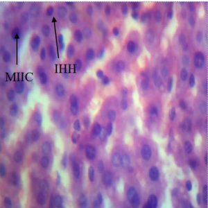
Fig. 14. The photomicrography section of the liver’s 30% cassava flour and 70% wheat flour in third week, stained with H/E (X 600) shows mild infiltration of inflammatory cells (MIIC) with intra-hepatic haemorrhage (IHH)
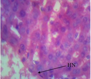
Fig. 15. The photomicrography section of the liver’s 40% cassava flour and 60% wheat flour in third week, stained with H/E (X 600) shows hepatic necrosis (HN).
Substitution of wheat flour with cassava flour in bread making is desirable. But this should be done at 10 – 20 % substitution. This is because substitution of more than 20 % will lead to granulated cytoplasm, hypertrophic hepatocyte and hepatic necrosis of the liver.
- Selomulyo, V. O and Zhou, W. Effects of Freezing Storage and Dough Improvers. Journal of Cereal Science, 2007; 45(1): 200-2007.
- Okafor, J. N., Okafor, G. I., Ozumba, A. U and Elemo, G. N. Ouality Characteristics of Bread Made from Wheat and Nigerian Oyster Mushroom (Pleurotus plumonrius) Powder. Pakistan Journal of Nutrition, 2012; 11(1): 5-10.
- Agu, H.O., J.A. Ukonze and K.A. Paul, Quality characteristics of bread made from wheat and fluted pumpkin seed flour. Nig. Food J., 2010; 28: 188-198.
- Hofmann, T., Lindenmeier, M. and Somoza, V. Pronyl-lysine-A Novel Protein Modification in Bread Crust Melanoidins Showing In Vitro Antioxidative and Phase1/11 Enzyme Modulating Activity. Annals of the New York Academy of Sciences, 2005; 1043: 887.
- Idowu, M. A.,Oni, A and Amusa, B. M. Bread and Biscuit Making Potentials of Some Nigerian Cocoyam Cultivars. Nigerian Food Journal, 1996; 14: 1-12.
- Giami, G. Y, Amasisi, T and Eklyor, G. Comparison of Bread Making Properties of Composite Flour from Kernels of Roasted and Boiled African Bread Fruit (Treculla africana seed). J. M. Res., 2004; 1(1): 16-25.
- IITA (International Institute of Tropical Agriculture (IITA). Competitiveness Workshop. In opportunities for Cassava in Nigeria: Bookanga, IITA, Ibadan 2005.
- FAO/WHO 2004. Provision of Scientific Advice to Codex and Member Countries. Report of Joint FAO/WHO Workshop. WHO Headquarters, Geneva, Switzerland. 27-29 January 2004.
- David M.O., Nigeria, No. 1 market for U. S. wheat; potential for other grains and feeds, USAD Foreign Agric. Serv. Bull., 2006; pp. 1-2.
- AOAC. Official Methods of Analysis of Association of Official Analytical Chemists, Washington D.C, USA 1990.
- Apea-Bah F.B, Oduro I., Ellis W.O. and O. Safo-Kantanka, Factor Analysis and Age at Harvest Effect on the Quality of Flour from Four Cassava Varieties. World Jr. of Dairy and Fd Sc. 2011; 6(1): 43-54.
- Masamba K and H. Jinazali, Effect of Cassava Flour Processing Methods and Substitution Level on Proximate Composition, Sensory Characteristics and Overall Acceptability of Bread Made From Wheat- Cassava Flour Blends. Afr.Jr. of Food, Agric., Nutr and Devpt, 2014; 14(6).
- Eddy, N. O., Udofia, P. G. and Eyo, D. Sensory evaluation of wheat/cassava composite bread and effect of label information on acceptance and preference. African Journal of Biotechnology, 2007; 6(20): 2415-2418.
- Koteish A, A.M. Diehl, Animal models of steatosis. Semin. Liver. Dis. 2001; 21(1):89-104
- Orisakwe, O.E., A.A. Njan, O.J. Afonne, D.D. Akumka, V.N. Orish and O.O. Udemezue, Investigation into the nephrotoxicity of Nigerian Bonny Light Crude Oil in Albino Rats. International Journal of Environmental Research and Public Health, 2004; 1(2): 106-110.
- Odutuga, A.A., Obaleye, J.A. and Ologan, F.O. Themoxidized Soyabean oil: Spectroscopic investigation and the effects on selected rat tissues. Biokemistri, 1997; 7: 45-58.
- Ologan, F.O., Some physicochemical properties of thermally oxidized groundnut oil and their toxicological effects on selected rat tissues. Ph.D Thesis. Dept of Biochemistry, University of Ilorin, 2002; Nigeria.
- Van Ginkel, G and Sevanian, A. Lipid Peroxidation Induced Membrane Structural Alteration. Metho. Enzymol. 1994; 233: 273-288.
- Palanivel G.M., Rajkapoor, B., Senthil K. R, Einstein J.W., Kumar E.P,Rupesh Kumar M, Kavitha K, Pradeep Kumar M and B. , Jayakar, Hepatoprotective and antioxidant effect of Pisonia aculeataL. against CCl4- induced hepatic damage in rats. Sci. Pharm. 2008; 76(2): 203–215.
- Khan AA, Alzohairy, M. Hepatoprotective effects of camel milk against CCl4-induced hepatotoxicity in Rats. Asian J Biochem 2011; 6(2):171–180
- Cardoso A.P., Mirione, E., Ernesto, M., Massaza, F., Cliff, J., M.R. Haque, Processing of cassava roots to remove cyanogens. J Food Comp Anal, 2004; 18:451– 460.
- Mlingi, N., Poulter, N.H. and, H. Rosling. An outbreak of acute intoxications from consumption of insufficiently processed cassava in Tanzania. Nutr Res, 1992; 12:677–687.
- Osuntokun, B. O. Cassava Diet, Chronic Cyanide Intoxication and Neuropathy in Nigeria. World Rev. Nutr Diet, 1981; 36:141-173.
- Longman-Adman, N., Renal effects of Environmental and occupational lead exposure. Environmental Health Perspective, 1997; 105(9): 938.
- Adhal N.H., D. Manning, Diagnosis of Liver Fibrosis. Gastroenterol Clin. Biol. 2008; 32: 88-90.
- Valade M.P. Central Nervous System Lesions in Chronic Experimental Poisoning with Gaseous Hydrocyanic acid. Bull. Acad. Nat. Med. 1952; 136: 280-285.
© The Author(s) 2018. Open Access. This article is distributed under the terms of the Creative Commons Attribution 4.0 International License which permits unrestricted use, sharing, distribution, and reproduction in any medium, provided you give appropriate credit to the original author(s) and the source, provide a link to the Creative Commons license, and indicate if changes were made.