


ISSN: 0973-7510
E-ISSN: 2581-690X
The crude alcoholic extract of Spirugera sp. algae with concentrations (250, 500, 1000) mg/ml and albendazole with concentrations (250, 500, 1000) mg/ml effects was studied in the viability of protoscolices in vitro, the viability of the protoscolices in 10 microliters and the three replicates calculated in the fixed-size method. There were no significant differences in the first three treatments (0,24,48 hour), whereas the coefficients of 72-144 hour showed significant differences . The mean S.D. ± viability of the protoscolices at the highest rate at treatment at 0 hour (33 ± 5.29) protoscolices, while the mean of the protoscolices dropped to the lowest ratio at 144 to 14 ± 1 protoscolices. While the mean S.D.± mean of the protoscolices viability of the treatments 24-120 hours between 31.33 ± 1.52-16.66 ± 1.52 protoscolices. The total number of protosclices ranged from 177-193. The average number of live protoscolices ranged between 170-177 protoscolices / 100 µl , While the number of dead protoscolices ranged from 6-16 protoscolices / 100µl. Sixty-nine chemical compounds were extracted from the crude alcoholic extract of Spirogyra sp. Varied between phenols, turbines, fatty acids, hydrocarbons and hetero cyclic compounds. The results of this study showed a decrease in the percentage of the viability of the protoscolices when exposed to the concentrations of the crude alcoholic extract of the Spirugera sp. The concentration of 250 mg / ml resulted in a decrease in the percentage of protoscolices from 92.75±0.62 at 0 hour to 27.06±0.48 at 144 hour compared with control group (76.56 ± 0.79), while the concentration of 500 mg/ml decreased from 92.59±0.58 per hour to 0.14±0.25% at 144 hours compared with the control group, while the concentration was 1000 mg /ml has led to the killing of all protoscolices at 144 hour compared with the control group, The results of used albendazole drug showed significant differences. concentrations reduced the percentage of viability of protoscolices. When exposed to 250 mg / ml, The result was a decrease from 93.75 ± 0.77 at 0 hour to 4.15 ± 0.36 at 144 hour, while the concentration of 500 mg / ml reduced the percentage from 48 ± 0.85 at 0 hour to 0,4±0.48 at 144 hour, while The concentration of 1000 mg / ml resulted in the killing of all protpscolices indications at 120 hour compared to 0 hour where the percentage of viability of protoscolices was 45.5 ± 0.98. The results of this study showed that a similar effect of a Spirogyra sp extract and albendazole drug on the viability of the protoscolices with increased concentration and duration of exposure to the drug or alcoholic extract.
Green Algae, Spirugera, Hydatid cysts Disease
Hydatid cyst disease (HCD) or cystic echinococcosis (CE) is a serious and important zoonotic disease (Zhang et al., 2017; De NV & Le van D 2017), an endemic disease (Al-Mayah et al., 2012). This disease results in the infection of the intermediated host (sheep, cattle and other livestock as well as humans) in the larval stage of Echinococcus granulosus of the Taeniidae family (Duman et al., 2016).
The disease spreads in livestock breeding areas and carnivorous animals that are in close contact with humans and which help to complete the life cycle of the parasite causing the disease (Hayajneh et al., 2014). The life cycle is complete with two speies of hosts, the intermediated host such as Sheep, Cows, Horses, Camels, Pigs and other animals (WHO, 2017) and the final host are Dogs, Wolves, Foxes and other animals where the adult parasite is present in the intestines and has no symptoms, (Duman et al., 2016) As well as the human who is sometimes an accidental host interrupted by the life cycle of the parasite(Rampal et al, 2016; WHO, 2017; Zhang et al., 2017; Mestrovic, 2016).
The disease affects many organs in the human body and the intermediate host, The most common organs are the liver, lung, spleen, kidney, heart, brain, urethra, uterus, fallopian tube, pancreas, mesenteric membrane, muscle and other organs.and injury The brain is more common in children (Khurana et al., 2012).
Echinococcosis is an endemic disease in large parts of the world (Pradhan et al., 2017), such as some of the countries of South America and the Middle East and North Africa, which are Hippendemicregions of the disease (McManus et al., 2012). Including the Arab countries, such as Egypt, Sudan, Libya, Algeria, Lebanon, Palestine, and Iraq (Sadadjadi, 2006).
In the seventh decade of the 20th century, researchers aimed to use benzemidazole and its derivatives such as benzadazole (ABZ), mebendazole (MBZ), flopendazole, and other drugs as a treatment for hydatid cysts disease (Bekhtiet al., 1977). Because of its high solubility and its entry into the hydatid cyst before the completion of its layers compared with other treatments, resulting in a decrease in the size of the cyst (Brunetti, 2015). It is the main routine treatment after the surgical removal of hydatid cyst. Studies show that about 30% Aqueous has completely treated Andasul Alps, while 40-50% have responded after a short period of treatment (Hejazi et al., 2016), then the effects of this treatment appeared in the body, especially when used for a long time. Because of the negative effects of chemotherapy, such as impaired liver function, diarrhea, nausea and abdominal pain, as well as the possibility of the return of infection in some cases and the resistance of parasite to some therapeutic materials, so many researchers went to the use of medicinal plants trying to find alternative treatments with less or without side effects, Cost and research worldwide (Koshkiet al., 2017).
Several algae species, which can be of a very high nature, have been used for new sales of biological activity (Athbi, 2017), including micro-algae, which are important in medical and commercial terms for containing high-value active materials and processing molecules. Disease and may act as antibiotics for a large number of microorganisms including parasites (Monoz and Fernandez, 2017), being able to produce a wide range of active biochemical compounds, immunosuppressant, cytotoxic and enzyme inhibitors (Kassemi et al., 2004).
Most of the compenentes isolates from algae or plants belong to the group of phenols, tannins, peptides, Saponins, triterpenes, and alkaloids (Molera and Semesi, 1996). One of these algae is Spirogyra sp. . (Liani and Katoch, 2017), a rich source of natural chemicals that can be derived from simple methods (Verma and Khan, 2016; Pacheco et al., 2015).
The present study extracted medically material contained in Spirogyra Sp.by Gas Chromatography-Mass Spectrometry (GC/MS). As well as a statement of the effect of the extract in reducing the viability of the protoscolices of the parasite Echinococcusgranulosus compared with the albendazole drug in vitro.
Collection and preparation of algae samples
Spirpgyra sp. (Fig. 1) Collected during the of September , 2017 from the rice fields in the Qadisiyah and Manathira districts area in Najaf province , then samples transfer in plastic bottles after immersion in distilled water to laboratory in the Department of biology / faculty of Education / University of AL- Qadisiyah university .The algae filaments were isolated under an inverted microscope( Optika , Italy) and then placed in test tubes, and then washed 8 times per tube using a cold centrifuge 3000 cycles / min for 15 minutes during which the water was replaced after each wash.
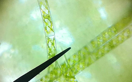
Fig. 1. Spirugera sp. (X400)
After ensuring no impurities, algae filaments were placed in a 500 mL conical container containing BG11 feed medium, placed in a culture chamber below 25 ° C and using fluorescent lamps for two months. Then the living mass of Spirogyra sp. It’s ready to extract.
Preparation of the extract
Active substances were extracted according to Taskinet al. (2007) with some modulation. Algae were dried at room temperature and dry matter was weighed. 50g dry weight was obtained for Spirogyra sp. . The dry mass was then kept at a temperature of 20 ° C, followed by 1g dry matter per moss and placed in the Thamble. then put the container in the place allocated to the Soxhlet and added 250 ml of ethanol 70% and left the sample for two hours to solvent saturated solvent, after the extraction was carried out in the Soxhlet until the leaky leachate near the water. Then the liquid leachate was dried using the incubator device at a temperature of 37 °C and then the resulting dry matter was weighed. 12 g dry weight was obtained for the extract.
Determination of active substances in the extract
The chemical compounds in the crude alcoholic extract of the Spirugera sp. were detected using (GC-Mass) gas chromatography Mass type (Japanese-made Agilent Technologies GC-mass 7890 AGC System) in the Ministry of Science and Technology / Baghdad. The machine works with the GC clarus 500 Perkin Elmer system which includes the [AOC-20i + s] auto sampler for vehicles and the gas chromatography associated with mass spectromrtry and gas chromatography for compounds.
Collection of Hydatid Cysts
The hydatid cysts (Fig. 2) were collected from sheep from the Al-Najaf abattoir was brought to the Department of biology / Faculty of Education / Al-Qadissiyah University immediately after the slaughter of infected sheep in plastic containers chilled with ice so that the protoscolices inside the bag are not affected by ambient temperature (Smyth, 1976).
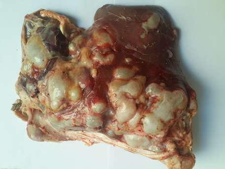
Fig. 2. Severe infected with hydatid cyst in liver of sheep
Preparation of Protocolices
Estimate of Protoscolices Viability
The viability of protoscolices was assessed By method of Samyth and Barett (1980) Where it takes a certain size of the protoscolices suspension in the PBS solution with the same volume of Eosin Aqueous Stain (0.1%) and using the Micropipette, then stir well and took a drop and examined it under the light microscope, Olympus (Japan) and under the 10×40 magnification force were calculated. The percentage of live protoscolices, which are bright green, where the stain did not enter its membranes, compared to the protoscolices red-colored that were dead because their membranes had become pigmented (Fig. 4) the number of protpscolices live in the sample divided on the total number of calculated protoscolices x 100 and three replicates.
Percentage of viability = [Number of live primary visions/Total number of primary visions in the calculated sample] x 100
Smyth (1985) method was used to obtain the protoscolices. The surface of the hydatid cysts was sterilized using 70% ethyl alcohol, then the fluid was withdrawn with the protoscolices using a 10 ml syringe with a 21 gauge needle to reduce pressure inside the hydatid cyst , then opened the hydatid cysts with a sterile medical scalpel and extracted the secondary hydatid cysts and the germinated layer and placed in a sterile Petri dish containing a solution of Phosphate Buffered Saline (PBS) (Figure 3). The generated layer was washed with solution (PBS) to collect the protoscolices, This suspension contains the protoscolices in the test tubes,A centrifugation process was carried out using the centrifuge, LWS M24 Combo three times at 3000 cycles per minute for 15 minutes per deposition. In the first wash, use the PBS solution and add 2000 units / L of penicillin and 1 G / l of streptomycin before starting the second wash. In the third wash, the Phosphate Buffered Saline (PBS) was used only. after the washing process to remove the leachate and add a little Phosphate Buffered Saline to the precipitate then to preserve the suspension of the protoscolices with Kerb’s Ringer solution. For the purpose of estimating the viability of the protoscolices and calculating their number.
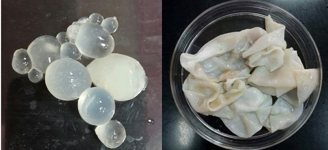
Fig. 3. Brood capsules and germinal layer isolated from sheep hydatid cysts
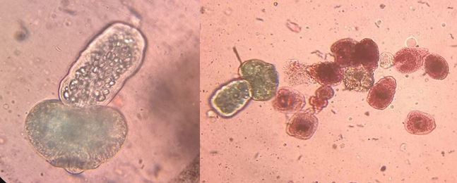
Fig. 4. living protoscolices (green) and dead protoscolices (red) used eosin stain
Count of Viable Protoscolices
The number of protoscolices was calculated using a 10 microliter pipette and a fixed size transfer method aftershaking the protoscolices suspension and Phosphate Buffered Saline. The number of protoscolices in 10 µl = 31 was as shown in Table (1), so the number of protoscolices in (1 ml) = 100 x 31 = 3100 scolice. Therefore, the approximate number of 2000 protoscolices is approximately 0.7 ml (Wangooet al., 1989).
Table (1):
Determination of the viability of the protoscolices in 10 microliter using the fixed size method.
| Duration of the transaction (hour) |
Number of protoscolices in 10µl For three repli-cates | Average number of protoscolices S.D. ± | Standard errorS.E. | ||
|---|---|---|---|---|---|
| 1 | 2 | 3 | |||
| 0 | 39 | 29 | 31 | 33±5.29 | 3.05 |
| 24 | 30 | 31 | 33 | 31.33±1.52 | 0.88 |
| 48 | 32 | 28 | 29 | 29.66±2.08 | 1.20 |
| 72 | 30 | 25 | 21 | 25.33±4.5 | 2.60 |
| 96 | 22 | 18 | 19 | 19.66±2.08 | 1.20 |
| 120 | 15 | 17 | 18 | 16.66±1.52 | 0.88 |
| 144 | 13 | 14 | 15 | 14±1 | 0.57 |
| LSD0.05 | 5.52 | ||||
Study of the effect of crude alcoholic extract of Spirugera sp. In the viability of the protoscolices in vitro
The crude alcoholic extract of Spirugera sp. was studied with concentrations (250, 500, 1000) mg / mL in the viability of protoscolices in vitro, based on Al-Nakeeb (2004) and Barzanjiet al. (2009). Concentrations were distributed on test tubes, each containing the Kerb’s Ringer medium with hydatid cyst fluid and by 1:4 respectively.The protoscolices were then shoved to ensure the distribution of protoscolices in the tubes.Each tube contained nearly 3,000 protoscolices, One of the tubes was leave without extract and was considered a control group. The number of live protoscolices in the suspension was then calculated with three replicates per concentration and using Eosin Aqueous Stain (0.1%) and the readings of the three replicates were taken from the first hour for 7 days.
Study of the effect of albendazole In the viability of the protoscolices in vitro
The efficacy of albendazole was studied in the viability of protoscolices with concentrations (250, 500, 1000) mg / ml in vitro. The drug was emulsified and in a 20 ml pack with a concentration of 20 mg / ml. The property was purchased from local pharmacies and is produced by the Egyptian International Company for Pharmaceutical Industries CO. E.I.P.I.CO. , The concentrations were distributed to test tubes containing the Kerb’s Ringer medium with the hydatid cyst fluid at 1:4, respectively, and the same steps were performed in the previous experiment.
Statistical Analysis
The results were analyzed using Prism (SAS Institute, Inc. USA) Graph Pad, the fifth version where the ANOVA test was applied with the least significant difference of LSD for this purpose. Confidence interval was equal to 95% The probability level is less than 0.05 (P <0.05).
Estimating the vitality of the protoscolices
The results of the study, as shown in Table (1), showing the viability of the protoscolices in 10 microliters and the three replicates calculated in the fixed-size method. There were no significant differences in the first three treatments, whereas the coefficients of 72-144 hour showed significant differences. If the mean S.D. ± viability of the protoscolices at the highest rate at treatment at 0 hour (33 ± 5.29) protoscolices, while the mean of the protoscolices dropped to the lowest ratio at 144 to 14 ± 1 protoscolices. While the mean S.D.± mean of the protoscolices viability of the treatments 24-120 hours between 31.33 ± 1.52-16.66 ± 1.52 protoscolices. thus showed that the vitality of the protoscolices begin to decrease over time.
Table (2):
Determination of the percentage of protoscolices in 100 microliters on the first day.
| Duplicates | The average | 1 | 2 | 3 | Average calculation S.D. ± |
|---|---|---|---|---|---|
| Average number of live protoscolices | 172.66 | 170 | 171 | 177 | 172.66±3.05 |
| Average number of dead protoscolices | 10.66 | 10 | 6 | 16 | 10.66±2.08 |
| Average number of headings | 183.33 | 180 | 177 | 193 | 183.33±8 |
| Percentage | 94.25 | 94.44 | 96.61 | 91.70 | |
| LSD0.05 | 9.38 | ||||
Percentage of viability of protoscolices
The results showed that the percentage of the viability of protoscolices in vitro was 100 µl and three replicates. As shown in Table (2), the total number of protosclices ranged from 177-193. The average number of live protoscolices ranged between 170-177 protoscolices / 100 µl, While the number of dead protoscolices ranged from 6-16 protoscolices / 100µl. Thus, the average percentage of the viability of the protoscolices on the first day was 94.25%. The results showed that the percentage of the viability of the protoscolices gradually decreased for seven days as shown in Table (3).
Table (3):
Determination of the percentage of protoscolices in 100 microliters within seven days.
| Duplicates | Percentage of viability of protoscolices (%) | ||||||
|---|---|---|---|---|---|---|---|
| 0 | 24 | 48 | 72 | 96 | 120 | 144 | |
| 1 | 95 | 93.86 | 89.23 | 88.1 | 82 | 75.06 | 77.09 |
| 2 | 93.5 | 90.6 | 87.82 | 83.19 | 80.25 | 80.12 | 78.2 |
| 3 | 94.25 | 87.4 | 85.45 | 86.5 | 81.5 | 79.18 | 74.4 |
| Rate (control) | 94.25± 0.31 |
90.62± 1.31 |
87.5± 0.77 |
85.93± 1.02 |
81.25± 0.36 |
78.12± 1.09 |
76.56± 0.79 |
Determination of chemical compounds in crude alcoholic extract for Spirogyra sp
GC-MS was analyzed for the crude alcoholic extract of algae. The algal chemical compounds were classified. Quantitative and qualitative abundance was observed in the chemical content of the algae. Sixty-nine chemical compounds were extracted from the crude alcoholic extract of Spirogyra sp. Varied between phenols, turbines, fatty acids, hydrocarbons and hetero cyclic compounds as shown in Fig. 5. The current study showed variation in species and time of retention of GC-MS-detected algal-chemical compounds.
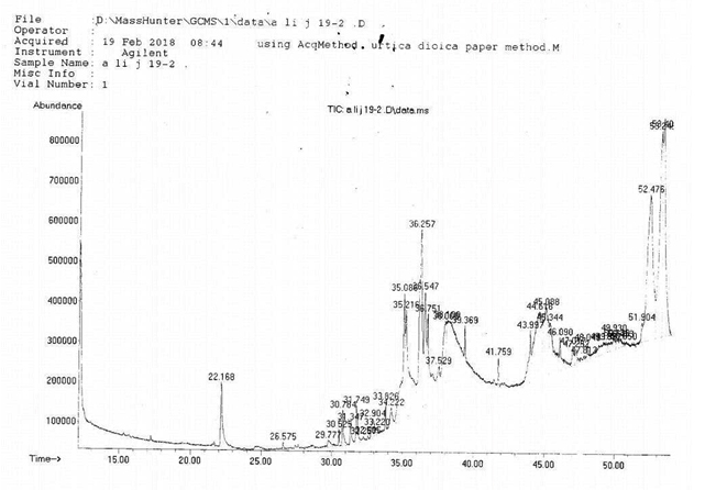
Fig. 5. Determination of chemical compounds in the crude extract of Spirogyra sp
Study of the effect of crude alcoholic extract of Spirogyra sp. In the viability of the protoscolices in vitro
The results of this study showed a decrease in the percentage of the viability of the protoscolices when exposed to the concentrations of the crude alcoholic extract of the Spirugera sp. The concentration of 250 mg/ml resulted in a decrease in the percentage of protoscolices from 92.75±0.62 at 0 hour to 27.06±0.48 at 144 hour compared with control group (76.56 ± 0.79), while the concentration of 500 mg/ml decreased from 92.59±0.58 per hour to 0.14±0.25% at 144 hours compared with the control group, while the concentration was 1000 mg / Ml has led to the killing of all protoscolices at 144 hour compared with the control group, has been shown (L.S.D.) showed significant differences of concentration of 1000 mg/ml compared with the rest of concentrations and the most important concentration as in Table (4) Figure (6) (7).
Table (4):
The percentage of the viability of the protoscolices after exposure to different concentrations of the crude alcohol extract of Spirugera sp. and for different time periods in vitro.
| Time (hour) concentrate |
Percentage rate of protoscolices, standard er-ror± | ||||||
|---|---|---|---|---|---|---|---|
| 0 | 24 | 48 | 72 | 96 | 120 | 144 | |
| Control | 94.25± 1.01 |
90.62± 0.41 |
87.5± 0.83 |
85.93± 0.90 |
81.25± 1.47 |
78.12± 1.12 |
76.56± 0.90 |
| 250mg/ml | 92.75± 0.62 |
89.56± 0.83 |
81.25± 0.46 |
72.90± 1.21 |
53.12± 0.78 |
39.56± 1.66 |
27.06± 0.48 |
| 500 mg/ml | 92.59± 0.58 |
82.70± 0.99 |
67.88± 0.48 |
55.55± 1.98 |
28.37± 1.02 |
12.33± 0.70 |
6.14± 0.25 |
| 1000 mg/ml | 90.9± 1.55 |
71.18± 1 |
57.54± 1.61 |
43.90± 1.03 |
10.59± 0.83 |
1.50± 0.11 |
0±0 |
| The least significant difference LSD0.05 |
2.746 | ||||||
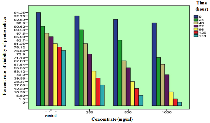
Fig. 6. Effect of the crude alcoholic extracts of Spirogyra sp. (1000,500,250) mg/ml in the viability of the protoscolices
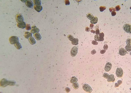
Fig. 7. Control group on the seventh day (144). (Power x100 magnification)
The rate of decrease in the percentage of all in vitro treatments was 76.56% after seven days of conservation. The results of the study of the effect of crude alcoholic extract of Spirogyra sp. reduces the viability of protoscolices with increased concentration. The concentration of 250 mg/ml of Spirogyra sp. was reduced to decreased viability protoscolices to 27.06% at 144 hour, while the concentration of 500 mg/ml for Spirogyra sp. was reduced to decreased viability protoscolices to 6.14% at 144 hours. Concentration of 1000 mg/ml for the crude alcoholic extract of the Spirogyra sp. resulted to killed to all the protoscolices at 144 hour.
Effectiveness of the crude alcoholic extract of Spirogyra sp. To the active substances in its chemical content such as phenols, turbines, fatty acids, hydrocarbons and hetero cyclic compounds. This extract had a significant effect in reducing the viability of the protoscolices of the E. granulosus parasite with increasing concentration. The presence of these active substances explains this effect as these substances have The inhibition effect (Adekunle, 2000). It also inhibits the metabolism of carbohydrates through its effect on mitochondria and thus the breathing process and thus leads to parasite death (Delorenziet al., 2001). Phenols also have an effect on the acetyl-choline esterase, which controls the permeability and elasticity of the cell membrane and thus the loss of the membrane of the competing property, which reduces to the entry of various substances, including uncontrolled nitrogenous substances into the parasite and accumulates in it leading to its death (Naguleswaranet al. , 2006). Some studies indicate that phenols have the ability to bind to the cytoplasm and cytoskeletal fat, thereby altering their functional structure and then the death of the living cell (Mani &Chitra, 1989; Diaz &Abeger, 1986)It also works to deposit the proteins in or inside the membrane of the cell when they are permeated through the membrane and the formation of hydrogen bonds between free and multiple hydroxyl phenols and nitrogen compounds or proteins and thus inhibiting certain enzymes needed by the organism (Covington, 1997; Reed, 1995). Some of the substances in the chemical content of this algae are fatty acids, some laboratory studies have indicated that some natural fatty acids possess antimicrobial and parasite properties, such as the study of Rayanet al. (2005), which proved that fatty acids play an important role in Giardia parasite resistance Especially Dodecanoic acid, through its effect on the trophozoite stage of the parasite, accumulating inside cytoplasmic cell and thus rupturing the cell membrane and parasite death. The fatty acids needed by parasitic tapeworms, including the E. coli parasite, E. granulosus, can’t be synthesized by the parasites, so they get them from the environment or host (Chabalgoity et al., 1997) It has been shown that a special protein called E.granulosus Fatty acids binding protein (EGFABp1) has a role that remains unclear in the transmission of fatty acids (Jakobsson, 2005). It has been shown to be important in the absorption and transfer of fatty acids into the parasite (Storch&Thumser, 2000; Zimmerman &Veerkamp, 2002; Haunerland&Spener, 2004). Fatty acids can bind with these proteins and penetrate the parasite membrane, causing high concentrations of fatty acids And the ability of the transport system to identify different fatty acids (Storch&Thumser, 2000; Haunerland&Spener, 2004). The Krugliaket al. (1995) study also showed that fatty acids have a disincentive to Plasmodiom sp. In white mice. Another study, Zuo et al. (2012), showed that fatty acids play a major role in increasing diseases resistance and regulating immunity after parasitic infection, as fatty acids have a significant protective role against Cryptocaryon irritants. Among the substances in the chemical content of this extract is the hetero cyclic compounds. Some studies such as Yanagimotoet al. (2002) have shown that heterocyclic compounds found in coffee have an antioxidant activity. Hetero cyclic compounds can also be used to synthesize drugs as antiviral agents for chronic diseases or infections (Cirillo et al., 2000)
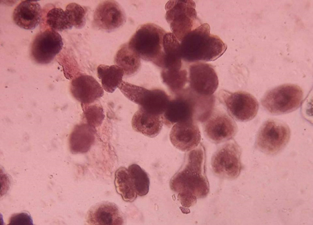
Fig. 8. Effect of the alcoholic extract of the Spirogyra sp. mulberry at a concentra-tion of 1000 mg / ml in viability of protoscolices on the seventh day (144) (magnification power X400)
Study of the effect of albendazole in viability of protoscolices in vitro
The results shown in Fig. 9 and 10 and Table 5 showed significant differences. Albendazole concentrations reduced the percentage of viability of protoscolices. When exposed to 250 mg / mL, The result was a decrease from 93.75 ± 0.77 at 0 hour to 4.15 ± 0.36 at 144 hour, while the concentration of 500 mg/ml reduced the percentage from 48 ± 0.85 at 0 hour to 0,4±0.48 at 144 hours, while The concentration of 1000 mg/ml resulted in the killing of all protpscolices indications at 120 hours compared to 0 hour where the percentage of viability of protoscolices was 45.5 ± 0.98.
Table (5):
The percentage of the viability of the protoscolices after exposure to different concentrations of albendazolefor difrent times periods in vitro.
| Time (hour) concentrate |
Percentage rate of protoscolices, standard er-ror± | ||||||
|---|---|---|---|---|---|---|---|
| 0 | 24 | 48 | 72 | 96 | 120 | 144 | |
| Control | 94.25± 1.01 |
90.62± 0.41 |
87.5± 0.83 |
85.93± 0.90 |
81.25± 1.47 |
78.12± 1.12 |
76.56± 0.90 |
| 250mg/ml | 93.75± 0.77 |
36.43± 1.06 |
25± 0.97 |
25± 1.18 |
21.87± 0.54 |
6.24± 0.44 |
4.15± 0.36 |
| 500 mg/ml | 48± 0.85 |
32± 1.29 |
28± 1.02 |
24± 1.26 |
13.32± 0.70 |
9.7± 0.25 |
4±0.48 |
| 1000 mg/ml | 45.5± 0.98 |
35.29± 1.14 |
17.64± 0.72 |
15.64± 0.80 |
1.94± 0.30 |
0±0 | 0±0 |
| The least significant difference LSD0.05 |
2.434 | ||||||
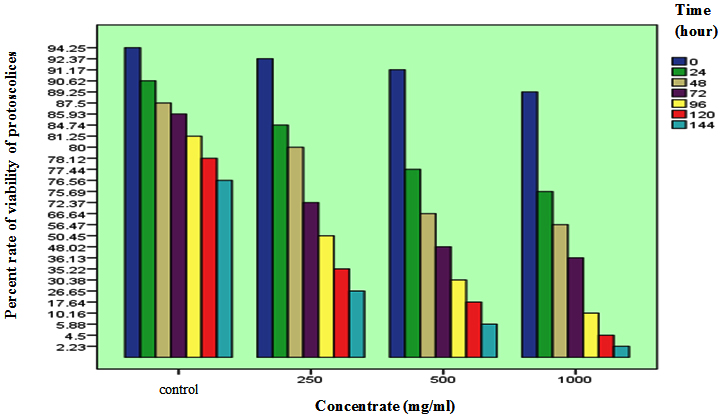
Fig. 9. Effect of albendazole concentrations (1000,500,250) mg/ml in the viability of exoge-nous primates
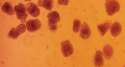
Fig. 10. Effect of albendazole 1000 mg / ml in the viability of protoscolices on day 6 (hour 120). (Power zoom X400)
The results of this study showed that this drug has a significant effect on the viability of the protoscolices with increased concentration and duration of exposure to the drug. This result is in line with the findings of Imad (2002), Al-Nakeeb (2004), Al-Bayati, Arslan (2009), Al-Hamiary (2010) and Khalaf (2012), which is of medical importance against microorganisms and parasites. The best medicines derived from benzimidazole. The drug has the ability to paralyze and kill the parasite and has the ability to stimulate the immune system in humans and animals (Bayati and Arslan 2009). Albendazole also has the ability to bind to B-tubulin, causing damage to the transport and growth functions of parasite cells Polymerization of this protein stops in the microtubules. This association impairs the absorption of glucose sugar in the parasites and their larvae, and depletes the stock of the animal starch (glycogen) in the parasite. The rate of ATP decreases and the energy levels decrease and thus the parasite dies. It is also a preferred drug in the treatment of HCD for its high ability to break down the layer created in the wall of the cyst and its decay and shrinkage as well as the disappearance of secondary hydatid cysts inside the hydatid cyst (Yarsanet al., 2003; Wilson et al., 1987.
ACKNOWLEDGMENTS
None.
CONFLICT OF INTEREST
The authors declare that there is no conflict of interest.
- Adekunle, A. A. Antifungal property of crude extracts of Brachystegiaeurystegia and Richardiabrasitiensis. AJOL : Nigerian journal Natural of prod medical., 2000; 4: 1-9.
- Al-Mayah, K. S., Al-Bashir, N. M., and Al-Azzawi, B. M. In Vivo Efficacy of Nigella Sativa Aqueous Seed Extract Against Metacestode of Echinococcusgranulosus. Medical Journal Babylon, 2012; 9: 1.
- Al-Nakeeb, S. A. R. Seroepidemiologicaland therapeutic study onhydatid cystinfection in Kirkuk and Tikrit provinces. M. Sc. Thesis College of Medicine, University of Tikrit 2004.
- Athbi, A. M. Atimicrobial Bioactive Compound Isolated from Cyanobacteria Nostoclinkia. Al-Qadisiyah Medical journal, 2017; 10(17), 239-247.
- Barzinji,A.K. R., Mothana, R.A. and Nasher, A.K. Effect of leaf extracts of Dendrosicyossocotrana and Jatrophaunicostata on the viability of Echinococcus granulosus protoscolices. Europa. Asia. Journal of Biology Sciences., 2009; 3: 122-129.
- Bekhti, A., Schaaps, J. P., Capron, M., Dessaint, J. P., Santoro, F., and Capron, A. Treatment of hepatic hydatid disease with mebedazole : preliminary results in four cases. Br. Medical. Journal, 1977; 2(6094): 1047-1051.
- Brunetti, E. Echinococcosis hydatid cyst medication 2015. Medscape. http://emedicine.medscape.com/article/216432-medication
- Chabalgoity , A., Harrison, J. , Esteves, R., Demarco, H. and Ricardo,E. Expression and Immunogenicity of an E. granulosusFatty Acid-Binding Protein in Live Attenuated Salmonella Vaccine Strains. Infectious and Immunology., 1997; 7: 2402–2412.
- Cirillo, P. F., Dinallo, R., Regan, J. R., Riska, P. S., Swinamer, A. D., Tan, Z., and Walter, B. A. (2003). U.S. Patent No. 6,525,046. Washington, DC: U.S. Patent and Trademark Office.
- Covington, A. D. Plant products as antimicrobial agents Clinical Microbiology. Rev., 1997; 12(4): 564-582.
- De, N. V. The first report of two cases of cystic echinococcosis in the lung by Echinococcus ortleppi infection, in Vietnam. Research and Reports in Tropical Medicine, 2016; 55(51), 45-51.
- Delorenzi, J. C., Attias, M., Gattas, C. R., Andrade, M., Rezende, C., Pinto, A. C., Henriques, A. T., Bou-Habib, D. C. and Saraiva, E. M.. Anti-Leishmanial activity of an indolealkaloid from peschieraaustralis. Antimicrob. Agents. Chemother., 2001; 45(5): 1349-1354.
- Diaz, A. M. and Abeger, A. Quantitative determination of the tannin content of the leaves of M. communis. Ann. R. Acad. Farm., 1986; 52(1): 117-122.
- Duman, K., Girgin, M., and Hamcan, S. Uncomplicated Hydatid Cysts of the Liver: Clinical Presentation, Diagnosis and Treatment. Journal Gastrointest and Digstive System, 2016; 6(430): 2-6.
- Ghasemi, Y., Yazdi, M. T., Shafiee, A., Amini, M., Shokravi, S., and Zarrini, G. Parsiguine, a novel antimicrobial substance from Fischerella ambigua. Pharmaceutical biology, 2004; 42(4-5): 318-322.
- Haunerland , N.H. and Spencer , F. Fatty acids binding proteins – insight genetic manipulation. Prog. Lipid. Research., 2004; 43: 328- 349.
- Hejazi, M. E., Tekantapeh, S. T., Noorabadi, P.,andHejazi, Y. Successful Medical Treatment of Multiple Pulmonary Hydatid Cysts with. Albendazole. Asian Journal of Medical Pharmacology. Research, 2016; 6(1): 01- 03.
- Jakobsson , E. Structural studies of Echinococcusgranulosus fatty acid binding protein 1 and human semicarbazide – sensitive amine oxidase . Ph.D. Thesis .Uppsala University, 2005; Sweden : 65 .
- Koshki, M. H. K., Nourian, A., Rahimi, M. T., Daryani, A., Spotin, A, Natural products applied against hydatid cyst protoscolices review of past to present. Elsevier. Acta tropical. 2017; 176: 385-394.
- Krugliak, M., Deharo, E., Shalmiev, G., Sauvain, M., Moretti, C., and Ginsburg, H. Antimalarial effects of C18 fatty acids on Plasmodium falciparum in culture and on Plasmodium vinckeipetteri and Plasmodium yoeliini geriensis in vivo. Experimental parasitology, 1995; 81(1), 97-105.
- Liani, C., and Katoch, S. S. Biosorption of Malathion pesticide using Spirogyra sp. International Journal of Environmental and Agriculture Research (IJOEAR) 2017; 15-20.
- Mani, A. and Chitra K. C. Toxicity of certain plant extracts to Meloidogyne incognita. Nematol. Medit., 1989; 17: 43-44.
- McManus, D. P., Gray, D. J., Zhang, W., and Yang, Y. Diagnosis, treatment, and management of echinococcosis. British Medical Journal. 2012; 344: 3866.
- Mestrovic, T. (2016). Alveolar echinococcosis. News Medical Life Sciences. https://www.news-medical.net/health/Alveolar-Echinococcosis.aspx
- Metcalf, J. A., Gallin, J. I., Nanseef PWM and Root, R. K. Labroatory mannal of Neitrophil. Function Raven press, New York. 1986; 84-90.
- Molera, M. S. P. and Semesi, A. K. Antimicrobial activity activty of extracts from six green algae from Tanzania. Curr. Trends. Mar. Bot. Research. East Africa . Reg., 1996; 211-217.
- Munoz, R., Fernandez, C. G. Microalgae-Based Biofuels and Bioproducts : From Feedstock Cultivation to End-Products. 2017; 356-357.
- Naguleswaran, A., Spicher, M., Vonlaufen, N., Ortega-Mora, L. M., Torgerson, P., Gottstein, B. and Hemphill, A. (2006). In vitrometacestodicidal activities of genisteinand other isoflavones against Echinococcus multilocularis and E. granulosus.
- Pacheco, R., Ferreira, A. F., Pinto, T., Nobre, B. P., Loureiro, D., Mour, P.,Gouveia, L., Silva, C. M. Energy convers. Manage. 2015; 89: 789-797.
- Pradhan , S., Dahal, R., phuyal, S., Ghimira, B., Singh, Y. Primary Hydatid Cyst of Pancreas: Case Report. Acta Scientific Medical Sciences., 2017; 101: 25-27.
- Rayan, P., Stenzel, D., and McDonnell, P. A. The effects of saturated fatty acids on Giardia duodenalistrophozoites in vitro. Parasitology research, 2005; 97(3): 191-200.
- Rampal, K., Prajapati, D. K., and Sharma, M. Hydatid Disease-A Clinical Perspective with A Review of Primary Hydatidosis of Spleen. International Journal Intg Medical Sciance, 3(7): 337-40.
- Reed, J. D. Nutritional Toxiclogy of Tannins and Releated Polyphenols in forage legumes. Journal of Animal society, 1995; 73: 1516-1528.
- Smyth, J. D. (1976). Introduction to Animal Parasitology. 2nd ed. Hodder and Stronghton. Ltd., London 466 pp.
- Smyth, J. D. (1985). In vitro culture of Echinococcusspp. In: Proceeding of the 13th . Tnt. Conger of hydatilogy. Madrid.
- Smyth, J. D. and Barrett, N. J. Procedure for testing the viability of human hydatid cysts following surgical removal, especially after chemotherapy. Trans. Roy. Soci. Tropical. Medical. Hyg. Journal., 1980; 74(5): 649-652.
- Storch , J. and Thumser , A. E. The fatty acid transport function of the fatty acids binding proteins. Biochem. Biophys. Acta., 2000; 36: 28- 44.
- Taskin, E. , Ozturk, M. and Kurt, O. Antibacterial activities of some marine algae from the Aegean Sea (Turkey). African Journal of Biotechnology, 2007; 6: 2746-2751.
- Verma, D. K., and Khan, F. Green approach to corrosion inhibition of mild steel in hydrochloric acid medium using extract of spirogyra algae. Green Chemistry Letters and Reviews, 2016; 9(1): 52-60.
- Wangoo, A., Ganguly, N. and Mahjan, R. C. Phagocytic Function of Monocytes in murine Model of Echinococcus granulosus of human origin. Indian. Journal of Midical Research., 1989; 89: 40-42.
- World Health Organization WHO. (2017). Obesity and Overweight factsheet from the WHO. Health.
- Yanagimoto, K., Lee, K. G., Ochi, H., and Shibamoto, T. Antioxidative activity of heterocyclic compounds found in coffee volatiles produced by Maillard reaction. Journal of agricultural and food chemistry, 2002; 50(19), 5480-5484.
- Zhang, R., Chen, X., and Wen, H. An improved experimental model of cystic hydatid disease in liver resembling natural infection route with stable growing dynamics and immune reaction. bioRxiv, 2017; 155168.
- Zimmerman , A.W. and Veerkamp , J. H. New insight into the structure and function of fatty acids binding protein. Cell. Mol. Life sci., 2002; 59: 1096 – 1116.
- Zuo, R., Ai, Q., Mai, K., Xu, W., Wang, J., Xu, H., and Zhang, Y. Effects of dietary n-3 highly unsaturated fatty acids on growth, nonspecific immunity, expression of some immune related genes and disease resistance of large yellow croaker (Larmichthyscrocea) following natural infestation of parasites (Cryptocaryonirritans). Fish & shellfish immunology, 2012; 32(2): 249-258.
© The Author(s) 2018. Open Access. This article is distributed under the terms of the Creative Commons Attribution 4.0 International License which permits unrestricted use, sharing, distribution, and reproduction in any medium, provided you give appropriate credit to the original author(s) and the source, provide a link to the Creative Commons license, and indicate if changes were made.