


ISSN: 0973-7510
E-ISSN: 2581-690X
Plastic in our environment is a serious threat to the life on earth. Bioremediation is the only way out to mitigate the present problem. Actinomycetes strains from two different locations were isolated within Kerala state. Efficiency of strains for biodegradation collected from both the locations was compared using weight loss experiment. Experimental evidence on the basis of percentage degradation of LDPE strip shows that strains collected from waste dump yard are more efficient degraders of polyethylene waste. (DSR2-24%, DSR1-20%, DSR3-8%) than those collected from virgin soil. (VSR3-13.2%, VSR2-8%, VSR1-4.8%). Actinomycetes are potential group of microorganisms which can be utilized for biodegradation of plastics. Bio-augmenting the soil with potential degraders will enhance the rate of degradation even more.
Biodegradation, Actinomycetes, Bioaugmentation
Plastics are anthropogenic polymers of carbon, hydrogen and oxygen. Almost 140 million tons of polymers are utilized worldwide and their usage is shooting up at a rate of 12%1. Every year an estimated 500 billion to 1trillion plastic bags are consumed worldwide2.Polythene could sometimes affect the entire marine life badly and may pave the way for their extinction3-4.
Low density polyethylene is one of the major environmental pollutants. The degradation of this pollutant is only possible if the microorganism is able to produce enzymes that cause cleavage of the polymer chains in to monomers and oligomers.These water soluble products are consumed by microorganisms as a source of carbon and broken down further to simpler products. Aerobic metabolism produces Carbon Dioxide and Water as end product5.
Microorganisms have a naturally occurring catabolic mechanism to degrade, transform or accumulate a huge range of compounds including hydrocarbons (PAHs), pharmaceutical substances and metals. The durability, light weight and processability of these polymers causes them to stay longer in the environment for centuries and end up in landfills and natural water resources creating a severe threat to the environment and its ecosystems .
The nature and ease of use of plastics make it very close to us and thereby its careless and excessive use leads to a great damage to the ecosystem, thereby raising severe ecological catastrophes. As microorganisms have the capacity to degrade the polymers, the only way to overcome the nightmare is by using efficient biodegraders from the soil.
Actinomycetes are gram-positive organisms that tend to grow slowly as branching elements. In addition, Actinomycetes are prokaryotes, produces spores and exhibit powdery growth. Their similarity to fungi is in the formation of branched aerial mycelium, which profusely sporulates,and is clearly noticeable. They are sometimes called higher bacteria, hence the name Actinobacteria. Actinomycetes are seen in a diverse and wide range of habitats as they are both mesophilic and thermophilic.Actinomycetes also produce a wide range of bioactive compounds of commercial importance6.
Plastic materials have been increasingly becoming popular in our day to day life as it is durable and can be moulded in to any form with much ease. Attempt has been made in this paper to study bioremediation methods to treat plastic waste accumulation and to compare the degradation capacity of Actinomycetes isolated from different locations.
Low Density Polyethylene powder (LDPE) was received from Sigma Aldrich Chemical Company. LDPE sheets were obtained from Shaz polymers, Calicut, Kerala.
Sample collection
Soil samples were obtained from two topographical locations viz. Garbage dumping site of Kozhikode Corporation and Virgin soil of Wayanad wildlife sanctuary. Using sterile sampling bags soil adjacent to plastic waste bags in the plastic waste dumping site was taken for the study and the other sample from non-polluted virgin soil. The soil samples were dried at room temperature for 2 hrs.
Isolation of Actinomycetes
Serial dilution method was carried out for isolating microorganisms from one gram of soil sample taken from both the sites. The agar used for isolating Actinomycetes was starch casein Agar, and isolation was performed by spread plate method. 3 strains from each location were selected for further studies. Identification of bacteria was done both by Macroscopic and Microscopic tests based on Bergeys Manual7.
Screening of Actinomycetes for biodegradability- by Clear zone method
Mineral salt medium constituted by adding polyethylene powder at a concentration of 1%(w/v)was used. It was then sonicated for 1 hour at 120RPM. Agar was then added to the sonicated medium and sterilized by autoclaving at 120°C and 15lbs pressure for 15 mins. The sterilized medium was then allowed to cool to 45°C and then poured in to sterile petriplates. After solidification, the isolated colonies were inoculated and plates were kept for incubation at 30-35°C for 2-4 weeks. The organisms producing zone of clearance were selected for further study8.
Biodegradation of LDPE by weight loss experiment
LDPE sheets were cut into 5cmx2cm equal pieces and then surface sterilized with ethanol and air dried. The strips were then introduced into conical flasks containing 100ml Mineral salt medium and later inoculated with polythene degrading microbes under study. It was mandatory to keep a control flask without inoculation of microorganism to confirm biodegradation.
The flasks were then kept in orbital shaker incubator at 30oC, 120RPM for 3 months. After every month, the strips were taken out, washed with 70% ethanol to remove bioflilms, then air dried and checked for weight. Weight loss was reported and compared5.
Colonization of Actinomycetes on the LDPE sheet
Mineral salt agar medium containing no carbon source was prepared and autoclaved at 120°C, and15lbs pressure for 15mins. After sterilization the medium was poured into petriplates and Actinomycetes were spread evenly over the medium. On to the seeded plate LDPE sheet (2cmx2cm) was placed at the centre. Plates were then incubated at 30-35°C for 3 months and morphological changes noted twice a month for a period of 3 months (Plates 1, 2, 3)
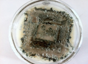
Plate 1. Attachment of Dsr2 Strain On Ldpe Sheet (15days)
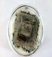
Plate 2. Attachment of Dsr2 Strain On Ldpe Sheet (30days)
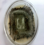
Plate 3. Attachment of Dsr2 Strain On Ldpe Sheet (after 45 Days)
Determination of variation in pH value of the medium
The pH value variation in Mineral salt medium containing LDPE sheet were observed, recorded and compared with its initial pH 6.8 value after an incubation period of 90 Days.
Evaluation of cell surface hydrophobicity
Bacterial Cell Surface Hydrophobicity is determined by [BATH] test9. With more and more bacteria becoming hydrophobic, their affinity towards hydrocarbon also increased resulting in change of turbidity of the culture due to the migration of cells from aqueous phase to organic phase. For BATH assay, Actinomycetes were cultured in Starch casein broth until logarithmic phase and then centrifuged and washed twice with PUM buffer (per litre)17gK2HPO4,7.26g KH2PO4,1.8g UREA and 0.2 gMgSO47H2O.The washed cells were resuspended in PUM buffer until they reached an OD value of 1.0 to 1.2 at 400nm. 1.2ml of suspension was poured to a set of test tubes to which hexadecane is added in progressive volume(range 0 to .2ml).The test tubes were shaken for 10mins and allowed for Phase separation of 2mins. The OD400 value of the aqueous suspension was measured. Cell free buffer served as the blank.
Six strains of Actinomycetes were considered for the present study. According to the location, strains were named as from dump yard DSR1, DSR2, DSR3 and other three from virgin soil VSR1, VSR2, and VSR3.
Clear zone formation
Growth began within 3-4 days of inoculation. Initially an opaque zone was seen around the colony, but gradually a clear and transparent zone was visualized with in 15-20- days at 30-35OC in polythene incorporated mineral salt medium.
Morphological changes on sheet
Microbial growth was visible over the sheet after 4-5 days of incubation, and the growth extended around the sheet in 10-15 days. Within 30-35 days a clear zone halo appeared around the sheet and the colonization was very evident on the sheet when compared to the uninoculated sample.
Biodegradation of LDPE by Weight loss method
After one week of incubation, the conical flasks were checked for visible growth. Thin bio film was observed over the surface of LDPE sheet and every month weight of the sheet was taken for a period of 3 months.
Table (1):
Determination of Weight Loss (SAMPLE 1- CALICUT).
| S. No |
Polyethylene Treated with Iso-lates | Weight of Polyethylene (mg) | Percentage of Polyethylene Degraded(%) | |||
|---|---|---|---|---|---|---|
| Initial wt | Aug 2016 | Sep 2016 | Oct 2016 | |||
| 1 | Control | 25 | 25 | 25 | 24.8 | .66 |
| 2 | DSR 1 | 25 | 23 | 21 | 20 | 20 |
| 3 | DSR2 | 25 | 22 | 20 | 19 | 24 |
| 4 | DSR 3 | 25 | 24 | 23.8 | 23 | 8 |
Table (2):
Determination of Weight Loss (SAMPLE 2- WAYAND).
| S.No | Polyethylene Treated with Isolates | Weight of Polyethylene (mg) | Percentage of Polyethylene Degraded(%) | |||
|---|---|---|---|---|---|---|
| Initial wt | Aug 2016 | Sep 2016 | Oct 2016 | |||
| 1 | Control | 25 | 25 | 25 | 24.9 | 0.4 |
| 2 | VSR 1 | 25 | 24 | 24 | 23.8 | 4.8 |
| 3 | VSR2 | 25 | 24.5 | 24 | 23 | 8 |
| 4 | VSR 3 | 25 | 24 | 22.8 | 21.7 | 13.2 |
Based on results given on (Table 1&2) it confirms that Actinomycetes utilized polyethylene as a sole source of carbon. The strains isolated from Dumpyard, where they were already associated and exposed to polyethylene wastes, showed a good biodegradation capacity (DSR2-24%, DSR1-20%, DSR3-8 %( Fig. 1). When compared to the Actinomycetes isolated (VSR3-13.3%, VSR2-8%, VSR1-4.8%) from virgin soil (Fig. 2).
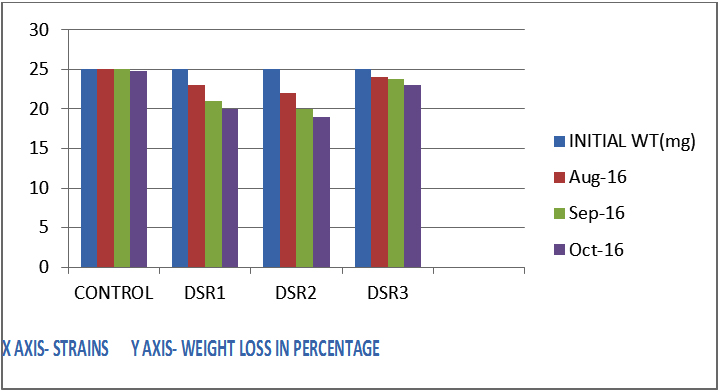
Fig. 1: Determination of weight loss (sample 1 )
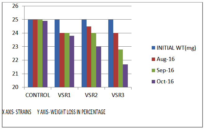
Fig. 2. Determination of weight loss (sample 2)
Variation of pH of the medium
The initial pH of the medium was 6.8 and it was seen reduced to acidic side over a period of 90 days of incubation. The reduction in pH proved that the culture was still metabolically active and it was utilizing LDPE for its growth. (Table 3 & 4).
Table (3):
Variation in pH(SAMPLE 1).
Sl.no |
ISOLATES |
pH(initial) |
pH(30days) |
pH(60days) |
pH(90 days) |
|---|---|---|---|---|---|
1 |
CONTROL |
6.8 |
6.6 |
6.4 |
6.3 |
2 |
DSR1 |
6.8 |
6.5 |
6.1 |
5.9 |
3 |
DSR2 |
6.8 |
6.3 |
6.0 |
5.5 |
4 |
DSR3 |
6.8 |
6.6 |
6.3 |
6.1 |
Table (4):
Variation in pH(SAMPLE 2).
Sl.no |
ISOLATES |
pH(intial) |
pH(30days) |
pH(60days) |
pH(90 days) |
|---|---|---|---|---|---|
1 |
CONTROL |
6.8 |
6.8 |
6.8 |
6.7 |
2 |
VSR1 |
6.8 |
6.6 |
6.6 |
6.5 |
3 |
VSR2 |
6.8 |
6.6 |
6.3 |
6.0 |
4 |
VSR3 |
6.8 |
6.5 |
6.2 |
5.9 |
Cell surface hydrophobicity
DSR 2 strain exhibited 11% OD where other strains from the same location showed much higher percentage of optical density, DSR1-39%, DSR3-32% and the samples taken from Virgin soil exhibited VSR3-30%, VSR2-33%, and VSR1-42%. The results clearly indicated that the strain DSR2-11% had more adhesion capacity to hydrocarbon than all the other strains. The migration of hydrophobic cells from aqueous phase to the organic phase (hexadecane) is shown as a decrease in turbidity of bacterial suspension. (Table 5 & 6)
Table (5):
Cell surface Hydrophobicity (sample 1).
sl no |
Vol of culture (ml) |
Vol of hexa decane (ml) |
DSR1 od400 |
DSR1 % of Initial od |
DSR2 od400 |
DSR2 % of Initial od |
DSR3 od400 |
DSR3 % of Initial od |
|---|---|---|---|---|---|---|---|---|
1 |
1.2 |
0 |
1.26 |
100 |
1.12 |
100 |
1.24 |
100 |
2 |
1.2 |
0.04 |
.96 |
88 |
0.50 |
41 |
0.89 |
71 |
3 |
1.2 |
0.08 |
0.89 |
55 |
.29 |
22 |
0.85 |
67 |
4 |
1.2 |
0.16 |
.71 |
53 |
0.21 |
16 |
0.72 |
60 |
5 |
1.2 |
0.20 |
0.88 |
39 |
0.13 |
11 |
0.76 |
32 |
Table (6):
Cell surface hydrophobicity (sample 2).
Sl no |
Vol of culture (ml) |
Vol of hexadecane (ml) |
VSR1 od400 |
VSR1 % of Initial od |
VSR2 od400 |
VSR2 % of Initial od |
VSR3 od400 |
VSR3 % of initial od |
|---|---|---|---|---|---|---|---|---|
1 |
1.2 |
0 |
1.30 |
100 |
1.23 |
100 |
1.20 |
100 |
2 |
1.2 |
0.04 |
0.93 |
86 |
0.89 |
71 |
0.48 |
49 |
3 |
1.2 |
0.08 |
0.87 |
73 |
0.84 |
67 |
0.50 |
37 |
4 |
1.2 |
0.16 |
0.71 |
53 |
0.72 |
60 |
0.49 |
36 |
5 |
1.2 |
0.20 |
0.81 |
42 |
0.75 |
33 |
0.23 |
30 |
Six strains of Actinomycetes were isolated by using Starch Casein Agar. Three strains from dump yard soil (DSR1,2 and 3) and three from virgin soil (VSR1,2,and3) were considered for the entire study.
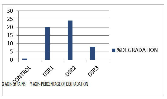
Fig. 3. Percentage of degradation capacity of strains (sample 1)
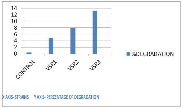
Fig. 4. Percentage of degradation capacity of strains (sample 2)
The total average count of identified actinomycetes was 12×105in Dumpyard samples and 7×10⁵ in virgin soil ones. The results were at par with previous studies done by Vijaya & Reddy10 in which the average number of organisms associated with polythene film was 37.08×10⁴.
All the Six strains(DSR1,2,3and VSR1,2,3) were considered for weight loss studies in which it was shown that the Actinomycetes colonized the surface of polythene forming films (Plates 1,2, and 3). The loss in weight of LDPE strip suggested the usage of polythene strip as an alternative source of carbon in the absence of readily available carbon. In a previous study conducted by Imam11 observed that biodegradation was triggered only after the colonization of microbes on the plastic sheet. The more the microbial load on the plastic, the higher is the chance for its biodegradation.
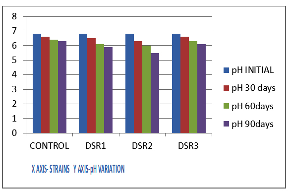
Fig. 5. Variation in ph (sample 1)
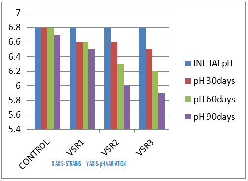
Fig. 6. Variation in ph (sample 2)
The weight loss study conducted clearly showed that, DSR2 strain obtained from dumpyard was the most efficient strain, degrading the polymer by 24% over a period of 3 months when compared with other strains under study.
Table 3 and 4 shows the variation in pH of the medium during biodegradation studies. The actinomycetes isolates in the present study produced some enzymes or metabolites which made pH to drop supporting the viability or growth of actinomycetes on LDPE substrate.DSR2 from dump yard could reduce the pH from 6.8 to 5.5 over a period of 90 days. In a similar study carried out by Dey U et al12, he showed that microorganisms secreted a wide range of enzymes which were utilized for breaking down the complex polymers into monomers that were small enough to permeate through the cell wall of organisms to be utilized as carbon and energy source .
For degradation of polythene, the microbes should adhere to the polythene sheet. The hydrophobic nature of the cell surface of Actinomycetes was responsible for their affinity towards hydrocarbon. When the microbes become more hydrophobic, their affinity for hydrocarbons also increased. DSR2 strain (11%) was the most hydrophobic Actinomycete and could be regarded as the most potential plastic degrading Actinomycete among all in the present study.
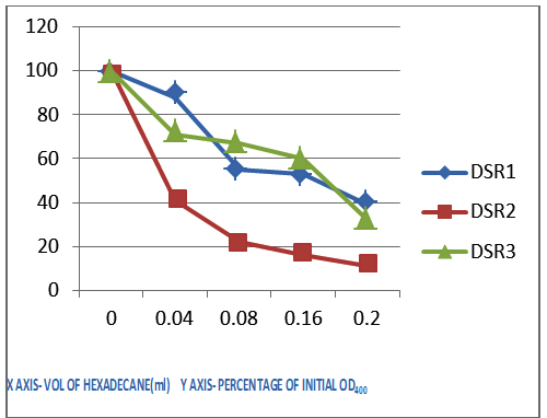
Fig. 7. Cell surface hydrophobicity (sample 1)
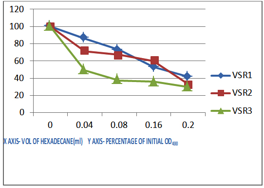
Fig. 8. Cell surface hydrophobicity (sample 2)
The growth, viability, weight loss and pH change clearly proved that the isolates taken from dump yard soil, where they were already associated with plastics, were more efficient degraders compared with the isolates from virgin soil, where they were not at all exposed to plastics.
By closely examining these results, we can conclude that Actinomycetes are a potential group of microorganisms which can be utilized for biodegradation of plastics, if isolated from a place where they are seen associated with plastic waste in soil rather than isolating them from virgin soil. Bio augmenting the soil with potential degraders will enhance the rate of degradation even more.
ACKNOWLEDGMENTS
None.
CONFLICT OF INTEREST
The authors declare that there is no conflict of interest.
- Shimao M. Biodegradation of plastics. Current Opinion in Biotechnology 2001; 12: 242-247.
- Roy PK, Titus S, Surekha P, Tulsi E, Deshmukh C, Rajagopal C. Degradation of abiotically aged LDPE films containing pro-oxidant by bacterial consortium. Polymer Degradation and Stability 2008; 93: 1917-1922. http:// dx.doi.org/10.1016/j.polymdegradstab.2008.07.016.
- Spear LB, Ainley DG, Ribic CA. Incidence of plastic in seabirds from the tropical pacific l984-1991: Relation with distribution of species, sex, age,season, year and body weight. Mar Environ Res l995; 40: 123-146.
- Secchi ER, Zarzur S. Plastic debris ingested by a Blainville’s beaked whale, Mesoplodon-densirostris, washed ashore in Brazil. Aquatic Mammals l999; 25: 21-24.
- Usha R, Sangeetha T, Palaniswamy M. Screening of Polythene Degrading microorganisms from garbage soil. Libyan Agricultural Research Center Journal International 2011; 2: 200-204.
- Prescot LM, Harley JP, Klein DA. Microbiology, Sixth edition, McGraw Hill International edition, New York. (2005).
- Holt JG, Krieg NR, Sneath PHA, Staleyand JT, Williams ST. Gram Positive Cocci. In: Hensyl WR (Ed.) Bergey’s Manual of Determinative Microbiology, 9th Ed., Williams and Wilkins, Baltimore, USA, 1994; Pp: 527-558.
- Augusta J, MullerRJ, Widdecke H. A rapid evaluation plate test for the biodegradability of plastics. Applied Microbiology and Biotechnology 1993; 39: 673-678.
- Rosenberg M, Gutnick D, Rosenberg E. Adherence of bacteria to hydrocarbons:A simple method for measuring cell surface hydrophobicity: FEMS Microbiology letters, 1980; 29-33.
- Vijaya C, Reddy RM. Impact of soil composting using municipal solid waste on biodegradation of plastics. Indian Journal of Biotechnology 2008; 7: 235-239.
- Imam SH, Gordon SH, ShogrenRL, Tosteson TR, Govind NS, Greene RV. Degradation of Starch- poly Hydoxybutyrate- co-beta –Hydroxyvalerate) Bioplastic in tropical coastal waters. Applied and Enviromental Microbiology 1999; 65: 431-43.
- Dey U., Mondal N.K.,Das,K.andDutta,S.An approach to polymer degradation through microbes. IOSRPHR. Vol. 2. pp.385-388.2012.
© The Author(s) 2018. Open Access. This article is distributed under the terms of the Creative Commons Attribution 4.0 International License which permits unrestricted use, sharing, distribution, and reproduction in any medium, provided you give appropriate credit to the original author(s) and the source, provide a link to the Creative Commons license, and indicate if changes were made.