

ISSN: 0973-7510
E-ISSN: 2581-690X
 , Marwah Nasir Ahmad1 , Saaid Mouazen1,
Mohammed Alsubhi1
, Elaf Muawwadh1, Raghad Alqamshi2 and Manal El Said3,4Although primarily known for its respiratory involvement, SARS-CoV-2 infection has increasingly been associated with significant effects on multiple organ systems, including the hepatobiliary tract. Over the course of the pandemic, it was shown that COVID-19 can trigger a variety of liver and cholangiocyte impairments through many overlapping mechanisms. These include entry of the virus into liver cells that express angiotensin-converting enzyme 2 receptors, impaired immune responses, ischemic injury, and adverse reactions to therapeutic drugs. These may lead to persistent hepatic enzyme abnormalities, biliary stasis, secondary sclerosing cholangitis, or fibrosis. Individuals who have underlying liver disease, such as liver cirrhosis or non-alcoholic fatty liver disease (NAFLD), are especially susceptible to serious complications and unfavorable outcomes. Uncommon immune-mediated conditions, such as post-COVID-19 vaccination autoimmune-like hepatitis, have been reported. Therefore, ongoing assessment of liver function and careful long-term follow-up are recommended for patients with post-COVID-19 syndrome, especially those at high risk. This review discusses current evidence to provide an overview of the mechanisms, manifestations, and long-term consequences of COVID-19 involving the hepatobiliary tract.
Post-Acute COVID-19 Syndrome, Cholangiopathy, Secondary Sclerosing Cholangitis, Autoimmune Hepatitis, Drug-Induced Liver Injury, Liver Fibrosis
The novel coronavirus causes severe acute respiratory syndrome, which emerged in late 2019 and was given the name coronavirus disease (COVID-19), leading to a significant global health crisis. It is a classic type of respiratory virus that primarily affects the upper and lower respiratory system.1,2 Recent evidence has shown the involvement of extrapulmonary manifestations in other organs such as the skin, eyes, kidneys, and endocrine organs in addition to the central nervous system and cardiovascular system.1 The long-term complications of gastrointestinal (GI) symptoms are commonly observed; emerging evidence suggests that SARS-CoV-2 infection can lead to significant liver injury and exacerbate preexisting liver conditions. Abnormal liver function tests (LFTs) have been discovered in 14%-65% of hospitalized COVID-19 patients with symptoms. In severe cases, these abnormalities are more significant and can account for up to 80% of fatal cases.1,3 Hepatobiliary involvement is increasingly recognized as one of the most common extrapulmonary manifestations of COVID-19, involving derangement of liver enzymes, cholestasis, and in severe cases, secondary sclerosing cholangitis.1,4,5 In a multinational population-based cohort study spanning South Korea, Japan, and the UK, SARS-CoV-2 infection was associated with a 30% higher risk of hepatobiliary disease relative to uninfected controls beyond the first thirty days of infection.6 Among the critically ill, invasively ventilated COVID-19 patients, roughly one in forty-three went on to develop secondary sclerosing cholangitis.1 Patients with preexisting liver disease, such as cirrhosis or non-alcoholic fatty liver disease (NAFLD), along with older adults, appear to be at increased risk of severe hepatobiliary complications and poorer outcomes following COVID-19 infection.7,8 Globally, SARS-CoV-2 has caused millions of infections and deaths, and the virus has continued to evolve into new circulating variants that differ in transmissibility and clinical behavior.9,10
Therefore, this review aims to summarize the current evidence on the mechanisms, clinical manifestations, and long-term hepatobiliary consequences of COVID-19, with particular attention to the populations at greatest risk of complications.
PubMed/MEDLINE, Web of Science, and Google Scholar were used as electronic databases; the search included case reports and series, case-control, cohort, and cross-sectional studies, as well as reviews from the inception of the databases to March 2024. The inclusion criteria were studies published in English that addressed hepatobiliary involvement or complications associated with SARS-CoV-2 infection, without any restrictions regarding time, population category, and/or detection assay. The search was performed by using terms related to “SARS-CoV-2” OR “COVID-19” OR “coronavirus infection” AND “Hepatobiliary” OR “Liver” OR “Complications”. Studies reporting patients with underlying liver injury or liver pathology, or deaths with confirmed COVID-19, were included in this review.
Etiology
The novel coronavirus first appeared in December 2019 and rapidly spread throughout the world, initiating the COVID-19 pandemic. The virus was identified as SARS-CoV-2, a member of the β-coronavirus family. There are seven strains of coronaviruses known to infect humans. While four of these generally result in mild cold-like illness, the remaining three, SARS-CoV, Middle East Respiratory Syndrome Coronavirus (MERS-CoV), and SARS-CoV-2, are associated with higher morbidity and mortality rates, with case fatality rates of 10%, 37%, and 5%, respectively.9,10
SARS-CoV-2 is an enveloped, single-stranded RNA virus. Its entire genome, which is 29,881 base pairs long, has been mapped using RNA sequencing. The virus’s genes code for various proteins, both structural and non-structural, with the S protein being particularly significant in causing the disease. These proteins are crucial for the life cycle of the virus and are important targets for possible therapeutic treatments.11
S Gene (Spike Protein) and the Emergence of SARS-CoV-2 Variants
The spike (S) gene encodes the S glycoprotein responsible for receptor binding and cell entry, and it is one of the most variable regions of the SARS-CoV-2 genome and represents a major driver of the emergence of new viral variants worldwide.2,10 Mutations accumulating in the S genome, specifically within the receptor-binding domain, can alter the conformation of the S protein in ways that increase its affinity for angiotensin-converting enzyme 2 (ACE2), enhance transmissibility, and enable partial escape from neutralizing antibodies generated by prior infection or vaccination.12,13 Several variants of concern first identified in the United Kingdom, South Africa, and Brazil carry characteristic S gene mutations linked to more efficient spread.12 Molecular surveillance studies have used the presence or absence of specific S gene targets on RT-PCR assays as a practical marker to track the emergence of these variants; in one such study, a rapid rise in S-mutant variants, that is, S-gene-negative variants, in a Lebanese population closely paralleled a surge in COVID-19 case prevalence.12 Because the spike protein is also the principal target of most COVID-19 vaccines, ongoing monitoring of S gene mutations remains crucial for anticipating shifts in transmissibility and vaccine effectiveness, which may in turn influence the pattern of extrapulmonary complications.13
Pathogenesis of hepatobiliary involvement
The study of complications following COVID-19 infection has evolved into a complex and multifaceted area, particularly as research has revealed that the virus responsible for COVID-19 can have detrimental effects on liver function.1,4 Although the precise mechanism behind hepatobiliary tract complications remains unclear, different pathways have been suggested, including direct viral replication within liver tissues, systemic inflammation, ischemic alterations in vascular supply, injury secondary to drugs, and exacerbation of underlying illnesses.5,14
Viral infection
One of the possible mechanisms of the coronavirus is direct viral infection, through direct cytotoxicity resulting from viral replication within hepatocytes.15 The virus enters the host cells through the binding of its spike (S) protein to ACE2, which is expressed on the surface of target cells6; furthermore, the host transmembrane protease serine 2 (TMPRSS2) activates the S protein.1 Once the viral RNA is released inside the cell, the host cell then translates the RNA genome into polyproteins. These polyproteins are cleaved and assembled to build a replication-transcription complex (RTC). The cleavage and assembly of the RTC are required for replication and transcription of the viral RNA genome. The replication of viral RNA, together with the synthesis, assembly, and packaging of structural proteins within the host cell, results in viral particles being released.5 In the series by Lagana et al., the polymerase chain reaction (PCR) for viral genes was positive in 55% of liver autopsies.16 The autopsies of COVID-19 patients have shown viral antigens by electron microscopy and in situ hybridization (ISH), demonstrating that SARS-CoV-2 may directly infect the liver, causing histopathological changes such as hepatocyte degeneration, focal necrosis, and inflammatory cell infiltration, including neutrophils and lymphocytes.17,18
These pathological changes suggest that the virus may directly damage the integrity of the liver, which could be the root cause of the increased levels of liver enzymes such as alanine aminotransferase (ALT) and aspartate aminotransferase (AST).19 Additionally, individuals who already have cirrhosis or non-alcoholic fatty liver disease (NAFLD) are more susceptible to severe liver injury after being infected with COVID-19.7
Systemic cytokine release
Another suggested pathway involving gastrointestinal (GI) and hepatobiliary damage is contributed by inflammatory responses and immune mediators. A significant elevation in inflammatory markers and cytokines, such as tumor necrosis factor-alpha (TNF-α), interleukin-1 (IL-1), and interleukin-6 (IL-6), has been linked to COVID-19 disease and may trigger a phenomenon called the cytokine storm, an accumulation of immune cells that minimizes hepatobiliary uptake and excretory systems, causing hepatocellular cholestasis. This immune overreaction may lead to reactive hepatitis. According to some authors, immunosuppression helps to improve outcomes by reducing cytokine release.1,5
Liver impairment in viral infections that do not primarily target hepatic tissue may arise from systemic cytokine release. For example, in influenza A, Kupffer cells regulate the cytotoxic T-cell response, which, despite the absence of direct viral invasion, causes hepatitis. These immune-mediated inflammatory processes, which originate from extrahepatic sites, can lead to collateral liver injury via infiltration of cytotoxic T cells. This could play a significant part in the novel coronavirus.5
Vascular and ischemic complications
A third possible mechanism contributing to hepatobiliary damage in the novel coronavirus involves hypoxic and thrombotic mechanisms. Hepatic hypoxia may be triggered by complications such as respiratory failure, right-sided heart failure, and systemic coagulopathy. In addition, viral endothelial infection leads to endothelial dysfunction and enhances the release of von Willebrand factor, factor VIII, and fibrinogen, which are prothrombotic factors and facilitate thrombogenesis. In severe cases of the novel coronavirus, extensive thrombosis has been reported in both macrovascular and microvascular beds, causing ischemic events like acute limb ischemia and pulmonary thrombosis. Thus, the presence of hepatic vascular thrombosis supports the role of hypercoagulable states and thrombotic injury of the liver.5 Figure 1 provides an overview of the primary mechanisms involved in the hepatobiliary pathogenesis of COVID-19.
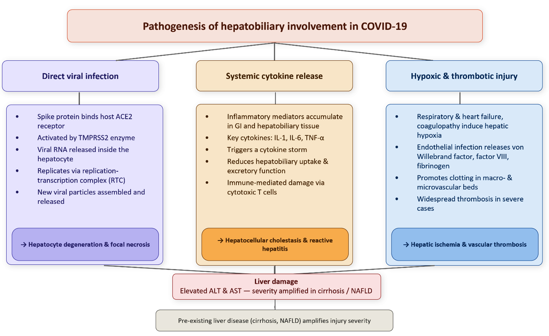
Figure 1. Key pathways involved in COVID-19-related liver injury and the resulting outcomes
Clinical presentation
Liver damage can persist even after the acute infection has been resolved. Recovered patients might display abnormalities in LFTs, such as increased levels of alanine transaminase (ALT) and aspartate transaminase (AST), as well as elevated total bilirubin levels, due to which there will be hepatic jaundice.20-23 Moreover, liver impairment on admission acts as an independent prognostic factor, predicting greater chances of severe outcomes, even in patients who initially presented with mild symptoms of COVID-19.24
One of the emerging conditions of particular significance is post-COVID-19 cholangiopathy. It is characterized by injury and dysfunction of bile duct cells. Cytokine storm, or immune-mediated liver injury, can directly damage the liver, which can worsen chronic liver disease and give rise to cholestatic syndromes, such as secondary sclerosing cholangitis, especially in patients with preexisting conditions.24-27
According to recent analysis, the risk of hepatobiliary complications is also higher in older adults, particularly those with preexisting liver diseases. Interestingly, although hepatic enzymes may alter, the severity of these changes remains mild to moderate in the majority of cases.28
Long-term complications of the hepatobiliary system
Many of the complications are observed during the acute phase of COVID-19, particularly in patients with severe disease. However, some symptoms may develop later, even after recovery, and are now being recognized as part of long-term SARS-CoV-2 complications, or chronic symptoms of the illness. Additionally, SARS-CoV-2 can affect patients with preexisting gastrointestinal or hepatic conditions, raising concerns about the use of immunologic therapies, the high potential for severe SARS-CoV-2 infection, and unfavorable prognosis. Furthermore, gastrointestinal or hepatic maladies may not always be directly attributable to SARS-CoV-2 itself but could result from the various treatments used to manage or prevent the disease.29 The main hepatobiliary complications associated with SARS-CoV-2 are summarized in Table 1.
Table 1. Summary of the main hepatobiliary complications associated with COVID-19
No. |
Long-term complications of the hepatobiliary system |
Mechanism |
Ref. |
|---|---|---|---|
1. |
Chronic liver injury and fibrosis |
Direct cellular damage caused by the virus, drug-induced hepatotoxicity, and hyper-inflammatory responses related to the novel coronavirus. |
30,31 |
2. |
Complications in patients with preexisting liver conditions |
Hepatic viral infection, acute liver disease, NAFLD, and liver cirrhosis. |
37,39 |
3. |
Complications of the biliary system (persistent jaundice and secondary sclerosing cholangitis) |
Prolonged hyperbilirubinemia and SSC in a COVID-19 patient are potentially due to biliary ischemia from microthrombi. |
37,40 |
4. |
Complications of drugs used for COVID-19 treatment |
Mucosal irritation, altered gut motility, microbiota disruption, direct hepatocellular toxicity, biliary injury, and immunosuppression leading to opportunistic infections. |
44-48 |
5. |
Complications of COVID-19 vaccination |
Molecular mimicry, epitope spreading, bystander activation, and adjuvant-driven immune enhancement. |
49,54, 55,56 |
Chronic liver injury and fibrosis
Chronic liver injury and fibrosis in SARS-CoV-2 patients are thought to arise from several mechanisms, including direct cellular damage caused by the virus, drug-induced hepatotoxicity, worsening of preexisting liver conditions, and hyper-inflammatory responses related to COVID-19. These factors collectively account for why more than half of patients may exhibit signs of liver fibrosis, particularly after the acute phase, as indicated by elevated fibrosis index (FIB-4) levels.30,31
Complications in patients with preexisting liver conditions
In patients with SARS-CoV-2, the observed rates of organ tissue, cholestatic, and combined liver injury were 75.0%, 29.2%, and 43.4%, respectively.32,33 The rate of previously existing liver disease among these patients ranges from 2.0% to 11%.34-36 Those with conditions such as hepatic viral infection, acute liver disease, NAFLD, and liver cirrhosis face a high risk of severe SARS-CoV-2 and have a high mortality rate, up to 10 times greater than that of individuals without such comorbidities.37
Advanced liver cirrhosis is associated with a higher mortality rate.8 However, NAFLD was predominantly linked with higher SARS-CoV-2 severity, with cirrhosis ranking second. Additionally, SARS-CoV-2 patients with hepatic viral infection had a low chance of being at higher risk of getting a severe form of SARS-CoV-2.38 A meta-analysis conducted by Vancsa et al., which covered fifty studies, indicated that previously diagnosed liver diseases or acute liver injury associated with severe SARS-CoV-2 infection were major determinants in the estimate of mortality.39
Complications of the biliary system
SARS-CoV-2 may lead to persistent jaundice and secondary sclerosing cholangitis (SSC) in some patients, particularly those with severe illness.37 A reported case showed that prolonged hyperbilirubinemia and SSC in a COVID-19 patient were potentially due to biliary ischemia from microthrombi.40
These cases highlight the possibility of long-term liver complications in COVID-19 survivors. While not directly related to COVID-19, a case of recurrent fever, jaundice, and right upper quadrant pain has been reported, emphasizing how important it is to consider various causes for these symptoms.41 Early identification and treatment of post-COVID-19 liver complications are crucial, as SSC is linked with significant morbidity and mortality if left untreated.42 Figure 2 summarizes the pathways that lead to long-term hepatobiliary complications of COVID-19 and their associated outcomes.
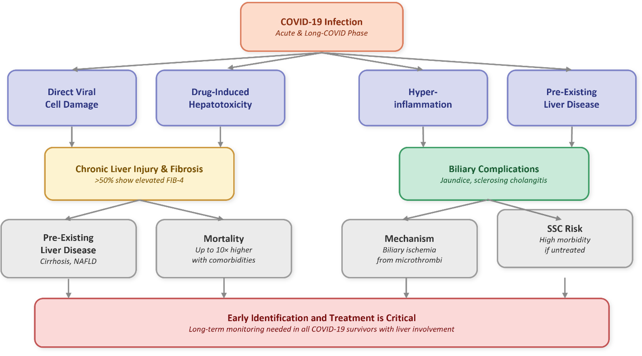
Figure 2. Long-term hepatobiliary outcomes of COVID-19 infection, showing how viral, drug-related, inflammatory, and preexisting liver factors converge on chronic liver injury/fibrosis and biliary complications
Hepatobiliary complications secondary to COVID-19 therapies
Drug complications
Differentiating GI and hepatic symptoms in patients with COVID-19 can be complex due to overlapping side effects from the virus and its treatments; for example, lopinavir-ritonavir can induce hepatic damage,43 oseltamivir is associated with a high prevalence of diarrhea, affecting nearly half of patients,44 and other antivirals like lopinavir, remdesivir, chloroquine phosphate, and Chinese-patented medications (such as lianhuaqingwen capsules) can also cause diarrhea.5 Additionally, controversial treatments such as chloroquine and hydroxychloroquine can cause GI side effects in about one-fifth of patients, including symptoms like abdominal pain, nausea, vomiting, and diarrhea,45 while ivermectin, despite ongoing debates regarding its effectiveness, also poses potential risks.46 Furthermore, ketamine, used for sedation during intubation, has been linked to rare cases of secondary sclerosing cholangitis, including a case that demonstrated a rapid rise in alkaline phosphatase levels accompanied by imaging findings of intrahepatic bile duct dilation and beading.47 Finally, some COVID-19 therapies can increase the risk of opportunistic infections, such as cytomegalovirus colitis in patients receiving tocilizumab,48 and broad-spectrum antibiotic use can predispose patients to Clostridium difficile colitis.
Vaccine complications
Recent studies have shown a possible association between vaccination against SARS-CoV-2 and the onset of autoimmune hepatitis (AIH). Zheng et al. conducted a comprehensive review summarizing 27 reported cases of autoimmune hepatitis (AIH) that occurred after administration of different COVID-19 vaccines, including mRNA-based (Moderna, Pfizer-BioNTech), inactivated (CoronaVac) vaccines, and adenoviral vector-based (ChAdOx1 nCoV- 19) vaccines in a thorough review.49 Ranging in age from 27 to 82, these incidents mostly affected women. Usually within one day to one month following immunization, clinical symptoms appeared most often as jaundice, itching, and raised transaminases. Often, histopathological studies showed interface hepatitis and lymphoplasmacytic infiltration matching classical AIH. Especially in a few liver biopsies, eosinophils, often connected to drug-induced liver damage, were found, which called into question overlapping mechanisms.49-51 Though two people died, almost all responded to corticosteroid treatment with or without azathioprine.52,53 In spite of these results, causality is still unproven, and these observations are confined to case reports and series.
Among the several immunological mechanisms suggested to account for this phenomenon are molecular mimicry, epitope spreading, bystander activation, and adjuvant-driven immune enhancement.54-56 Including its lipid nanoparticle (LNP) components, the mRNA vaccine platform itself may act as both antigen and adjuvant, provoking strong innate and adaptive immune responses.57,58 Sex-based variations in immune response could also help to explain the observed female predominance in these instances; X-linked immune genes like TLR7 and estrogen-related enhancement would increase vulnerability to vaccine-triggered autoimmunity in women.59-61 The study also suggests that, rather than directly causing de novo AIH, vaccination might expose latent autoimmunity in predisposed people.62 In the end, although these studies deserve careful clinical attention and more mechanistic investigation, the authors underline that the advantages of COVID-19 vaccination still surpass the hazards.
Diagnosis of complications
Liver function tests
Abnormalities in LFTs were observed in 14%-65% of hospitalized patients, according to an observational study conducted in Germany. The elevations observed were in alanine transaminase, aspartate transaminase, alkaline phosphatase, and gamma-glutamyl transferase. The first two indicate liver cell injury, whereas the latter two indicate cholestatic injury. Higher elevations indicate a more severe case.1
Imaging studies
There is limited information about ultrasound (US), but it is generally used for biliary pathologies. In patients presenting with jaundice, especially those with elevated ALP and bilirubin levels can be evaluated using US, which identifies conditions such as biliary dilation or abnormalities in the intrahepatic ducts.63
Abdominal computed tomography (CT) usually shows multiple hepatobiliary imaging findings, such as liver hypodensity, pericholecystic fat stranding, and gallbladder wall thickening, that might indicate acute cholangitis or thrombosis leading to ischemic changes.1,64 Biliary injuries due to COVID-19 resemble injuries associated with primary or secondary sclerosing cholangitis (SSC).63 MRI is less frequently mentioned, but it is used to assess liver architecture and the integrity of the biliary tree. MRIs performed in patients with SSC may show inflammatory changes and strictures within the bile ducts.6,63
Disease management
Mild hepatic dysfunction
Using liver-protecting or enzyme-lowering medications preventively for COVID-19 patients with mild liver biochemical abnormalities is not recommended. Antiviral and supportive management should be given concurrently with primary disease management to prevent viral replication, relieve inflammation, and boost immunity.29
Acute hepatic damage
When dealing with acute hepatocellular injury, medical professionals should closely monitor albumin, INR/PTA, total bilirubin, direct bilirubin, ALT, and AST while also analysing and assessing the reasons for the injury and taking the necessary action by directly stopping the cause of injury; suggested management in these cases includes administration of fresh frozen plasma or albumin infusion.29
Severe Liver Failure
Patients who present with severe liver dysfunction require vigilant monitoring and intensive care, with a focus on correcting hypoproteinemia. In cases of progression to multi-organ failure, including liver and lung failure with severe hypoxemia, extracorporeal membrane oxygenation (ECMO) may be used to support oxygenation.29
Prognosis
Recent evidence suggests that being male, aged 60 years or above, or having underlying conditions such as hypertension, diabetes, or cardiovascular disease is associated with poorer outcomes in patients with COVID-19. In addition, the development of secondary acute respiratory distress syndrome and other contributing factors further worsens prognosis.65
Higher neutrophil counts and an elevated neutrophil-to-lymphocyte ratio are generally indicative of more severe disease and a poorer clinical outlook. Conversely, severe COVID-19 does not seem to show a direct association with individual laboratory markers such as ALT, AST, total bilirubin, alkaline phosphatase, or albumin. Patients with severe disease, particularly older adults with multiple comorbidities, often require closer clinical observation and more tailored therapeutic strategies.66
As evidenced, hepatic impairment can persist well beyond the acute phase of COVID-19. The infection of hepatocytes and cholangiocytes by the virus occurs through binding to ACE2 receptors directly, thus leading to cellular injury and inflammation. The presence of viral RNA in liver tissue on autopsy supports the notion that the virus exhibits direct hepatotropism.16,67 However, it is not one factor that leads to hepatocellular damage in COVID-19; rather, a multiple, and so involves cytokine storms resulting in immune-mediated damage, coagulopathy leading to ischemic injury, as well as antiviral and immunomodulatory therapies resulting in drug-induced hepatotoxicity.1,63
One of the most serious complications is post-COVID cholangiopathy, which resembles secondary sclerosing cholangitis. This has been documented in critically ill patients, especially those undergoing extended periods of mechanical ventilation. Imaging studies also documented biliary abnormalities such as bile duct strictures and intrahepatic cholestasis.24,26 In addition, emerging research suggests that liver fibrosis has emerged as a potential sequela in patients recovering from COVID-19, which can be measured by markers such as the FIB-4 index.31 Lastly, patients who already have preexisting liver conditions experience much worse outcomes, with meta-analyses showing an increase in mortality risk of up to ten-fold, for example, non-alcoholic fatty liver disease and liver cirrhosis.8,38
Drug-induced liver injury (DILI) is a critical aspect of post-COVID complications. DILI was reported after the use of certain medications like remdesivir, immune modulators, and corticosteroids. In addition, following COVID-19 vaccination, cases of autoimmune-like hepatitis have been documented. This suggests that immune dysregulation plays an important role in liver and bile dysfunction.33,68 With more evidence emerging, it is important to monitor hepatic function in recovering patients, particularly by incorporating LFTs, imaging, and fibrosis assessment in post-COVID care. Table 2 summarizes the main studies highlighting the hepatobiliary complications. This might help in finding out those at risk of progressive complications. It is recommended that future research focus on understanding the molecular mechanisms behind these hepatobiliary complications and assessing targeted therapies to reduce long-term hepatic damage.
Table 2. Summary of the main included studies, their findings, and complications
No. |
Study |
Type of Study |
Key Findings |
Complication |
|---|---|---|---|---|
1. |
Lagana et al.16 |
Autopsy |
55% of livers positive for COVID-19 |
Direct hepatic viral injury |
2. |
Fassan et al.67 |
Autopsy/biopsy |
Replicating virus in 25% of livers |
Direct hepatotropism |
3. |
Roth et al.24 |
Case series |
Identified cholangiopathy resembling SSC |
Post-COVID cholangiopathy |
4. |
Kolesova et al.31 |
Cross-sectional |
Elevated FIB-4 fibrosis index scores in post-COVID patients |
Liver fibrosis |
5. |
Middleton et al.8 |
Meta-analysis |
Mortality risk increased by up to 10-fold in cirrhosis patients |
Worsening of condition in preexisting liver disease |
6. |
Zheng et al.49 |
Review of case reports |
Onset of AIH post-vaccination, responded to corticosteroids |
Vaccine-induced AIH |
7. |
Butikofer et al.26 |
Case series |
Jaundice persisted, SSC in critically ill patients, biliary ischemia |
Ischemic cholangiopathy |
8. |
Gan et al.20 |
Cohort study |
Patients discharged with abnormal LFTs |
Persistently elevated liver enzymes |
Abbreviations: SSC = Secondary Sclerosing Cholangitis; AIH = Autoimmune Hepatitis; LFTs = Liver Function Tests
COVID-19 has shown long-term effects on the hepatobiliary system, with complications ranging from mild abnormal liver function tests and enzyme elevations to severe conditions like fibrosis and cholangiopathy. These impacts are worse in patients with chronic liver conditions or those who had a severe form of the coronavirus infection. Continuous liver function monitoring and early recognition of symptoms of post-infection are important in order to provide effective long-term care. Clinicians therefore should remain vigilant when managing patients with liver conditions. Furthermore, more longitudinal studies are required to clarify the mechanisms behind such complications and to identify those at high risk.
ACKNOWLEDGMENTS
The authors would like to thank their colleague Zyad Waleyd Jmalddin for his contribution to the study.
CONFLICT OF INTEREST
The authors declare that there is no conflict of interest.
AUTHORS’ CONTRIBUTION
All authors listed have made a substantial, direct and intellectual contribution to the work, and approved it for publication.
FUNDING
None.
DATA AVAILABILITY
All datasets generated or analyzed during this study are included in the manuscript.
ETHICS STATEMENT
Not applicable.
- Leonhardt S, Jürgensen C, Frohme J, et al. Hepatobiliary long-term consequences of COVID-19: dramatically increased rate of secondary sclerosing cholangitis in critically ill COVID-19 patients. Hepatol Int. 2023;17(6):1610-1625.
Crossref - Walls AC, Park YJ, Tortorici MA, Wall A, McGuire AT, Veesler D. Structure, function, and antigenicity of the SARS-COV-2 spike glycoprotein. Cell. 2020;181(2):281-292.e6.
Crossref - Aprilicia G, Syarif S, Kalista KF, et al. Survival COVID-19 in Adult Patients with Liver Cirrhosis. Indones J Gastroenterol Hepatol Dig Endosc. 2021;22(2):124-129.
Crossref - Florea CE, Bălaș-Maftei B, Obreja M, et al. Hepatitis B virus associated with severe acute respiratory syndrome coronavirus 2 infection: a case report.
J Med Case Rep. 2025;19(1):80.
Crossref - Shih AR, Misdraji J. COVID-19: gastrointestinal and hepatobiliary manifestations. Hum Pathol. 2022;132:39-55.
Crossref - Lee K, Park J, Lee J, et al. Long-term gastrointestinal and hepatobiliary outcomes of COVID-19: A multinational population-based cohort study from South Korea, Japan, and the UK. Clin Mol Hepatol. 2024;30(4):943-958.
Crossref - Rammohan R, Joy M, Saggar T, et al. Investigating the Impact of COVID-19 Vaccines on Liver Function: Insights From a Single-Institute Study. Cureus. 2023. 15(3): e36588.
Crossref - Middleton P, Hsu C, Lythgoe MP. Clinical outcomes in COVID-19 and cirrhosis: A systematic review and meta-analysis of observational studies. BMJ Open Gastroenterology. 2021;8:e000739.
Crossref - Zhu N, Zhang D, Wang W, et al. A Novel Coronavirus from Patients with Pneumonia in China, 2019. N Engl J Med. 2020;382:727-733.
Crossref - Huang Y, Yang C, Xu XF, Xu W, Liu SW. Structural and functional properties of SARS-CoV-2 spike protein: potential antivirus drug development for COVID-19. Acta Pharmacol Sin. 2020;41(9):1141-1149.
Crossref - Chen L, Liu W, Zhang Q, et al. RNA based mNGS approach identifies a novel human coronavirus from two individual pneumonia cases in 2019 Wuhan outbreak. Emerging Microbes & Infections. 2020;9(1):313-319.
Crossref - Noureddine FY, Chakkour M, El Roz A, et al. The Emergence of SARS-CoV-2 Variant(s) and Its Impact on the Prevalence of COVID-19 Cases in the Nabatieh Region, Lebanon. Medical Sciences. 2021;9(2).
Crossref - Cosar B, Karagulleoglu ZY, Unal S, et al. SARS-CoV-2 Mutations and their Viral Variants. Cytokine Growth Factor Rev. 2022;63:10-22.
Crossref - Ali FEM, Abd El-Aziz MK, Ali MM, et al. COVID-19 and hepatic injury: cellular and molecular mechanisms in diverse liver cells. World J Gastroenterol. 2023;29(3):425-449.
Crossref - Zhao Y, Tang Y, Wang QY, et al. Ocular neuroinflammatory response secondary to SARS-CoV-2 infection-a review. Front Immunol. 2025;16:1515768.
Crossref - Lagana SM, Kudose S, Iuga AC, et al. Hepatic pathology in patients dying of COVID-19: a series of 40 cases including clinical, histologic, and virologic data. Mod Pathol. 2020;33(11):2147-2155.
Crossref - Xie H, Zhao J, Lian N, et al. Clinical characteristics of non-ICU hospitalized patients with coronavirus disease 2019 and liver injury: A retrospective study. Liver Int. 2020;40(6):1321-1326.
Crossref - Fiel MI, El Jamal SM, Paniz-Mondolfi A, et al. Findings of Hepatic Severe Acute Respiratory Syndrome Coronavirus-2 Infection. Cell Mol Gastroenterol Hepatol. 2020;11(3):763-770.
Crossref - Zdanowicz K, Bobrus-Chociej A, Kopiczko A, et al. Autoimmune sclerosing cholangitis might be triggered by SARS-CoV-2 infection in a child – a case report. Cent Eur J Immunol. 2022;47(2):183-187.
Crossref - Gan Q, Gong B, Sun M, et al. A High Percentage of Patients Recovered From COVID-19 but Discharged With Abnormal Liver Function Tests. Front Physiol. 2021;12:1-7.
Crossref - Ipek S, Gelgec SA, Turker G, Catakli SA. Association of liver function tests with severity of disease in patients with COVID-19. Int J Res Med Sci. 2024;12(3):710-715.
Crossref - Chaibi S, Boussier J, El Hajj W, et al. Liver function test abnormalities are associated with a poorer prognosis in Covid-19 patients: Results of a French cohort. Clin Res Hepatol Gastroenterol. 2021;45.
Crossref - Xu W, Huang C, Fei L, Li Q, Chen L. Dynamic Changes in Liver Function Tests and Their Correlation with Illness Severity and Mortality in Patients with COVID-19: A Retrospective Cohort Study. Clin Interv Aging. 2021;16:675-685.
Crossref - Roth NC, Kim A, Vitkovski T, et al. Post-COVID-19 Cholangiopathy: A Novel Entity. Am J Gastroenterol. 2021;116(5):1077-1082.
Crossref - Wu Y, Li H, Guo X, et al. Incidence, risk factors, and prognosis of abnormal liver biochemical tests in COVID-19 patients: a systematic review and meta-analysis. Hepatol Int. 2020;14(5):621-637.
Crossref - Bütikofer S, Lenggenhager D, Wendel Garcia PD, et al. Secondary sclerosing cholangitis as cause of persistent jaundice in patients with severe COVID-19. Liver Int. 2021;41(10):2404-2417.
Crossref - Hartl L, Haslinger K, Angerer M, et al. Progressive cholestasis and associated sclerosing cholangitis are frequent complications of COVID-19 in patients with chronic liver disease. Hepatology. 2022;76(6):1563-1575.
Crossref - Kumar-M P, Mishra S, Jha DK, et al. Coronavirus disease (COVID-19) and the liver: a comprehensive systematic review and meta-analysis. Hepatol Int. 2020;14(5):711-722.
Crossref - Tian D, Ye Q. Hepatic complications of COVID-19 and its treatment. J Med Virol. 2020;92(10):1818-1824.
Crossref - Mundra P, Kailani Z, Yaghoobi M, Albashir S. A254 Hepatic Sequelae of Post-Acute Covid-19 Syndrome (Pacs): A Systematic Review of The Current Literature. J Can Assoc Gastroenterol. 2023;6(Suppl 1):76.
Crossref - Kolesova O, Vanaga I, Laivacuma S, et al. Intriguing findings of liver fibrosis following COVID-19. BMC Gastroenterol. 2021;21(1):370.
Crossref - Boettler T, Marjot T, Newsome PN, et al. Impact of COVID-19 on the care of patients with liver disease: EASL-ESCMID position paper after 6 months of the pandemic. JHEP Rep. 2020;2(5):100169.
Crossref - Ortiz GX, Lenhart G, Becker MW, et al. Drug-induced liver injury and COVID-19: A review for clinical practice. World J Hepatol. 2021;13(9):1143–1153.
Crossref - Zhang C, Shi L, Wang FS. Liver injury in COVID-19: management and challenges. Lancet Gastroenterol Hepatol. 2020;5(5):428-430.
Crossref - Xu XW, Wu XX, Jiang XG, et al. Clinical findings in a group of patients infected with the 2019 novel coronavirus (SARS-Cov-2) outside of Wuhan, China: retrospective case series. BMJ;368. 2020.
Crossref - Chen TL, Dai Z, Mo P, et al. Clinical Characteristics and Outcomes of Older Patients with Coronavirus Disease 2019 (COVID-19) in Wuhan, China: A Single-Centered, Retrospective Study. J Gerontol A Biol Sci Med Sci. 2020;75(9):1788-1795.
Crossref - Taye BW, Valery PC, Clark PJ. Protecting persons at-risk of viral hepatitis and alcohol-related liver disease during the pandemic and beyond: mitigating the impact of coronavirus disease 2019 on liver health.
J Glob Health Rep. 2021;5:e2021060.
Crossref - Nagarajan R, Krishnamoorthy Y, Rajaa S, et al. COVID-19 Severity and Mortality Among Chronic Liver Disease Patients: A Systematic Review and Meta-Analysis. Prev Chronic Dis. 2022;19:E53.
Crossref - Váncsa S, Hegyi PJ, Zádori N, et al. Pre-existing Liver Diseases and On-Admission Liver-Related Laboratory Tests in COVID-19: A Prognostic Accuracy Meta-Analysis With Systematic Review. Front Med (Lausanne) 2020;7:572115.
Crossref - Weaver M, McHenry S, Das KK. COVID-19 and Jaundice. Gastroenterology. 2021;160(7):e1-e3.
Crossref - Blombery PA, Russell PA, Daffy JR. An unusual case of recurrent fever, jaundice and right upper quadrant pain. Med J Aust. 2009;191(7):396-7.
Crossref - Kobeszko M, Kumar N. S1655 COVID-19-Induced Persistent Jaundice With Secondary Sclerosing Cholangitis. American Journal of Gastroenterology. 2021;116:S741-S741.
Crossref - Lory P, Combret S, Michot J, Veyrac G, Chouchana L, Grandvuillemin A. Safety profile of the lopinavir/ritonavir combination before and during the SARS-CoV-2 pandemic. Therapies. 2022;78(4):419-425.
Crossref - Mohamed DZ, El-Sayed Abu-Risha S, El-Sayed Ghoneim M, et al. Gastrointestinal and hepatic diseases during the COVID-19 pandemic: Manifestations, mechanism and management. World J Gastroenterol. 2021;27(28):4504-4535.
Crossref - Doyno C, Sobieraj DM, Baker WL. Toxicity of chloroquine and hydroxychloroquine following therapeutic use or overdose. Clin Toxicol. 2021;59(1):12-23.
Crossref - Temple C, Hoang R, Hendrickson RG. Toxic Effects from Ivermectin Use Associated with Prevention and Treatment of Covid-19. N Engl J Med. 2021;385(23):2197-2198.
Crossref - Knooihuizen SAI, Aday A, Lee WM. Ketamine-Induced Sclerosing Cholangitis (KISC) in a Critically Ill Patient With COVID-19. Hepatology. 2021;74(1):519-521.
Crossref - Khatib MY, Shaik KS, Ahmed AA, et al. Tocilizumab-induced cytomegalovirus colitis in a patient with COVID-19. Clin Case Rep. 2020;9(1):148–152.
Crossref - Zheng H, Zhang T, Xu Y, Lu X, Sang X. Autoimmune hepatitis after COVID-19 vaccination. Front Immunol. 2022;13:1035073.
Crossref - Vuille-Lessard É, Montani M, Bosch J, Semmo N. Autoimmune hepatitis triggered by SARS-CoV-2 vaccination. J Autoimmun. 2021;123:102710.
Crossref - Goulas A, Kafiri G, Kranidioti H, Manolakopoulos S. A typical autoimmune hepatitis (AIH) case following Covid-19 mRNA vaccination. More than a coincidence? Liver Int. 2022;42(1):254-255.
Crossref - Erard D, Villeret F, Lavrut PM, Dumortier J. Autoimmune hepatitis developing after COVID-19 vaccine: presumed guilty? Clin Res Hepatol Gastroenterol. 2022;46(3):101841.
Crossref - Rela M, Jothimani D, Vij M, Rajakumar A, Rammohan A. Auto-immune hepatitis following COVID vaccination. J Autoimmun. 2021;123:102688.
Crossref - Segal Y, Shoenfeld Y. Vaccine-induced autoimmunity: the role of molecular mimicry and immune crossreaction. Cell Mol Immunol. 2018;15(6):586-594.
Crossref - Pacheco Y, Acosta-Ampudia Y, Monsalve DM, et al. Bystander activation and autoimmunity. J Autoimmun. 2019;103:102301.
Crossref - Pulendran B, S. Arunachalam P, O’Hagan DT. Emerging concepts in the science of vaccine adjuvants. Nat Rev Drug Discov. 2021;20(6):454-475.
Crossref - Teijaro JR, Farber DL. COVID-19 vaccines: modes of immune activation and future challenges. Nat Rev Immunol. 2021;21:195-197.
Crossref - Ndeupen S, Qin Z, Jacobsen S, Bouteau A, Estanbouli H, Igyártó BZ. The mRNA-LNP platform’s lipid nanoparticle component used in preclinical vaccine studies is highly inflammatory. iScience. 2021;24(12):103479.
Crossref - Schurz H, Salie M, Tromp G, Hoal EG, Kinnear CJ, Möller M. The X chromosome and sex-specific effects in infectious disease susceptibility. Hum Genomics. 2019;13(1):2.
Crossref - Meier A, Chang JJ, Chan ES, et al. Sex differences in the Toll-like receptor-mediated response of plasmacytoid dendritic cells to HIV-1. Nat Med. 2009;15(8):955-9.
Crossref - Pradhan A, Olsson PE. Sex differences in severity and mortality from COVID-19: are males more vulnerable? Biol Sex Differ. 2020;11(1):53.
Crossref - Akinosoglou K, Tzivaki I, Marangos M. Covid-19 vaccine and autoimmunity: Awakening the sleeping dragon. Clin Immunol. 2021;226:108721.
Crossref - Yadlapati S, Jarrett SA, Baik D, et al. COVID-19 related biliary injury: A review of recent literature. World J Gastroenterol. 2023;29:2127-2133.
Crossref - Lei P, Zhang L, Han P, et al. Liver injury in patients with COVID-19: clinical profiles, CT findings, the correlation of the severity with liver injury. Hepatol Int. 2020;14(5):733–742.
Crossref - Cheng H, Wang Y, Wang G-Q. Organ-protective effect of angiotensin-converting enzyme 2 and its effect on the prognosis of COVID-19. J Med Virol. 2020;92(7):726-730.
Crossref - Zhang Y, Zheng L, Liu L, et al. Liver impairment in COVID-19 patients: A retrospective analysis of 115 cases from a single centre in Wuhan city, China. Liver Int. 2020;40(9):2095-2103.
Crossref - Fassan M, Mescoli C, Sbaraglia M, et al. Liver histopathology in COVID-19 patients: A mono-Institutional series of liver biopsies and autopsy specimens. Pathol Res Pract. 2021;221.
Crossref - Palla P, Vergadis C, Sakellariou S, Androutsakos T. Letter to the editor: autoimmune hepatitis after COVID-19 vaccination: a rare adverse effect? Hepatology. 2022;75(2):489-490.
Crossref
© The Author(s) 2026. Open Access. This article is distributed under the terms of the Creative Commons Attribution 4.0 International License which permits unrestricted use, sharing, distribution, and reproduction in any medium, provided you give appropriate credit to the original author(s) and the source, provide a link to the Creative Commons license, and indicate if changes were made.