


ISSN: 0973-7510
E-ISSN: 2581-690X
This research investigates the impact of ten medicinal plant extracts on the growth and sporulation of prominent industrial fungal strains-Trichoderma harzianum, Aspergillus niger, and Penicillium chrysogenum. The phytochemical analysis revealed a diverse array of bioactive compounds, hinting at potential antimicrobial and antioxidant properties. Particularly, Terminalia arjuna consistently exhibited positive effects on fungal growth, implying a lower degree of antifungal activity, whereas Phyllanthus emblica showed variable impacts. These findings were supported by radial growth rates, highlighting Terminalia arjuna’s consistent stimulation of fungal growth. Sporulation indices and spore production further affirmed substantial sporulation in Terminalia arjuna. Comparative analyses with previous studies emphasized the unique characteristics of this extract. In conclusion, Terminalia arjuna shows distinct characteristics compared to other samples, indicating the need for additional investigation, suggests promising applications in both industrial and medicinal contexts, warranting further exploration for optimal utilization.
Medicinal Plants, Sporulation, Trichoderma harzianum, Aspergillus niger, Penicillium chrysogenum, Terminalia arjuna
The induction of growth of fungal sporulation by plant extracts refers to the process of promoting or enhancing the formation and release of fungal spores using extracts derived from plants. Fungal sporulation is an important reproductive process for many types of fungi, and it plays a crucial role in their life cycle and dispersal. Plant extracts can contain various compounds that may affect fungal growth and development, including phytohormones, secondary metabolites, and other bioactive substances. When these plant extracts are applied to fungal cultures, they can stimulate or modulate the production and release of fungal spores. This induction of sporulation can be beneficial for research purposes, such as studying the fungal life cycle or obtaining a higher yield of spores for experimentation.
The intricate interplay between medicinal plants and fungi, characterized by bioactive compounds, holds significant promise for therapeutic applications. This investigation explores ten medicinal plants acknowledged for their historical use in traditional medicine and well-documented bioactive profiles. Chlorophyta, Phyllanthus emblica, Withania somnifera, Plantago ovata, Hordeum vulgare, Rhizophora mangle, Mucuna pruriens, Azadirachta indica, Asparagus racemosus and Terminalia arjuna boast diverse phytochemical compositions, forming a robust foundation for interactions with fungal growth. T. arjuna is recognized for its notable hydrolipidemic properties. It is believed that the inotropic effects of T. arjuna can be attributed to its saponin glycosides, whereas the antioxidant activity and vascular amplification activity are associated with its flavonoids/phenolics. These characteristics validate the plant’s potential to serve a cardio-protective function.1-3 The primary focus of this study is to unveil the specific impacts of these plant extracts on the induction of growth and sporulation among selected fungi. Fundamentally, the induction of growth signifies the stimulation and enhancement of fungal development, while sporulation refers to the reproductive process involving the formation of spores. The highly complex process of fungal sporulation is influenced by both endogenous and exogenous factors.4
The fungi under this research include Aspergillus niger, Penicillium chrysogenum, and Trichoderma harzianum, which are significant industrial fungi. Many enzymes, including pectinases and amylases, as well as organic acids, such as citric acid, are produced by Aspergillus niger and used for biotransformations and waste treatment.5 Penicillium chrysogenum secondary metabolites have noteworthy and beneficial pharmacological activities, including cytotoxic, antifungal, antibacterial, and other miscellaneous activities. Many plant interaction effectors have been identified through research on Trichoderma, including small (20-30 nucleotide long) non-coding RNAs, secondary metabolites (lactones, peptaibols, polyketides, terpenes, trichothecenes, VOCs, and phytohormones), and proteins (cerato-platanins, glycoside hydrolases, hydrophobins, and small secreted cysteine-rich proteins).6 Trichoderma’s resistance to a wide range of toxic chemicals, both organic and inorganic, as well as its ability to boost plant tolerance to stress factors in xenobiotic-contaminated environments and offer total protection without the need for chemical pesticides, make it a promising candidate for use as the foundation of new phytoremediation technologies.7 In the realm of biological control, which involves utilizing natural enemies of pests and pathogens, fungi like various species of Trichoderma have proven successful in controlling soil-borne pathogens and plant-feeding nematodes.8,9
This investigation is not solely of academic interest; it holds substantial implications for various industries, especially the pharmaceutical and agricultural sectors. By comprehending how plant extracts influence fungal growth, this research seeks to contribute to the establishment of a compound that helps to induce the growth and sporulation of industrially important fungi and hence increase their valuable products. In conclusion, this study aims to shed light on the intricate dynamics between medicinal plant extracts and industrial fungi, with potential outcomes leading to the development of new fungal growth promoting agents and the promotion of sustainable agricultural practices, plant pathology, and pharmaceutical research, underscoring the versatility of natural resources.
Test Samples and organisms
Medicinal plants used as test samples in the study were: Chlorophyta, Phyllanthus emblica, Withania somnifera, Plantago ovata, Hordeum vulgare, Rhizophora mangle, Mucuna pruriens, Azadirachta indica, Asparagus racemosus and Terminalia arjuna.
Test Microorganism and growth media
The clinical and pharmacological relevance of Aspergillus niger ATCC 6275, Trichoderma harzianum ATCC 90237, and Penicillium chrysogenum ATCC 66564 was considered. The suspension was made and cultured on potato dextrose broth, and the culture was incubated at 27°C for 24-48 hours. Using 0.5 McFarland standards, cell density was adjusted to 1 × 10⁶ cfu/mL.
Preparation of extracts
To prepare extracts, the plant was air-dried at 50°C, ground into powder, and dissolved in sterile water, methanol, and petroleum ether (1:5 ratio). The mixture was placed in a covered 250 mL beaker and subjected to a 50°C hot water bath for 4 hours. After incubation, the extract was filtered using Whatman paper, and the filtrate was collected in a 50 mL beaker. After discarding residue, the filtrate was retained, and dried at
50°C, yielding a semi-solid state. The weight of this sample was measured, recording the yield.10
Preliminary phytochemical analysis
The extracts underwent initial phytochemical screening to identify various chemical groups of compounds. The following tests were conducted to analyse the phytochemical composition of the medicinal plant extracts. Alkaloids (Dragendorff’s Test),11 Flavonoids (Alkaline Reagent Test),12 Saponins (Foam Test),13 Steroids (Lieberman-Burckhardt test),14 Test for Glycosides (Prakash and Vedanayaki),15 Test for Terpenoid (Salkowski’s test),16 Tannins (Braymer’s test),17 Carbohydrates (Molisch’s Test),18 Mucilage Test (Bhatia et al),19 Starch (Iodine test), Protein (Biuret test), Phenols (Katalinic et al.).20
Fungal growth measurement
Point inoculation method
To evaluate fungal growth using the point inoculation method, control plates were prepared without test samples, involving a single point inoculation on 90 mM Petri plates. The plant extracts, which showed almost negative results for antifungal activity are taken for this experiment. The positive control comprised 10% methanol extracts in water (samples with potential fungal growth) mixed with PDA liquid medium at <40°C before solidification, followed by a single point inoculation on 90 mM Petri plates. For the negative control, 10% methanol extracts in water (two samples without potential fungal growth) were mixed with PDA liquid medium at <40°C before solidification and single-point inoculation on 90 mM Petri plates. Incubation at room temperature for 7 days followed, with subsequent measurement and tabulation of colony growth coverage for comparison with control plates.21
Radial growth rate
Harvested spores for inoculation were treated with 0.1% (v/v) Tween 80 from previously grown cultures at room temperature, and the spore density was adjusted to 1×10⁵ cfu/mL. Agar plates, each containing 20 mL of agar medium with 10% methanol extracts (dissolved in water), underwent inoculation at a single point using a pipette (10 µl), while control plates were maintained without any test samples. All plates were then incubated at room temperature for 7 days. The radial growth rates of the colonies were measured in millimetres on day 1, day 3, day 5, and day 7 of incubation.21
Sporulation index and spore production
Sporulation,22 determined spores per gram of substrate in the culture medium. Recovery involved incubating agar medium for 5-7 days, using an electromagnetic stirrer in 0.01% Tween 80 solution for 30 minutes. For counting, a 1/1000 dilution used 0.01% Tween. In counting, 10 µl of the suspension was placed on a Haemocytometer, covered, and observed at 40X under an optical microscope. Valid counts ranged from 10 to 30 spores per field. The average spores per unit cell were used to calculate spores per 1 mL.23
No. of spores per cubic mM suspension = [ Avg of no. of spores counted x dilution (1000) / No. of smallest square counted (64) ] × 4000
Phytochemical analysis results
Methanol extracts from diverse medicinal plant samples exhibited varying outcomes, revealing the presence or absence of specific chemical compounds. The phytochemical analysis of ten distinct medicinal plant extracts was conducted to elucidate their composition and potential bioactive constituents. The findings, outlined in Table 1, reveal a diverse array of phytochemicals across the examined samples.
Table (1):
Phytochemical analysis of the sample extracts
Tests |
Chlorophyta |
Phyllanthus emblica |
Withania somnifera |
Plantago ovata |
Hordeum vulgare |
Rhizophora mangle |
Mucuna pruriens |
Azadirachta indica |
Asparagus racemosus |
Terminalia arjuna |
|---|---|---|---|---|---|---|---|---|---|---|
Alkaloid |
present |
present |
present |
present |
present |
present |
Present |
present |
present |
present |
Glycoside |
present |
Absent |
Partially present |
Absent |
Partially present |
present |
Absent |
Absent |
Partially present |
Absent |
Steroid |
Absent |
Absent |
Absent |
Absent |
Absent |
present |
Absent |
present |
Absent |
present |
Terpenoid |
Absent |
present |
present |
Absent |
Partially present |
present |
Absent |
present |
Partially present |
Absent |
Proteins |
Absent |
present |
Absent |
Absent |
present |
present |
Absent |
Absent |
Absent |
present |
Flavanoid |
present |
Absent |
present |
present |
Absent |
Absent |
Present |
Absent |
Absent |
Absent |
Saponin |
present |
Absent |
Absent |
Absent |
Absent |
present |
Absent |
present |
Absent |
Absent |
Tannin |
present |
Absent |
present |
Absent |
Absent |
present |
Absent |
present |
Absent |
Partially present |
Carbohydrate |
Absent |
Absent |
present |
present |
present |
Absent |
Present |
Absent |
Absent |
present |
Mucilage Test |
Absent |
Absent |
Absent |
Absent |
Absent |
Absent |
Absent |
Absent |
Absent |
Absent |
Starch |
Absent |
Absent |
present |
Absent |
Absent |
Absent |
Absent |
Absent |
Absent |
Absent |
Phenol |
present |
present |
present |
Absent |
Absent |
Absent |
Absent |
present |
Absent |
Absent |
The examination of phytochemicals in a range of medicinal plant extracts aimed to investigate their potential bioactive components. Alkaloids were present in all extracts, suggesting potential antimicrobial properties. Glycosides exhibited variability, being entirely absent in Phyllanthus emblica, Plantago ovata, Mucuna pruriens, Azadirachta indica, Asparagus racemosus, and Terminalia arjuna. Steroids were identified in specific extracts, indicating potential anti-inflammatory effects. Terpenoids, found in multiple extracts, contributed to antioxidant and antimicrobial attributes. Proteins were notably present in extracts of, Phyllanthus emblica, Hordeum vulgare, Rhizophora mangle, and Terminalia arjuna, hinting at potential nutritional benefits. Flavonoids, observed in Chlorophyta, Withania somnifera, Plantago ovata, and Mucuna pruriens, are associated with diverse therapeutic effects. Saponins, tannins, phenols and carbohydrates displayed varying patterns among the extracts. All the ten medicinal plants exhibit negative results for mucilage test and the starch is present only in Withania somnifera. The comprehensive phytochemical profile highlights the diverse array of bioactive compounds in these medicinal plants, suggesting their potential influence on fungal growth and sporulation, crucial for industrial applications. These findings provide valuable insights into the pharmacological potential of the investigated plant extracts, prompting further exploration in both industrial and medicinal contexts (Table 1).
Fungal growth measurement
Point inoculation method
This study aimed to evaluate the impact of medicinal plant extracts on the growth of Trichoderma harzianum (T. harzianum), Aspergillus niger (A. niger), and Penicillium chrysogenum (P. chrysogenum) utilizing the Point Inoculation Method (Table 2). The outcomes, illustrating growth percentages and spreadability on agar plates on Day 3, Day 5, and Day 7 as percentage for two experiments (n = 1, n = 2), yield valuable insights into the potential antifungal properties of these extracts (Figure 1 to Figure 3). In terms of P. chrysogenum growth, both Control and Terminalia arjuna-treated samples consistently displayed 100% spreadability by Day 7. Mucuna pruriens exhibited slower growth, while Chlorophyta, Plantago ovata, and Phyllanthus emblica showcased progressively slower rates, culminating in minimal growth by Day 7 (Figure 4).
Table (2):
Results of the impact of medicinal plant extracts on Trichoderma harzianum (T. harzianum), Aspergillus niger (A. niger), and Penicillium chrysogenum (P. chrysogenum) using the Point Inoculation Method
| Growth/spreadability of P.chrysogenum in (%) | ||||||
|---|---|---|---|---|---|---|
| Experiment | n=1 | n=2 | ||||
| Sample – Scientific. Name | Day 3 | Day 5 | Day 7 | Day 3 | Day 5 | Day 7 |
| Control | 50 | 70 | 100 | 50 | 70 | 90 |
| Terminalia Arjuna | 50 | 75 | 100 | 50 | 75 | 100 |
| Mucuna Pruriens | 40 | 75 | 100 | 40 | 65 | 100 |
| Chlorophyta | 10 | 30 | 70 | 10 | 40 | 70 |
| Plantago Ovata | 10 | 20 | 50 | 20 | 30 | 50 |
| Phyllanthus Emblica | – | 5 | 10 | – | 5 | 10 |
| Growth/spreadability of A.niger on agar plate (%) | ||||||
| Experiment | n=1 | n=2 | ||||
| Sample – Scientific Name | Day 3 | Day 5 | Day 7 | Day 3 | Day 5 | Day 7 |
| Control | 30 | 75 | 95 | 30 | 60 | 80 |
| Phyllanthus Emblica | 50 | 80 | 100 | 40 | 80 | 100 |
| Terminalia Arjuna | 40 | 80 | 90 | 45 | 70 | 85 |
| Mucuna Pruriens | 10 | 20 | 50 | 10 | 30 | 40 |
| Chlorophyta | 5 | 10 | 30 | 5 | 10 | 30 |
| Growth/spreadability of T.harzianum on agar plate (%) | ||||||
| Experiment | n=1 | n=2 | ||||
| Sample – Scientific Name | Day 3 | Day 5 | Day 7 | Day 3 | Day 5 | Day 7 |
| Control | 40 | 75 | 95 | 40 | 80 | 100 |
| Terminalia Arjuna | 45 | 80 | 95 | 40 | 85 | 95 |
| Phyllanthus Emblica | – | 5 | 10 | – | 5 | 10 |
| Chlorophyta | 60 | 85 | 100 | 50 | 80 | 90 |
| Plantago Ovata | 5 | 5 | 10 | 5 | 5 | 10 |
| Asparagus Racemosus | – | – | 10 | – | – | 10 |
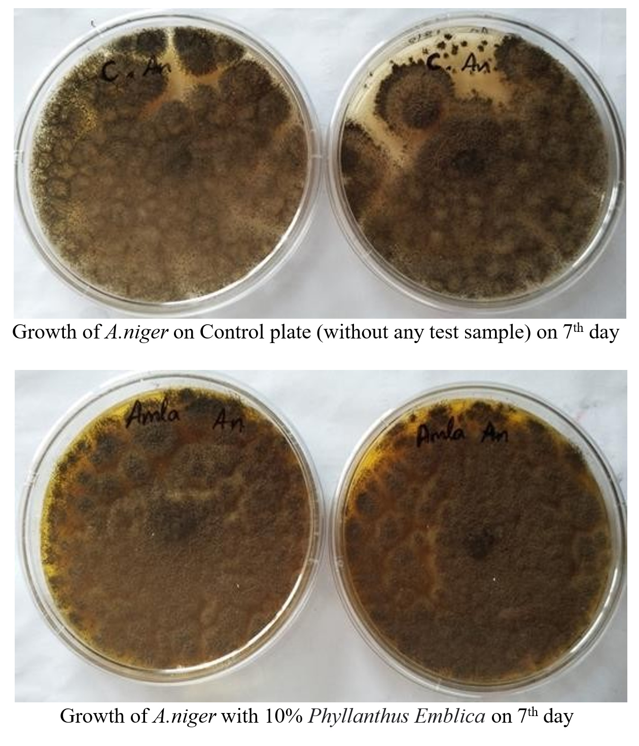
Figure 1. Growth of A. niger
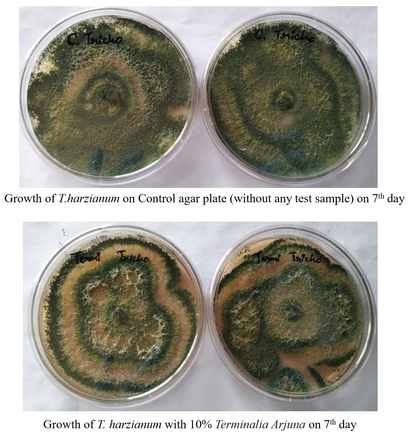
Figure 2. Growth of T. harzianum
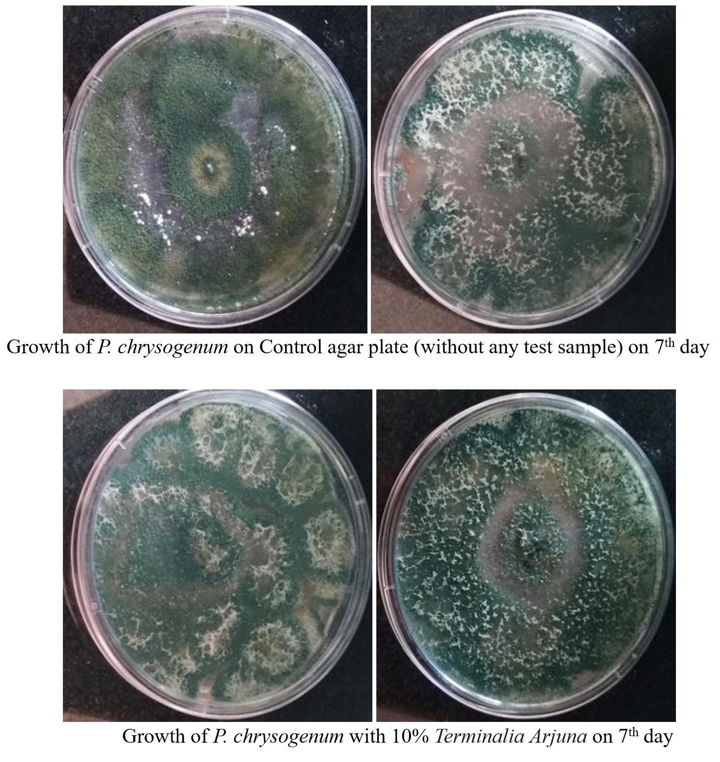
Figure 3. Growth of P. chrysogenum

Figure 4. Growth of P. chrysogenum
Concerning A. niger growth, the Control achieved 95% spreadability by Day 7, but Phyllanthus emblica demonstrated robust growth, Terminalia arjuna exhibited good growth, Mucuna pruriens displayed moderate growth, and Chlorophyta showed slower growth (Figure 5). For T. harzianum growth, both Control and Terminalia arjuna-treated samples consistently displayed robust growth (95-100% spreadability by Day 7). Phyllanthus emblica exhibited minimal growth, Chlorophyta showcased robust growth, Plantago ovata displayed minimal growth, and Asparagus racemosus demonstrated minimal to moderate growth (Figure 6).
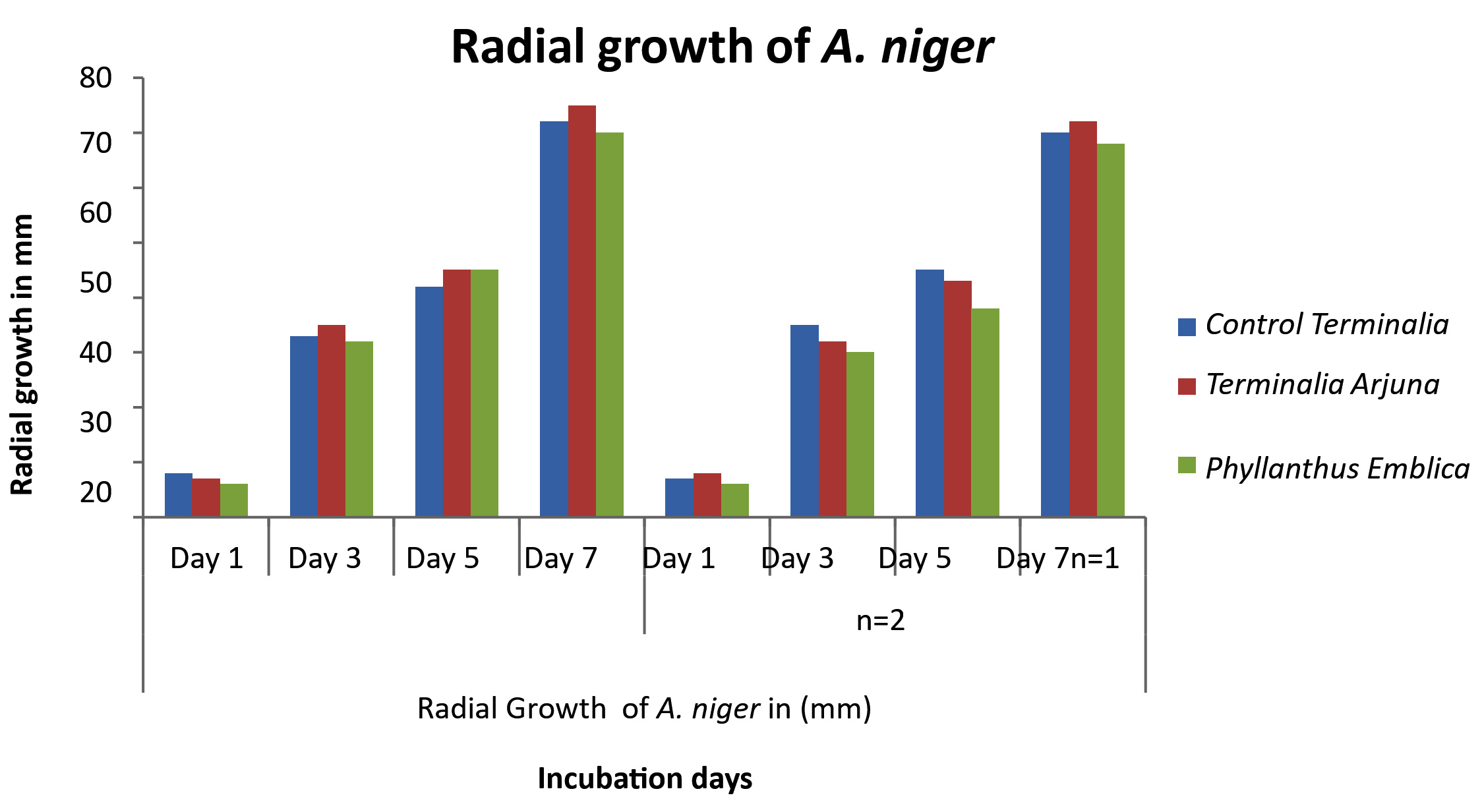
Figure 5. Growth of A. niger
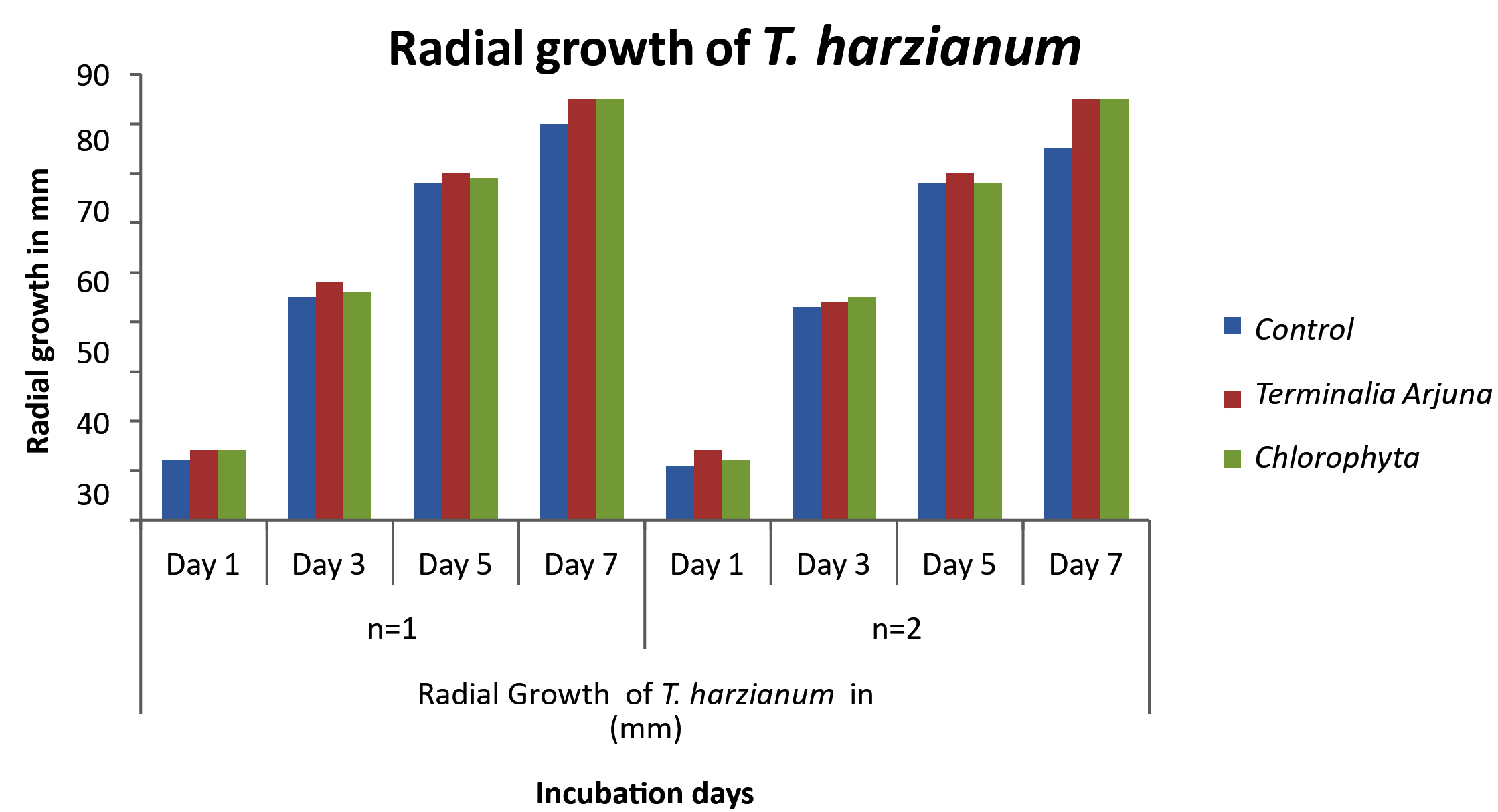
Figure 6. Growth of T. harzianum
In summary, distinct plant extracts manifested diverse effects on fungal growth. Terminalia arjuna consistently demonstrated positive effects suggesting it to be less antifungal, while the impacts of Phyllanthus emblica and Chlorophyta were varied. These findings provide valuable insights into the fungal proliferation while using the plant extracts.
Radial growth rate
The investigation into medicinal plant extract effects on Trichoderma harzianum, Aspergillus niger, and Penicillium chrysogenum involved assessing four-day radial growth. Differences in growth rate were evident (Figure 7 to Figure 9). Control growth of P. chrysogenum increased from Day 1,3,5 to 7 were 8 ± 2 mM, 25 ± 2 mM, 62 ± 2 mM, and 78 ± 2 mM, respectively, while Terminalia arjuna and Mucuna pruriens showed potential robust growth than control. In the case of radial Growth of T. harzianum, the control showed progressive growth of 12 ± 2 mM, 45 ± 2 mM, 68 ± 3 mM and 80 ± 5 mM, while Terminalia arjuna and Chlorophyta extracts stimulated T. harzianum growth, surpassing the control. For the radial Growth of A. niger the control showed as 8 ± 1 mM, 33 ± 1 mM, 42 ± 2 mM and 72 ± 2 mM, while Terminalia arjuna and Phyllanthus emblica extracts slightly elevated growth.
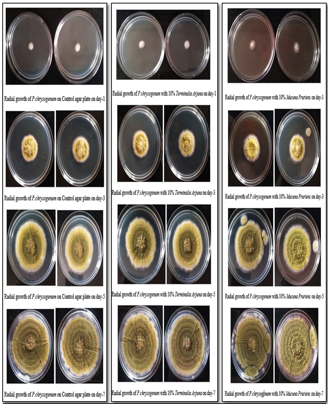
Figure 7. Radial growth of P. chrysogenum
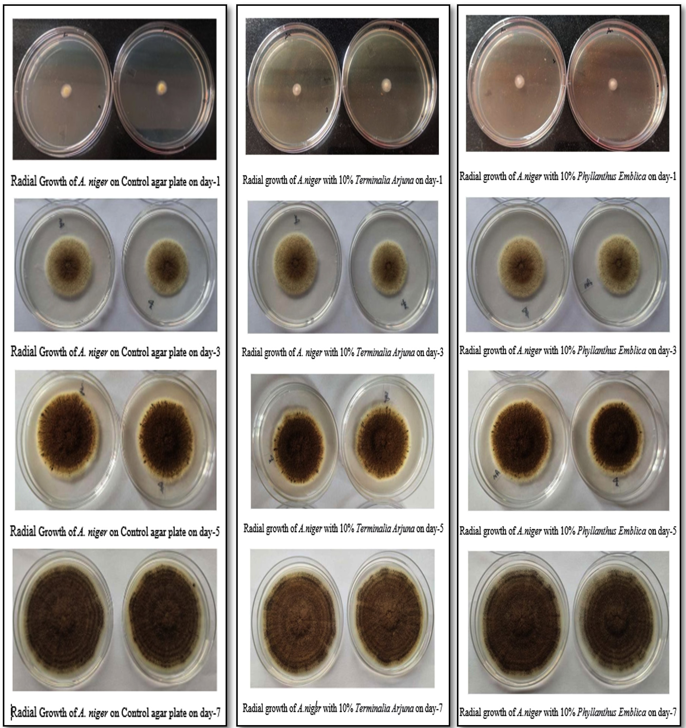
Figure 8. Radial growth of A. niger
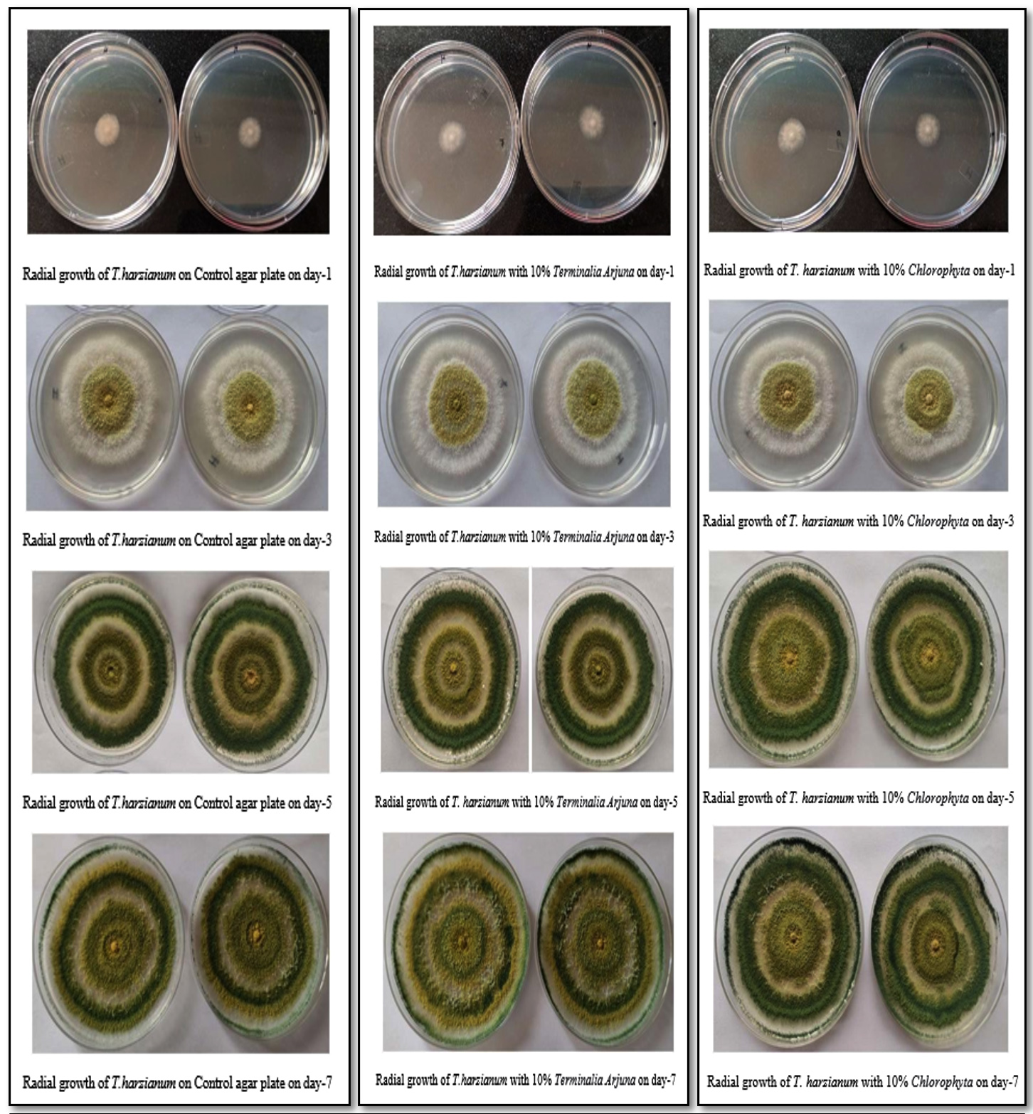
Figure 9. Radial growth of T. harzianum
Terminalia arjuna consistently stimulated radial growth across all fungi. Fungal plates treated with Terminalia arjuna extract promoted better radial colony growth than the control for A. niger, T. harzianum, and P. chrysogenum. These changes imply potential applications of these plant extracts in modulating the growth and sporulation of industrially significant fungi (Table 3).
Table (3):
Results showing the radial growth rate of fungi
| Radial Growth of P.chrysogenum in (mm) | ||||||||
|---|---|---|---|---|---|---|---|---|
| Scientific Name | Day 1 | Day 3 | Day 5 | Day 7 | Day 1 | Day 3 | Day 5 | Day 7 |
| Control | 8 ± 2 | 25 ± 2 | 62 ± 2 | 78 ± 2 | 9 ± 2 | 25 ± 2 | 60 ± 2 | 80 ± 2 |
| Terminalia Arjuna | 9 ± 2 | 27 ± 2 | 60 ± 2 | 82 ± 2 | 8 ± 2 | 26 ± 2 | 60 ± 2 | 82 ± 2 |
| Mucuna Pruriens | 8 ± 5 | 23 ± 5 | 58 ± 5 | 82 ± 5 | 9 ± 5 | 25 ± 5 | 59 ± 5 | 80 ± 5 |
| Radial Growth of T.harzianum treated with test samples | ||||||||
| Control | 12 ± 2 | 45 ± 2 | 68 ± 3 | 80 ± 5 | 11 ± 2 | 43 ± 2 | 68 ± 3 | 75 ± 5 |
| Terminalia Arjuna | 14 ± 2 | 48 ± 2 | 70 ± 3 | 85 ± 5 | 14 ± 2 | 44 ± 2 | 70 ± 3 | 85 ± 5 |
| Chlorophyta | 14 ± 2 | 46 ±2 | 69 ± 3 | 85 ± 5 | 12 ± 2 | 45 ± 2 | 68 ± 3 | 85 ± 5 |
| Radial Growth of A.niger treated with test samples. | ||||||||
| Control | 8 ± 1 | 33 ± 1 | 42 ± 2 | 72 ± 2 | 7 ± 1 | 35 ± 1 | 45 ± 2 | 70 ± 2 |
| Terminalia Arjuna | 7 ± 1 | 35 ± 1 | 45 ± 2 | 75 ± 2 | 8 ± 1 | 32 ± 1 | 43 ± 2 | 72 ± 2 |
| Phyllanthus Emblica | 6 ± 1 | 32 ± 1 | 45 ± 2 | 70 ± 2 | 6 ± 1 | 30 ± 1 | 38 ± 2 | 68 ± 2 |
Sporulation index and spore production
Phyllanthus emblica and Terminalia arjuna samples demonstrate robust sporulation for A. niger (good to excellent), while Mucuna pruriens and Chlorophyta samples exhibit fair to poor sporulation (Table 4). Terminalia arjuna and Chlorophyta samples show noteworthy sporulation for T. harzianum (good to excellent), while Phyllanthus emblica, Plantago ovata, and Asparagus racemosus exhibit inconsistent results. Terminalia arjuna sample displays commendable sporulation for P. chrysogenum (good), whereas Mucuna pruriens and Chlorophyta exhibit fair sporulation. Plantago ovata demonstrates poor sporulation.
Table (4):
Results showing Sporulation Index and Spore Production
| Organism | Sample – Scientific Name | No. of spores per microscopic field | Sporulation capacity (Number of spores/ml) |
|---|---|---|---|
| A.niger | Control | 95 | 5.9*109 |
| Phyllanthus Emblica | 112 | 7.0*109 | |
| Terminalia Arjuna | 102 | 6.3*109 | |
| Mucuna Pruriens | 25 | 1.5*109 | |
| Chlorophyta | 12 | 0.75*109 | |
| T. harzianum | Control | 95 | 5.9*109 |
| Terminalia Arjuna | 92 | 5.7*109 | |
| Phyllanthus Emblica | – | – | |
| Chlorophyta | 93 | 5.8*109 | |
| Plantago Ovata | 6 | 0.37*109 | |
| Asparagus Racemosus | 12 | 0.75*109 | |
| P. chrysogenum | Control | 81 | 5.0*109 |
| Terminalia Arjuna | 94 | 5.8*109 | |
| Mucuna Pruriens | 62 | 3.8*109 | |
| Chlorophyta | 40 | 2.5*109 | |
| Plantago Ovata | 25 | 1.5*109 | |
| Phyllanthus Emblica | – | – |
Based on the findings of the study, it can be concluded that Terminalia arjuna exhibits superior growth and relative sporulation across all three fungi tested. This suggests that Terminalia arjuna shows a positive effect on the growth of these fungi. Therefore, Terminalia arjuna holds potential for further investigations and studies.
Trichoderma harzianum, Aspergillus niger, and Penicillium chrysogenum are well-known fungi, and their growth and sporulation are of interest in various fields such as agriculture, pharmaceuticals, and biotechnology. Based on the findings of experiments in the current study, it can be inferred that Terminalia arjuna stands out among all the samples and warrants further investigation. This conclusion is drawn from the absence of notable inhibitory activity against A. niger, T. harzianum, and P. chrysogenum in the case of Terminalia arjuna.
The examination of phytochemicals revealed a diverse array of bioactive compounds within the medicinal plant extracts, including alkaloids, glycosides, steroids, terpenoids, proteins, flavonoids, saponins, tannins, and carbohydrates. The presence of alkaloids in all extracts suggests potential antimicrobial properties, while the involvement of terpenoids and flavonoids suggests contributions to antioxidant and antimicrobial characteristics. These compounds hold promise for influencing fungal growth and sporulation, positioning the plant extracts as potential candidates for various industrial applications.
The results from the Point Inoculation Method showed varied effects of medicinal plant extracts on the growth of Trichoderma harzianum, Aspergillus niger, and Penicillium chrysogenum. Notably, Terminalia arjuna consistently exhibited positive effects, implying a lower degree of antifungal activity, while the impacts of Phyllanthus Emblica varied. This finding is particularly significant in industrial contexts where precise control of fungal growth is pivotal. The analysis of radial growth rates further supported these observations, indicating that Terminalia arjuna consistently stimulated radial growth across all fungi. Fungal plates treated with Terminalia arjuna extract facilitated superior radial colony growth compared to the control for A. niger, T. harzianum, and P. chrysogenum. These outcomes suggest potential applications of these plant extracts in influencing the growth and sporulation of fungi that hold industrial significance.
The evaluation of sporulation indices and spore production indicated commendable sporulation in Terminalia arjuna for all three fungi. In contrast, Phyllanthus emblica and Chlorophyta exhibited inconsistent results in terms of sporulation. This implies that the Terminalia arjuna extract may exert a less inhibitory effect on fungal sporulation when compared to other extracts.
The diversity, composition, and abundance of fungi found in sapwood align with the findings of Evans et al.24 and Tejesvi et al.25 a significant presence of Trichoderma isolates in T. gileri and Terminalia arjuna, respectively. The antifungal activity of Terminalia arjuna was assessed in a study, the methanol extract of T. arjuna leaves exhibited moderate activity against Microsporum canis, but showed no significant activity against Aspergillus flavus. The methanol extract of T. arjuna fruit displayed non-significant activity against Aspergillus flavus, Microsporum canis, and Fusarium solani. However, no antifungal activity was observed with the methanol extract of T. arjuna bark. Additionally, the extracts from T. arjuna leaves, stem bark, and fruit in dichloromethane (DCM) demonstrated no antimicrobial activity.26
Some of the identified fungal endophytic species have the potential to be tested as biocontrol agents for combating H. brasiliensis diseases. Previous studies on the use of biological control against Hevea diseases27-29 have focused on different approaches and have not specifically explored the use of fungal endophytes as antagonistic agents. In future antagonistic assays, it would be worthwhile to consider three genera: Colletotrichum, Pestalotiopsis, and Trichoderma. These genera hold promise for their potential effectiveness in combating Hevea diseases and warrant further investigation as biocontrol candidates.
Certain strains of Trichoderma, such as T. harzianum, T. stromaticum, and T. asperellum, have demonstrated their ability to act antagonistically against fungal diseases.30-32 Trichoderma employs various mechanisms to protect its host, including direct parasitism, antibiosis, nutrient competition, enhanced plant growth, and induced resistance.33-37 These attributes, combined with its strong competitiveness, make Trichoderma species highly recommended for further assessment of their biocontrol potential.
The comprehensive findings suggest that while the Terminalia arjuna extract is not entirely devoid of antifungal properties, it may exhibit a lesser inhibitory effect on fungal growth and sporulation compared to other extracts. This has potential implications for applications in both industrial and medicinal contexts, where maintaining specific levels of fungal growth may be desirable. Further investigation and validation of these findings are essential to fully exploit the pharmacological potential of medicinal plant extracts in specific applications.
The empirical exploration into the impact of medicinal plant extracts on the growth and sporulation of industrially significant fungal strains yields valuable insights. Phytochemical analysis uncovers a diverse range of bioactive compounds, indicating potential applications in antimicrobial, antioxidant, and nutritional domains. Findings from the Point Inoculation Method and radial growth rates consistently indicate positive effects of Terminalia Arjuna, suggesting lower antifungal activity compared to alternative extracts. Sporulation indices confirm commendable sporulation in Terminalia Arjuna, contrasting with variable results in other extracts. A comparative examination with previous studies underscores the distinctive attributes of Terminalia Arjuna. While not entirely devoid of antifungal properties, Terminalia Arjuna may exert a milder inhibitory effect on fungal growth, holding potential implications for industrial and medicinal purposes. Further research is essential to fully exploit its pharmacological potential.
ACKNOWLEDGMENTS
The authors would like to thank Dr. D. Kalpana, Principal, Sree Narayana Guru College, and Skanda Life Sciences, Bangalore, for the facilities provided.
CONFLICT OF INTEREST
The authors declare that there is no conflict of interest.
AUTHORS’ CONTRIBUTION
Both authors listed have made a substantial, direct and intellectual contribution to the work, and approved it for publication.
FUNDING
None.
AVAILABILITY OF DATA
All datasets generated or analyzed during this study are included in the manuscript.
ETHICS STATEMENT
This article does not contain any studies with human participants or animals performed by any of the authors.
- Dwivedi S. Terminalia arjuna Wight & Arn.—a useful drug for cardiovascular disorders. J Ethnopharmacol. 2007;114(2):114-129.
Crossref - Maulik SK, Talwar KK. Therapeutic potential of Terminalia arjuna in cardiovascular disorders. Am J Cardiovasc Drugs. 2012;12(3):157-163.
Crossref - Kapoor D, Vijayvergiya R, Dhawan V. Terminalia arjuna in coronary artery disease: ethnopharmacology, pre-clinical, clinical & safety evaluation. J Ethnopharmacol. 2014;155(2):1029-1045.
Crossref - Wendland J. Sporulation in Ashbya gossypii. J Fungi. 2020;6(3):157.
Crossref - Schuster E, Dunn-Coleman N, Frisvad JC, Van Dijck PWM. On the safety of Aspergillus niger—a review. Appl Microbiol Biotechnol. 2002;59(4-5):426-435.
Crossref - Shaaban R, Elnaggar MS, Khalil N, Singab ANB. A comprehensive review on the medicinally valuable endosymbiotic fungi Penicillium chrysogenum. Arch Microbiol. 2023;205(6):240.
Crossref - Tyskiewicz R, Nowak A, Ozimek E, Jaroszuk-Scisel J. Trichoderma: The Current Status of Its Application in Agriculture for the Biocontrol of Fungal Phytopathogens and Stimulation of Plant Growth. Int J Mol Sci. 2022;23(4):2329.
Crossref - Howell CR. Mechanisms Employed by Trichoderma Species in the Biological Control of Plant Diseases: The History and Evolution of Current Concepts. Plant Dis. 2003;87(1):4-10.
Crossref - Knudsen JR, Dandurand LMC. Ecological Complexity and the Success of Fungal Biological Control Agents. Adv Agric. 2014;2014(1):542703.
Crossref - Sen A, Batra A. Evaluation of antimicrobial activity of different solvent extracts of medicinal plant: Melia azedarach L. Int J Curr Pharm Res. 2012;4(2):67-73.
- Coe FG, Parikh DM, Johnson CA, Anderson GJ. The good and the bad: alkaloid screening and brineshrimp bioassays of aqueous extracts of 31 medicinal plants of eastern Nicaragua. Pharm Biol. 2012;50(3):384-392.
Crossref - Hossain MA, Nagooru MR. Biochemical profiling and total flavonoids contents of leaves crude extract of endemic medicinal plant Corydyline terminalis L. Kunth. Pharmacogn J. 2011;3(24):25-30.
Crossref - El Aziz MMA, Ashour AS, Melad AG. A review on saponins from medicinal plants: chemistry, isolation, and determination. J Nanomed Res. 2019;8(1):282-288.
Crossref - Packirisamy S, Magdaline BL, Dhanavel A, Rajendiran D, Vijayashree RJ. Preliminary screening and evaluation of Commiphora caudata leaf extract on cariogenic pathogens. IJPSR. 2022;13(10):4159-4165.
- Prakash J, Vedanayaki S. Organoleptic, fluorescence, qualitative and quantitative analysis of bulb extract of Zephyranthes citrina. J Pharmacogn Phytochem. 2019;8(3):2531-2536.
- Malik SK. Qualtitative and quantitative estimation of terpenoid contents in some important plants of Punjab, Pakistan. Pak J Sci. 2017;69(2).
Crossref - Manzo LM, Moussa I, Ikhiri K. Phytochemical screening of selected medicinal plants used against diarrhea in Niger, West Africa. Int J Herb Med. 2017;5(4):32-38.
Crossref - Yadav RNS, Agarwala M. Phytochemical analysis of some medicinal plants. J Phytol. 2011;3(12).
- Bhatia NM, Salunkhe SS, Mali SS, et al. Extraction and characterization of mucilage from Lepidium sativum Linn. seeds. Scholars Research Libraryder Pharmacia Lettre. 2014;6(1):65-70.
- Katalinic V, Milos M, Kulisic T, Jukic M. Screening of 70 medicinal plant extracts for antioxidant capacity and total phenols. Food Chem. 2006;94(4):550-557.
Crossref - Quiroga EN, Sampietro, A. R., & Vattuone, M. A. Screening antifungal activities of selected medicinal plants. J Ethnopharmacol. 2001;74(1):89-96.
Crossref - Ramírez-Guzmán, N., Roussos, S., Martinez-Medina, G.A. et al. Selection of a biocontrol agent based on a comparative spore production evaluation. Syst Microbiol and Biomanuf, 2024;4;794–800.
Crossref - Ngegba PM, Enikuomehin OA, Afolabi CG, et al. Evaluation of some plant extracts on mycelial growth and sporulation density of fungal pathogens of groundnut (Arachis hypogaea L.) in-vitro. Int J Dev Res, 2017;7:13808-13814.
- Evans HC, Holmes KA, Thomas SE. Endophytes and mycoparasites associated with an indigenous forest tree, Theobroma gileri, in Ecuador and a preliminary assessment of their potential as biocontrol agents of cocoa diseases. Mycol Prog. 2003;2(2):149-60.
Crossref - Tejesvi MV, Mahesh B, Nalini MS, et al. Endophytic fungal assemblages from inner bark and twig of Terminalia arjuna W. & A.(Combretaceae). World J Microbiol Biotechnol. 2005;21:1535-1540.
Crossref - Javed T, Riaz S, Uzair M, Mustafa G, Mohyuddin A, Ahmad B. Biological activity of Terminalia arjuna on human pathogenic microorganisms. Pak J Pharm Res. 2016;2(1):23-27.
Crossref - Sudirman LI, Housseini AII, Le Febvre G, Kiffer E, Botton B. Screening of some basidiomycetes for biocontrol of Rigidoporus lignosus, a parasite of the rubber tree Hevea brasiliensis. Mycol Res. 1992;96(8):621-625.
Crossref - Mello SC, Santos MD, Silva JB. Dicyma pulvinata isolates colonizing Microcyclus ulei stromata in rubber. Pesquisa Agropecuaria Brasileira. 2006;41:359-63.
Crossref - Evueh GA, Ogbebor NO. Use of phylloplane fungi as biocontrol agent against Colletotrichum leaf disease of rubber (Hevea brasiliensis Muell. Arg.). Afr J Biotechnol. 2008;7(15).
- De Meyer G, Bigirimana J, Elad Y, Hofte M. Induced systemic resistance in Trichoderma harzianum T39 biocontrol of Botrytis cinerea. Eur J Plant Pathol. 1998;104:279-86.
Crossref - Watanabe S, Kumakura K, Izawa N, et al. Mode of action of Trichoderma asperellum SKT-1, a biocontrol agent against Gibberella fujikuroi. J Pestic Sci. 2007;32(3):222-228.
Crossref - De Souza JT, Bailey BA, Pomella AW, et al. Colonization of cacao seedlings by Trichoderma stromaticum, a mycoparasite of the witches’ broom pathogen, and its influence on plant growth and resistance. Biological Control. 2008;46(1):36-45.
Crossref - Hanada RE, de Jorge Souza T, Pomella AW, et al. Trichoderma martiale sp. nov., a new endophyte from sapwood of Theobroma cacao with a potential for biological control. Mycol Res. 2008;112(11):1335-1343.
Crossref - Harman GE, Howell CR, Viterbo A, Chet I, Lorito M. Trichoderma species-opportunistic, avirulent plant symbionts. Nat Rev Microbiol. 2004;2(1):43-56.
Crossref - Holmes KA, Schroers HJ, Thomas SE, Evans HC, Samuels GJ. Taxonomy and biocontrol potential of a new species of Trichoderma from the Amazon basin of South America. Mycol Prog. 2004;3(3):199-210.
Crossref - Khan J, Ooka JJ, Miller SA, Madden LV, Hoitink HA. Systemic resistance induced by Trichoderma hamatum 382 in cucumber against Phytophthora crown rot and leaf blight. Plant Dis. 2004;88(3):280-286.
Crossref - Bailey BA, Bae H, Strem MD, et al. Antibiosis, mycoparasitism, and colonization success for endophytic Trichoderma isolates with biological control potential in Theobroma cacao. Biological Control. 2008;46(1):24-35.
Crossref
© The Author(s) 2024. Open Access. This article is distributed under the terms of the Creative Commons Attribution 4.0 International License which permits unrestricted use, sharing, distribution, and reproduction in any medium, provided you give appropriate credit to the original author(s) and the source, provide a link to the Creative Commons license, and indicate if changes were made.