


ISSN: 0973-7510
E-ISSN: 2581-690X
For millennia, people have successfully treated a wide range of illnesses, including bacterial infections, with different plant parts or extracts. “MDR strain” typically refers to a “multi-drug resistant strain” of a bacteria. In the context of infectious diseases, a multi-drug resistant strain refers to a strain of the pathogen that has acquired resistance to multiple drugs that are commonly used to treat infections caused by that specific pathogen. MDR strains can present significant challenges in healthcare settings as they limit the effectiveness of standard treatments and may require more aggressive or specialized approaches to manage the infection. The discdiffusion method was used in this investigation to test the antimicrobial properties of Petroleum ether, acetone, ethanol, methanol, and aqueous extracts of Ficus auriculata leaves against four bacterial strains namely Salmonella enteric serovar typhi, Salmonella enteric ser Paratyphi (MDR strain) Pseudomonas aeruginosa and Escherichia coli. The findings showed that all of the investigated organisms (zone of inhibition of 0.5 ± 0.15 & 18 ± 1.7 mm) were significantly inhibited by the petroleum ether, ethanol, and methanol extracts, with the exception of Salmonella typhi (an 18 mm inhibitory zone). The restricted area (≤5) indicated moderate activity in the aqueous extracts. It’s crucial to remember that antimicrobial activity analysis of plant extracts is just one step in the process of identifying potential natural antimicrobial agents. Further studies, including the identification and isolation of specific bioactive compounds, toxicology assessments, and clinical trials, are required before any plant extract can be considered for use as a drug development.
MDR Strain, Ficus auriculata, Disc Diffusion Method, Antimicrobial Activity, Drug Development
Ficus auriculata, referred to as the Roxburgh fig in common or elephant ear fig, is a type of flowering plant being a part of the family Moraceae.1 Here are some key characteristics and information about Ficus auriculata:
Habitat
This species comes from Southeast Asia and is commonly found in nations like India, Bangladesh, Sri Lanka, Myanmar, and Thailand. It typically develops in a tropical and subtropical region.2,3
Tree Characteristics
Ficus auriculata is a large, evergreen tree that can reach heights of up to 30 meters. It has a spreading canopy and distinctive, large, ear-shaped leaves, which give it the common name “elephant ear fig.”
Leaves
The leaves of Ficus auriculata are glossy, dark green, and can grow up to 30 centimeters in length. The shape of the leaves is somewhat reminiscent of an elephant’s ear, hence the common name.4
Fruits
The figs produced by this species are small, green to yellowish-brown, and typically measure about 1 to 1.5 centimeters in diameter. The fruits are often used by birds and other wildlife as a food source.5
Ecological importance
Ficus species, in general, play a crucial role in various ecosystems. They are known for their mutualistic relationship with fig wasps, which are essential for the pollination of fig flowers.
Cultural significance
In some cultures, Ficus auriculata may have cultural or religious significance. Ficus trees, in general, are associated with sacred sites and are often planted near temples or other religious places.
Landscaping
Due to its large and attractive foliage, Ficus auriculata is sometimes used in landscaping for its ornamental value. However, its size needs to be considered when planting in smaller spaces.
Propagation
Ficus auriculata can be propagated through seeds or stem cuttings. The plant is relatively hardy and can adapt to a variety of soil types, but it prefers well-drained soil.
Always be sure to check local regulations and guidelines before introducing non-native plant species to new areas, as they can potentially become invasive and disrupt local ecosystems.
While Ficus auriculata, commonly known as the Roxburgh fig or elephant ear fig, is not as extensively documented in traditional medicine as some other Ficus species, certain plant parts have been utilised in customary practices in some regions. It’s important to note that the information provided here is based on traditional knowledge, and scientific evidence may be limited. Always consult with healthcare professionals before using any plant for medicinal purposes.6-8
Leaves and latex
In traditional medicine, Ficus auriculata leaves and latex have been used for various purposes. The latex, in particular, is known for its potential wound-healing properties. In some cultures, it has been applied topically to wounds or skin conditions.
Antimicrobial properties
Some Ficus species, including Ficus auriculata, are believed to possess antimicrobial properties. The leaves and latex may be utilised in conventional medicine to treat skin infections or wounds.9
Anti-inflammatory effects
The plant’s components, when applied externally, are thought to have anti-inflammatory effects. This may be relevant in traditional remedies for conditions such as skin inflammation or joint pain.
Digestive issues
In certain traditional systems, Ficus species, including Ficus auriculata, have been used to address digestive issues. This might include the use of specific parts of the plant to alleviate symptoms like indigestion.
Antioxidant potential
Some Ficus species are known to contain compounds with antioxidant properties. Antioxidants are believed to help combat oxidative stress in the body, and in traditional medicine, plants with such properties are sometimes used to promote overall health.
It’s important to emphasize that while there may be anecdotal evidence supporting the use of Ficus auriculata in traditional medicine, scientific studies validating its efficacy and safety are often lacking. If you are considering using Ficus auriculata or any other plant for medicinal purposes, it’s crucial to consult with healthcare professionals or traditional medicine practitioners who are knowledgeable about the specific uses and potential risks associated with the plant. Additionally, be aware that traditional practices can vary widely, and the effectiveness of traditional remedies may not be universally accepted or supported by scientific evidence.6,13,14
The term “MDR strain” typically refers to a strain of a pathogenic microorganism that has developed resistance to multiple drugs. MDR stands for multidrug-resistant. This phenomenon is a significant concern in the context of bacterial, viral, fungal, or parasitic infections where the microorganism evolves to withstand the effects of multiple drugs that were originally effective in treating the infection.
Plant Sample collection and authentication
The foliage of F. auriculata were collected from Tehri region of Uttarakhand, India. The BSI (Botanical Survey of India) validated and taxonomically identified the plant samples. A voucher specimen (872) of F. auriculata Lour were accessioned at herbarium BSI, Dehradun. Wiped the sample to get rid of dust. Cut the leaves and stem into small pieces after washing. Plant stems and leaves were air dried for two weeks in shaded area before being packaged in paper bags and kept in storage. To prevent any contamination from growing, samples were constantly monitored. To create a fine powder sample, dried leaves and stems were ground up in an electric grinder.
Preparation of plant extracts
The sequential Soxhlet extraction technique was used for the extraction.
Soxhlet extraction
This method was applied to get extracts for pharmaceutical and biological testing, as well as phytochemical screening. To obtain a uniformly sized powder, the leaves and stem were ground together in a grinder. A homogenous 25 gm of powered plant substance was placed in a thimble, and 250 mL of numerous solvents were extracted from each separately. After that, the thimble was situated inside the Soxhlet apparatus, where extraction was carried out using petroleum ether, acetone, ethanol, methanol, as well as water as solvents in a sequential order from non-polar to polar. The extraction process lasts for a whole day, or until the solvent in the extractor’s siphon tube turns colorless.10 After the petroleum ether extract was collected, acetone was extracted in a further step using powder made from the thimble, and the yield was estimated. The same process was used for drying, and thimbles were filled with powder that was utilized for ethanol, methanol, and water extraction. Finally, soluble fractions in water, methanol, ethanol, acetone, and petroleum ether were obtained. Crude extracts were then left behind as the extract was allowed to concentrate in a vacuum using a rotating evaporator. The dehydrated mixture was stored in a refrigerator at 4°C for their future use in different analysis.11,12 All extracts obtained from Tehri regions of Uttarakhand.
Test organism collection: used microorganisms and growth conditions
For the experiment, MTCC 735 for Salmonella enterica ser. Paratyphi and MTCC 733 for Salmonella enterica serovar Typhi were provided by the “Chandigarh Institute of Microbial Technology (IMTECH)”. Human harmful bacterial strains, such as Escherichia coli and Pseudomonas aeruginosa, were gathered for antimicrobial screening and antibacterial research purposes from Medical & Health Sciences department, SGRR University Dehradun, India. Bacteria were cultivated using “Mueller-Hinton broth medium (MHB) as well Mueller-Hinton agar medium (MHA)” from Liofilchem, Italy.
Preparation of stock solutions
To avoid sterile filtration, the samples’ stock solutions were made by dissolving the raw desiccated extracts in (DMSO) pure dimethyl sulfoxide (Sigma-Aldrich) concentrate. Meanwhile, sterile distilled water was used to dilute the aqueous extracts and filtered using a Millipore 0.22 μm sterile filter.15
Preparation of bacterial inoculums
Bacterial inoculation for Escherichia coli, Typhi and Paratyphi are two types of Salmonella enterica and Pseudomonas aeruginosa made using the Luria-Bertani broth (HIMEDIA). After thoroughly dissolving nutrient agar in distilled water, after mixing it was autoclaved regarding 30 minutes at 120°C as well as 15 to 20 psi pressure for inoculate bacterial colonies. After being recovered in BHI, pathogenic cultures were cultured for an entire night at 36°C. Once the cultures were maintained at 0.5 of the McFarland scale which is equivalent to 108 CFU.ml-1, they were diluted to 105 CFU.ml-1 using casein peptone water.16
Preparation of Agar plates
For the purpose of cultivating bacteria, nutrient agar medium was prepared and then put into sterile plates. It ought to be carried out in an undisturbed, laminar flow environment to prevent medium contamination. Agar medium covered the sterile plates evenly. Agar nutrition plates were now positioned within an incubator set at 37°C for the entire night, or until the agar nutrient solidified in the sterile plates.17
Placements of plates
Following the even distribution of agar on sterile plates, a 5 mm sterile paper disc was immersed in a mixture of 50-100 μL of petroleum ether, acetone, ethanol, methanol, and water extracted from the leaves of Ficus auriculata. These plates are now placed back into the incubator to develop bacterial cultures overnight at 37°C. The following day, measure the inhibition zone at various concentrations and compare it to the conventional streptomycin (10 μg) zone of inhibition. Inhibition zone diameter as measured on a scale (mm).18
Disc diffusion
The isolates were inoculated onto Müller-Hinton Agar media using swabs. Following that, each plate received three sterile discs of Whatman filter paper, measuring 6 mm in diameter. 15 μl of the extracts were put to the paper discs. The negative control was sterile distilled water. For 24 hours, the plates were cultivated at 35 ± 1°C. Using a pachymeter, the inhibitory zone’ diameters were determined, and the outcomes was given in millimetres (mm). The extract’s antibacterial efficacy against the studied pathogens increases in the inhibition zone. Every test was run in triplicates.19,21
Minimum inhibitory concentration (MIC)
The plant extract’s minimum inhibitory concentration, or “MIC”, at which no growth emerged, was ascertained against two harmful and two multidrug-resistant strains of Salmonella were ascertained by the use of a macrobroth dilution experiment. In well plates, extracts were serially diluted using Mueller-Hinton Broth in a 2-fold range in accordance with the findings of the disc diffusion method. The same controls, both positive and negative were used in the MIC. Following a 20 µl inoculation with freshly made bacterial suspension (5 × 10⁸ CFU/ml), the plates were retained at 37°C for an entire day. It was determined what the minimal amount that inhibits growth “MIC” of the plant extract was at which no growth appeared.22
MBC value determination
Samples (5 μl) collected from the wells that did not show growth during the MIC determination were cultivated on agar media in order to determine the minimum bactericidal “MBC” of the plant crude extracts. The least amount of crude extracts that did not show signs of growth following a 24-hour incubation period at 37°C was designated as MBC.23
Determination of the antioxidant or Free radical scavenging activity of Ficus auriculata different extracts by DPPH Scavenging for Free Radicals
The activity of the antioxidant was tested with some minor modifications of method of Jothi and Jebamalar.22 Various sample concentrations (20%, 40%, 60%, 80%, and 100%) were combined with 0.1 mM of working DPPH reagent. Keep the mixture for incubation at ambient temperature for 2 hours. Discoloration of sample was predicted by spectrophotometer at 517 nm. Utilising the standard ascorbic acid, the outcomes were stated as mg ASC/g of extract.20
Hydrogen peroxide scavenging activity
0.1 ml of dilutions of the sample (20%, 40%, 60%, 80% and 100%) was added in 0.4 ml of 50 mM of phosphate buffer to which 2 mM of 0.6ml of H2O2 was added, vortex the sample for 10 minutes, absorbance was taken through spectrophotometer at 230 nm. The results were articulated as mg ASC/gm extract, using ascorbic acid as the benchmark.24
Data processing
The three replicates’ mean ± standard deviation was provided as the result. To examine the experimental data, SPSS version 16.0 was utilised (SPSS Inc. Chicago, IL). The mean differences were assessed utilising an ANOVA in one way.
Extract preparation
Ficus auriculata leaf extracts were made using the soxhlet method. After the extracts were ready, Table 1 tallied their yield, colour, and condition. The extracts containing water and ethanol had the highest yield, followed by those containing methanol, acetone, and P. ether.
Table (1):
Physical properties and Yield of Ficus auriculata leaf extract31
| No. | Solvent used | Ficus auriculate | ||
|---|---|---|---|---|
| Yield | Colour | State | ||
| 1 | Petroleum ether | 1.0 | Light green | Liquid |
| 2 | Acetone | 2.34 | Dark green | Viscous |
| 3 | Ethanol | 2.91 | Dark green | Viscous |
| 4 | Methanol | 2.84 | Dark green | Viscous |
| 5 | Water | 5.04 | Light green | Liquid |
Antibacterial activity
All leaf extracts, including petroleum ether, acetone, ethanol, methanol as well as water, were exposed to an antibacterial assay using the disc diffusion method against four different microorganisms. Of these, two were multidrug-resistant (MDR) bacteria, the two strains of Salmonella enterica are Typhi and Paratyphi and the other two were gram-negative bacteria, E. coli as well as Pseudomonas aeruginosa. ZOI was measured in order to determine microbial proliferation. Regarding Ficusauriculata, it was determined that at a dose of 1 mg/100 µl of methanolic extract, S. typhi was the most susceptible, followed by S. typhi, S. paratyphi, E. coli, and Pseudomonas aeruginosa, with ZOIs of 14 ± 1.5 mm, 18 ± 1 mm, 16 ± 1 mm, and 18 ± 1.7 mm, respectively.5,9,25 At a dose of 0.5 mg/100 µl, the highest ZOI was recorded by S. typhi, S. para typhi, E. coli, and Pseudomonas aeruginosa, measuring 6.0 ± 0.5 mm, 9.0 ± 1 mm, 4.0 ± 0.57 mm, and 6.1 ± 0.1 mm, respectively.7 S. typhi, S. paratyphi, E. coli, and Pseudomonas aeruginosa demonstrated the highest susceptibility to ethanol extract at 1 mg/100 µl dose, with ZOI values of 11 ± 2 mm, 12 ± 2 mm, 12 ± 1.5 mm, and 17 ± 1.1 mm, respectively; in contrast, at 0.5 mg/100 µl dose, the highest ZOI values were 4.0 ± 1 mm, 5.0 ± 0.17mm, 8.1 ± 1 mm, and 7.1 ± 0.57 mm for S. typhi, S. paratyphi, E. coli, and Pseudomonas aeruginosa. The highest ZOI for S. typhi, S. paratyphi, E. coli, and Pseudomonas aeruginosa in the case of Acetone extract for a 1 mg/100 µl dose was found to be 11 ± 1.1 mm, 10 ± 2 mm, 15 ± 1.5 mm, and 18 ± 0.5 mm, respectively.3 In contrast, the most susceptible bacteria in the case of a 0.5 mg/100 µl dose were S. typhi, S. paratyphi, E. coli, and Pseudomonas aeruginosa, with ZOIs of 5 ± 0.5 mm, 5.2 ± 0.5 mm, 10.1 ± 1mm, and 7 ± 1 mm.15,26 S. typhi, S. paratyphi, E. coli, and Pseudomonas aeruginosa demonstrated the highest susceptibility to P. ether extract at 1 mg/100 µl dose, with ZOI values of 3.0 ± 2 mm, 5.0 ± 1.5 mm, 0.8 ± 0.6 mm, and 7.0 ± 0.57 mm, respectively; meanwhile, at 0.5 mg/100 µl dose, the highest ZOI values were 1.5 ± 0.15 mm, 1.2 ± 0.1 mm, 0.6 ± 0.15 mm, and 0.8 ± 0.11 mm for S. typhi, S. paratyphi, E. coli as well as Pseudomonas aeruginosa. S. typhi, S. paratyphi, E. coli, as well as Pseudomonas aeruginosa demonstrated the highest susceptibility to water extract at a dose of 1 mg/100 µl, with ZOIs of 4 ± 2 mm, 2.8 ± 0.25 mm, 10 ± 1 mm, and 2.6 ± 0.2 mm, respectively; at a dose of 0.5 mg/100 µl, the highest ZOIs were 1.5 ± 0.17 mm, 1.2 ± 0.1 mm, 1.6 ± 0.2 mm, and 2.5 ± 0.05 mm for S. typhi, S. paratyphi, E. coli, and Pseudomonas aeruginosa6 (Table 2, Figure 1-5).
Table (2):
Antibacterial activity of Ficus auriculata leaves for various extract determined by disc diffusion24
| No. | Microbial culture | Concentration of Various Extracts | |||||||||
|---|---|---|---|---|---|---|---|---|---|---|---|
| Petroleum ether | Acetone | Ethanol | Methanol | Water | |||||||
| 0.5 mg/ 0.1 ml | 1 mg/ 0.1 ml | 0.5 m/0.1 ml | 1 mg/ 0.1 ml | 0.5 m/0.1 ml | 1 mg/ 0.1 ml | 0.5 mg/ 0.1 ml | 1 mg/ 0.1 ml | 0.5 mg/ 0.1 ml | 1 mg/ 0.1 ml | ||
| 1 | S. enterica ser typhi | 1.5 ± 0.1 | 3 ± 2 | 5 ± 0.5 | 11 ± 1.1 | 4 ± 1 | 11 ± 2 | 6 ± 0.5 | 14 ± 1.5 | 1.5 ± 0.1 | 4 ± 2 |
| 2 | S. enterica para typhi | 1.2 ± 0.1 | 5 ± 1.5 | 5 ± 0.5 | 10 ± 2 | 5.2 ± 0.1 | 12 ± 2 | 9 ± 1 | 18 ± 1 | 1.2 ± 0.1 | 2.8 ± 0.2 |
| 3 | E. coli | 0.6 ± 0.1 | 0.8 ± 0.6 | 10 ± 1 | 15 ± 1.5 | 8 ± 1 | 12 ± 1.5 | 4 ± 0.5 | 16 ± 1 | 1.6 ± 0.2 | 10 ± 1 |
| 4 | P. aeruginoa | 0.8 ± 0.11 | 7 ± 0.57 | 7 ± 1 | 18 ± 0.5 | 7 ± 0.57 | 17 ± 1.1 | 6.3 ± 0.1 | 18 ± 1.7 | 2.5 ± 0.05 | 2.6 ± 0.2 |
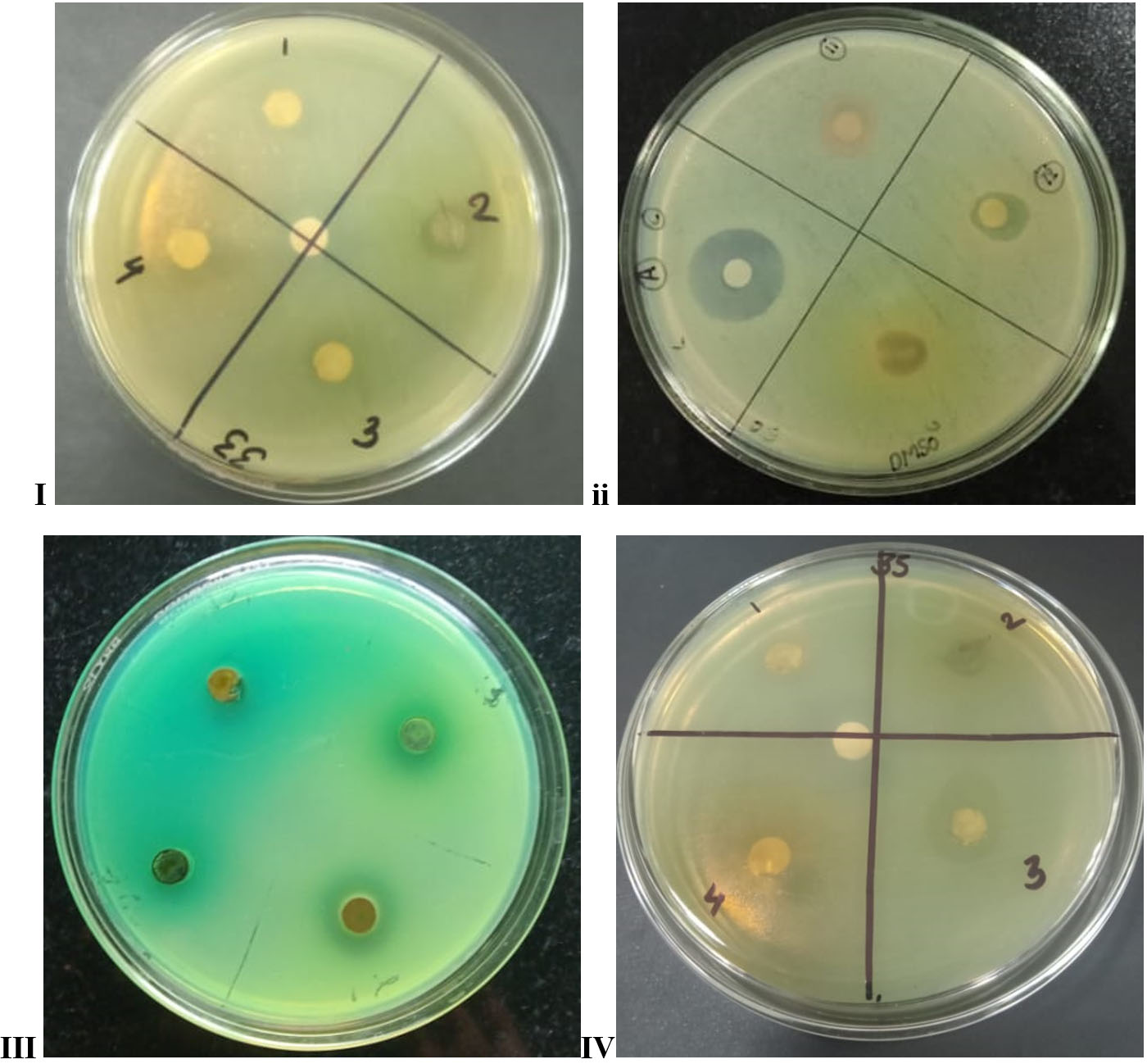
Figure 1. Showing antimicrobial activity of: i. Salmonella enterica typhi, ii. Escherichia coli, iii. Pseudomonas aeruginosa, iv. Salmonella enterica paratyphi
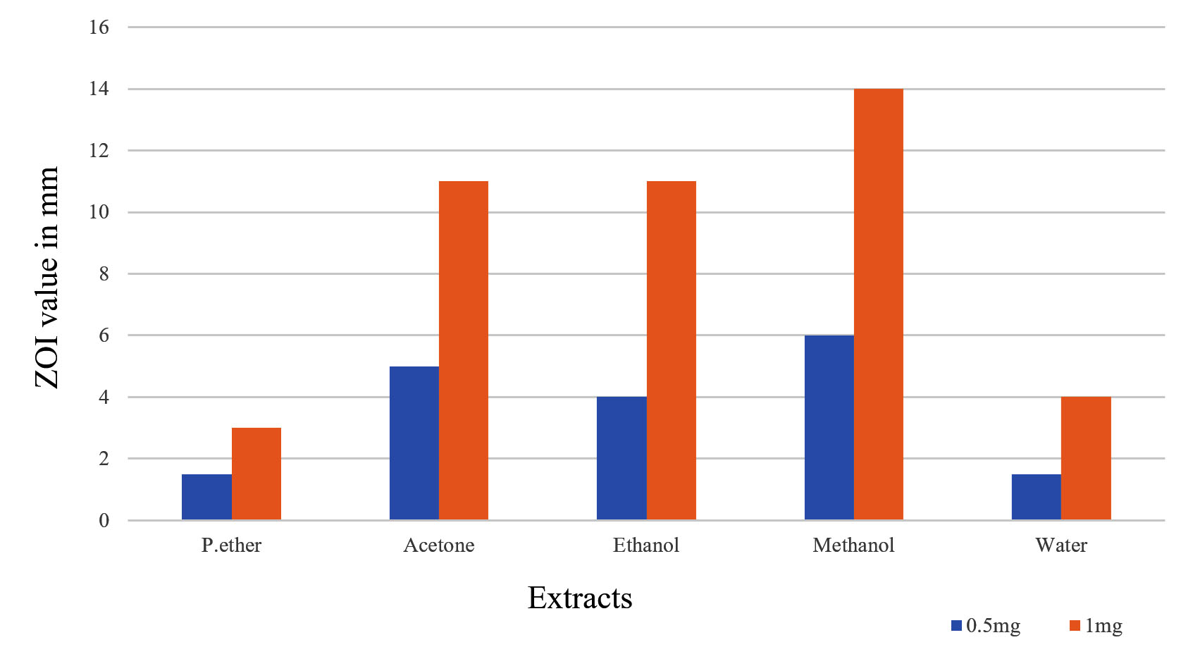
Figure 2. Shows antibacterial activity of F. auriculata extracts at quantities of 0.5 mg and 1 mg/100 mL against Salmonella enterica ser typhi cultures
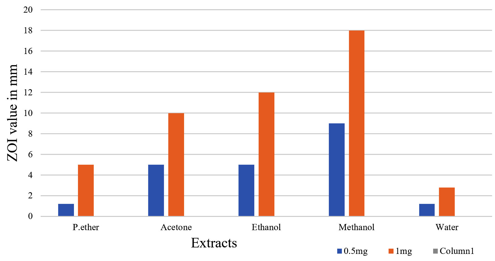
Figure 3. Shows antibacterial activity of F. auriculata extracts at quantities of 0.5 mg and 1 mg/100 mL against Salmonella enterica para typhi cultures
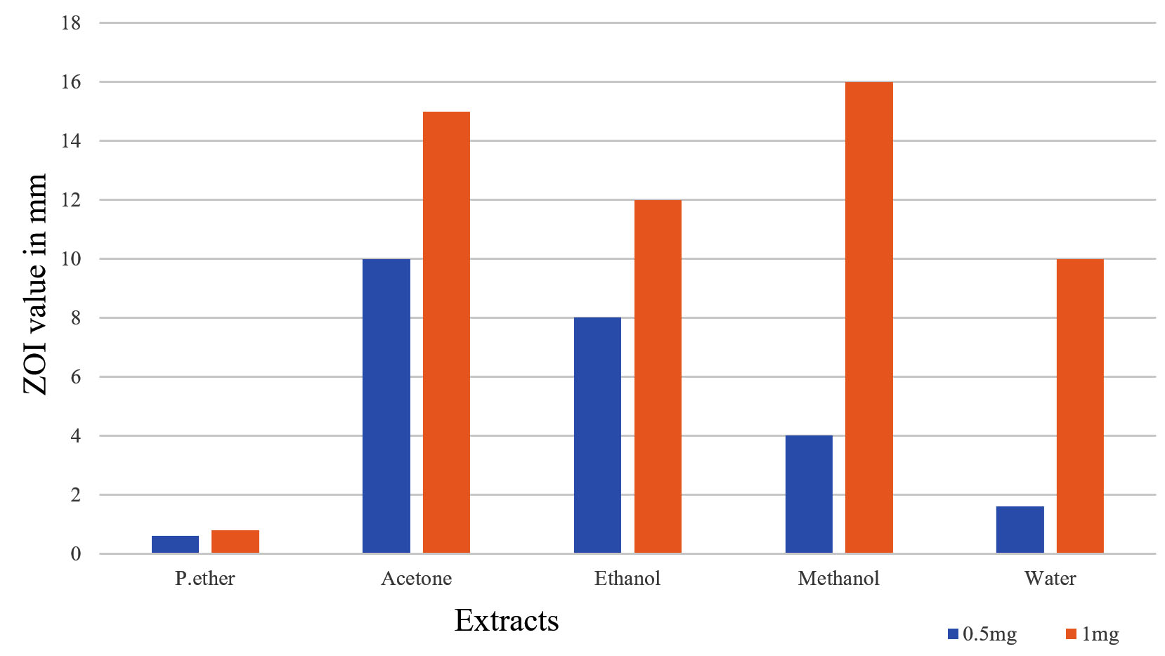
Figure 4. Shows antibacterial activity of F. auriculata extracts at quantities of 0.5 mg and 1 mg/100 mL against E. coli cultures
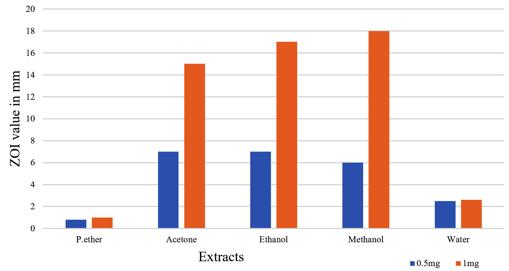
Figure 5. Shows antibacterial activity of F. auriculata extracts at quantities of 0.5 mg and 1 mg/100 mL against Pseudomonas aeruginosa cultures
MIC and MBC
All extracts were subjected to the MIC assay utilising a two-fold serial dilution method. The “minimum inhibitory concentration” (MIC) of an antibiotic or test sample needed in order to avoid or slow the rise of germs is known as this. The MIC and MBC control extracts were discovered to be 0.0156 mg/mL and 0.0312 mg/mL, in that order. All of the bacterial strains had MIC Indices of 2.0 for both the extracts and the control. The concentration of ZOI was highest in methanol plant extract, then in ethanol, acetone, petroleum ether, and aqueous. MBC levels varied from 0.5 to 0.0312 mg/mL, based on the MBC data (Table 3-6). The extract’s bactericidal properties were evaluated using the MIC Index.25,26
Table (3):
Ficus auriculata MIC and MBC extracts, as well “MIC/MBC” values in the index against MTCC 733 (Salmonella enterica ser. Typhi) bacterial strains19
Extract |
Range (mg/ml) |
Control of MIC (mg/ml) |
MIC extract (mg/ml) |
MBC control (mg/ml) |
MBC (mg/ml) extract |
Index of MIC (control) |
Index of MIC (extract) |
|---|---|---|---|---|---|---|---|
P. ether |
0.5 to 0.0156 |
0.0156 |
0.125 |
0.0312 |
0.25 |
2 |
2 |
Acetone |
0.5 to 0.0156 |
0.0156 |
0.125 |
0.0312 |
0.25 |
2 |
2 |
Ethanol |
0.5 to 0.0156 |
0.0156 |
0.0625 |
0.0312 |
0.125 |
2 |
2 |
Methanol |
0.5 to 0.0156 |
0.0156 |
0.0625 |
0.0312 |
0.125 |
2 |
2 |
Water |
0.5 to 0.0156 |
0.0156 |
0.25 |
0.0312 |
0.50 |
2 |
2 |
Table (4):
Ficus auriculata MIC and MBC extracts, as well “MIC/MBC” values in the index against MTCC 735 (Salmonella enterica ser. Paratyphi) bacterial strains
Extract |
Range (mg/ml) |
Control of MIC (mg/ml) |
MIC extract (mg/ml |
MBC control (mg/ml) |
MBC (mg/ml) extract |
Index of MIC (control) |
Index of MIC (extract) |
|---|---|---|---|---|---|---|---|
P. ether |
0.5 to 0.0156 |
0.0156 |
0.125 |
0.0312 |
0.5 |
2 |
2 |
Acetone |
0.5 to 0.0156 |
0.0156 |
0.125 |
0.0312 |
0.25 |
2 |
2 |
Ethanol |
0.5 to 0.0156 |
0.0156 |
0.0625 |
0.0312 |
0.125 |
2 |
2 |
Methanol |
0.5 to 0.0156 |
0.0156 |
0.0625 |
0.0312 |
0.012 |
2 |
2 |
Water |
0.5 to 0.0156 |
0.0156 |
0.25 |
0.0312 |
0.50 |
2 |
2 |
Table (5):
Ficus auriculata MIC and MBC extracts, as well “MIC/MBC” values in the index against E. coli bacterial strains22,23
Extract |
Range (mg/ml) |
Control of MIC (mg/ml) |
MIC extract (mg/ml) |
MBC control (mg/ml) |
MBC (mg/ml) extract |
Index of MIC (control) |
Index of MIC (extract) |
|---|---|---|---|---|---|---|---|
P. ether |
0.5 to 0.0156 |
0.0156 |
0.125 |
0.0312 |
0.25 |
2 |
2 |
Acetone |
0.5 to 0.0156 |
0.0156 |
0.125 |
0.0312 |
0.125 |
2 |
2 |
Ethanol |
0.5 to 0.0156 |
0.0156 |
0.0156 |
0.0312 |
0.125 |
2 |
2 |
Methanol |
0.5 to 0.0156 |
0.0156 |
0.0156 |
0.0312 |
0.125 |
2 |
2 |
Water |
0.5 to 0.0156 |
0.0156 |
0.25 |
0.0312 |
0.5 |
2 |
2 |
Table (6):
Ficus auriculata MIC and MBC extracts, as well “MIC/MBC” values in the index against Pseudomonas aeruginosa bacterial strains19,21
Extract |
Range (mg/ml) |
Control of MIC (mg/ml) |
MIC extract (mg/ml) |
MBC control (mg/ml) |
MBC (mg/ml) extract |
Index of MIC (control) |
Index of MIC (extract) |
|---|---|---|---|---|---|---|---|
P. ether |
0.5 to 0.0156 |
0.0156 |
0.25 |
0.0312 |
0.5 |
2 |
2 |
Acetone |
0.5 to 0.0156 |
0.0156 |
0.125 |
0.0312 |
0.25 |
2 |
2 |
Ethanol |
0.5 to 0.0156 |
0.0156 |
0.0156 |
0.0312 |
0.125 |
2 |
2 |
Methanol |
0.5 to 0.0156 |
0.0156 |
0.0625 |
0.0312 |
0.0312 |
2 |
2 |
Water |
0.5 to 0.0156 |
0.0156 |
0.25 |
0.0312 |
0.5 |
2 |
2 |
Free radical scavenging activity
The primary cause of plant’s antioxidant activity is the presence of bioactive compounds within them. The reason for this may not solely be attributed to the large proportion of primary elements, but also to the existence of other constituents in minimal amounts.28 The hydrogen peroxide assay and the “DPPH” assay were utilised to measure antioxidant activity. Methanolic extract of Ficus auriculata shows a strong free radical activity shown in Figure 6. DPPH assay and H2O2 assay show strong activity at FP 100% whereas FP 80% shows strong activity for hydrogen peroxide assay13,27 (Table 7 and 8) (Figure 6 and 7).
Table (7):
Ficus auriculata (Leaf) DPPH scavenging activity in various solvent extracts27,29
Concen. (µg/ml) |
Methanol |
Ethanol |
Acetone |
Petroleum ether |
Water |
|---|---|---|---|---|---|
20 |
25.71 ± 0.15 |
22.14 ± 0.012 |
23.80 ± 0.12 |
24.76 ± 0.12 |
27.14 ± 0.11 |
40 |
30.95 ± 0.13 |
31.19 ± 0.009 |
33.33 ± 0.11 |
35.23 ± 0.10 |
33.80 ± 0.10 |
60 |
46.66 ± 0.10 |
44.2 ± 0.014 |
45.47 ± 0.09 |
47.61 ± 0.08 |
47.14 ± 0.08 |
80 |
62.85 ± 0.06 |
58.33± 0.021 |
56.90 ± 0.06 |
59.52 ± 0.06 |
60.71 ± 0.05 |
100 |
72.38 ± 0.05 |
71.9 ± 0.007 |
71.19 ± 0.04 |
69.04 ± 0.04 |
70 ± 0.04 |
IC 50 |
3.18 |
3.35 |
3.32 |
3.24 |
3.2 |
Table (8):
Ficus auriculata (Leaf) H2O2 activity in various solvent extracts28,29
Concen. (µg/ml) |
Methanol |
Ethanol |
Acetone |
Petroleum ether |
Water |
|---|---|---|---|---|---|
20 |
13.22 ± 0.19 |
6.44 ± 0.20 |
24.06 ± 0.24 |
11.69 ± 0.01 |
10 ± 0.19 |
40 |
25.25 ± 0.16 |
21.86 ± 0.16 |
30.84 ± 0.18 |
34.74 ± 0.05 |
16.94 ± 0.16 |
60 |
41.01 ± 0.12 |
41.01 ± 0.13 |
39.66 ± 0.16 |
44.57± 0.008 |
38.64 ± 0.12 |
80 |
62.54 ± 0.09 |
59.32 ± 0.10 |
46.10 ± 0.12 |
49.83 ± 0.04 |
51.01 ± 0.09 |
100 |
72.37 ± 0.06 |
70.84 ± 0.06 |
65.93 ± 0.07 |
64.23 ± 0.04 |
67.62 ± 0.06 |
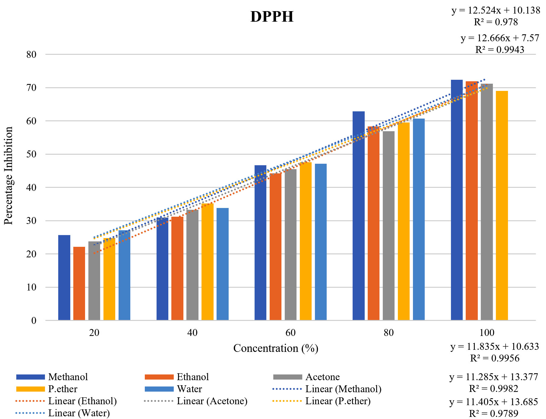
Figure 6. Percentage inhibition of DPPH for different solvent of Ficus auriculata
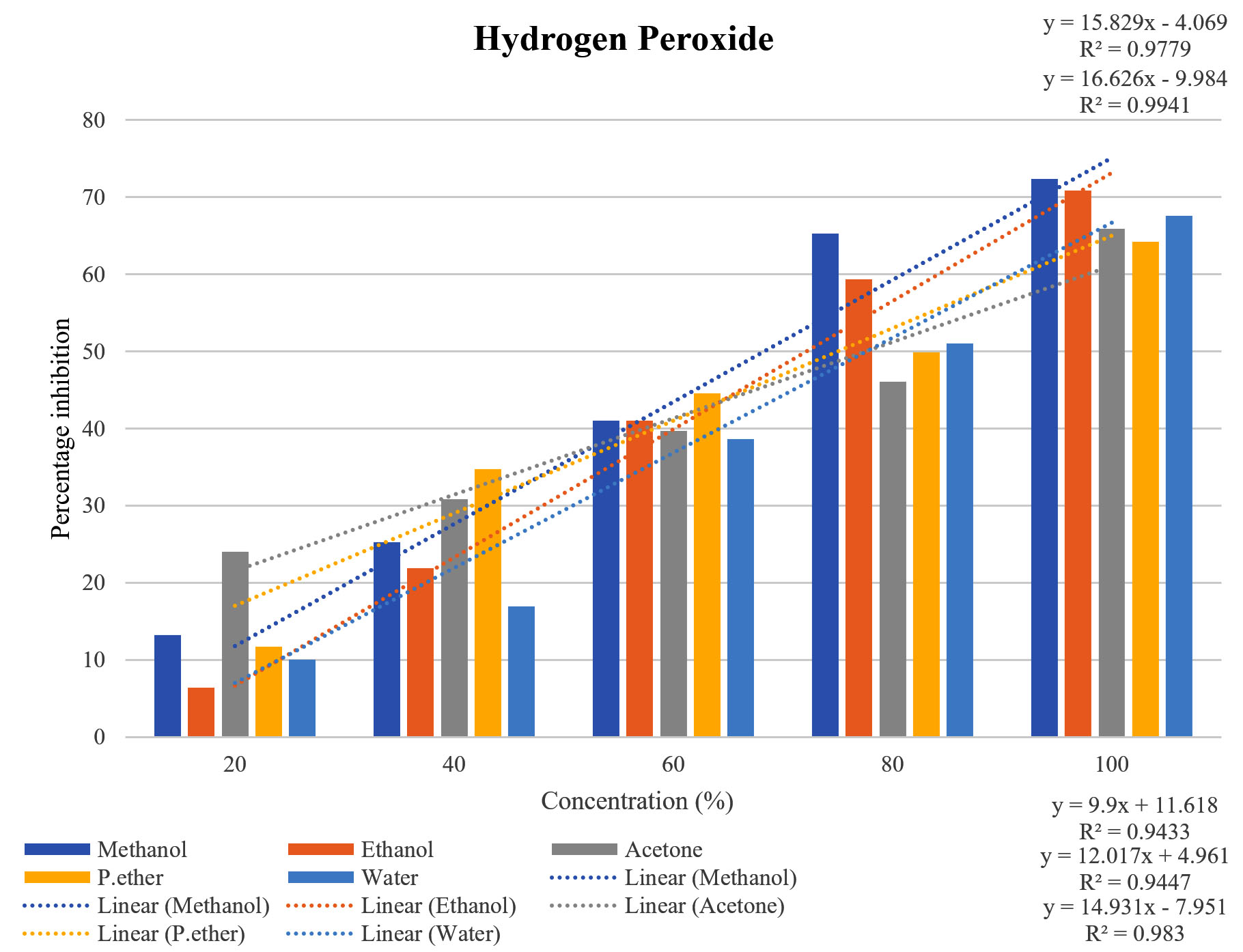
Figure 7. Percentage inhibition of Hydrogen peroxide for different solvent of Ficus auriculata
The IC50 values for each extract in DPPH are given in Table 6, where the methanol extract had 3.18 µg/mL, the petroleum ether extracts 3.24 µg/mL, the aqueous 3.32 µg/mL, the acetone 3.32 µg/mL, and the P. ether 3.24 µg/mL.30
Research on medicinal plants is crucial for the creation of new drugs and study on pharmaceuticals. They are employed directly as a medicinal agent, as well as a modern pharmacologically active chemical or as a raw material for drug synthesis. Compared to F. auriculata (leaf), a large portion of contemporary medications are depending on or coming from secondary metabolites of medicinal plants that have antibacterial activity against all four organisms. The highest ZOI found for several F. auriculata extracts was for E. coli, P. aeruginosa, S. (serovar) typhi, and S. paratyphi. While using antibiotics has brought about instant relief, MDR poses a major risk. Therefore, using different extracts from medicinal plants as an alternate form of therapy is the best way to cure illnesses caused by bacteria. The current investigation expandable to incorporate in vivo examinations to be able to determine the mechanism of the extracts’ activity. India is home to several medicinal plants with the potential to be used in a variety of medication formulations.
ACKNOWLEDGMENTS
The authors would like to thank SGRR University Dehradun, Uttarakhand, India,
for providing instrumentation facilities for the investigation of phytochemicals.
CONFLICT OF INTEREST
The authors declare that there is no conflict of interest.
AUTHORS’ CONTRIBUTION
NS, RV, RBA, AB and PR contributed to the study’s conceptualization, design, data collection and analysis. NS and RV verify the integrity of the raw data. NS, RV, RBA, AB and PR drafted the manuscript. NS wrote the manuscript. NS, RV, RBA, AB and PR reviewed and edited the manuscript. All authors read and approved the final manuscript for publication.
FUNDING
None.
DATA AVAILABILITY
All datasets generated or analyzed during this study are included in the manuscript.
ETHICS STATEMENT
Not applicable.
- Al-Fishawy A, Zayed R, Afifi S. Phytochemical and pharmacological studies of Ficus auriculata Lour. (Moraceae) cultivated in Egypt. Journal of Natural Products.2011;4:184-95.
Crossref - Rekka R. Ethnobotanical investigation of yercaud hills eastern ghatstamilnadu and phytochemical and pharmacological studies on ficus auriculata roxb. 2016.
Crossref - Gaire BP, Lamichhane R, Sunar CB, Shilpakar A, Neupane S, Panta S. Phytochemical screening and analysis of antibacterial and antioxidant activity of Ficus auriculata (Lour.) Pharmacognosy Journal .2011;3(21):49-55.
Crossref - Kumari A, Verma R, Sharma M, Chauhan P, Kumar A. Evaluation of phytochemical, antioxidant, antibacterial and anti-cancerous activity of Ficus auriculata Lour. and Osyris wightiana Wall. ex Wight. Bull Environ Pharmacol Life Sci. 2018;7:64-70.
- Khatun MJ, Rahman MM, Rahim MA, Jakariya M, Mirdah MH. Study on the ethnobotany and nutritional status of three edible Ficus species in hill district of Bangladesh. Int J Min Fruits Med Arom Plants. 2016;2(1):35-40.
- Saklani S, Chandra S. In vitro antimicrobial activity, nutritional profile and phytochemical screening of wild edible fruit of Garhwal Himalaya (Ficus auriculata). Int J Pharm Sci Rev Res. 2012;12(2):61-66.
- Shilpakar A, Gaire BP, Bahadur SC, Lamichhane R, Neupane S. Phytochemical screening and analysis of antibacterial and antioxidant activity of Ficus auriculata, Lour. Stem bark. Pokhara University, Pokhara, Nepal. 2009.
Crossref - Malesh B, Satish S. Antimicrobial Activity of some important medicinal plant against plant and human pathogen. World J Agric Sci. 2008;4(5):839-843.
- de Oliveira GF, Furtado NAJC, da Silva Filho AA, et al. Antimicrobial activity of Syzygiumcumini (Myrtaceae) leaves extract. Braz J Microbiol. 2007;38(2):381-384.
Crossref - Salem MZM, Salem AZM, Camacho LM, Ali HM. Antimicrobial activities and phytochemical composition of extracts of Ficus species: An over view. Afr J Microbiol. Res. 2013;7(33):4207-4219.
- Manandhar S, Luitel S, Dahal RK. In vitro antimicrobial activity of some medicinal plants against human pathogenic bacteria. J Trop Med. 2019.
Crossref - Murugan R, Parimelazhagan T. Comparative evaluation of different extraction methods for antioxidant and anti-inflammatory properties from Osbeckia parvifolia Arn.- An in vitro approach. J King Saud Univ Sci. 2014;26(4):267-275.
Crossref - Shi YX, Xu YK, Hu HB, Na Z, Wang WH. Preliminary assessment of antioxidant activity of young edible leaves of seven Ficus species in the ethnic diet in Xishuangbanna, Southwest China. Food Chem. 2011;128(4):889-894.
Crossref - Ostrosky EA, Mizumoto MK, Lima MEL, Kaneko TM, Nishikawa SO, Freitas BR. Methods for evaluation of the antimicrobial activity and determination of minimum inhibitory concentration (MIC) of plant extracts. Braz J Pharmacogn. 2008;18(2):301-3017.
Crossref - Pa R, Mathew L. Antimicrobial activity of leaf extracts of Justicia adhatoda L. in comparison with vasicine. Asian Pac J Trop Biomed. 2012;2(3):S1556-S1560.
Crossref - Oliveira BD, Rodrigues AC, Cardoso BMI, et al. Antioxidant, antimicrobial and anti-quorum sensing activities of Rubus rosaefolius phenolic extract. Industrial Crops and Products. 2016;84:59-66.
Crossref - Singh M, Khatoon S, Singh S, Kumar V, Rawat AKS, Mehrotra S. Antimicrobial screening of ethnobotanically important stem bark of medicinal plants. Pharmacognosy Res. 2010;2(4):254-257.
Crossref - Parekh J, Chanda S. In vitro antimicrobial activity and phytochemical analysis of some Indian medicinal plants. Turk J Biol. 2007;31(1):53-58.
- Bauer AW, Kirby WMM, Sherris JC, Turck M. Antibiotic susceptibility testing by a standardized single disk method. Am J Clin Pathol. 1966;45(4_ts):493-496.
Crossref - Oldoni TLC, Melo PS, Massarioli AP, et al. Bioassay-guided isolation of proanthocyanidins with antioxidant activity from peanut (Arachis hypogaea) skin by combination of chromatography techniques. Food Chem. 2016;192:306-312.
Crossref - Shahzad A, Ishtiaq M, Tanveer H, et al. Analysis of antimicrobial potential of some Ficus taxa from district Bhimber Azad Jammu and Kashmir, Pakistan. Appl Ecol Environ Res. 2016;14(5):159-176.
Crossref - Abraham J, Thomas TD. Antibacterial activity of medicinal plant Cyclea peltata (Lam) Hooks & Thoms. Asian Pac J Trop Dis. 2012;2(suppl 1):S280-S284.
Crossref - Preethi R, Devanathan VV, Loganathan M. Antimicrobial and antioxidant efficacy of some medicinal plants against food borne pathogens. Adv Biol Res. 2010;4(2):122-125.
- Bertoletti LL, Skoronski E, Schittler L, Kempka AP. Extracts of Leaves of Ficus auriculata Lour.: antioxidant, antimicrobial and phytotoxic activity. Agriculturae Conspectus Scientificus. 2018;83(4):321-328.
- Gupta C, Garg AP, Uniyal RC, Kumari A. Antimicrobial activity of some herbal oils against common food-borne pathogens. Afr J Microbiol Res. 2008;2(10):258-261.
- Kuete V, Ngameni B, Simo CCF, et al. Antimicrobial activity of the crude extracts and compounds from Ficus chlamydocarpa and Ficus cordata (Moraceae). J Ethnopharmacol. 2008;120(1):17-24.
Crossref - Annan K, Houghton PJ. Antibacterial, antioxidant and fibroblast growth stimulation of aqueous extracts of Ficus asperifolia Miq. and Gossypium arboreum L., wound-healing plants of Ghana. J Ethnopharmacol. 2008;119(1):141-144.
Crossref - Tchinda CF, Voukeng IK, Beng VP, Kuete V. Antibacterial activities of the methanol extracts of Albizia adianthifolia, Alchornea laxiflora, Laportea ovalifolia and three other Cameroonian plants against multi-drug resistant Gram-negative bacteria. Saudi J Biol Sci. 2017;24(4):950-955.
Crossref - Zhu W, Liu J, Ye J, Li G. Effects of phytotoxic extracts from peach root bark and benzoic acid on peach seedlings growth, photosynthesis, antioxidance and ultrastructure properties. Scientia Horticulturae. 2017;215:49-58.
Crossref - Thingbaijam R, Dutta BK, Paul SB. In vitro antioxidant capacity, estimation of total phenolic and flavonoid content of Ficus auriculata lour. Int J Pharm Pharm Sci. 2012;4(4): 518-521.
- Singh N, Verma R, Rawat P, Aziz RB, Kala A. Ficus palmata and Ficus auriculata Phytochemical Screening in Different Solvents by HPLC and FTIR Spectroscopic Analysis. Scope. 2023;13(4):459-472.
© The Author(s) 2024. Open Access. This article is distributed under the terms of the Creative Commons Attribution 4.0 International License which permits unrestricted use, sharing, distribution, and reproduction in any medium, provided you give appropriate credit to the original author(s) and the source, provide a link to the Creative Commons license, and indicate if changes were made.