


ISSN: 0973-7510
E-ISSN: 2581-690X
 , Bincy Joseph1 , Shivasharanappa Nayakvadi2, Sandeep Kumar Sharma1, Surendra Singh Shekhawat3, Taruna Bhati1, Archana Fozdar4, Rama Kumari1 and Vijay Kumar Meena1
Klebsiella pneumoniae, a critical pathogen with significant implications for global antimicrobial resistance (AMR), is a major threat to public health, particularly in regions with intensive human–animal–environment interfaces. This study investigated the AMR patterns of K. pneumoniae isolates from human–camel–environment interactions in Rajasthan, India, a region where camels are economically significant. A total of 138 samples were collected from camel populations, animal handlers, and their surrounding environments in the districts of Ajmer and Jaipur. Thirty-three isolates were confirmed to be Klebsiella pneumoniae by microbiological and molecular methods, as well as by using BD Phoenix M50. Antibiotic resistance was assessed using the Kirby-Bauer disc diffusion and BD Phoenix M50 AST methods. The study revealed that 66.66% of the isolates were multidrug-resistant (MDR), with the most pronounced resistance noted against ciprofloxacin (72.72%), as the organism exhibits intrinsic resistance to narrow-spectrum penicillins. Conversely, sulfafurazole, tetracycline, and trimethoprim–sulfamethoxazole exhibited 100% sensitivity. A significant proportion of the isolates (30.30%) showed a multiple antibiotic resistance (MAR) index greater than 0.2, indicating a high risk of spreading resistance. Thirty-three percent of the isolates exhibited phenotypic evidence of extended-spectrum beta-lactamase (ESBL) production, with the predominant ESBL gene identified as blaTEM. Notably, one isolate demonstrated phenotypic colistin resistance, representing the first such report in a camel ecosystem in India. However, mcr-1, which is commonly associated with colistin resistance, was not detected in the present study. The genetic mechanism underlying colistin resistance remains unclear. Additionally, none of the isolates possessed carbapenem resistance genes. These findings underscore the critical need for robust and comprehensive AMR surveillance in camel ecosystems to protect public health, food safety, and the livelihoods of camel-dependent communities.
Camel ecosystem, Colistin resistance, ESBL, MDR, One Health Approach
Antimicrobial resistance (AMR) is an increasing global health concern, responsible for an estimated 4.95 million deaths worldwide.1 The rapid emergence and dissemination of AMR endanger human and animal health, thereby necessitating a One Health approach to understand its transmission dynamics. According to the World Health Organization (WHO), there is a strong need to track antimicrobial use in humans, animals, and the environment to address this crisis effectively.2 The movement of antimicrobial-resistant bacteria and resistance genes among different ecological niches underscores the interconnected nature of AMR development and spread.3
A major contributor to the global AMR crisis is the group of ESKAPE pathogens (Enterococcus faecium, Staphylococcus aureus, Klebsiella pneumoniae, Acinetobacter baumannii, Pseudomonas aeruginosa, and Enterobacter species). Among these, K. pneumoniae is a Gram-negative opportunistic pathogen known for its extensive resistance gene repertoire. It exhibits high adaptability to diverse environments and plays a significant role in the transfer of antimicrobial resistance genes such as blaKPC, blaOXA-48, and blaNDM-1, facilitating the spread of resistance among clinically relevant pathogens.4 Although K. pneumoniae is primarily a human pathogen, it is also a commensal organism in animals, including livestock and wildlife, where it contributes to AMR transmission dynamics.
Despite the increasing global focus on AMR, limited research has been conducted on the prevalence of AMR in camels, particularly in regions where these animals play crucial socioeconomic and cultural roles. In Rajasthan, India, camels have a vital socioeconomic role, especially among nomadic pastoral communities, and are recognized as the state animal. However, the wandering nature of nomads also poses a significant risk for the spread of diseases and resistant pathogens. Despite their importance, research on camels in Rajasthan remains sparse, particularly in the context of AMR. The misuse and overuse of antibiotics in veterinary practice, exposure to contaminated environments, and close human–camel interactions contribute to the spread of resistant pathogens. These resistant bacteria can be transferred between camels, humans, and the environment, thereby affecting public health, food safety, and environmental health.
Given this background, the human–camel–environment interface in Rajasthan is a critical area for AMR research. Through the adoption of a One Health approach that integrates human, animal, and environmental health, highly effective strategies can be developed to monitor and combat the spread of AMR. This is essential for safeguarding not only public health but also the sustainable livelihoods of those dependent on camels and the health of the broader ecosystem.
Sample collection
Study samples were randomly sourced from two districts in Rajasthan, namely, Ajmer and Jaipur, during a 1-year period from December 2023 to December 2024, as detailed in Table 1. Samples were collected from camels (nasal and rectal swabs), the surrounding environment (soil, manger soil, and water), and camel handlers (hand swabs). All the samples were collected under sterile conditions and processed accordingly.
Table 1. Summary of sample collection from different sources in Ajmer and Jaipur districts, Rajasthan
| District | Source | Sample type | Number of samples |
|---|---|---|---|
| Ajmer | Camels | Nasal swabs | 8 |
| Rectal swabs | 9 | ||
| Jaipur | Camels | Nasal swabs | 29 |
| Rectal swabs | 30 | ||
| Environment | Soil samples | 25 | |
| Manger’s soil samples | 3 | ||
| Water samples | 3 | ||
| Camel Handler’s | Hand swabs | 31 | |
| Total | 138 | ||
Sample processing and isolation
Each sample was inoculated into nutrient broth and incubated overnight for further analysis. The following day, samples were streaked onto MacConkey agar for preliminary isolation. Klebsiella pneumoniae isolates were subcultured on Simmons citrate inositol agar (SCIA). The isolates were identified using biochemical tests. Primary tests included Gram staining and microscopy using immersion oil, whereas motility was assessed using a motility test medium and catalase and oxidase tests. Secondary biochemical tests were performed for further characterization.
Tentative Klebsiella spp. isolates were confirmed using molecular techniques targeting the 16S-23S rDNA internal transcribed spacer (ITS) region.
Molecular characterization of K. pneumoniae
Genomic DNA was extracted using a HipurA bacterial genomic DNA kit and the snap-chilling method. The 16S-23S rDNA ITS region of the isolates was amplified using species-specific primers.5 The PCR mixture consisted of 12.5 µL of 2X PCR Master Mix (Thermo Scientific, Waltham, MA, USA), 1 µL each of forward and reverse primers at 10 pM, and 2 µL of template DNA. Amplification was performed in a Nexus Gradient Thermal Cycler (Eppendorf, Hamburg, Germany) under the following conditions: initial denaturation at 95 °C for 5 min, followed by 35 cycles of 95 °C for 1 min, 45 °C for 1 min (annealing), and 72 °C for 1 min (extension), with a final extension at 72 °C for 10 min. The amplified product (approximately 130 bp) was verified by electrophoresis on a 1.5% agarose gel.
Testing of antibiotic susceptibility
Disc diffusion method
Antibiotic susceptibility of the isolates was determined using the Kirby-Bauer disc diffusion method with the following antibiotics: piperacillin/tazobactam, penicillin G, cefepime, cefoxitin, ciprofloxacin, gentamicin, amikacin, imipenem, tetracycline, ceftazidime, cefotaxime, cefixime, tobramycin, meropenem, sulfafurazole, trimethoprim, levofloxacin, ofloxacin, norfloxacin, doxycycline, and minocycline (HiMedia, Mumbai, India). Antibiotic discs were carefully placed on Mueller–Hinton agar (MHA) plates uniformly inoculated with test cultures. Plates were incubated at 37 °C for 16-18 hrs. Following incubation, the zones of inhibition were measured, and isolates were classified as susceptible, intermediate, or resistant according to the CLSI (2024) guidelines.4
BD Phoenix™ M50 automated system
Susceptibility and minimum inhibitory concentration (MIC) of antimicrobial agents were assessed using the BD Phoenix™ M50 automated system (Becton Dickinson, Franklin Lakes, NJ, USA), which determines MIC based on broth microdilution and automated reading.
Escherichia coli ATCC 25922 was used as a quality control strain for both methods used for antibiotic susceptibility testing.
Phenotypic resistance testing
ESBL production
The double-disc diffusion test (DDST) was used to detect ESBL production. The test was performed using third-generation cephalosporins (ceftazidime and cefotaxime) alone and in combination with clavulanic acid. An increase of >5 mm in the inhibition zone was interpreted as indicative of ESBL production.
Detection of carbapenemase production
A modified Hodge test (MHT) was used to detect carbapenemase production. A positive result was identified by the appearance of a cloverleaf shaped indentation around the meropenem disc placed on the MHA plate after overnight incubation.
Quinolone resistance testing
MIC testing for ciprofloxacin resistance was performed using the Ezy MIC™ strip (HiMedia, Mumbai, India) on MHA plates. The MIC values were determined by observing the intersection of the inhibition ellipse with a scale printed on the strip.
Colistin resistance
Colistin agar test: MHA plates were supplemented with 0.25-4 µg/mL colistin. After 24 hrs of incubation at 37 °C, growth on the plate was indicative of colistin resistance.
Polymyxin B MIC testing: Polymyxin B Ezy MIC™ strips were used, and the MIC was assessed following 16 hrs of incubation at 37 °C.
Detection of genes associated with Antibiotic Resistance Genes (ARGs)
The presence of specific ARGs was assessed by PCR. Genes for beta-lactamase (blaSHV, blaTEM, blaCTX-M), carbapenem resistance (blaVIM, blaIMP, blaKPC, blaNDM), quinolone resistance (qnrA, qnrB, qnrS), and colistin resistance (mcr-1) were amplified using specific primers. The PCR mixture consisted of 12.5 µL of 2X PCR Master Mix, 1 µL each of forward and reverse primers, and 2 µL of DNA template. Details of the primers and thermal cycling conditions are presented in Table 2.
Table 2. PCR conditions and primers used for characterization of producing K. pneumoniae
Gene |
Forward primer sequence (5′ to 3′) |
PCR conditions |
Amplicon Size |
Ref. |
|---|---|---|---|---|
16S- 23S rDNA ITS5 |
F: ATT TGA AGA GGT TGC AAA CGA T R: TTC ACT CTG AAG TTT TCT TGT GTT C |
Initial denaturation at 95 °C for 5 min; 35 cycles of 95 °C for 1 min, 45 °C for 1 min, and 72 °C for 1 min; final extension at 72 °C for 10 min; hold at 4 °C. |
130 bp |
5 |
blaTEM |
F: GCG GAA CCC CTA TTT G R: ACC AAT GCT TAA TCA GTG AG |
Initial denaturation at 95 °C for 5 min; 35 cycles of 95 °C for 1 min, 42 °C for 1 min, and 72 °C for 1 min; final extension at 72 °C for 10 min; hold at 4 °C. |
964 bp |
6 |
blaCTX-M |
F: ATG TGC AGY ACC AGT AAR GTK ATG GC R: TGG GTR AAR TAR GTS ACC AGA AYS AGC GG |
Initial denaturation at 95 °C for 5 min; 35 cycles of 95 °C for 1 min, 54 °C for 1 min, and 72 °C for 1 min; final extension at 72 °C for 10 min; hold at 4 °C. |
593 bp |
7 |
blaSHV |
F: AGC CGC TTG AGC AAA TTA AAC R: ATC CCG CAG ATA AAT CAC CAC |
95 °Cx5 m / 95 °Cx1 m-54 °Cx1m -72 °Cx1 m (35 Cycles) / 72 °Cx10 m-4 °C Initial denaturation at 95 °C for 5 min; 35 cycles of 95 °C for 1 min, 54 °C for 1 min, and 72 °C for 1 min; final extension at 72 °C for 10 min; hold at 4 °C. |
713 bp |
8 |
blaKPC |
F: CGT CTA GTT CTG CTG TCT TG R: CTT GTC ATC CTT GTT AGG CG |
Initial denaturation at 95 °C for 5 min; 35 cycles of 95 °C for 1 min, 54 °C for 1 min, and 72 °C for 1 min; final extension at 72 °C for 10 min; hold at 4 °C. |
498 bp |
9 |
blaNDM |
F: GGT TTG GCG ATC TGG TTT TC R: CGG AAT GGC TCA TCA CGA TC |
Initial denaturation at 95 °C for 5 min; 35 cycles of 95 °C for 1 min, 54 °C for 1 min, and 72 °C for 1 min; final extension at 72 °C for 10 min; hold at 4 °C. |
621 bp |
10 |
blaVIM |
F: GAT GGT GTT TGG TCG CAT A R: CGA ATG CGC AGC ACC AG |
Initial denaturation at 95 °C for 5 min; 35 cycles of 95 °C for 1 min, 52 °C for 1 min, and 72 °C for 1 min; final extension at 72 °C for 10 min; hold at 4 °C. |
390 bp |
10 |
blaIMP |
F: GGA ATA GAG TGG CTT AAY TCT C R: GGT TTA AYA AAA CAA CCA CC |
95 °Cx5 m / 95 °Cx1 m-52 °Cx1m -72 °Cx1 m (35 Cycles) / 72 °Cx10 m-4 °C Initial denaturation at 95 °C for 5 min; 35 cycles of 95 °C for 1 min, 52 °C for 1 min, and 72 °C for 1 min; final extension at 72 °C |
232 bp |
10 |
qnrA1 to qnrA6 |
F: AGA GGA TTT CTC ACG CCA GG R: TGC CAG GCA CAG ATC TTG AC |
Initial denaturation at 95 °C for 5 min; 35 cycles of 95 °C for 1 min, 54 °C for 1 min, and 72 °C for 1 min; final extension at 72 °C |
580 bp |
11 |
qnrB1 to qnrB6 |
F: GGM ATH GAA ATT CGC CAC TG R: TTT GCY GYY CGC CAG TCG AA (M= A/C, H= A/C, Y=C/T) |
95 °Cx5 m / 95 °Cx1 m-54 °Cx1m -72 °Cx1 m (35 Cycles) / 72 °Cx10 m-4 °C Initial denaturation at 95 °C for 5 min; 35 cycles of 95 °C for 1 min, 54 °C for 1 min, and 72 °C for 1 min; final extension at 72 °C |
264 bp |
11 |
qnrS1 to qnrS2 |
F: GCA AGT TCA TTG AAC AGG GT R: TCT AAA CCG TCG AGT TCG GCG |
Initial denaturation at 95 °C for 5 min; 35 cycles of 95 °C for 1 min, 54 °C for 1 min, and 72 °C for 1 min; final extension at 72 °C |
428 bp |
11 |
gyrA |
F: AAA TCT GCC CGT GTC GTT GGT R: GCC ATA CCT ACG GCG ATA CC |
Initial denaturation at 95 °C for 5 min; 35 cycles of 95 °C for 1 min, 50 °C for 1 min, and 72 °C for 1 min; final extension at 72 °C |
344 bp |
12 |
parC |
F: CTG AAT GCC AGC GCC AAA TT R: GCG AAC GAT TTC GGA TCG TC |
Initial denaturation at 95 °C for 5 min; 35 cycles of 95 °C for 1 min, 49 °C for 1 min, and 72 °C for 1 min; final extension at 72 °C |
168 bp |
12 |
mcr1 |
F: AGT CCG TTT GTT CTT GTG GC R: AGA TCC TTG GTC TCG GCT TG |
Initial denaturation at 95 °C for 5 min; 35 cycles of 95 °C for 1 min, 53 °C for 1 min, and 72 °C for 1 min; final extension at 72 °C |
320 bp |
13 |
Samples (n = 138) were collected from camels, their handlers, and the surrounding environment. Of these, 34 isolates initially exhibited pink mucoid colonies on MacConkey agar and yellow, dome-shaped mucoid colonies on SCIA. Microscopy after Gram staining indicated small, pink, Gram-negative rods. All the isolates were non-motile. Further, all the isolates tested positive for catalase and negative for oxidase activity, followed by an IMViC pattern is –++. On triple sugar iron (TSI) testing, all isolates displayed yellow butt, yellow slant, and gas production without H2S production. The results of the primary and biochemical characterizations are displayed in Figure 1. Based on these preliminary findings and the amplification of the 16S-23S rDNA ITS region, 33 isolates were confirmed to be K. pneumoniae at the molecular level.
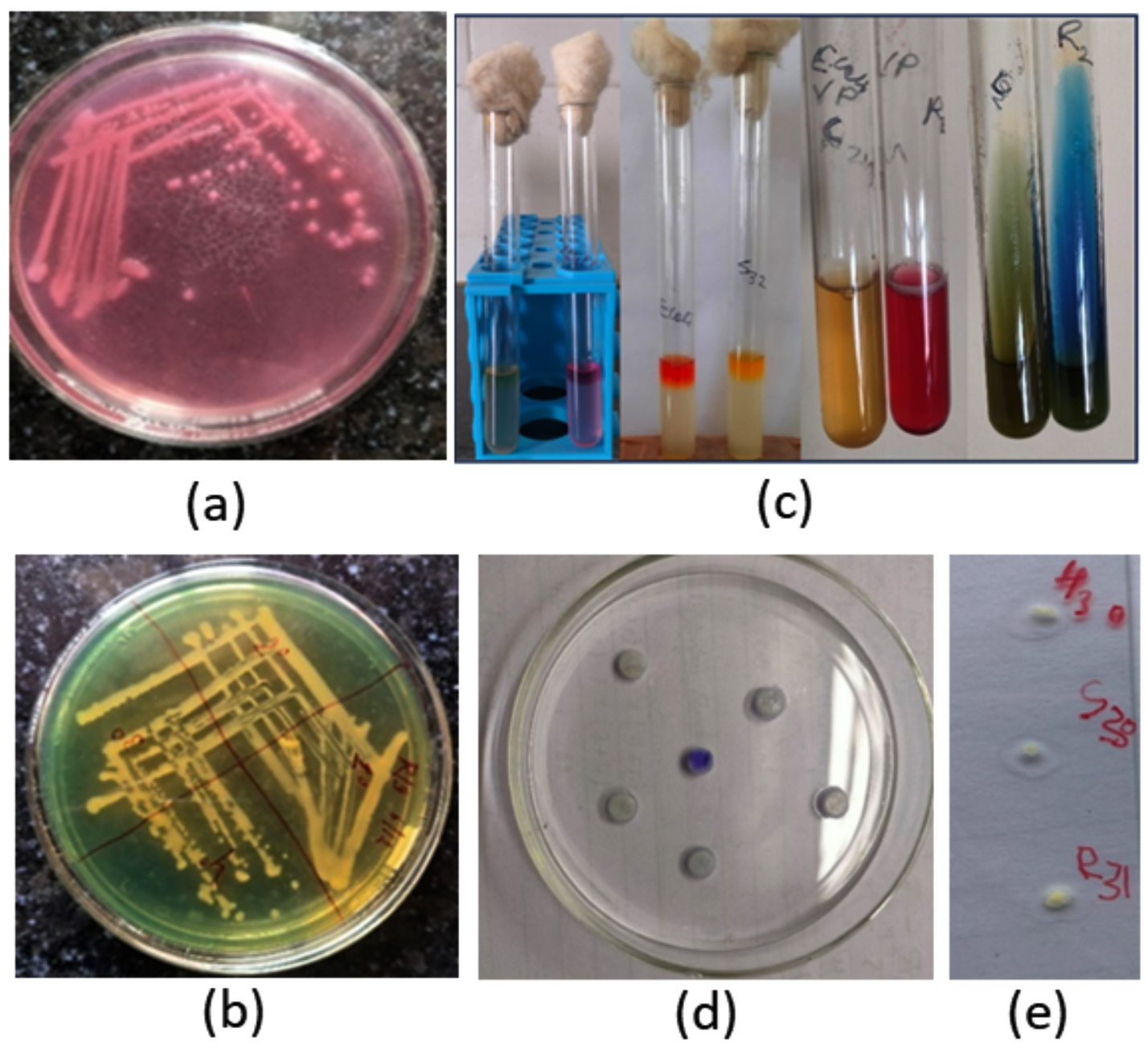
Figure 1. Cultural and biochemical characterization of Klebsiella pneumoniae isolates. (a) McConkey Agar: pink lactose fermenting mucoid colonies. (b) Simmons Citrate Inositol Agar (SCIA): Yellow mucoid colonies. (c) IMViC reactions: Indole (-), Methyl red (-), Voges-Proskauer (+), Citrate (+) (- – + +). (d) Oxidase Test: Negative. (e) Catalase Test: Positive
Occurrence and distribution of K. pneumoniae
The overall occurrence of Klebsiella pneumoniae infection in the camel ecosystem was 23.9% (33/138). Among the camels, 22 isolates were recovered from 76 samples (28.95%), including 11 of the 37 nasal swabs (29.73%) and 11 of the 39 rectal swabs (28.20%). From the surrounding environment, eight isolates were obtained from 31 samples (25.80%), with five out of 25 soil samples (20%) and three out of three manger soil samples (100%) testing positive, while none of the three water samples (0%) yielded isolates. In contrast, three isolates were recovered from 31 samples (9.68%) collected from camel handlers. The stacked bar chart in Figure 2 shows K. pneumoniae positivity across different sample types and locations.
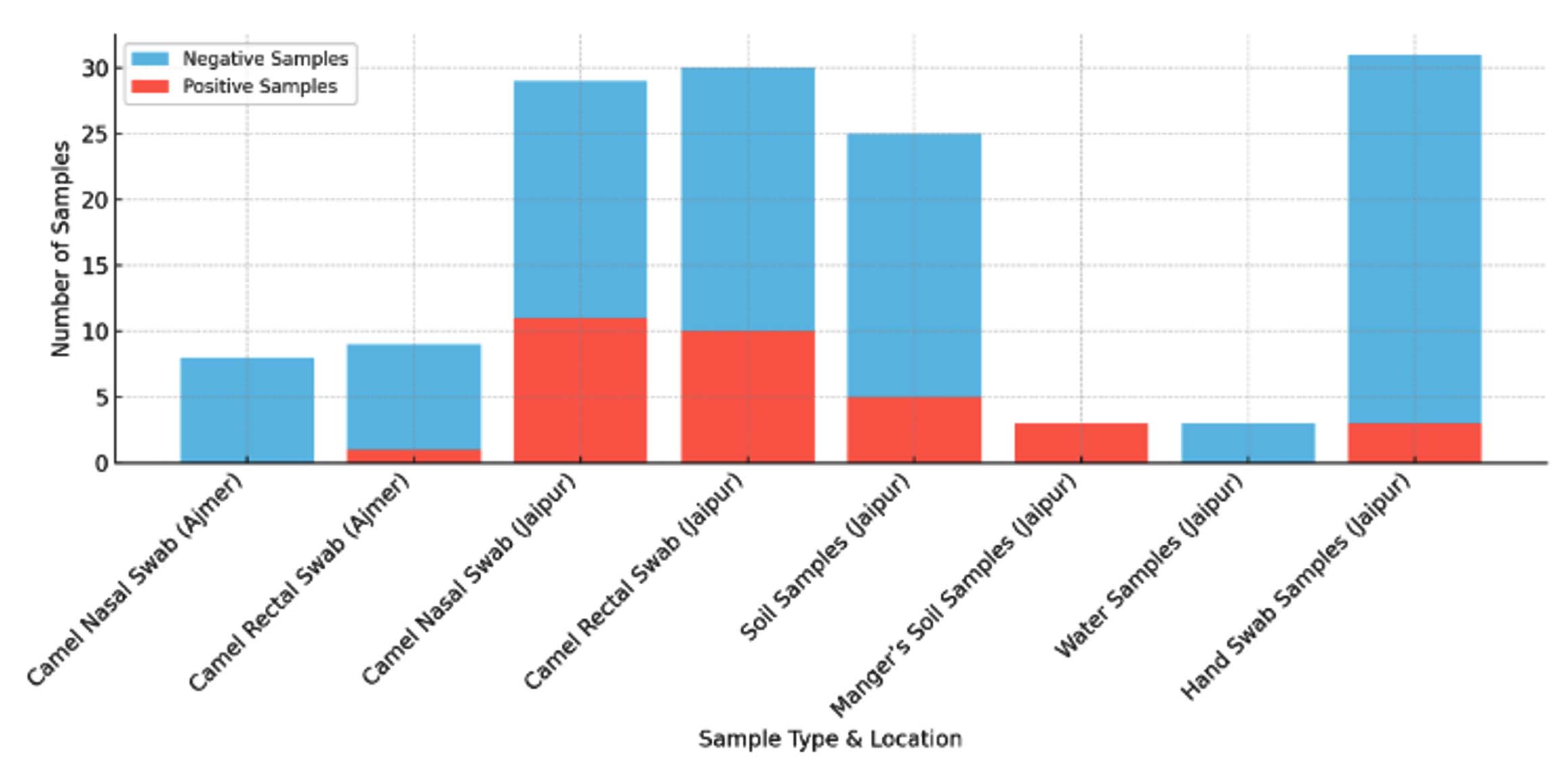
Figure 2. Stacked bar chart visualizing Klebsiella pneumoniae positivity across different sample types and locations. Red (salmon) bars indicate positive samples, whereas blue (skyblue) bars indicate negative samples
Antibiotic susceptibility profile of K. pneumoniae isolates
All isolates (100%) exhibited resistance to penicillin and ampicillin, which is attributable to the intrinsic resistance of K. pneumoniae resulting from constitutive production of a chromosomal class A β-lactamase. Antibiotic sensitivity pattern of Klebsiella pneumoniae isolates against different classes of antibiotics by Kirby-Bauer Disc diffusion method and BD phoenix M50 method is mentioned in the supplementary Table S1 and S2, respectively. High resistance against ciprofloxacin (CIP5) (72.72%) and piperacillin (PIP) (51.51%) was observed. A clustered heatmap of the antibiotic resistance patterns of the isolates is shown in Figure 3. Antibiotic resistance profile of Klebsiella pneumoniae isolated from Camels, their handlers and surrounding environment by Kirby-Bauer disc diffusion method/MIC/ BD Phoenix M50 is mentioned in the supplementary Table S3.
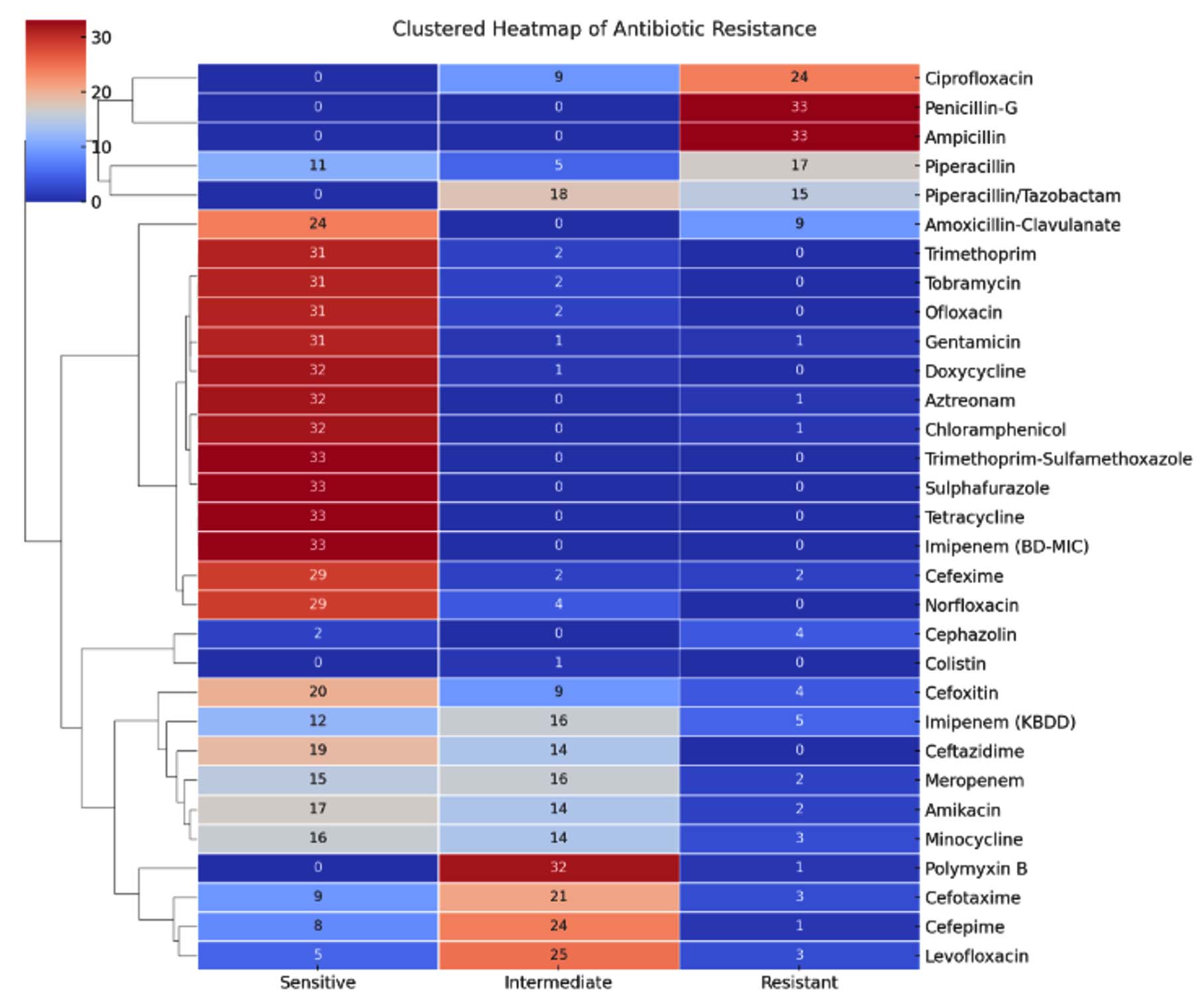
Figure 3. Clustered heat map of antibiotic resistance pattern of Klebsiella pneumoniae isolates
Multidrug-resistance
Of the isolates, 22 (66.67%) were MDR and exhibited resistance to three or more classes of antibiotics, as summarized in Figure 4. The multiple antibiotic resistance (MAR) index was calculated for each isolate. Detection and distribution of multiple antibiotic resistance (MAR) Index of the isolates are presented in the supplementary Table S4. The MAR index reflects the potential risk of each isolate acquiring or transmitting drug resistance to other bacterial pathogens. MAR typing of the isolates was conducted based on the MAR index, which resulted in nine MAR categories. In this study, ten isolates (RCKR2, RCKN12, RCKR13, RCKR15, RCKR18, RCKN24, RCKM26, RCKS27, RCKH30, and RCKS30) exhibited MAR indices exceeding 0.2%. All these isolates were from camels (six isolates: RCKR2, RCKN12, RCKR13, RCKR15, RCKR18, and RCKN24), humans (one isolate: RCKH30), or the environment (three isolates: RCKM26, RCKS27, and RCKS30), indicating the potential risk of camel rearers picking up MDR K. pneumoniae infection if they do not follow proper sanitary precautions.
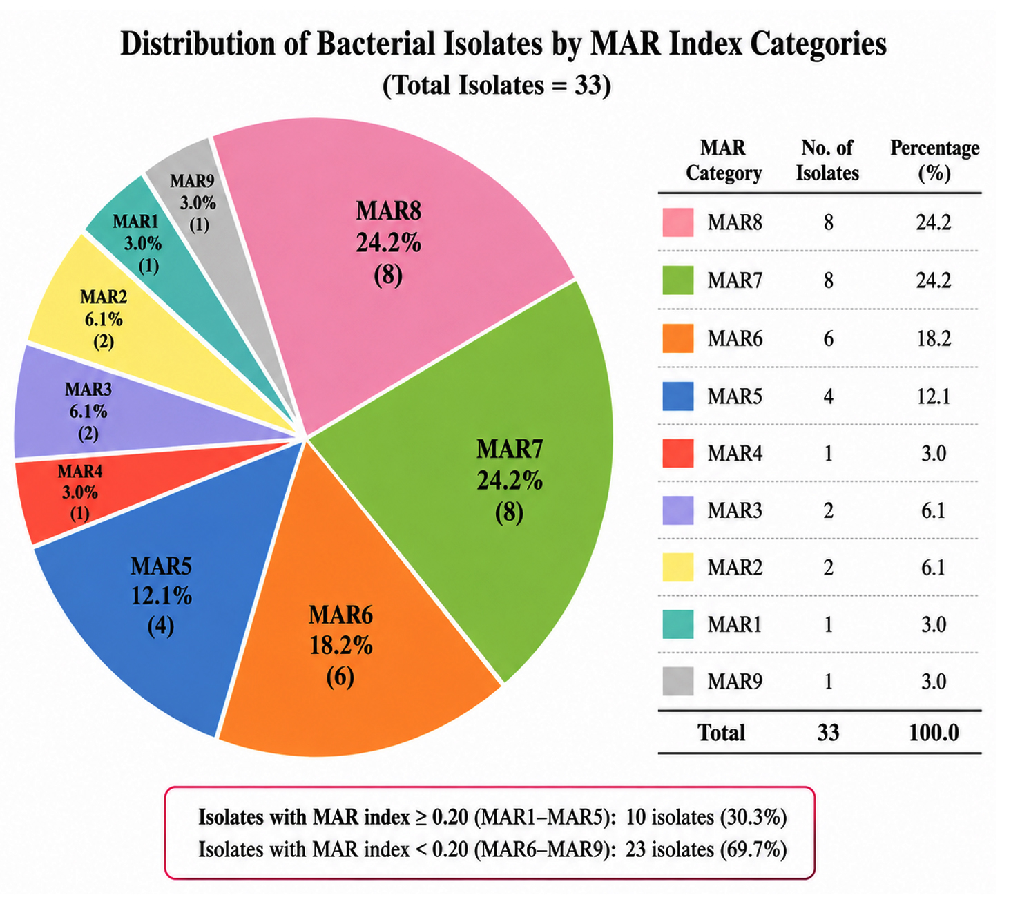
Figure 4. Distribution of 33 bacterial isolates among nine Multiple Antibiotic Resistance (MAR) index categories. MAR7 and MAR8 were the most prevalent categories, each comprising 24.2% of the isolates, followed by MAR6 (18.2%) and MAR5 (12.1%). Isolates with MAR Indices ≥ 0.20 (MAR1-MAR5) represented 30.3% of the total isolates, indicating a substantial proportion of multidrug-resistant strains
Carbapenem resistance
Although five isolates were resistant to imipenem (IPM10) upon disc diffusion, all isolates were confirmed to be susceptible to carbapenems via BD Phoenix M50.
Colistin resistance
One isolate (RCKR3) was phenotypically resistant to colistin based on the colistin agar test and BD Phoenix M50 MIC testing (MIC >2 µg/mL) (Figure 5).
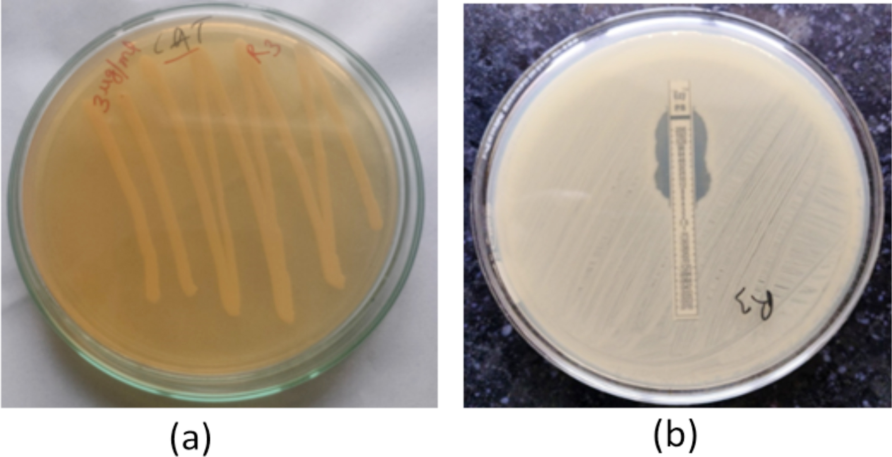
Figure 5. Colistin resistance by colistin agar test. (a) Isolate growth on 3 µg/mL colistin agar indicating resistance. (b) Colistin resistance by polymyxin-B MIC testing. Polymyxin isolate RCKR3 shows a MIC of 6 mcg/mL
ESBL detection in K. pneumoniae
Phenotypic ESBL production was confirmed in 11 isolates (33.33%) using the double-disc diffusion test (Figure 6). Genotypic screening identified ESBL-associated genes, of which blaTEM was the most prevalent gene, detected in 31/33 isolates (93.9%) (Figure 7); blaSHV was detected in 22/33 isolates (66.66%) (Figure 8), and blaCTX-M was absent in all isolates.
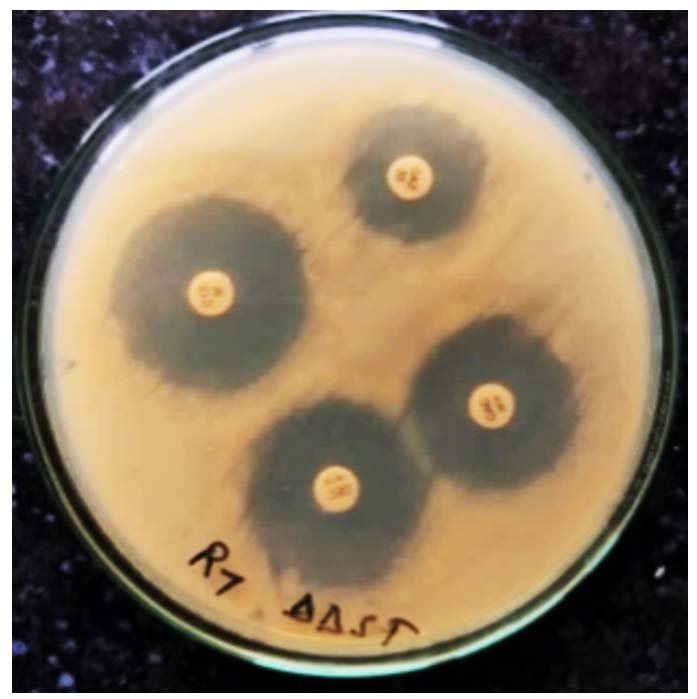
Figure 6. ESBL production was assessed phenotypically by the double disc diffusion method
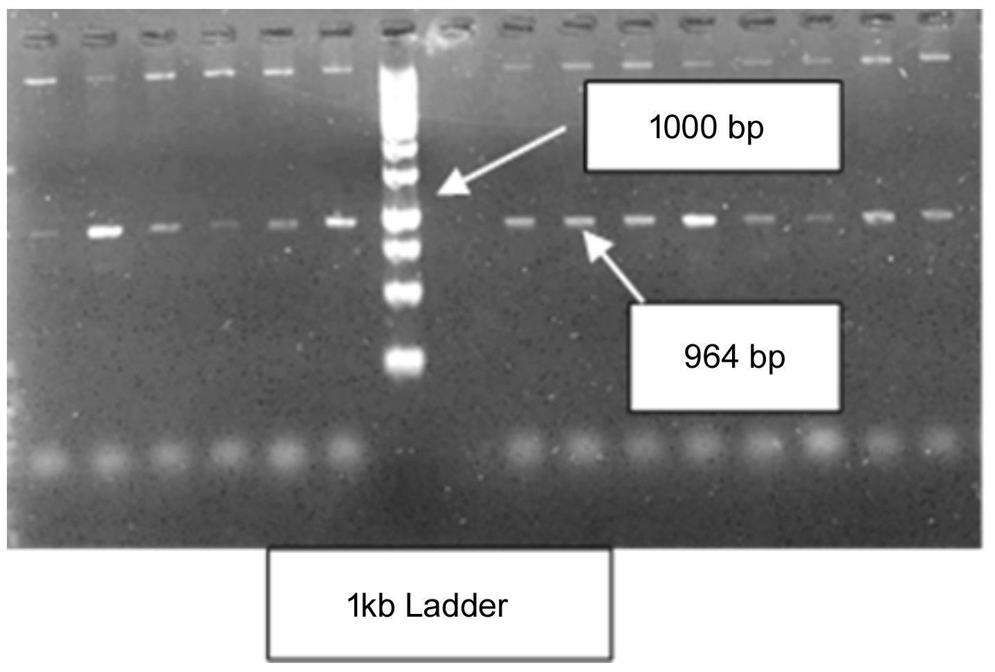
Figure 7. Amplification of the blaTEM gene of Klebsiella pneumoniae by PCR
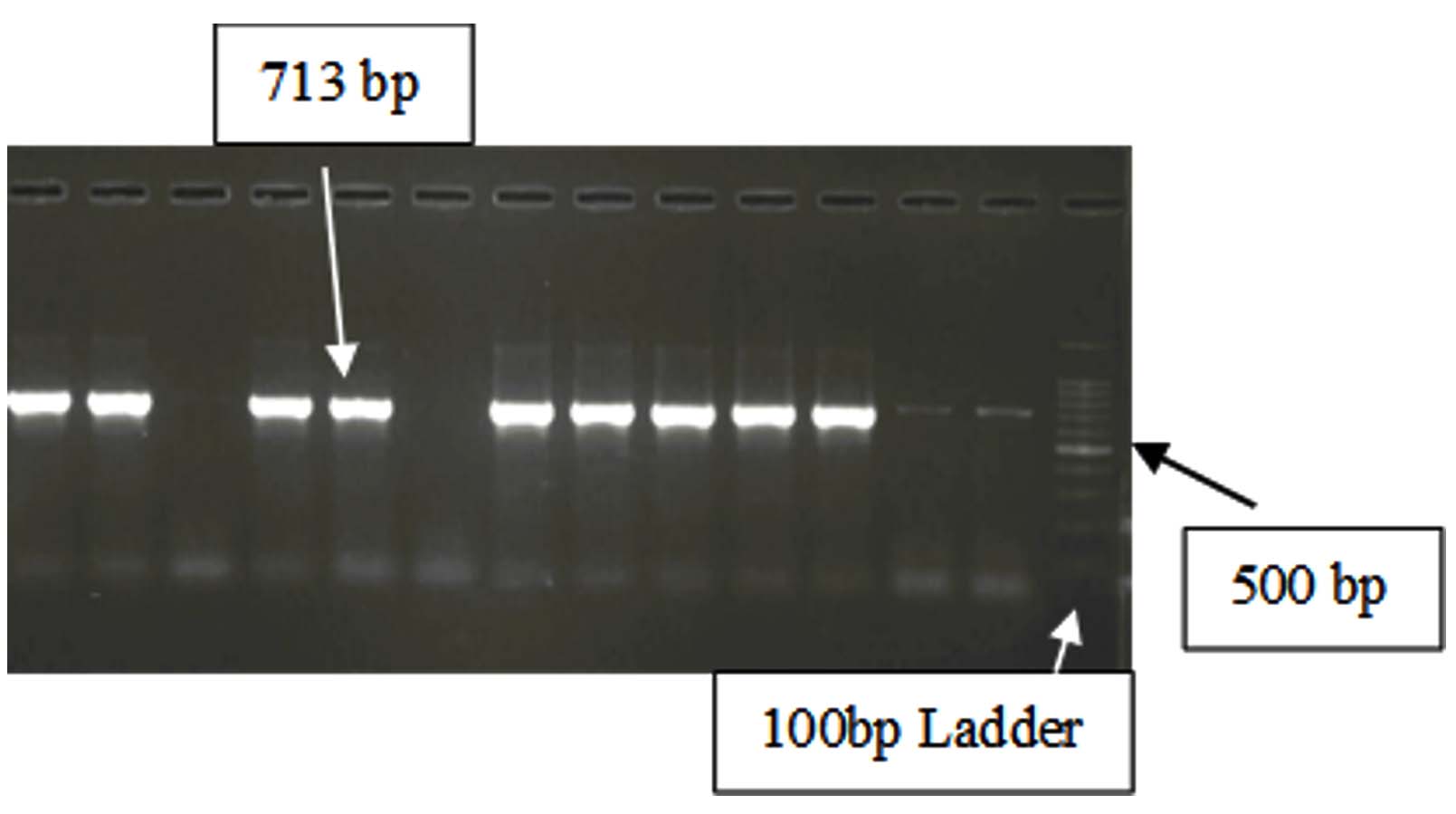
Figure 8. PCR amplification of the blaSHV gene of Klebsiella pneumoniae
Detection of carbapenem and quinolone resistance genes
Carbapenem resistance genes (blaVIM, blaIMP, blaKPC, blaNDM) were not detected in any isolate. None of the isolates harbored plasmid-mediated quinolone resistance genes (qnrA, qnrB, qnrS). Given the high resistance to ciprofloxacin (84.84%) (Figure 9), mutations in gyrA may be responsible for this resistance.
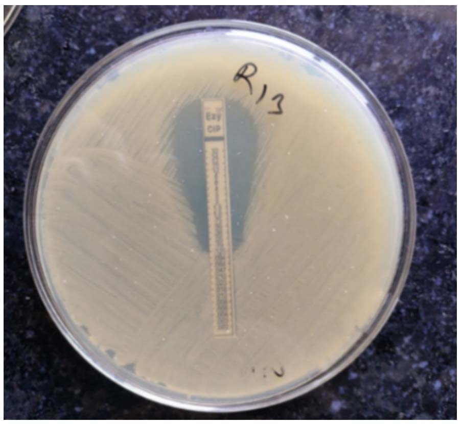
Figure 9. Phenotypic detection of quinolone resistance by ciprofloxacin MIC testing
Colistin resistance and mcr-1 gene screening
Only one isolate (RCKR3) was colistin resistant. Screening for the mcr-1 gene by PCR was negative, indicating that resistance could be mediated by other mcr-1-10 variants or chromosomal mutations. Further whole-genome sequencing of this isolate is required to identify the exact mechanism underlying the colistin resistance.
The present study provides key insights into the prevalence, antimicrobial resistance, and genetic determinants of K. pneumoniae at the camel–human–environment interface, emphasizing its significance within the One Health framework. The findings indicate that K. pneumoniae is not only prevalent in camels but also in their associated environment and handlers, with an overall isolation rate of 23.9%. These results are consistent with earlier observations on K. pneumoniae from camels and other livestock, highlighting its ubiquitous nature and potential role in cross-contamination and zoonotic transmission.
Comparative prevalence and epidemiological significance
The isolation rate recorded in the current investigation resembles prior findings in camels, with a reported prevalence of 26.71% in healthy camels in Egypt,14 of which the isolation rate from nasal and lung swabs from camels in Gharbia was 7.31%.15 Similarly, another One Health study reported K. pneumoniae isolation rates of 11.6% in animals, 8.4% in humans, and 7.0% from environmental sources in Punjab, Pakistan.16 The detection of K. pneumoniae across multiple reservoirs shows its remarkable adaptability in diverse ecological contexts.
Multidrug resistance and ESBL production in Camel ecosystem
One of the most significant findings was the predominance of ESBL-producing K. pneumoniae in apparently healthy camels (31.8%). This is particularly concerning, as previous studies have reported no ESBL producers in healthy camels (0.0%) and 31.9% in diseased camels.17 The increasing prevalence of ESBL-producing strains in healthy animals suggests an increasing trend of AMR, possibly owing to environmental contamination, unregulated antibiotic use, or horizontal gene transfer.
The predominant ESBL genotype detected in this study was blaTEM (93.9%), followed by blaSHV (66.66%), which is consistent with the findings of other studies from Rajasthan.18,19 These results further support the notion that blaTEM is the dominant genotype in this region. The high prevalence of blaTEM (93.9%) may reflect either the clonal expansion of a successful ESBL-producing lineage or widespread dissemination mediated by mobile genetic elements. Given the recovery of isolates from multiple ecological niches within the camel–human environment interface, horizontal gene transfer under antimicrobial selection pressures appears plausible. However, molecular typing studies are required to delineate clonal relatedness and transmission dynamics. The absence of the globally dominant ESBL blaCTX-M in our study contrasts with previous reports, which found a higher blaCTX-M prevalence in K. pneumoniae from bovines in Northeast India and West Bengal.20,21 The absence or very low prevalence of blaCTX-M in Rajasthan’s camel ecosystem may be due to limited third-generation cephalosporin pressure and reduced spillover of hospital-origin strains. Due to the limited use of third-generation cephalosporins in camel ecosystems, there is insufficient selective pressure to drive blaCTX-M expansion. This geographical variation in ESBL gene distribution warrants further genomic surveillance to track the development and dissemination of resistance determinants.
Resistance to quinolone and role of chromosomal mutations
This study identified a high ciprofloxacin resistance rate (84.84%). Since no plasmid-mediated quinolone resistance genes (qnrA, qnrB, qnrS) were found, chromosomal alterations, particularly in gyrA and parC, likely contributed to the quinolone resistance in these isolates. These findings are consistent with reports of 80% resistance to quinolones in K. pneumoniae isolates from poultry farms in coastal Karnataka.22 In contrast, one study reported no ciprofloxacin-resistant isolates in healthy camels and only 8.5% resistance in diseased camels, highlighting the emerging resistance trend over time.17
Colistin resistance and implications for last-resort antibiotics
The detection of the colistin-resistant isolate RCKR3 is notable because colistin is a last-line antibiotic used against MDR Gram-negative infections. Although mcr-1 was not detected, the resistance might be mediated by other mcr variants (mcr-1 to mcr-10) or chromosomal mutations in mgrB or pmr. Reports on colistin-resistant K. pneumoniae in camels are extremely rare, and this study is the first to document colistin resistance in K. pneumoniae in camels in India. Resistance to colistin has been previously reported in pigs (Portugal),23 turkey meat (Czech Republic),24 and camels (Dubai),25 where E. coli with mcr-1 was detected and colistin-resistant Enterobacter cloacae (Tunisia)26 was negative for mcr-1. Phenotypic colistin resistance in a camel-associated ecosystem, despite the absence of mcr-1, highlights the need for ongoing genomic surveillance to detect emerging resistance mechanisms, risk assessment studies to evaluate zoonotic transmission pathways, and investments in research on resistance in nontraditional livestock systems.
Public health and one health implications
These results have significant public health relevance, particularly in areas where camels are central to milk production, meat supply, and transportation. Our findings provide empirical evidence that camel ecosystems can serve as reservoirs of clinically significant resistant pathogens, particularly in regions where camels are a key source of milk, meat, and transportation.
Given the rapid evolution of AMR, integrating the One Health approach into future research is critical for understanding the transmission dynamics between animals, humans, and the environment.
This study provides the first comprehensive analysis of ESBL-producing, quinolone- and colistin-resistant K. pneumoniae isolates from a camel ecosystem in India. The high prevalence of MDR isolates across camels, handlers, and the environment underscores the need for continuous surveillance, strict antimicrobial stewardship, and interdisciplinary collaboration to mitigate the spread of AMR within the One Health Framework.
Additional file: Table S1-S4.
ACKNOWLEDGMENTS
The authors are grateful to the Vice Chancellor of RAJUVAS, Bikaner, and the Dean of PGIVER, Jaipur, for their support through the provision of laboratory facilities and research consumables. The authors also sincerely acknowledge the Director of ICAR-NIVEDI for granting permission to use the institute’s laboratory facilities.
CONFLICT OF INTEREST
The authors declare that there is no conflict of interest.
AUTHORS’ CONTRIBUTION
YS, BJ, SN, SKS, SSS, TB, AF, RK and VKM conceptualized and designed the study. YS, RK and VKM performed material preparation. YS, BJ, SN and SKS designed the methodology. YS, AF, TB and SSS performed data collection and analysis. YS wrote the manuscript. All authors reviewed, revised and approved the final manuscript for publication.
FUNDING
None.
DATA AVAILABILITY
All datasets generated or analyzed during this study are included in the manuscript and/or in the supplementary files.
ETHICS STATEMENT
Not applicable.
INFORMED CONSENT
Written informed consent was obtained from the participants before enrolling in the study.
- Lahariya Upadhyay A, Garg R, Singh SK. Antimicrobial resistance: An emerging Global challeng waiting for urgent actions. Prev Med Res Rev.2024; 1(4):169-171.
Crossref - World Health Organization (WHO). Global Action Plan on Antimicrobial Resistance. Geneva, Switzerland: World Health Organization. 2016. https://www.who.int/publications/i/item/9789241509763. Accessed September 5, 2025.
- Queenan K, Hasler B, Rushton J. A One Health approach to antimicrobial resistance surveillance: is there a business case for it? Int J Antimicrob Agents. 2016;48(4):422-427.
Crossref - Clinical and Laboratory Standards Institute (CLSI). Performance Standards for Antimicrobial Susceptibility Testing. 34th ed. CLSI Supplement M100. Wayne, PA: CLSI. 2024.
- Liu Y, Liu C, Zheng W, et al. PCR detection of Klebsiella pneumoniae in infant formula based on 16S–23S internal transcribed spacer. Int J Food Microbiol. 2008;125(3):230-235.
Crossref - Olesen I, Hasman H, Muller Aarestrup F. Prevalence of β-lactamases among ampicillin-resistant Escherichia coli and Salmonella isolated from food animals in Denmark. Microb Drug Resist. 2004;10(4):334-340.
Crossref - Hasman H, Mevius D, Veldman K, Olesen I, Aarestrup FM. Beta-Lactamases among extended-spectrum β-lactamase-resistant Salmonella from poultry, poultry products, and human patients in The Netherlands. J Antimicrob Chemother. 2005;56(1):115-121.
Crossref - Dallenne C, Da Costa A, DecreD, Favier C, Arlet G. Development of a set of multiplex PCR assays for the detection of genes encoding important beta-lactamases in Enterobacteriaceae. J Antimicrob Chemother. 2010;65(3):490-495.
Crossref - Begum N, Shamsuzzaman SM. Emergence of carbapenemase-producing urinary isolates at a tertiary care hospital in Dhaka, Bangladesh. Tzu Chi Med J. 2016;28(3):94-98.
Crossref - Poirel L, Walsh TR, Cuvillier V, Nordmann P. Multiplex PCR for detection of acquired carbapenemase genes. Diagn Microbiol Infect Dis. 2011;70(1):119-123.
Crossref - Cattoir V, Poirel L, Rotimi V, Soussy CJ, Nordmann P. Multiplex PCR for detection of plasmid-mediated quinolone resistance qnr genes in ESBL-producing enterobacterial isolates. J Antimicrob Chemother. 2007;60(2):394-397.
Crossref - Rodriguez-Martinez JM, Velasco C, Pascual A, Garcia I, Martinez-Martinez L. Correlation of quinolone resistance levels and differences in basal and quinolone-induced expression from three qnrA-containing plasmids. Clin Microbiol Infect. 2006;12(5):440-445.
Crossref - Rebelo AR, Bortolaia V, Kjeldgaard JS, et al. Multiplex PCR for detection of plasmid-mediated colistin resistance determinants, mcr-1, mcr-2, mcr-3, mcr-4, and mcr-5 for surveillance purposes. Euro Surveill. 2018;23(6):17-000672.
Crossref - Wareth G, Murugaiyan J, Khater DF, Moustafa SA. Subclinical pulmonary pathogenic infection in camels slaughtered in Cairo, Egypt. J Infect Dev Ctries. 2014;8(7):909-913.
Crossref - Moawad AA, Ammar AM, Dapgh AN, Ragab DA. Identification and antibiogram of Klebsiella pneumoniae isolated from camels with molecular recognition of some virulence and antibiotic resistance encoding genes. Pak J Zool. 2023;57(3):1059-1065.
Crossref - Yasmeen N, Aslam B, Fang LX, Baloch Z, Liu Y. Occurrence of extended-spectrum β-lactamase harboring Klebsiella pneumoniae in various sources: a One Health perspective. Front Cell Infect Microbiol. 2023;13:1103319.
Crossref - Sharma SK, Patel K, Maherchandani S, Shringi BN. ESBL detection and comparison of antibiotic resistance pattern of Klebsiella pneumoniae isolated from healthy and acute respiratory tract infected camels. Adv Anim Vet Sci. 2017;5(2):83-91.
Crossref - Yadav SK. Phenotypic and Genotypic Studies on Antibiotic Resistance Pattern of Klebsiella pneumoniae Associated with Various Clinical Conditions of Animals [MVSc thesis]. Rajasthan University of Veterinary and Animal Science. 2020.
- Choudhary S, Joseph B, Sharma SK, Shekhawat SS, Mohammed N. Detection of extended-spectrum beta-lactamase and carbapenemase resistant Klebsiella pneumoniae in bovine milk. Ruminant Sci. 2022;11(1):93-100.
- Koovapra S. Characterization of Extended-Spectrum Beta-Lactamase Producing Klebsiella spp. of Bovine Mastitis Origin [MVSc thesis]. Indian Veterinary Research Institute; 2015.
- Bandyopadhyay S, Banerjee J, Bhattacharyya D, et al. Genomic identity of fluoroquinolone-resistant blaCTX-M-15-type ESBL and pMAmpC β-lactamase-producing Klebsiella pneumoniae from buffalo milk, India. Microb Drug Resist. 2018;24(9):1345-1353.
Crossref - Bhardwaj K, M SS, Baliga S, B U, Baliga BS, Shetty VK. Research note: characterization of antibiotic resistant phenotypes and linked genes of Escherichia coli and Klebsiella pneumoniae from healthy broiler chickens, Karnataka, India. Poult Sci. 2021;100(6):101094.
Crossref - Fournier C, Aires-de-Sousa M, Nordmann P, Poirel L. Occurrence of CTX-M-15 and MCR-1-producing Enterobacterales in pigs, Portugal: evidences of direct links with antibiotic selective pressure. Int J Antimicrob Agents. 2020;55(2):105802.
Crossref - Gelbicova T, Kolackova I, Krutova M, Karpiskova R. The emergence of mcr-1-mediated colistin-resistant Escherichia coli and Klebsiella pneumoniae in domestic and imported turkey meat in the Czech Republic, 2017–2018. Folia Microbiol (Praha). 2020;65:211-216.
Crossref - Ghazawi A, Strepis N, Anes F, et al. First report of colistin-resistant Escherichia coli carrying mcr-1 IncI2(delta) and IncX4 plasmids from camels (Camelus dromedarius) in the Gulf region. Antibiotics. 2024;13(3):227.
Crossref - Rhouma M, Bessalah S, Salhi I, Theriault W, Fairbrother JM, Fravalo P. Screening for fecal presence of colistin-resistant Escherichia coli and mcr-1 and mcr-2 genes in camel-calves in southern Tunisia. Acta Vet Scand. 2018;60(1):35.
Crossref
© The Author(s) 2026. Open Access. This article is distributed under the terms of the Creative Commons Attribution 4.0 International License which permits unrestricted use, sharing, distribution, and reproduction in any medium, provided you give appropriate credit to the original author(s) and the source, provide a link to the Creative Commons license, and indicate if changes were made.