


ISSN: 0973-7510
E-ISSN: 2581-690X
 , Phong Xuan Huynh1, Lieu Thuy Thi Nguyen3,
Long Dang Hoang Bui1
, Thanh Ngoc Nguyen1 and Dung Ngoc Tran4 Cutibacterium acnes is a dominant member of the human skin microbiota and is closely associated with the pathogenesis of acne vulgaris. The increasing emergence of antibiotic-resistant C. acnes strains has raised concerns regarding the long-term effectiveness of current therapies. This study aimed to characterize antibiotic susceptibility patterns and genomic features of C. acnes isolates obtained from healthy individuals and acne patients with different disease severities. A total of 45 participants were enrolled, including healthy controls and patients with mild and moderate-severe acne. C. acnes isolates recovered from pilosebaceous follicles were subjected to antibiotic susceptibility testing using the Kirby-Bauer disk diffusion method against five antibiotics including clindamycin, tetracycline, azithromycin, trimethoprim-sulfamethoxazole (TMP-SMX), and doxycycline. Representative isolates were selected for whole-genome sequencing, followed by multi-locus sequence typing (MLST) and antimicrobial resistance gene annotation. The results showed that most isolates remained highly susceptible to tetracycline-class antibiotics and TMP-SMX, whereas resistance to macrolides and clindamycin was detected, particularly among moderate-severe acne isolates. Genomic analysis revealed notable genetic diversity among the isolates and identified a single antimicrobial resistance gene, erm(51), consistent with the observed resistance phenotype. Overall, antimicrobial resistance in the analyzed C. acnes population was limited but detectable. The integration of phenotypic susceptibility testing with whole-genome analysis provides valuable insights for antimicrobial resistance surveillance and supports rational antibiotic use in acne management.
Cutibacterium acnes, Acne Vulgaris, Antibiotic Susceptibility, Antimicrobial Resistance, Whole-genome Sequencing, MLST
Acne vulgaris is a chronic inflammatory disorder of the pilosebaceous unit that affects a large proportion of adolescents and young adults worldwide. Its pathogenesis involves sebum overproduction, follicular hyperkeratinization, inflammation, and skin microbiota dysbiosis.1 Among skin-resident microorganisms, C. acnes plays a central role, acting as a physiological commensal, while also contributing to inflammation under dysbiosis conditions.2,3
Cutibacterium acnes is a Gram-positive anaerobic bacterium that preferentially colonizes sebaceous follicles due to its ability to metabolize sebum-derived lipids. Recent evidence indicates that acne severity is more closely associated with the genetic diversity and strain composition of C. acnes rather than its absolute abundance.4 Specific sequence types and phylotypes, particularly those belonging to phylotype IA1, have been strongly associated with inflammatory acne, whereas other lineages are more frequently detected on healthy skin.3,5
Antibiotics, especially macrolides, lincosamides, and tetracyclines, have been widely used in acne treatment for decades. However, prolonged and sometimes inappropriate antibiotic use has contributed to the increasing emergence of antibiotic-resistant C. acnes strains, leading to reduced treatment efficacy and growing public health concerns.1 Recent epidemiological studies have reported high resistance rates to macrolides and clindamycin, with substantial geographic variation and an overall increasing global trend.5-7
In parallel with phenotypic data, genomic studies have shown that antimicrobial resistance in C. acnes may arise through target gene mutations or acquisition of resistance genes mediated by mobile genetic elements.8,9 Nevertheless, integrated studies combining antibiotic susceptibility testing with whole-genome characterization of C. acnes isolates from different clinical groups remain limited, particularly in developing countries.2,5 Therefore, the present study aimed to evaluate the antibiotic susceptibility profiles of C. acnes isolates from healthy individuals and acne patients with varying disease severities and to characterize representative isolates using whole-genome sequencing and multi-locus sequence typing.
Antibiotic susceptibility testing of Cutibacterium acnes
The study included 45 participants, comprising 30 patients with acne vulgaris and 15 healthy controls, stratified into healthy (n = 15), mild acne (n = 15), and moderate-severe acne (n = 15) groups. Cutibacterium acnes isolates obtained from pilosebaceous follicles were evaluated for antibiotic susceptibility using the Kirby-Bauer disk diffusion method with five antibiotics including clindamycin, tetracycline, azithromycin, TMP-SMX, and doxycycline. The experiment consisted of 45 isolates, including one reference strain and 44 clinical isolates, each performed in triplicate. Isolates were cultured in Fluid Thioglycolate Medium for 5 days, inoculated onto Mueller–Hinton agar, and incubated anaerobically at
37 °C for 5 days. Inhibition zone diameters were measured and interpreted as susceptible (S) or resistant (R) according to EUCAST breakpoints for Staphylococcus spp. and CLSI (2024) guidelines, as EUCAST species-specific criteria for C. acnes are currently unavailable. Based on the antibiotic susceptibility results, one C. acnes isolate exhibiting the highest level of antibiotic resistance was selected from each study group. In addition, one isolate from the healthy control group that was fully susceptible to all tested antibiotics was included as a comparator. These representative isolates were subjected to whole-genome sequencing to investigate the presence and genetic basis of antimicrobial resistance determinants.
Data processing and quality control
Raw sequencing data were initially assessed for quality using fastQC to examine key parameters, including per-base quality score distribution, GC content, adapter contamination, and sequence duplication levels.10 Following preliminary evaluation, reads were cleaned and optimized using fastQ by trimming low-quality regions, removing adapter sequences, and filtering out substandard reads to ensure high reliability for downstream analyses.11
Genome assembly and quality assessment
Quality-filtered reads were assembled de novo using Unicycler, a hybrid assembly algorithm specifically optimized for bacterial genome reconstruction.12 The quality of the assembled genomes was comprehensively evaluated using QUAST based on key metrics, including total genome length, number of contigs, N50 value, and overall genome completeness.13
Functional annotation and molecular typing
The assembled genomes were functionally annotated using Prokka to identify structural genes, protein-coding sequences, RNA genes, and functional genomic regions.14 Genome structure visualization and comparative analysis were performed using the Proksee platform. Strain-level identification was subsequently conducted using multi-locus sequence typing (MLST), based on allele profiling of standardized housekeeping genes.
Antimicrobial resistance gene analysis
Genes and variants associated with antimicrobial resistance were identified using AMRFinderPlus,15 which detects resistance-associated genetic markers through comparison with a curated reference database.
This study was approved by the Ethics Committee of Biomedical Research, Can Tho University of Medicine and Pharmacy (Ref. No. 24.004/PCT-H׀׀׀). Written informed consent was obtained from the participants before enrolling in the study.
Antibiotic susceptibility of Cutibacterium acnes isolates from healthy individuals
The results showed that most C. acnes isolates (n = 16, including 15 clinical isolates obtained from healthy individuals and one reference strain used as a control) were susceptible to antibiotics belonging to the macrolide, lincosamide and tetracycline classes; however, antibiotic resistance was still observed (Table 1). Notably, azithromycin exhibited the highest resistance rate, with 7 out of 16 isolates (43.75%) classified as resistant, while resistance to clindamycin (18.75%) and tetracycline (6.25%) was also detected, as indicated by the absence or marked reduction of inhibition zones (Figure 1). In contrast, all 16 isolates remained fully susceptible to doxycycline and TMP-SMX (100%), highlighting substantial inter-strain variability in antibiotic resistance phenotypes among C. acnes isolates, consistent with previous reports describing heterogeneous resistance profiles across different phylogenetic lineages and clinical isolates.16
Table 1. Antibiotic susceptibility of Cutibacterium acnes isolates from healthy individuals
Isolate |
Clindamycin IZ (mm) |
I |
Tetracycline IZ (mm) |
I |
Azithromycin IZ (mm) |
I |
TMP-SMX IZ (mm) |
I |
Doxycycline IZ (mm) |
I |
|---|---|---|---|---|---|---|---|---|---|---|
C. acnes ATCC 6919 |
50.00 ± 0.00 |
S |
50.00 ± 5.00 |
S |
50.00 ± 10.00 |
S |
39.50 ± 1.50 |
S |
36.67 ± 2.89 |
S |
Hn1-1 |
47.00 ± 3.00 |
S |
43.33 ± 2.89 |
S |
42.67 ± 2.52 |
S |
40.00 ± 0.00 |
S |
42.33 ± 2.52 |
S |
Hn2 |
47.00 ± 0.00 |
S |
54.00 ± 1.00 |
S |
47.50 ± 7.50 |
S |
40.00 ± 0.00 |
S |
31.00 ± 1.00 |
S |
Hn4 |
0.00 ± 0.00 |
R |
44.33 ± 1.16 |
S |
0.00 ± 0.00 |
R |
33.00 ± 1.00 |
S |
43.67 ± 1.53 |
S |
Hn5-1 |
38.00 ± 5.20 |
S |
15.00 ± 0.00 |
R |
24.00 ± 1.00 |
S |
39.50 ± 0.50 |
S |
22.67 ± 2.52 |
S |
Hn7-2 |
50.00 ± 0.00 |
S |
40.00 ± 0.00 |
S |
40.00 ± 10.00 |
S |
31.00 ± 1.00 |
S |
35.00 ± 5.00 |
S |
Hn8-1 |
0.00 ± 0.00 |
R |
49.00 ± 4.00 |
S |
0.00 ± 0.00 |
R |
38.50 ± 1.50 |
S |
22.50 ± 2.50 |
S |
Hn8-2 |
21.50 ± 1.50 |
S |
52.00 ± 5.00 |
S |
0.00 ± 0.00 |
R |
29.67 ± 4.73 |
S |
63.00 ± 3.00 |
S |
Hn9-2 |
45.50 ± 0.50 |
S |
27.00 ± 2.65 |
S |
10.67 ± 2.52 |
R |
45.00 ± 0.00 |
S |
30.00 ± 0.00 |
S |
Hn10-1 |
50.00 ± 3.61 |
S |
32.00 ± 5.00 |
S |
30.00 ± 5.00 |
S |
35.00 ± 4.36 |
S |
32.50 ± 2.50 |
S |
Hn10-2 |
32.00 ± 0.00 |
S |
51.00 ± 1.00 |
S |
48.50 ± 1.50 |
S |
30.00 ± 0.00 |
S |
51.67 ± 2.89 |
S |
Hn12-1 |
0.00 ± 0.00 |
R |
46.33 ± 1.16 |
S |
0.00 ± 0.00 |
R |
42.00 ± 0.00 |
S |
46.33 ± 1.16 |
S |
Hn13 |
51.00 ± 1.00 |
S |
48.67 ± 3.22 |
S |
57.50 ± 2.50 |
S |
42.00 ± 0.00 |
S |
53.33 ± 5.77 |
S |
Hn14 |
31.50 ± 0.50 |
S |
55.67 ± 0.58 |
S |
0.00 ± 0.00 |
R |
30.00 ± 0.00 |
S |
64.67 ± 3.22 |
S |
Hn15-1 |
43.50 ± 0.50 |
S |
38.00 ± 6.93 |
S |
50.00 ± 0.00 |
S |
40.00 ± 0.00 |
S |
46.33 ± 3.22 |
S |
Hn15-2 |
21.50 ± 1.50 |
S |
50.00 ± 0.00 |
S |
0.00 ± 0.00 |
R |
41.00 ± 1.00 |
S |
54.33 ± 0.58 |
S |
Notes: IZ: inhibition zone diameter (mm); I: interpretation; S: susceptible; R: resistant. Susceptibility was interpreted according to EUCAST criteria for Staphylococcus spp.
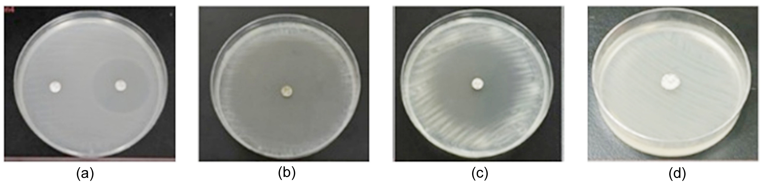
Figure 1. Inhibition zones of antibiotics against Cutibacterium acnes isolates obtained from healthy individuals
Note: (a) left – Clindamycin, right – TMP-SMX; (b) Doxycycline; (c) Tetracycline; (d) Azithromycin
The detection of C. acnes strains resistant to azithromycin and clindamycin in a healthy population indicates that antibiotic resistance genes can persist and potentially disseminate even in the absence of overt disease.1 This observation is concerning because it implies an accumulation of resistant bacteria in the community reservoir and underscores the need for vigilant monitoring of antibiotic use as well as systematic surveillance of the dynamics of resistant bacterial strains.3
Antibiotic susceptibility of Cutibacterium acnes isolates from mild acne patients
Seventeen C. acnes isolates were evaluated for susceptibility to five commonly used antibiotics, including clindamycin, tetracycline, azithromycin, TMP-SMX, and doxycycline (Table 2; Figure 2). All isolates were fully susceptible to TMP-SMX (100%), with inhibition zone diameters ranging from 27 ± 4.5 mm to 47.5 ± 2.5 mm. Doxycycline also showed high efficacy, with 94% of isolates (16/17) remaining susceptible. Susceptibility rates for clindamycin and tetracycline were both 82% (14/17 isolates), whereas azithromycin exhibited the lowest susceptibility rate at 76% (13/17 isolates). Overall, resistance to clindamycin, tetracycline, and azithromycin was observed in 22%-28% of the isolates.
Table 2. Antibiotic susceptibility of Cutibacterium acnes isolates from mild acne patients
Isolate |
Clindamycin IZ (mm) |
I |
Tetracycline IZ (mm) |
I |
Azithromycin IZ (mm) |
I |
TMP-SMX IZ (mm) |
I |
Doxycycline IZ (mm) |
I |
|---|---|---|---|---|---|---|---|---|---|---|
Mn1 |
0.00 ± 0.00 |
R |
44.00 ± 3.61 |
S |
0.00 ± 0.00 |
R |
38.00 ± 0.00 |
S |
41.00 ± 9.00 |
S |
Mn2-2 |
0.00 ± 0.00 |
R |
65.00 ± 5.00 |
S |
0.00 ± 0.00 |
R |
39.00 ± 1.00 |
S |
35.00 ± 0.00 |
S |
Mn2-3 |
39.50 ± 5.50 |
S |
47.33 ± 2.52 |
S |
0.00 ± 0.00 |
R |
32.50 ± 0.50 |
S |
56.67 ± 5.77 |
S |
Mn3-2 |
45.50 ± 0.50 |
S |
52.50 ± 2.50 |
S |
49.67 ± 2.52 |
S |
35.00 ± 1.00 |
S |
51.67 ± 2.89 |
S |
Mn4 |
44.50 ± 0.50 |
S |
24.00 ± 0.00 |
S |
35.00 ± 0.00 |
S |
27.50 ± 4.50 |
S |
33.50 ± 0.50 |
S |
Mn5-1 |
43.00 ± 1.00 |
S |
67.50 ± 2.50 |
S |
55.50 ± 0.50 |
S |
44.00 ± 1.00 |
S |
56.50 ± 0.50 |
S |
Mn7-1 |
43.00 ± 0.00 |
S |
20.00 ± 0.00 |
R |
35.00 ± 0.00 |
S |
35.00 ± 0.00 |
S |
41.00 ± 1.00 |
S |
Mn9-1 |
51.00 ± 1.00 |
S |
30.00 ± 0.00 |
S |
40.00 ± 0.00 |
S |
38.00 ± 2.00 |
S |
45.00 ± 0.00 |
S |
Mn9-2 |
59.50 ± 0.50 |
S |
60.00 ± 0.00 |
S |
45.00 ± 0.00 |
S |
47.50 ± 0.50 |
S |
41.00 ± 3.61 |
S |
Mn11-1 |
47.00 ± 0.00 |
S |
45.00 ± 5.00 |
S |
40.00 ± 5.00 |
S |
38.00 ± 0.00 |
S |
45.00 ± 0.00 |
S |
Mn11-2 |
0.00 ± 0.00 |
R |
12.33 ± 2.52 |
R |
0.00 ± 0.00 |
R |
27.00 ± 0.00 |
S |
18.50 ± 1.50 |
R |
Mn12-1 |
46.00 ± 0.00 |
S |
45.00 ± 0.00 |
S |
51.67 ± 2.89 |
S |
35.00 ± 0.00 |
S |
55.00 ± 0.00 |
S |
Mn12-2 |
45.00 ± 5.00 |
S |
55.00 ± 0.00 |
S |
55.00 ± 0.00 |
S |
39.00 ± 0.00 |
S |
50.00 ± 0.00 |
S |
Mn13-2 |
0.00 ± 0.00 |
R |
60.00 ± 5.00 |
S |
0.00 ± 0.00 |
R |
39.00 ± 1.00 |
S |
57.50 ± 2.50 |
S |
Mn14-1 |
42.00 ± 2.00 |
S |
16.00 ± 1.00 |
R |
23.00 ± 0.00 |
S |
34.00 ± 1.00 |
S |
22.50 ± 2.50 |
S |
Mn15-1 |
41.50 ± 0.50 |
S |
48.67 ± 5.51 |
S |
47.50 ± 2.50 |
S |
37.00 ± 0.00 |
S |
52.50 ± 2.50 |
S |
Mn15-2 |
49.50 ± 0.50 |
S |
20.67 ± 0.58 |
R |
22.33 ± 2.52 |
S |
39.50 ± 0.50 |
S |
30.00 ± 0.00 |
S |
Notes: IZ: inhibition zone diameter (mm); I: interpretation; S: susceptible; R: resistant. Susceptibility was interpreted according to EUCAST criteria for Staphylococcus spp.
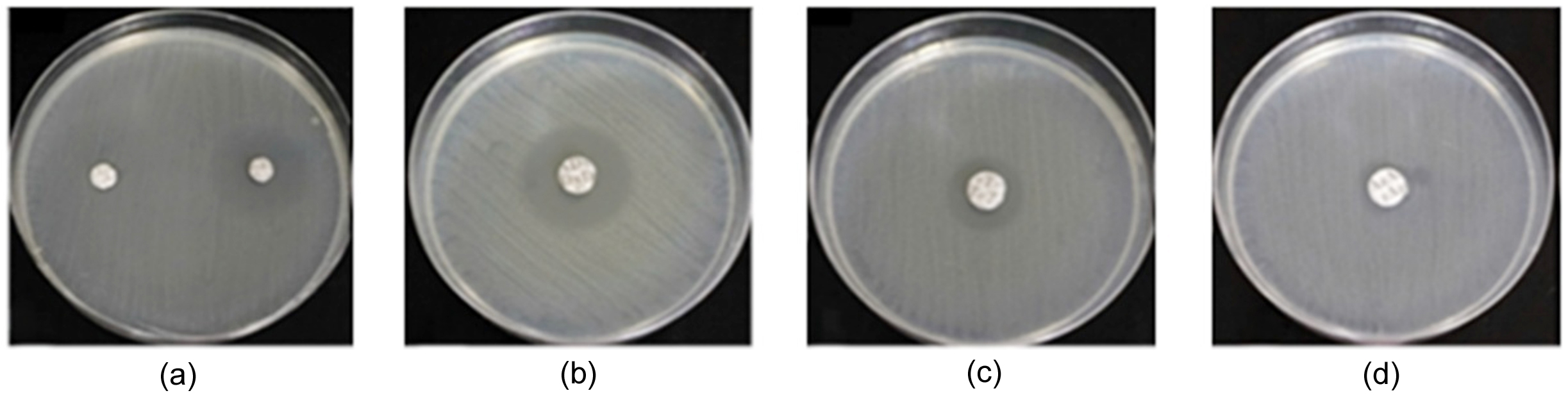
Figure 2. Inhibition zones of antibiotics against Cutibacterium acnes isolates obtained from mild acne patients
Note: (a) left – Clindamycin, right – TMP-SMX; (b) Doxycycline; (c) Tetracycline; (d) Azithromycin
The notable resistance rates observed for clindamycin, tetracycline, and particularly azithromycin indicate a declining effectiveness of several antibiotics commonly used in acne treatment. These findings are consistent with previous studies reporting that prolonged use or monotherapy with clindamycin and azithromycin contributes to the selection and persistence of resistant C. acnes strains.1 In contrast, the sustained susceptibility to TMP-SMX and doxycycline suggests that these agents remain effective therapeutic options. Collectively, these results underscore the importance of prudent antibiotic use and the implementation of antimicrobial resistance surveillance to limit the further spread of resistant C. acnes within the community.17,18
Antibiotic susceptibility of Cutibacterium acnes isolates from moderate-severe acne patients
Twelve C. acnes isolates obtained from patients with moderate-severe acne were evaluated for susceptibility to five commonly used antibiotics, including clindamycin, tetracycline, azithromycin, TMP-SMX, and doxycycline (Table 3; Figure 3). Antibiotics belonging to the tetracycline class (tetracycline and doxycycline) and the trimethoprim-sulfamethoxazole combination (TMP-SMX) showed complete susceptibility, with all isolates (100%) classified as susceptible. The inhibition zone diameters ranged from 22.00 ± 0.00 mm to 62.67 ± 0.58 mm for tetracycline, from 27.67 ± 0.58 mm to 45.00 ± 0.00 mm for TMP-SMX, and from 29.33 ± 1.16 mm to 64.00 ± 0.00 mm for doxycycline. In contrast, four isolates (Sn3, Sn4-2, Sn14-1, and Sn14-2) exhibited resistance to both clindamycin and azithromycin, accounting for 33.33% of the tested isolates.
Table 3. Antibiotic susceptibility of Cutibacterium acnes isolates from moderate-severe acne patients
Isolate |
Clindamycin IZ (mm) |
I |
Tetracycline IZ (mm) |
I |
Azithromycin IZ (mm) |
I |
TMP-SMXIZ IZ (mm) |
I |
Doxycycline IZ (mm) |
I |
|---|---|---|---|---|---|---|---|---|---|---|
Sn1-1 |
48.67 ± 1.53 |
S |
22.00 ± 0.00 |
S |
37.67 ± 7.51 |
S |
37.00 ± 0.00 |
S |
43.67 ± 0.58 |
S |
Sn3 |
0.00 ± 0.00 |
R |
32.67 ± 2.52 |
S |
0.00 ± 0.00 |
R |
45.00 ± 0.00 |
S |
32.33 ± 6.43 |
S |
Sn4-2 |
0.00 ± 0.00 |
R |
58.00 ± 0.00 |
S |
0.00 ± 0.00 |
R |
33.00 ± 9.85 |
S |
49.00 ± 1.00 |
S |
Sn9-1 |
41.00 ± 1.00 |
S |
62.67 ± 0.58 |
S |
65.00 ± 0.00 |
S |
30.67 ± 0.58 |
S |
64.00 ± 0.00 |
S |
Sn9-2 |
40.00 ± 0.00 |
S |
45.00 ± 3.00 |
S |
45.00 ± 0.00 |
S |
32.67 ± 2.52 |
S |
51.67 ± 1.53 |
S |
Sn10 |
50.00 ± 0.00 |
S |
43.67 ± 1.53 |
S |
45.00 ± 0.01 |
S |
33.00 ± 0.00 |
S |
48.00 ± 0.00 |
S |
Sn11-1 |
39.00 ± 1.00 |
S |
41.67 ± 0.58 |
S |
33.67 ± 1.53 |
S |
35.00 ± 0.00 |
S |
37.67 ± 2.52 |
S |
Sn11-2 |
42.00 ± 0.00 |
S |
50.00 ± 0.00 |
S |
46.67 ± 1.53 |
S |
41.00 ± 0.00 |
S |
48.00 ± 0.00 |
S |
Sn12 |
43.67 ± 0.58 |
S |
55.67 ± 0.58 |
S |
51.00 ± 4.00 |
S |
34.67 ± 0.58 |
S |
49.00 ± 3.61 |
S |
Sn13-2 |
49.00 ± 5.00 |
S |
43.67 ± 1.53 |
S |
50.33 ± 2.59 |
S |
39.00 ± 6.00 |
S |
48.33 ± 5.69 |
S |
Sn14-1 |
0.00 ± 0.00 |
R |
30.00 ± 0.00 |
S |
16.00 ± 1.00 |
R |
27.67 ± 0.58 |
S |
29.33 ± 1.16 |
S |
Sn14-2 |
8.33 ± 1.53 |
R |
52.67 ± 0.58 |
S |
0.00 ± 0.00 |
R |
43.00 ± 0.00 |
S |
52.67 ± 0.58 |
S |
Notes: IZ: inhibition zone diameter (mm); I: interpretation; S: susceptible; R: resistant. Susceptibility was interpreted according to EUCAST criteria for Staphylococcus spp.
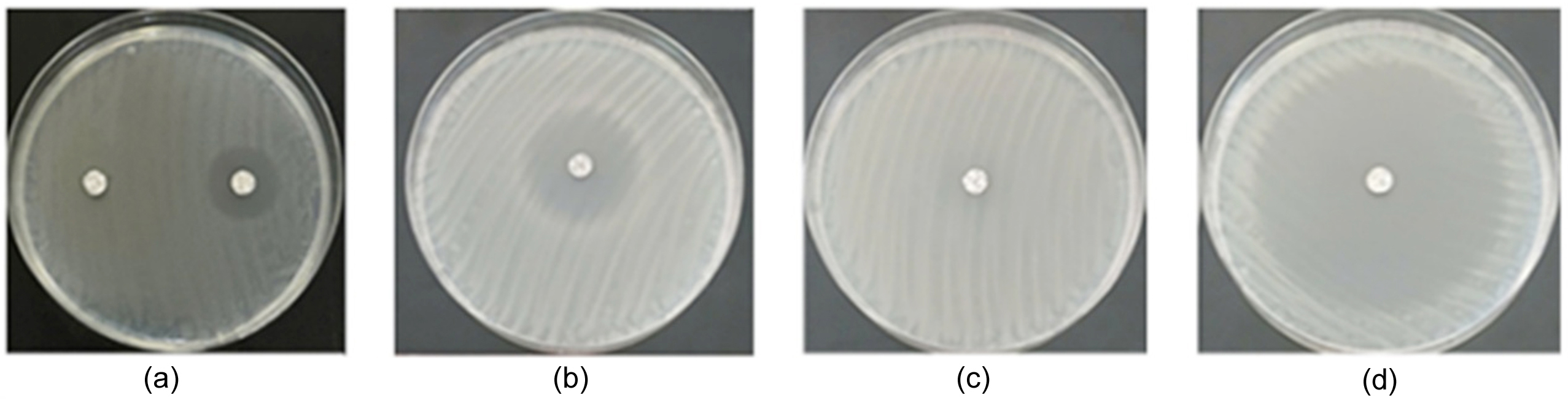
Figure 3. Inhibition zones of antibiotics against Cutibacterium acnes isolates obtained from moderate–severe acne patients
Note: (a) left – Clindamycin, right – TMP-SMX; (b) Doxycycline; (c) Tetracycline; (d) Azithromycin
The present findings indicate that C. acnes isolates from patients with moderate–severe acne remain highly susceptible to TMP-SMX, doxycycline, and tetracycline, suggesting that these antibiotics continue to be effective options for bacterial control. In contrast, the considerable resistance observed to clindamycin and azithromycin reflects a declining efficacy of antibiotics that have been widely and frequently used in acne treatment.1,7 The presence of resistant C. acnes strains in isolates derived from both diseased and healthy populations raises concerns regarding the accumulation and dissemination of antimicrobial resistance within the community and underscores the importance of prudent antibiotic use and strengthened resistance surveillance in clinical practice.1,5
Optimization of sequencing data
All four samples exhibited high sequencing quality, and data cleaning using fastQ effectively improved read quality (Table 4). Prior to filtering, %Q30 values exceeded 91% for all samples, with three samples (Hn4, Hn13, and Mn1) showing values above 93%, indicating reliable raw data. After adapter trimming and removal of low-quality reads, %Q30 increased across all samples, confirming the efficiency of the cleaning process. Approximately 3% of reads were removed, suggesting low background noise and preservation of most high-quality reads for downstream analyses.
Table 4. Sequencing quality metrics before and after data cleaning
| Sample | No. of reads | Total bases (bp) | %GC | %Q30 |
|---|---|---|---|---|
| Before quality filtering | ||||
| Hn4 | 2,818,280 | 422,742,000 | 60.09 | 94.47 |
| Hn13 | 3,674,966 | 551,244,900 | 60.06 | 93.71 |
| Mn1 | 8,140,776 | 1,221,116,000 | 60.17 | 95.41 |
| Sn3 | 3,578,368 | 536,755,200 | 59.54 | 91.75 |
| After quality filtering | ||||
| Hn4 | 2,743,442 | 410,671,862 | 60.17 | 95.23 |
| Hn13 | 3,578,334 | 535,627,374 | 60.13 | 94.48 |
| Mn1 | 7,924,356 | 1,184,617,000 | 60.23 | 96.18 |
| Sn3 | 3,474,936 | 519,854,798 | 59.6 | 92.59 |
Note: %Q30 represents the percentage of bases with a Phred quality score ≥30
GC content (%GC) remained highly stable before and after filtering, with minimal variation (0.05%-0.08%), indicating the absence of nucleotide composition bias. The similar %GC values across samples (59.5%-60.2%) are consistent with the genomic characteristics typically reported for C. acnes and may reflect overall genomic similarity among the analyzed isolates.
Genome assembly
Key statistics describing the de novo assembly quality of four C. acnes genomes (Hn4, Hn13, Mn1, and Sn3) are summarized in Table 5. In terms of fragmentation, Mn1 (13 contigs) and Hn13 (14 contigs) exhibited the lowest contig numbers, indicating the most complete assemblies; notably, all contigs in Hn13 were longer than 1,000 bp. Assemblies of Hn4 (24 contigs) and Sn3 (19 contigs) showed slightly higher fragmentation but remained acceptable.
Table 5. De novo genome assembly statistics
Metric |
Hn4 |
Hn13 |
Mn1 |
Sn3 |
|---|---|---|---|---|
Number of contigs |
24 |
14 |
13 |
19 |
Number of contigs >1,000 bp |
17 |
14 |
12 |
17 |
%GC |
60.04 |
60.06 |
60.17 |
60.1 |
Total contig length (bp)* |
2,541,425 |
2,470,568 |
2,483,967 |
2,516,009 |
Longest contig length (bp) |
572,461 |
683,404 |
873,047 |
722,801 |
N50 (bp) |
323,417 |
288,109 |
717,330 |
216,077 |
L50 |
3 |
3 |
2 |
3 |
Note: *Calculated based on contigs ≥1,000 bp
The estimated genome sizes were highly consistent across isolates, ranging from approximately 2.47 Mb to 2.54 Mb, with stable GC contents (60.04%-60.17%), consistent with raw sequencing data and indicating the absence of assembly-induced nucleotide bias. Regarding continuity, Mn1 showed superior performance with an N50 of 717,330 bp and an L50 of 2 and also contained the longest contig (873,047 bp). Hn4 and Hn13 demonstrated good continuity, whereas Sn3 had the lowest N50 (216,077 bp). Overall, all four genomes were assembled with high quality and were suitable for downstream functional annotation, with Mn1 representing the highest-quality assembly.
Multilocus sequence typing based on whole-genome data
MLST analysis using the pacnes_3 scheme (eight loci: aroE, atpD, gmk, guaA, lepA, sodA, tly, and CAMP2) was conducted on four C. acnes genomes (Hn4, Hn13, Mn1, and Sn3). All isolates shared a common genetic background but differed in sequence types (STs). Hn4 was assigned to ST53 and Sn3 to ST1, whereas Hn13 and Mn1 could not be assigned to existing STs, suggesting potential novel STs or variants closely related to known lineages (Table 6).
Table 6. MLST-based sequence typing results of four Cutibacterium acnes genomes
Isolate |
MLST scheme |
Sequence type (ST) |
Allele profile |
|---|---|---|---|
Hn4 |
pacnes_3 |
53 |
aroE(1), atpD(1), gmk(9), guaA(4), lepA(1), sodA(4), tly(8), CAMP2(6) |
Hn13 |
pacnes_3 |
– |
aroE(1), atpD(~1), gmk(1), guaA(3), lepA(1), sodA(1), tly(1), CAMP2(1) |
Mn1 |
pacnes_3 |
– |
aroE(1), atpD(1), gmk(1), guaA(5), lepA(1), sodA(~4), tly(8), CAMP2(2) |
Sn3 |
pacnes_3 |
1 |
aroE(1), atpD(1), gmk(1), guaA(3), lepA(1), sodA(1), tly(1), CAMP2(1) |
Notes: The MLST analysis was performed using the pacnes_3 scheme comprising eight loci (aroE, atpD, gmk, guaA, lepA, sodA, tly, and CAMP2); “–” indicates that the sequence type could not be assigned based on the current MLST database; Approximate allele matches are indicated by “~”
All isolates shared aroE(1) and lepA(1) alleles, indicating strong conservation of housekeeping genes, while variability was mainly observed at gmk, guaA, sodA, tly, and CAMP2. Sn3 (ST1) displayed a typical phylotype IA1 allele profile, which represents a lineage commonly associated with acne lesions, heightened inflammatory responses, and increased virulence potential,19,20 and is frequently reported in severe acne with increased virulence or antibiotic resistance potential.9 In contrast, ST53 (Hn4) is commonly associated with phylotypes IB/IA2 and has been linked to healthy skin or opportunistic infections.21,22
The inability to assign STs to Hn13 and Mn1 is consistent with the high discriminatory power of MLST and the frequent emergence of new allelic profiles during lineage diversification.23 Mn1 exhibited a mosaic allele pattern combining features of ST1 and ST53, suggesting possible allelic diversification within related C. acnes lineages rather than assignment to a single established ST.24 Moreover, diversity at the tly and CAMP2 loci, genes implicated in virulence, immune modulation, and biofilm formation, suggests potential phenotypic differences among C. acnes lineages.25,26 The coexistence of ST1, ST53, and putative novel STs reflects a complex population structure that may influence inflammatory outcomes on human skin.20
Basic functional annotation
Structural genome annotation of four C. acnes isolates (Hn4, Hn13, Mn1, and Sn3) revealed a high level of conservation in core genomic features (Table 7). The number of predicted coding sequences ranged from 2,289 to 2,354, while tRNA gene counts were highly conserved (46-47 genes). In contrast, rRNA gene copy numbers varied more substantially, ranging from 3-6 among the isolates. Overall, the predicted proteome size and translational machinery were largely stable across the isolates, consistent with the conserved genomic organization commonly reported for C. acnes, whereas minor differences in CDS and rRNA counts suggest strain-level variation.
Table 7: Overview of genomic components of four Cutibacterium acnes isolates
Sequence type |
Hn4 |
Hn13 |
Mn1 |
Sn3 |
|---|---|---|---|---|
CDS (Coding sequences) |
2,354 |
2,289 |
2,304 |
2,343 |
tRNA |
47 |
46 |
46 |
46 |
rRNA |
3 |
3 |
4 |
6 |
Circular genome maps generated using Proksee demonstrated highly similar genome organization across the four isolates, characterized by dense CDS distribution and interspersed tRNA/rRNA regions, consistent with the annotation statistics (Figure 4).
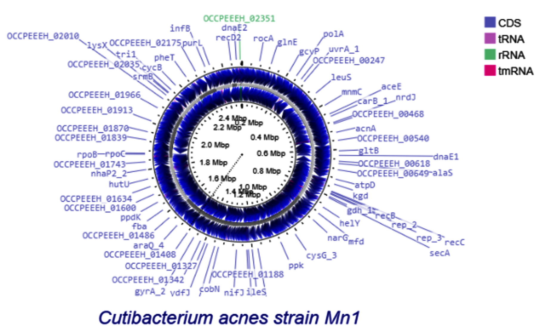
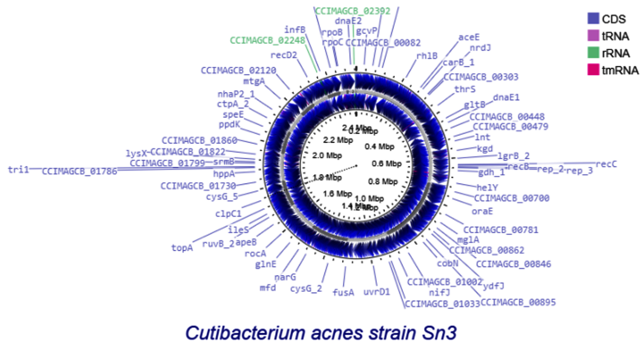
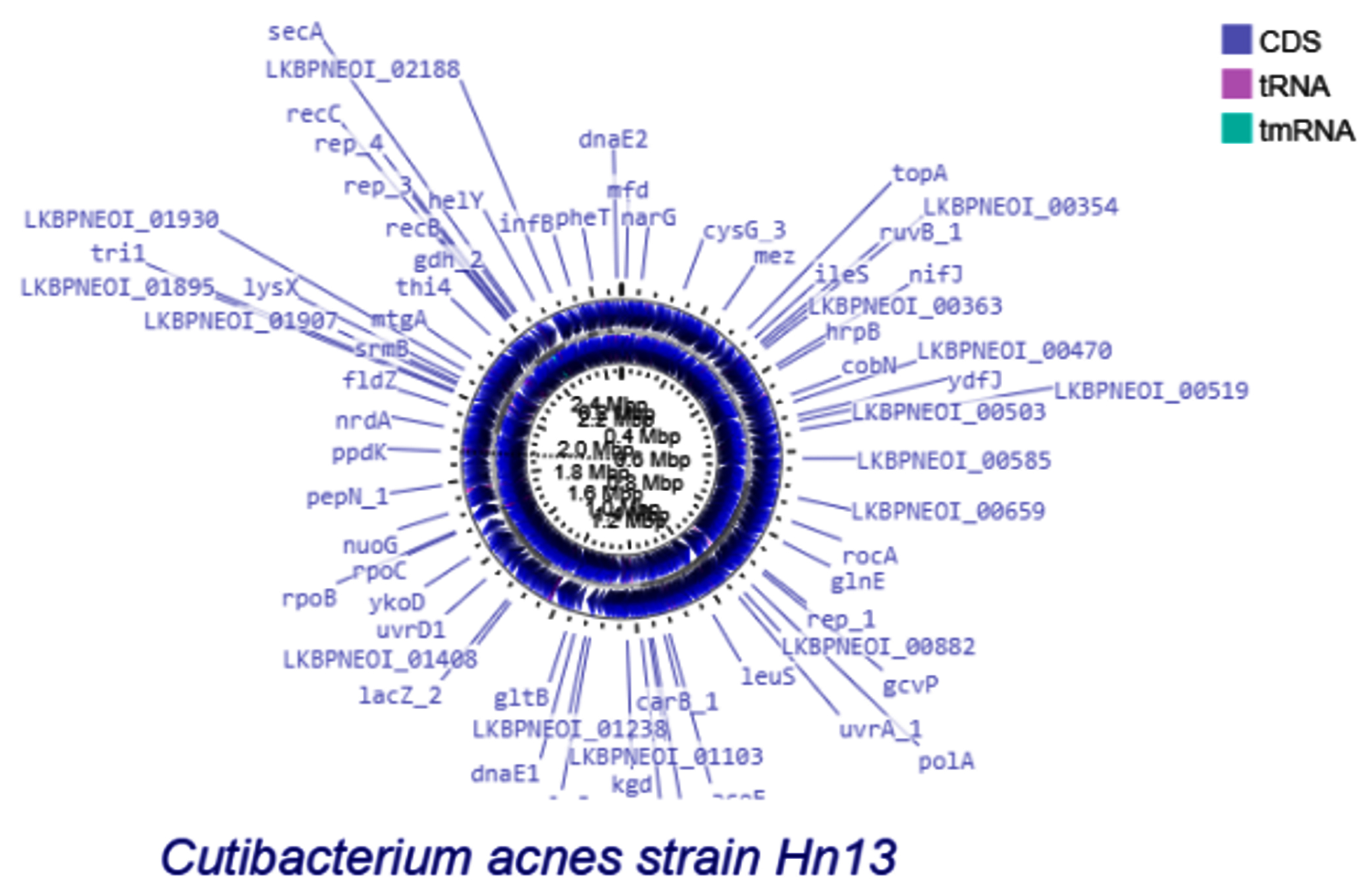
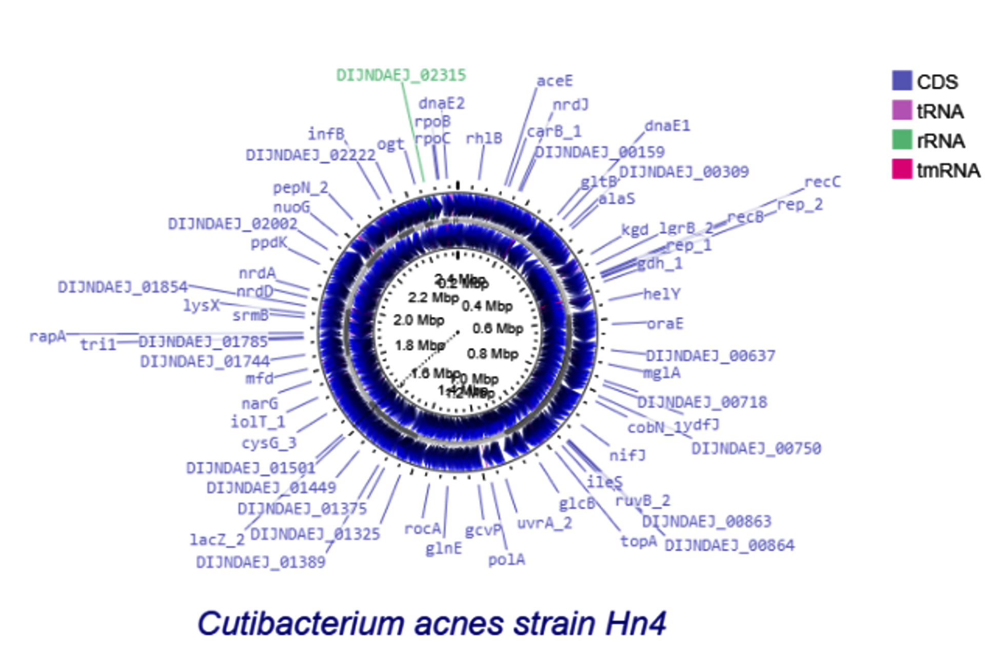
Figure 4. Circular genome maps of four Cutibacterium acnes isolates
Notes: Circular genome maps of Cutibacterium acnes strains Hn4, Hn13, Mn1, and Sn3. Colored rings represent coding sequences (CDS), tRNA, rRNA, and tmRNA genes. Gene annotations displayed on the outermost ring indicate the distribution of functional coding regions across the genomes of each strain
Annotation of virulence and antimicrobial resistance genes
Genome-wide annotation of four C. acnes isolates was performed using the ABRicate pipeline on the Galaxy platform against the CARD, NCBI AMR, and VFDB databases. Using minimum thresholds of 80% nucleotide identity and 80% reference sequence coverage, no antimicrobial resistance or virulence-associated genes were detected in the analyzed genomes. In contrast, analysis with AMRFinderPlus identified a single antimicrobial resistance gene, erm(51), exclusively in the Mn1 genome (Table 8). The erm(51) gene was located on a chromosomal contig and showed complete sequence coverage with high similarity to the reference sequence, suggesting the presence of an intact resistance-associated gene. No additional resistance genes or resistance-associated mutations were identified in the remaining genomes. The presence of erm(51) in Mn1 was consistent with the antimicrobial susceptibility profile observed for this isolate.
Table 8. Annotation results of antimicrobial resistance genes identified in the Mn1 genome
Parameter |
Description |
|---|---|
Contig ID |
12 |
Start position |
763 |
End position |
1551 |
DNA strand |
+ |
Target gene |
erm(51) |
Gene product |
23S rRNA (adenine(2058)-N(6))-methyltransferase Erm(51) |
Antibiotic class |
Lincosamide/macrolide/streptogramin |
Antibiotics affected |
Azithromycin, clarithromycin, clindamycin, erythromycin |
Gene length (bp) |
263 |
Reference coverage (%) |
100 |
Reference identity (%) |
99.62 |
Alignment length (bp) |
263 |
The detection of erm(51) as the sole antimicrobial resistance gene in the genome of C. acnes Mn1, in the absence of other resistance markers such as tet(W)/tet(M) (tetracycline resistance), erm(X) (macrolide–clindamycin resistance), gyrA/parC mutations (fluoroquinolone resistance), or rpoB mutations (rifampicin resistance), suggests a relatively narrow resistance spectrum primarily restricted to the macrolide–lincosamide–streptogramin B (MLSB) group, rather than a broad multidrug-resistant (MDR) phenotype, typically defined as resistance to multiple classes of antimicrobial agents, as reported for some C. acnes or Cutibacterium spp. strains.8
Over the past decade, increasing resistance to macrolides and clindamycin in C. acnes has been widely reported, with marked geographic variability. Epidemiological data from Jordan showed resistance rates of 73% to erythromycin and 59% to clindamycin,6 while recent global analyses indicate that more than 50% of C. acnes isolates are resistant to these antibiotics, exceeding 90% in certain regions.5,6 These trends are consistent with recent reviews emphasizing prolonged macrolide use as a major driver of resistance selection.27
Mechanistically, MLSB resistance in C. acnes is mainly mediated by point mutations in 23S rRNA or acquisition of erm methylase genes via plasmids or other mobile genetic elements.9 Aoki et al. described the transferable plasmid pTZC1 carrying erm(50) and tet(W), highlighting the role of plasmid-borne erm genes in the emergence of MDR strains. In this context, erm(51), a member of the Erm 23S rRNA methyltransferase family, has been characterized as a functional MLSB resistance determinant with potential for horizontal transfer.27
Notably, the Mn1 genome lacked resistance markers for tetracyclines or rifampicin that have been reported in MDR C. acnes strains,9,28 suggesting that macrolide–lincosamide resistance may currently be evolving independently without progression to a broad MDR phenotype. Nevertheless, the presence of an erm gene remains epidemiologically relevant, as it may facilitate further resistance accumulation under sustained antibiotic pressure.8 Clinically, these findings reinforce the importance of antimicrobial stewardship by limiting unnecessary macrolide and clindamycin exposure and prioritizing tetracycline-class antibiotics or non-antibiotic therapeutic approaches in accordance with current acne management guidelines.1
This study provides an integrated assessment of antibiotic susceptibility patterns and genomic characteristics of C. acnes isolates obtained from healthy individuals and patients with different severities of acne vulgaris. Phenotypic analysis showed that most isolates remained highly susceptible to tetracycline-class antibiotics and trimethoprim, whereas resistance to macrolides and clindamycin was detected, particularly among moderate–severe acne isolates. Whole-genome sequencing combined with MLST revealed notable genetic diversity, including both established sequence types and putative novel variants. The identification of a single resistance-associated gene, erm(51), consistent with the observed resistance phenotype, suggests that the analyzed C. acnes population has not yet developed widespread multidrug resistance. However, interpretation of these findings should consider the relatively small number of sequenced isolates included in this study. Overall, these results highlight the value of integrating phenotypic susceptibility testing with genomic analysis to support ongoing resistance surveillance programs and evidence-based antibiotic stewardship in acne management.
ACKNOWLEDGMENTS
The authors sincerely thank the Institute of Food and Biotechnology, Can Tho University, Vietnam, for their kind support and for providing access to their laboratories. The authors also thank Can Tho Hospital of Dermato-Venereology for allowing sample collection.
CONFLICT OF INTEREST
The authors declare that there is no conflict of interest.
AUTHORS’ CONTRIBUTION
XMT and DNT conceptualized the study. DNT developed the methodology. XMT performed the software-related work. XMT and LTTN conducted the formal analysis. LTTN and DNT validated the data. LDHB and DNT carried out the investigation. XMT, PXH, and TNN performed data curation. PXH, LTTN, LDHB, and TNN contributed to data visualization. LTTN, XMT, LDHB, and TNN interpreted the data. PXH and DNT supervised the study. XMT prepared the original draft. XMT, PXH, and DNT wrote, reviewed and revised the manuscript. All authors read and approved the final manuscript for publication.
FUNDING
None.
DATA AVAILABILITY
The genome assemblies and associated metadata generated in this study have been submitted to the NCBI database under BioProject accession number PRJNA1466956. The corresponding BioSample accession numbers are SAMN60158190 (Hn4), SAMN60158191 (Hn13), SAMN60158192 (Mn1), and SAMN60158193 (Sn3). The additional datasets generated and/or analysed during the current study are included in the article, and the rest can be obtained from the corresponding author on reasonable request.
ETHICS STATEMENT
This study was approved by the Ethics Committee of Biomedical Research, Can Tho University of Medicine and Pharmacy (Ref. No. 24.004/PCT-H׀׀׀).
INFORMED CONSENT
Written informed consent was obtained from the participants before enrolling in the study.
- Dessinioti C, Katsambas A. Antibiotics and antimicrobial resistance in acne: Epidemiological trends and clinical practice considerations. Yale J Biol Med. 2022;95(4):429-443
- Legiawati L, Halim PA, Fitriani M, Hikmahrachim HG, Lim HW. Microbiomes in acne vulgaris and their susceptibility to antibiotics in Indonesia: A systematic review and meta-analysis. Antibiotics. 2023;12(1):145.
Crossref - Boyanova L, Dimitrov G, Raykova V, Patrikov K, Gergova R, Markovska R. Cutibacterium acnes phylotyping and antibiotic resistance to six antibiotics: A Bulgarian study. Microorganisms. 2025;13(9):2185.
Crossref - Yu T, Xu X, Liu Y, et al. Multi-omics signatures reveal genomic and functional heterogeneity of Cutibacterium acnes in normal and diseased skin. Cell Host & Microbe. 2024;32(7):1129-1146.e8.
Crossref - Beig M, Shirazi O, Ebrahimi E, Banadkouki AZ, Golab N, Sholeh M. Prevalence of antibiotic-resistant Cutibacterium acnes (formerly Propionibacterium acnes) isolates, a systematic review and meta-analysis. J Glob Antimicrob Resist. 2024;39:82-91.Crossref
- Alkhawaja E, Hammadi S, Abdelmalek M, Mahasneh N, Alkhawaja B, Abdelmalek SM. Antibiotic resistant Cutibacterium acnes among acne patients in Jordan: a cross-sectional study. BMC Dermatol. 2020;20(1):17.
Crossref - Zhu C, Wei B, Li Y, Wang C. Antibiotic resistance rates in Cutibacterium acnes isolated from patients with acne vulgaris: a systematic review and meta-analysis. Front Microbiol. 2025;16:1565111.
Crossref - Aoki S, Nakase K, Nakaminami H, Wajima T, Hayashi N, Noguchi N. Transferable multidrug-resistance plasmid carrying a novel macrolide-clindamycin resistance gene, erm(50), in Cutibacterium acnes. Antimicrob Agents Chemother. 2020;64(3):e01810-19.
Crossref - Beirne C, McCann E, McDowell A, Miliotis G. Genetic determinants of antimicrobial resistance in three multi-drug resistant strains of Cutibacterium acnes isolated from patients with acne: a predictive in silico study. Access Microbiol. 2022;4(8):acmi000404.
Crossref - Andrews S. FastQC: A quality control tool for high throughput sequence data. Babraham Bioinformatics. 2010. https://www.bioinformatics.babraham.ac.uk/projects/fastqc/
- Chen S, Zhou Y, Chen Y, Gu J. fastp: an ultra-fast all-in-one FASTQ preprocessor. Bioinformatics. 2018;34(17):i884-i890.
Crossref - Wick RR, Judd LM, Gorrie CL, Holt KE. Unicycler: Resolving bacterial genome assemblies from short and long sequencing reads. PLoS Comput Biol. 2017;13(6):e1005595.
Crossref - Gurevich A, Saveliev V, Vyahhi N, Tesler G. QUAST: quality assessment tool for genome assemblies. Bioinformatics. 2013;29(8):1072-5.
Crossref - Seemann T. Prokka: rapid prokaryotic genome annotation. Bioinformatics. 2014;30(14):2068-9.
Crossref - Feldgarden M, Brover V, Gonzalez-Escalona N, et al. AMRFinderPlus and the Reference Gene Catalog facilitate examination of the genomic links among antimicrobial resistance, stress response, and virulence. Sci Rep. 2021;11(1):12728.
Crossref - Dagnelie M, Corvec S, Saint-Jean M, et al. Decrease in diversity of Propionibacterium acnes phylotypes in patients with severe acne on the back. Acta Dermo Venereol. 2018;98(2):262-267.
Crossref - Thiboutot D, Gollnick H, Bettoli V, et al. New insights into the management of acne: an update from the Global Alliance to Improve Outcomes in Acne group. J Am Acad Dermatol. 2009;60(5 Suppl):S1-50.
Crossref - Walsh TR, Efthimiou J, Dreno B. Systematic review of antibiotic resistance in acne: an increasing topical and oral threat. Lancet Infect Dis. Mar 2016;16(3):e23-33.
Crossref - Dreno B, Pecastaings S, Corvec S, Veraldi S, Khammari A, Roques C. Cutibacterium acnes (Propionibacterium acnes) and acne vulgaris: a brief look at the latest updates. J Eur Acad Dermatol Venereol. 2018;32(S2):5-14.
Crossref - Dagnelie MA, Corvec S, Saint-Jean M, Nguyen JM, Khammari A, Dreno B. Cutibacterium acnes phylotypes diversity loss: a trigger for skin inflammatory process. J Eur Acad Dermatol Venereol. 2019;33(12):2340-2348.
Crossref - Lomholt HB, Scholz CFP, Bruggemann H, Tettelin H, Kilian M. A comparative study of Cutibacterium (Propionibacterium) acnes clones from acne patients and healthy controls. Anaerobe. 2017;47:57-63.
Crossref - Both A, Huang J, Hentschke M, et al. Genomics of invasive Cutibacterium acnes isolates from deep-seated infections. Microbiol Spectr. 2023;11(2):e04740-22.
Crossref - Dagnelie MA, Khammari A, Dreno B, Corvec S. Cutibacterium acnes molecular typing: time to standardize the method. Clin Microbiol Infect. 2018;24(11):1149-1155.
Crossref - Sripath K, Bangperng J, Chumnanpeun P, Teerasak EK. Unique genetic diversity of Propionibacterium acnes isolated from human facial skins in Thailand revealed by the multi-locus sequence typing scheme. BMC Microbiol. 2025;25(1):643.
Crossref - Mayslich C, Grange PA, Dupin N. Cutibacterium acnes as an opportunistic pathogen: An update of its virulence-associated factors. Microorganisms. 2021;9(2):303.
Crossref - Chudzik A, Migdal P, Pasciak M. Different Cutibacterium acnes phylotypes release distinct extracellular vesicles. Int J Mol Sci. 2022;23(10):5797.
Crossref - Lund D, Kieffer N, Parras-Molto M, et al. Large-scale characterization of the macrolide resistome reveals high diversity and several new pathogen-associated genes. Microb Genom. 2022;8(1):000770.
Crossref - Zhang N, Lu Z, Ma Y. Draft genome sequences of three multidrug-resistant Cutibacterium (formerly Propionibacterium) acnes strains isolated from acne patients, China. J Glob Antimicrob Resist. 2017;11:114-115.
Crossref
© The Author(s) 2026. Open Access. This article is distributed under the terms of the Creative Commons Attribution 4.0 International License which permits unrestricted use, sharing, distribution, and reproduction in any medium, provided you give appropriate credit to the original author(s) and the source, provide a link to the Creative Commons license, and indicate if changes were made.