


ISSN: 0973-7510
E-ISSN: 2581-690X
Chicken drumstick mushroom (Coprinus comatus (O.F. Mull) Pers.) is a medicinal mushroom containing various mycochemical compounds, such as flavonoids, terpenoids, vitamins C and E, quercetin, and rutin. These bioactive compounds have pharmacological activities, including anti-nephrotoxic, decreasing uric acid (UA) level, anti-inflammatory, and antioxidant, capable of reducing free radicals. Therefore, this study aims to investigate anti-inflammatory and antioxidant activity of C. comatus ethanol extract on carrageenan-induced inflammatory rats. A completely randomized design (CRD) was used, and the test rats were divided into 6 groups. These included 1 healthy group (HG), 5 groups induced by 0.5 mL of 1% carrageenan, in which 3 were administered extract at doses of 250 (TA1), 500 (TA2), and 750 mg/kg BW (TA3), 1 given sodium diclofenac (PG), and 1 served as a negative control (NG). During the procedure, extract administration was carried out for 14 days. The results showed that 500 mg/kg BW dose reduced IL-1β (12.38%), MDA (20.72%), and UA (16.67%), as well as increased superoxide dismutase (SOD) (12.79%). Meanwhile, 250 mg dose increased total protein (TP) (5.33 g/dL) and globulin (GLB) (1.58 g/dL), with 750 mg dose also elevating albumin (ALB) (3.89 g/dL). Increased enzymatic antioxidants can prevent oxidative stress and peroxidation reactions. Antioxidant effects of C. comatus are expected to decrease the release of inflammatory mediators, leading to the prevention of cell and tissue damage caused by oxidative stress.
Anti-inflammatory, Coprinus Comatus, Extract, Pro-inflammatory Cytokine, In Vivo
Mushroom metabolite compounds are known to possess a wide range of biological activities, including anti-inflammatory, anti-tumor, and anti-oxidative stress properties. These activities can either enhance or decrease the body’s immunity under specific circumstances.1 Coprinus comatus is a medicinal and edible mushroom,2 also known as chicken drumstick or the shaggy ink cap mushroom. In addition, it has anti-inflammatory, nephroprotective, and antioxidant properties that reduce reactive oxygen species (ROS). Several studies have shown that ROS often cause oxidative stress, lipid peroxidation reactions, cell death and damage.3 In the current study, carrageenan was used as an induction agent, and its injection had been described as a biphasic, age-dependent event in which various mediators act sequentially to release and produce inflammatory response. Local neutrophil activation and infiltration also contribute to this response by generating free radicals derived from superoxide anion (O2-) and hydroxyl radicals (OH–), among other mediators.4 Over the years, there have been limited studies on the acute anti-inflammatory effects due to carrageenan induction by administering C. comatus extract. Consequently, this study focused on the role of C. comatus in reducing pro-inflammatory cytokines and increasing enzymatic antioxidant levels in carrageenan-induced rat models. The aim of increasing antioxidant levels is important because, during inflammation, the formation of free radicals also increases. To minimize the risk of oxidative stress, exogenous antioxidants are needed, such as flavonoids contained in C. comatus.
In addition to an increase in ROS, inflammation causes a rise in pro-inflammatory cytokines. Pro-inflammatory cytokines such as interleukin (IL-1β, IL-6, IL-8, & IL-5) and prostaglandin E2 (PGE2) enzyme are produced and released by proliferative FLS (fibroblast-like synoviocytes), which play essential roles in tissue damage caused by inflammation. Immune responses induced by pro-inflammatory mediators, such as IL-6, occur in in-vivo animal models. Interleukin-1 (IL-1β) and tumor necrosis factor (TNF-α) also activate and stimulate transcription factors to produce IL-6, while enhanced IL-1β exacerbates inflammatory conditions.5 Carrageenan induction in experimental animals has been introduced to investigate acute inflammatory effects. It is characterized by inflammatory signs, including edema, which results from intraplantar injection, leading to hyperalgesia and erythema.6 Carrageenan induction has been widely used by various studies in evaluating the effects of certain drug administration on inflammatory effects, such as an increase in free radicals, pro-inflammatory mediators, and a decrease in endogenous antioxidants.7 Previous studies have shown that administration of C. comatus nanogel to experimental animals with inflammation induced by Complete Freund’s Adjuvant (CFA) reduces inflammatory mediators, including TNF-α, interleukin [IL]-6, and IL-1β. In silico studies also indicate that C. comatus compounds can inhibit the activity of enzymes iNOS and COX-2 with binding affinities of -7.9 and -8.8 kcal/mol, respectively.8 This result was demonstrated in an in vivo experiment where C. comatus nanogel inhibited NOS release by 23.94% with a dose of 750 mg/kg BW. Despite the potential, evaluation of the final lipid peroxidation product (MDA) has not been previously studied.9 Therefore, this study investigated the levels of cytokines (pro-inflammatory), endogenous antioxidants, and lipid peroxidation product, namely MDA.
Materials, chemicals, and tools
The materials used included C. comatus fruiting bodies that were cultivated and obtained from Asa Agro Corporation CV. (AAC), Cianjur City, West Java Province, Indonesia. A total of 36 male Wistar rats, such as rats for the drop estimation group (obtained from UD. Wistar), Yogyakarta Province, uric acid (UA) assay kit (No. MAK077-1KT) were from Sigma-Aldrich @Merck. Rat ELISA Kit IL-1β, malondialdehyde (MDA) kit (Catalogue ID E0156Ra), while superoxide dismutase (SOD) Kit (Catalogue ID E0168Ra) was from BT-Laboratory, Shanghai, China. Colorimetric Assay Kits of BCA/TP Protein (Catalogue ID E-BC-K117-S) and albumin (ALB) (for BGM (Bromocresol Green Method) with Catalogue ID: E-BC-K057-S) were obtained from Elabscience Biotechnology Inc, United States (Elabscience®), feed pellets, and dried husks.
The chemicals used were 0.9% physiological NaCl, Alcohol 70%, and 0.012 g/200 g BW sodium diclofenac (Dexa Medica PT.), 5000 mL ethanol (PA), 500 µL carrageenan (Catalogue No. CAS 11114-20-8), and other materials such as ether and distilled water. Furthermore, the tools used were a 50 x 30 x 20 cm rearing cage, a vacuum rotary evaporator (RE-2010 Henan Lanphan Industry Co. Ltd.), an ELISA reader (BioTek 800 TS), a water bath (Yamato Scientific), a microscope (Olympus CX-23), a haemocytometer, a calibrated DFAL-001 plethysmometer, and a hematocrit capillary.
Mushroom extraction
Fruiting body of C. comatus was cut into ± 2-4 cm sizes and dried in an oven at 55 °C. In total, 200 g of the dried mushroom pieces were blended. The powder (simplicial) was transferred into a glass beaker, and ethanol pro-analysis was added at a ratio of 1:3. This mixture was stood for 24 hours, and mushroom powder was macerated again with a ratio of 3:1 and 1:1. Total macerate was evaporated on a rotary vacuum evaporator at 77 °C temperature.
Qualitative and quantitative GC-MS identification of C. comatus mycochemical compounds
Mycochemical compounds of ethanol extract of C. comatus mushroom were identified qualitatively using reagents to characterize compounds from flavonoid, alkaloid, terpenoid, and saponin groups. The reagents used for identification and flavonoid tests were magnesium (Mg) powder, amyl alcohol reagent, and hydrochloric acid (HCl). A positive indication of flavonoids was a color change to orange, reddish-yellow, or reddish-orange. Alkaloid tests were carried out using Mayer’s reagent, Wagner’s reagent, and Dragendorff’s reagent. A positive sign of alkaloids was a dark brown color change, or blackish brown. Terpenoid testing was performed using acetic anhydride (CH3COOH) and concentrated sulfuric acid (H2SO4). A positive indication of terpenoids was a color change to blackish purple or reddish orange. Saponin testing required boiling the sample extract with distilled water, adding methanol (CH3OH), and then shaking gently. This process showed a positive sign of stable foam formation for 5-10 minutes.
Mycochemical compound identification of C. comatus was carried out using GC-MS PerkinElmer (Gas Chromatography-Mass Spectrometry 2400). The detector used was an Agilent 5973 inert MSD (70 eV direct inlet). The amount of sample used was approximately 2 µL, which was injected into a J&W Scientific HP-5MS capillary column. Acetonitrile was used as the mobile phase. The identification results were then compared with the compound data contained in the database of Pubchem NCBI from National Institutes of Health’s (NIH) and National Library of Medicine (NLM), United States (www.pubchem.ncbi.nlm.nih.gov), Chemspider from Royal Society of Chemistry (www.chemspider.com), ChEBI (The Chemical Entities of Biological Interest) Part of EMBL (European Molecular Biology Lab.), United Kingdom (https://www.ebi.ac.uk/chebi/init.do), and NIST Chemistry WebBook The NIST (National Institute of Standards and Technology) United States (https://webbook.nist.gov/chemistry/).
Study method and animal treatment
This study using a control group and posttest-only method with a completely randomized design (CRD). Based on simple random sampling method, 36 rat models (divided into 6 groups randomly), healthy control group (HG = no treatment/healthy rat), negative control (NG = 0.5 mL of 1% carrageenan induction, received no treatment of extract or sodium diclofenac), and positive control (PG = induction of 0.5 mL carrageenan and administration treatment of 1 mL sodium diclofenac), as well as treatment group of extract administration, namely treatment 1 (TA1= 250 mg/kg dose), 2 (TA2 = 500 mg/kg BW dose), and 3 (TA3 = 750 mg/kg BW dose). Ethanol extract and sodium diclofenac were administered orally for 15 days, starting 6 hours after carrageenan injection.
Induction of carrageenan
Rats were first anesthetized with ketamine-xylazine 10 mg Kg/BW (KX) and placed on a surgical board, and then induced intra-plantar with up to 500 µL of 1% carrageenan in physiological NaCl solution with 0.9% concentration. Inflammatory state was detected immediately on the first day, specifically 6 hours after induction.
Ethical approval
The health study ethics committee approved the experimental method and animal treatment ethically with number 515/IV/HREC/2021 (Dr. Moewardi Regional General Hospital (RSUD), Surakarta City, Central Java, Indonesia). To prevent dying, rats were placed in a glass beaker filled with ketamine-xylazine (KX) 10 mg kg/BW, then closed and left to stand. Their necks were decapitated a few moments after appearing weak. The termination procedure was carried out by minimizing or even eliminating experimental animals’ suffering through Institutional Animal Care and Use Committee (IACUC) procedure and recommendations.
Blood sample collection
Initially, rat models were fasted for 10 hours, then blood was collected on day 15 from the orbital vein with a hematocrit capillary pipette. Blood was centrifugated at 8000 rpm (revolutions per minute) to obtain plasma which was the sample used for measurement of interleukin-1β (IL-1β), UA levels, total protein (TP) levels, SOD, ALB, globulin (GLB), and Malondialdehyde (MDA) levels, while leucocyte distribution was counted using fresh blood.
Measurement of main parameters
The levels of endogenous antioxidants, as well as TP, ALB, and GLB, were measured using Shimadzu 1800 (Spectrophotometer UV-Vis) at a wavelength of 546 nm, while SOD was measured at 340 nm, respectively. UA levels were calculated using UA assay kit MAK077-1KT (Sigma-Aldrich @Merck) and UV-Vis Spectrophotometer at 570 nm, following the manufacturer’s instructions. The IL-1β production was evaluated with an ELISA kit (BT Laboratory), and then the optical density was measured at 450-500 nm in 10 to 15 minutes in a microplate reader (ELISA reader), following the manufacturer’s instructions. Leucocyte distribution of neutrophil, eosinophil, lymphocyte, and monocyte was counted and observed using a hemocytometer and microscope at 400x magnification.
Measurement of oxidative stress parameters
Malondialdehyde (MDA) levels were used to measure the activity of C. comatus extract as anti-oxidative stress agent. MDA was measured using a Shimadzu 1800 (Spectrophotometer UV-Vis) at 532 nm.
Statistical data analysis
All parameters’ data were presented (mean ± standard error). One-way analysis of variance (ANOVA), Duncan’s multiple range tests (DMRT), and correlation tests were performed using SPSS v.28.0 to compare the main parameters, and the P-values obtained were <0.05, indicating statistical significance.
The results of identifying C. comatus compounds were carried out qualitatively and quantitatively to determine the type of mycochemical compounds in the extract. Identifying these compounds is very important because various compounds had specific anti-inflammatory and antioxidant activities in preventing cell lipid peroxidation reactions in inflammatory states. Qualitative identification results were presented in Table 1, and GC-MS identification results were presented in Table 2, while the chromatogram of GC-MS was presented in Figure 1.
Table (1): Qualitative identification results of mushrooms
No. |
Mycochemical Compounds |
Testing Reagent |
Colorimetric Results (Color Change) |
Description |
|---|---|---|---|---|
1 |
Flavonoids |
Magnesium, Hydrochloric Acid, Amyl Alcohol |
Dark Red |
+++ |
2 |
Alkaloids |
Mayer Reagent, Dragendroff Reagent |
Dark brown, white/red precipitate formed |
++ |
3 |
Saponins |
Distilled Water (Sterile), Boiling, Methanol |
Stable Foam Formed (5-10 minutes) |
++ |
4 |
Terpenoids |
Hydrochloric Acid, Sulfuric Acid |
Reddish-orange |
++ |
Note: + (moderate level), ++ (medium level), +++ (high level).
Qualitative identification showed that C. comatus extract contained high flavonoids (+++), while alkaloids, saponins, and terpenoids showed medium levels (++)
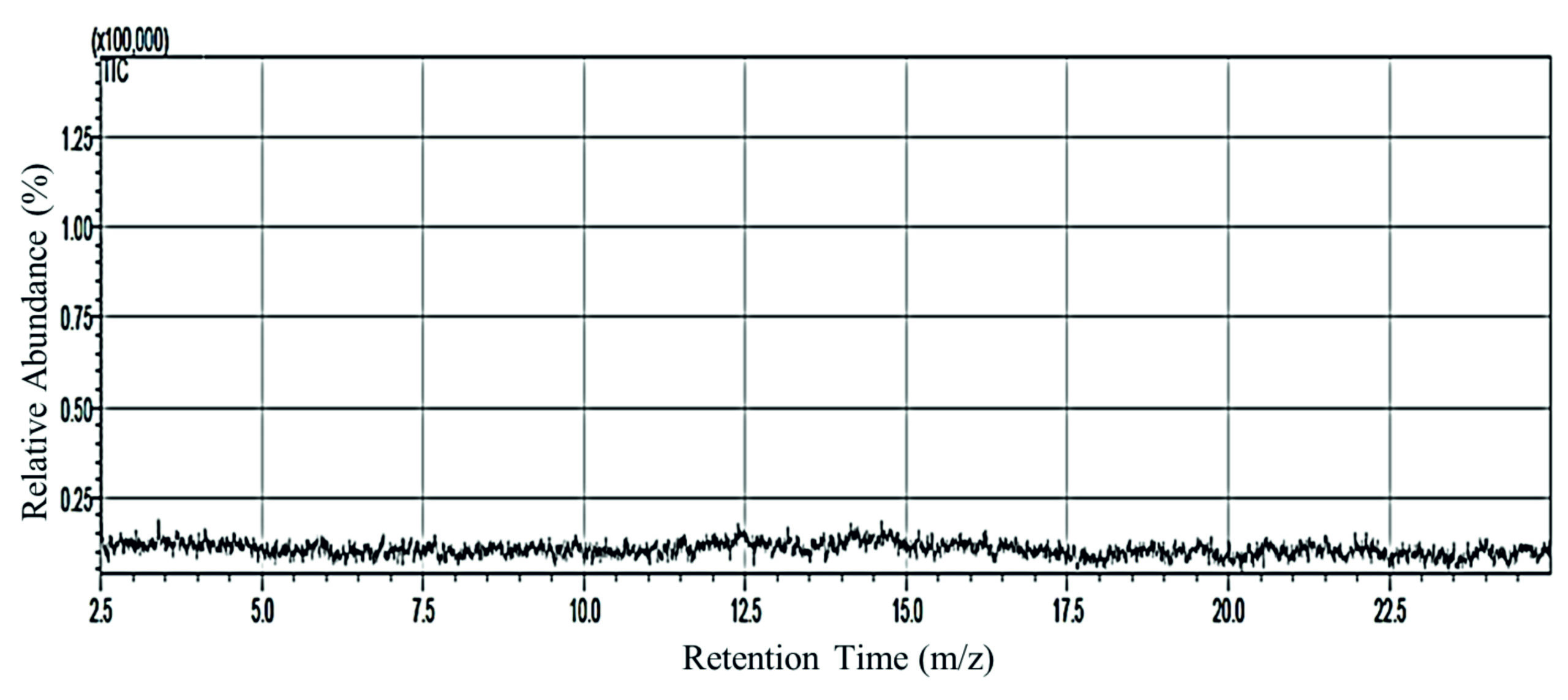
Figure 1. Chromatogram GC-MS of C. comatus extract
The identification results using GC-MS showed a more diverse type of compound. Table 2 showed that there were 14 compounds from GC-MS analysis. Furthermore, the highest molecular weight of C. comatus compound was TUNGSTEN TRIS(CYCLOOCTATETRAENE)BIS, while Thiazole-4-carboxylic acid was the compound with the lowest molecular weight. The % area range was between 0.09 to 0.21, and the retention time range of 14 compounds was 10.370-11.375 (Table 2).
Table (2): Mycochemical identification of C. comatus ethanol extract by GC-MS
GC-MS Compound |
Retention Time |
Compound Formula |
Molecular Weight (g.mol-1) |
% Area |
|---|---|---|---|---|
Tungsten. tris(cyclooctatetraene)bis |
10.37 |
C24H24W2 |
680.14 |
0.13 |
Thiazole-4-carboxylic acid |
10.439 |
C4H3NO2S |
129.14 |
0.12 |
Violerythrin |
11.375 |
C38H44O4 |
568.4 |
0.09 |
1.2.5-Oxadiazol 3 amine 4-[5-[1-(2-bromophenoxy) ethyl] 1.2.4 oxadiazol -3-yl] |
10.51 |
C12H10BrN5O3 |
352.14 |
0.15 |
Triflupromazine |
10.546 |
C18H19F3N2S |
352.4 |
0.13 |
5-Acetoxy-6-bromo-1-phenyl-2-(4-o-tolyl- piperazin-1-ylmethyl)-1h-indole-3-carboxylic acid ethyl ester |
10.615 |
C31H32BrN3O4 |
590.508 |
0.14 |
Dibenzothiophene 5.5-dioxide |
10.649 |
C12H8OS |
200.256 |
0.21 |
Bis(dipropylamido)dichloromethylphosphonate |
10.681 |
C13H29Cl2N2OP |
331.3 |
0.16 |
N-(Cyanomethyl)piperidine |
10.715 |
C8H13N3O |
167.21 |
0.11 |
2-isopropyl-. 1.4-dioxide (CAS) |
10.743 |
C7H14O2 |
130.18 |
0.15 |
Phosphonic acid [3 (acetylamino) propyl]bis(trimethylsilyl) ester (CAS) |
10.78 |
C11H28NO4PSi2 |
325.49 |
0.16 |
Octadecanoic acid, 2-oxo-, methyl ester |
6.185 |
C19H36O3 |
312.487 |
0.11 |
Sericealactone |
10.89 |
C16H20O5 |
292.327 |
0.13 |
2-(2-Methylphenyl)-2-bromo-3-hydroxyindan-1- one-3-carboxilic acid |
10.964 |
C17H13BrO4 |
361.2 |
0.18 |
This study was conducted to assess and evaluate anti-inflammatory activity and antioxidant potential of C. comatus fruiting body extract. The results showed a reduction in IL-1β and UA levels, and endogenous or enzymatic antioxidants such as SOD, TP, and ALB were increased. However, MDA levels as anti-oxidative stress effect decreased according to the following description.
The extract was qualitatively identified, showing the presence of flavonoids, saponins, terpenoids, and alkaloids, and the content with the highest qualitative test was flavonoids (+++). Previous studies showed C. comatus extracts contained flavonoids (+++) and polyphenols (+++),10 and the content of alkaloids (+) and saponins (+).11 The presence of flavonoid and polyphenol compounds could reduce the development of inflammatory reactions by reducing the free radicals formed. Flavonoids acted as electron donors and stabilized free radicals, thereby preventing cell lipid peroxidation.10 Terpenoid compounds (monoterpenoids and triterpenoids) had a role as anti-inflammatory agents.12
Levels of IL-1β
The extract significantly reduced IL-1β (P < 0.05), as presented in Table 1, and the highest levels of IL-1β were found in NC group, and the lowest were in HC. Furthermore, the group TA3 showed the highest decreasing effect of 13.64%. Carrageenan intraplantar induction significantly increased IL-1β in the NG group by more than 16.61% compared to HG (Table 3).
Table (3): Interleukin-1β (IL-1β) levels in carrageenan-induced inflammatory rats
No. |
Experimental group |
IL-1β level (µmol/L) |
% decrease |
|---|---|---|---|
1 |
HC |
11.50 ± 0.53a |
14.24 |
2 |
NG |
13.41 ± 0.63b |
+16.61 |
3 |
PG |
12.36 ± 0.83ab |
7.83 |
4 |
TA1 |
11.64 ± 1.16a |
13.20 |
5 |
TA2 |
11.75 ± 1.16a |
12.38 |
6 |
TA3 |
11.58 ± 0.68a |
13.64 |
Note: The different letters on each column in the table represented the significantly differences (P < 0.05), data were expressed as mean ± SE (n: 36). HG: healthy control; NG: negative control (carrageenan induction only); PG: positive control (administration of sodium diclofenac); TA1: administration of 250 mg C. comatus extract; TA2: administration of 500 mg C. comatus extract; TA3: administration of 750 mg C. comatus extract for 14 days
In this study, anti-inflammatory and antioxidant effects of C. comatus fruiting body ethanol extract were investigated and evaluated. The results showed a reduction in interleukin-1β (IL-1β) and UA levels, as well as an increase in endogenous antioxidant levels and a decrease in MDA. The administration of 250, 500, and 750 mg extract doses and diclofenac sodium significantly decreased IL-1β, UA, and MDA, but increased SOD, TP, ALB, and GLB levels. Furthermore, the extract affected leukocyte distribution, where the number of neutrophils, monocytes, and lymphocytes increased in the treatment (TA1, TA2, TA3) group compared to NC. The results were consistent with previous studies, showing that the triglyceride extract from fermented (TF) mushroom of C. comatus can reduce IL-1β, TNF-α, and IL-17 levels, compared to the saline group (control group) in inflammatory model Wistar rats induced by 1% carrageenan intrapleurally by 0.1 mL.13
Uric acid levels (nephroprotective effect)
UA measurement results were significant (P < 0.05) as carrageenan induction increased UA levels by >8% in NG group compared to HC. Furthermore, the best decrease was generated in TA2 with a 500 mg extract dose administration, as presented in Table 4.
Table (4): UA levels in carrageenan-induced inflammatory rats
No |
Experimental group |
Uric acid level (g/dL) |
% decrease |
|---|---|---|---|
1 |
HC |
4.83 ± 0.26b |
7.47 |
2 |
NG |
5.22 ± 0.36c |
+8.07 |
3 |
PG |
4.52 ± 0.37ab |
13.41 |
4 |
TA1 |
4.49 ± 0.10ab |
13.98 |
5 |
TA2 |
4.35 ± 0.24a |
16.67 |
6 |
TA3 |
4.47 ± 0.22ab |
14.37 |
Note: The different letters on each column in the table represented the significantly differences (P < 0.05), data were expressed as mean ± SE (n: 36). HG: healthy control; NG: negative control (carrageenan induction only); PG: positive control (administration of sodium diclofenac); TA1: administration of 250 mg C. comatus extract; TA2: administration of 500 mg C. comatus extract; TA3: administration of 750 mg C. comatus extract for 14 days
Carrageenan induction could lead to inflammation, increased ROS, and the release and production of pro-inflammatory mediators such as interleukin (IL-1β). Anion superoxide (O2-), nitric oxide radicals (NO–), and hydroxyl anion (HO–) played an essential role in inflammatory reactions because cell membrane lipid peroxidation reactions were caused by strong oxidizing reactions and activity.14 The first phase involved the release of serotonin, histamine, and kinins, which occur during the initial phase of edema. However, the second phase was characterized by the release of prostaglandins, proteases, and lysosomes.15 Sakthivel & Guruvayoorappan stated that due to the increased activity of cyclooxygenase enzyme (COX-2) and inducible nitric oxide synthase (iNOS), pro-inflammatory stimuli triggered the cellular response. This increased the synthesis of different cytokines, inflammatory mediators, and prostaglandins (PG).16 Carrageenan was a very strong substance that promoted the release of inflammatory and pro-inflammatory mediators. Furthermore, acute inflammation develops in 2 stages after a phlogistic chemical injection, namely the release of kinins as well as histamine, and the release of prostaglandin-like substances in 2-3 hours. This was sensitive to both nonsteroidal and steroidal anti-inflammatory medicines or agents that were clinically useful.17
Kidney disease was defined by elevated serum creatinine, urea, UA, minerals, protein, and severe proximal renal tubular necrosis effects, which led to renal failure. One of the primary consequences of protein catabolism and a waste product that kidneys filter out of the bloodstream was UA.18 In this study, UA levels decreased after the treatment with mushrooms. UA reacted with many oxidants, including singlet oxygen, peroxyl, and hydroxyl radicals.19
An increase in its levels was a potential indication of nephron inflammation and could lead to other comorbidities, namely hypertension. Elevated serum UA had recently emerged as a significant independent risk factor for hypertension and was common in hypertensive patients, particularly those with severe cases or any kidney disease.20 Previous study showed that 1% carrageenan-induced paw edema in rats in the saline group (control) had UA levels of 2.23 mg/dL and urea levels of 28.24 mg/dL.21
SOD and GLB levels
SOD was one of enzymatic antioxidants used as a parameter of C. comatus extract antioxidant effect in carrageenan-induced rats, and its measurement showed significant results (P < 0.05). Administration of 500 mg dose produced the highest effect, which increased SOD levels by >14%. Meanwhile, in NG group, SOD levels decreased by >17%, compared to HC, as presented in Table 5.
Table (5): SOD and GLB levels in carrageenan-induced inflammatory rats
No. |
Experimental group |
SOD level (U/mL) |
% increase SOD |
Globulin level (g/dL) |
% increase Globulin |
|---|---|---|---|---|---|
1 |
HC |
40.5 ± 0.66c |
21.25 |
1.59 ± 0.35bc |
84.88 |
2 |
NG |
33.4 ± 1.31a |
0 |
0.86 ± 0.48a |
0 |
3 |
PG |
36.4 ± 1.45b |
8.98 |
2.10 ± 0.40c |
104.42 |
4 |
TA1 |
37.1 ± 2.14b |
11.08 |
1.58 ± 0.22bc |
83.72 |
5 |
TA2 |
38.3 ± 1.07b |
14.67 |
1.33 ± 0.33ab |
54.65 |
6 |
TA3 |
37.6 ± 0.19b |
12.57 |
1.34 ± 0.17ab |
55.81 |
Note: The different letters on each column in the table represented the significant differences (P < 0.05), data were expressed as mean ± SE (n: 36). HG: healthy control; NG: negative control (carrageenan induction only); PG: positive control (administration of sodium diclofenac); TA1: administration of 250 mg C. comatus extract; TA2: administration of 500 mg C. comatus extract; TA3: administration of 750 mg C. comatus extract for 14 days
The results of GLB level measurement showed that NG group indicated a huge decrease of >50% compared to HC, which was significant (P < 0.05). Furthermore, the highest GLB increase was in TA1 group, namely 83.72%, which was almost the same as HC. In general, the extract administration had an increasing effect on GLB levels as presented in Table 5.
The administration of mushroom extract (C. comatus), which contained flavonoids, vitamins E and C, and rutin, increases the body’s defences against free radical attacks.22 Previous studies showed that this mushroom contained 16.40 mg/mL flavonoids, 132.3 mg/L ascorbic acid (Vit. C), 102.3 g/L vitamin E (α-tocopherol), and 351.1 ppm of rutin.2 Vitamin E was a critical agent in protecting cellular lipids from oxidation by ROS, which were potentially harmful byproducts of metabolic activity. Vitamin C converted α-tocopherol to its active form, besides being a free radical scavenger.23
SOD was an endogenous antioxidant that played a role as the first line of defense in preventing oxidative stress effects by reducing O2-, and in this study, its level increased. The role of SOD became essential in inflammatory conditions to suppress lipid peroxidation reactions capable of exacerbating tissue damage due to inflammation.24 SOD decrease caused an increase in O2-, and once O2- reacted with NO, it formed peroxynitrite, which was more reactive to cell membranes and was a strong oxidant.25 The level increase was possible in the treatment group because trace elements presented in C. comatus acted as cofactors for SOD biosynthesis, such as (Cu2+), zinc (Zn2+), and manganese (Mn2+).22 Furthermore, ethanol extract was previously reported to reduce free OH– (3.23 mg/mL) and O2- (25.3 mg/mL) radicals (EC50).26 According to another source, intraplantar induction of 1% carrageenan decreased SOD to < 0.6 nmol/g, while the levels became 1.1 nmol/g in healthy rats.7 Moreover, the group given diclofenac sodium had SOD elevation, and this was consistent with previous studies that stated 10 mg/kg sodium diclofenac administration in rats induced with 0.1 mL of 1% carrageenan showed a 0.54 U/mL SOD increase. The result found 0.37 U/mL in the negative control group and 0.56 U/mL in normal rats.27
TP and ALB levels
The results of TP and ALB level measurements were significant (p < 0.05). The 0.5 mL carrageenan induction in NG group showed a significant difference in TP and ALB compared to HC. TP levels in NG group decreased by 20.7% (6.15-4.88 g/dL), while ALB decreased by 39.04% (4.56-2.78 g/dL). The highest TP level was 5.33 g/dL in group TA1 with the administration of a 250 mg extract dose, and the highest ALB was in group TA2 with a 500 mg/kg BW dose, as demonstrated in Figure 2.
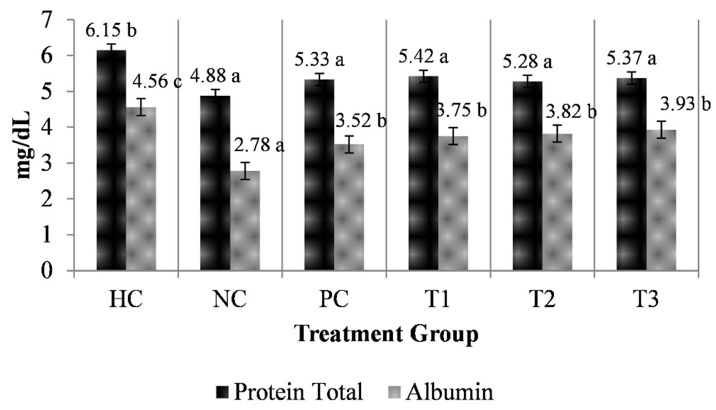
Figure 2. ALB and TP in carrageenan-induced inflammatory rats
Note: The different letters (a, b) on each bar in the figure represented the significant differences (p < 0.05), data were expressed as mean ± SE (n: 36). HG: healthy control; NG: negative control (carrageenan induction only); PG: positive control (administration of sodium diclofenac); TA1: administration of 250 mg C. comatus extract; TA2: administration of 500 mg C. comatus extract; TA3: administration of 750 mg C. comatus extract for 14 days
Figure 2 shows that the bar chart shows total protein and albumin levels (mg/dL) across six treatment groups (HC, NC, PC, T1, T2, T3). Total protein levels are consistently higher than albumin in all groups. The highest total protein is observed in the HC group (6.15 mg/dL), while the lowest albumin appears in the NC group (2.78 mg/dL). Treatment groups (T1-T3) show moderate total protein levels (around 5.28-5.42 mg/dL) and gradual increases in albumin compared to NC. Different superscript letters indicate statistically significant differences between groups (Figure 2).
Radical scavenging activity was one of the vital antioxidant effects, and one of the most prevalent and significant antioxidants that scavenged lipophilic radicals in vivo was vitamin E.28 Vitamin E only absorbed peroxyl radicals to break chain propagation efficiently, but could not act as a strong scavenger of nitric dioxide, hydroxyl, thiyl, and alkoxyl radicals according to its physiological molar ratios to substrates and the kinetic data based on in vivo experiment. Vitamin C acted as a reactive oxygen scavenger in the cytoplasm and was water-soluble. However, α-tocopherol protects cell membranes from peroxidation caused by oxidative stress and was fat-soluble. In terms of free radicals, the roles of vitamins C and E complemented each other.29 Vitamin E was considered a free radical and converted into an unreactive tocopherol, which was then recovered by vitamin C. Its antioxidant capacity was increased by a synergistic interaction with vitamin C.10 In vivo, the main antioxidants that scavenged free radicals were vitamins E and C, ubiquinol (reduced form) of coenzyme Q, carotenoids, UA, and bilirubin. Vitamins E and C, in particular, were essential lipophilic and hydrophilic scavengers.30 Vitamin C, along with biotin and niacin, was known to have preventive effects and minimize the effects of chronic diseases. One of its roles was as antihypertensive agent by lowering blood pressure, as well as antioxidant, anti-ischemic, and anti-cholesterolemic agent.31 In addition to acting as antioxidants, flavonoids inhibited COX enzyme activity increase and production to prevent exacerbation of inflammatory reactions.32 Ethanol and ethyl acetate extracts (C. comatus) contained flavonoids3 in rutin form.28 Studies reported that flavonoid administration inhibited COX-1 enzyme activity. The quercetin form of this compound at a dose of 1.5 mM inhibited 40% of COX-1 activity, rutin impeded 80.54% at 1.25 mM, and diosmin hampered 68.87% at 1.5 mM.33
The high levels exacerbated inflammation and tissue damage, specifically during a reaction with free radicals. Furthermore, the reaction with peroxynitrite (ONOO–) generated radicals, according to UA’s ability to be prooxidative under several conditions. Serum UA elevation had been shown in animal studies to cause hypertension by increasing lipid peroxidation reaction due to oxidative stress, stimulating the renin-angiotensin system, and endothelial dysfunction.34 The decreased level in the treatment group was possible due to C. comatus bioactive compound activity. C. comatus extract contained flavonoids and vitamins C and α-tocopherol, which could reduce oxidative stress due to carrageenan induction,7 thereby preventing systemic hypertension and UA elevation. Administration of Smilax glabra extract flavonoid fraction was reported to reduce UA levels by inhibiting renal oxidative stress.35
TP, ALB, and GLB levels were evaluated in this study since their concentrations were acceptable biomarkers for liver functional state assessment and could also show pathogenicity or inflammation.27 A TP and ALB level increase was discovered in the treatment group, while a decrease was observed in the negative group. ALB was one of the most abundant plasma proteins produced by hepatocytes, while TP and ALB level elevation after administration of the extract increased cell defence against free radicals.36 The results showed TP and ALB levels were normal, showing that the endoplasmic reticulum in charge of protein synthesis was stabilized. In cases of hepatotoxicity caused by defective protein synthesis in liver, TP and ALB levels could be reduced. A previous study performed with induction of 0.1 mL CCl4 and 1% carrageenan showed a decrease in TP (5.1 g/dL) and ALB (2.3 g/dL) in the treatment group, while healthy rats exhibited 8.4 g/dL TP and 4.1 g/dL ALB levels.37 Previous study showed that intraplantar induction of 1% carrageenan by 0.1 mL caused an increase in IL-1β and decreased ALB levels compared to the healthy group.7
MDA levels (anti-oxidative stress effect)
MDA was one of antioxidant parameters, and its levels after extract administration showed a significant decrease (P < 0.05), showing good anti-oxidative stress effect and activity in the body. Based on the experimental results, the lowest MDA occurred in TA2 group with a more than 20% decrease at a 500 mg dose. Meanwhile, the highest MDA levels were in NG, with an increase of more than 37% after carrageenan induction, as presented in Table 6.
Table (6): MDA levels in carrageenan-induced inflammatory rats
No. |
Experimental group |
MDA level (µmol/L) |
% decrease |
|---|---|---|---|
1 |
HC |
0.81 ± 0.13a |
27.07 |
2 |
NG |
1.11 ± 0.06c |
+37.03 |
3 |
PG |
0.95 ± 0.02b |
14.41 |
4 |
TA1 |
0.94 ± 0.02b |
15.31 |
5 |
TA2 |
0.88 ± 0.05ab |
20.72 |
6 |
TA3 |
0.93 ± 0.01b |
16.21 |
Note: The different letters on each column in the table represented the significant differences (P < 0.05), data were expressed as mean ± SE (n: 36). HG: healthy control; NG: negative control (carrageenan induction only); PG: positive control (administration of sodium diclofenac); TA1: administration of 250 mg C. comatus extract; TA2: administration of 500 mg C. comatus extract; TA3: administration of 750 mg C. comatus extract for 14 days
The levels of GLB, which was a liver health marker in addition to bilirubin, increased after extract administration, and in the meantime, NG group experienced a significant decrease. Furthermore, carrageenan-induced inflammation was a biphasic event, with a release of serotonin and histamine during the first phase. During the second phase of swelling, prostaglandin, lysozyme, and bradykinin are released. This was supported by Jisha et al., who reported an increase in GLB levels to 2.81 g/dL and 2.82 g/dL by Muntingia calabura extract administration at 100 and 200 mg, respectively, in inflammatory rat model induced with 1% carrageenan, compared to a normal level of 2.83 g/dL. TP, ALB, and GLB exerted anti-inflammatory effect by increasing lysozyme activity (LZM) with 121.89 U/mL, compared to 89.19 U/mL in the control group. The elevated TP, ALB, and GLB levels reduced prostaglandin E2 (PGE2) activity to 31.56 pg/mL, while the control group had 37.13 pg/mL.38 Besides increasing TP, ALB, and GLB, C. comatus extract decreased PGE2 levels. This mushroom contained flavonoid compounds,3 which acted as anti-inflammatory activity by reducing IL-1β, NO, IL-8, and PGE2 levels.39 A previous study showed that the injection of 1% carrageenan, 0.5 mL, caused an increase in IL-1β levels by 24.79 g/mL. While in the group that was given 500 mg of C. comatus ethanol extract, IL-1β levels were 7.4 g/mL.10 Administration of nanogel C. comatus extract could reduce the levels of mediator (IL-1β, IL-6, and TNF-α) as pro-inflammatory cytokines.40
Previous studies have also shown that microencapsulation of the Pleurotus cystidiosus mushroom can reduce TNF-α levels by >22% and IL-1β levels by >56% in diabetic mice with inflammation.41 C. comatus nanogel can reduce NOS levels in mice with inflammation induced by CFA.9
These results showed a decrease in MDA levels after C. comatus treatment, while there was a significant increase in NG. MDA decrease was consistent with the measured levels of endogenous antioxidants such as TP, ALB, SOD, and GLB. Furthermore, the increase in antioxidants allowed a rise in cell defence against free radical attack, thereby minimizing the possibility of oxidative stress and lipid peroxidation reactions to ensure a lesser MDA product. Increased ROS levels in inflammatory conditions caused oxidative stress and cell lipid peroxidation with MDA’s final product.36 Based on previous studies, increased free radicals that caused oxidative stress could increase pro-inflammatory cytokine levels, COX-2, and MDA.42 Decreased MDA levels also occurred because C. comatus contained vitamins C and E capable of breaking the peroxidation chain.2 According to Justil-Guerrero et al., the administration of Chuquiraga spinosa extract containing flavonoids, tannins, polyphenols, and triterpenes inhibited MDA release with an IC50 value of 28.80 nmol/mL in rats induced with 0.1 mL of 2% carrageenan.
In this study, there were decreasing MDA levels, while SOD, TP, ALB, and GLB increased. Meanwhile, vitamins E, C, and flavonoids in C. comatus extract provided better support to the reduction of free radical activity.22 During inflammation, ONOO– radicals increased due to the O2- and NO– reaction. During inflammatory process, inducible NO synthase and NADPH oxidase produced large amounts of both mediators. Furthermore, severe pain was induced by increasing hyperalgesia through NFΚB activation, such as TNF-α acting through COX-2/PGE2 axes and TNFR1, or by directly inducing neuronal depolarization.43
Leucocytes distribution
The distribution of leukocyte types, including neutrophils, eosinophils, lymphocytes, and monocytes in carrageenan-induced rats was calculated using a binocular light microscope at 40×10 magnification. The calculation and observation results showed the highest neutrophil levels in HC (62 cells/mm3) and the lowest in NC (52 cells/mm3). Furthermore, the highest number of eosinophils, lymphocytes, and monocytes was in PG group, namely 4, 65, and 12 cells/mm3, respectively. The highest leukocyte type was neutrophils, and the lowest was eosinophils, as demonstrated in Figure 3.
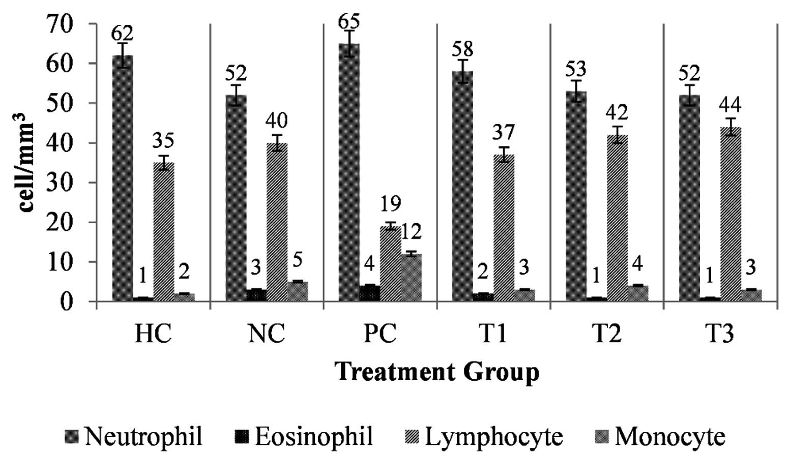
Figure 3. Leukocyte distribution in carrageenan-induced inflammatory rats
Note: The different letters on each bar in the figure represented the significant differences (p < 0.05), data were expressed as mean ± SE (n: 36). HG: healthy control; NG: negative control (carrageenan induction only); PG: positive control (administration of sodium diclofenac); TA1: administration of 250 mg C. comatus extract; TA2: administration of 500 mg C. comatus extract; TA3: administration of 750 mg C. comatus extract for 14 days
The observation results of leukocyte distribution varied, including neutrophils, eosinophils, lymphocytes, and monocytes. Neutrophil distribution was the highest compared to other leukocytes. Previous studies showed that carrageenan induction increased the number of lymphocytes, monocytes, total, and polymorphonuclear (PMN) leukocytes to 20.56 x 10³/mm3, 3.75 x 10³/mm3, 24.07 x 10³/mm3, and 1.59 x 10³/mm3, respectively. Meanwhile, C. spinosa extract administration reduced the total leukocyte count to 1.76 x 10³/mm3 and lymphocytes to 0.99 x 10³/mm3.44
Among the mediators of inflammatory response were prostaglandins, leukotrienes, interleukins, interferons, and tumor necrosis factors. Overcrowding of ROS, activation of complex enzymes, the release of inflammatory substances, and other factors potentially contributed to acute inflammatory response progression. The synthesis of prostaglandins, prostacyclin, and thromboxane by COX enzyme contributed significantly to inflammatory process by causing pain, inflammation, and platelet aggregation.21 Furthermore, the acute inflammatory triggered the activation of immune cells, including neutrophils. Neutrophils’ infiltration into inflammatory region was crucial for antigen inactivation. However, the excessive occurrence of these events, together with a rise in levels of MPO, which reflected the tissue content secreted from neutrophil azurophilic granules, caused tissue damage exacerbation.7,45
Flavonols inhibited iNOS protein expression and the activation of nuclear factor (NF-ΚB), signal transducer, and transcription 1 (STAT-1) activator, which were transcription factors for iNOS, thereby decreasing NO production. In chronic disorders, products that lowered NO generation showed promise in preventing inflammatory processes.44 Observation of the number of neutrophils, lymphocytes, and monocytes in this study was significant since their ratios could describe inflammation levels. An increase in this number indicated a marker for an elevated inflammatory reaction in a cell or tissue. Furthermore, the ratio and number of eosinophils affected inflammatory status.46 According to a previous study, eosinophils and a rise in pro-inflammatory mediators caused allergic asthma inflammation by serving as antigen-presenting cells (APG) and exposing T lymphocytes to allergen peptides.47
A previous study that induced 0.1 carrageenan 1% into animal models showed the bioactive compounds identified in Alstonia boonei ethyl acetate extract contained 0.98 mg/g phenol and 0.25 mg total flavonoids. This extract was then tested and reduced DPPH free radicals and warded off H2O2 and NO with IC50 values of 66.08 g/mL, 146.65 g/mL, and 65.47 g/mL, respectively. Furthermore, the results of A. boonei fraction administration at a dose of 250 mg decreased neutrophil numbers to 3.41, and lymphocytes to 2.68 x 10⁹/L, compared to the 1% carrageenan-induced group, where neutrophils were 8.5 x 10⁹/L, and lymphocyte count was 7.32 x 10⁹/L.48 The increase in neutrophil, eosinophil, lymphocyte, and monocyte numbers during inflammatory conditions and after C. comatus ethanol extract administration showed the presence of immunomodulatory activity. Immunomodulatory actions against infiltrating cells at the site of localized inflammation had been reported in conventional anti-inflammatory drugs.49
In conclusion, C. comatus fruiting body ethanol extract has the potential to be used as anti-inflammatory with the reduction of IL-1β, nephroprotective effect with the reduction of UA levels, antioxidant effects with an increase in the levels of SOD, TP, ALB, and GLB, and anti-oxidative stress with the reduction of MDA levels. The extract has the potential to be developed into anti-inflammatory herbal medicine. Characterizing single bioactive compounds can be further studied to determine, more specifically, the compounds with anti-inflammatory, antioxidant effects, and nephroprotective effects.
ACKNOWLEDGMENTS
The authors are grateful to CV. Asa Agro Corporation for providing all mushroom materials used.
CONFLICT OF INTEREST
The authors declare that there is no conflict of interest.
AUTHORS’ CONTRIBUTION
NIR and FH conceptualized, designed the study, performed the experiments and supervised the study. NIR, FH, DWK, JHS, DS and NE analyzed the data and provided intellectual input. NIR and FH interpreted the results and wrote the manuscript. NIR, FH, DWK, JHS, DS and NE reviewed, revised and approved the final manuscript for publication.
FUNDING
This study was funded by the Institute for Research and Community Service (LPPM) through the Applied Leading Research Grant (RUT) under the supervision of Jenderal Soedirman University Service and Business Agency (BLU), with grant number T/530/UN23.18/PT.01.03/2021.
DATA AVAILABILITY
All datasets generated or analyzed during this study are included in the manuscript.
ETHICS STATEMENT
This study was approved by the Ethics Committee, Dr. Moewardi Regional General Hospital (RSUD), Surakarta City, Central Java, Indonesia, with number 515/IV/HREC/2021.
- Dilfy SH, Al-bideri AWM, Hanawi MJ. Effect of Pleurotus ostreatus Extract on Epidermal Growth Factor Receptor Expression during Healing of Aspirin-Induced Peptic Ulcer in Male Rats. Jordan J Biol Sci. 2020;13(Suppl. Issue):669-674.
- Husen F, Hernayanti H, Ekowati N, Sukmawati D, Ratnaningtyas NI. Antidiabetic Effects and Antioxidant Properties of the Saggy Ink Cap Medicinal Mushroom, Coprinus comatus (Agaricomycetes) on Streptozotocin-Induced Hyperglycemic Rats. Int J Med Mushrooms. 2021;23(10):9-21.
Crossref - Ratnaningtyas NI, Hernayanti, Ekowati N, Husen F. Nephroprotective and Antioxidant Effects of Ethanol Extract of Coprinus comatus Mushroom Fruit-Bodies on Streptozotocin-Induced Diabetic Rat Models. In The 4th International Conference on Biosciences. IOP Conf Ser: Earth Environ Sci. 2021;948:012078.
Crossref - Necas J, Bartosikova L. Carrageenan: A Review. Vet Med – Czech. 2013;58(4):187-205.
Crossref - Liu JY, Hou YL, Cao R, et al. Protodioscin Ameliorates Oxidative Stress, Inflammation and Histology Outcome in Complete Freund’s Adjuvant Induced Arthritis Rats. Apoptosis. 2017;22(11):1454-1460.
Crossref - Parvathi A, Thangam EB. Local and Systemic Profiles of Inflammatory Cytokines in Carrageenan-Induced Paw Inflammation in Rats. Immunol Invest. 2017;46(3):274-283.
Crossref - Semis HS, Gur C, Ileriturk M, Kaynar O, Kandemir FM. Investigation of the Anti-Inflammatory Effects of Caffeic Acid Phenethyl Ester in a Model of l-Carrageenan–Induced Paw Edema in Rats. Hum Exp Toxicol. 2021;40(12_suppl):S721-738.
Crossref - Ratnaningtyas NI, Husen F. Therapeutic Potential of Coprinus comatus Nanogels: Antiarthritic and Anti-Inflammatory Effects in Rheumatoid Arthritis Models. Vet World. 2025;18(3):582-597.
Crossref - Husen F, Ratnaningtyas NI. Anti-Inflammatory Activity of the Shaggy Ink Cap Medicinal Mushroom Coprinus comatus (Agaricomycetes) Nanogel in Complete Freund’s Adjuvant – Induced Rheumatoid Arthritis:In Silico and in Vivo Approach. Int J Med Mushrooms. 2025;27(8):13-35.
Crossref - Ratnaningtyas NI, Husen F, Hernayanti H, Ekowati N, Budianto BH. Anti-Inflammatory and Immunosuppressant Activity of Coprinus comatus Ethanol Extract in Carrageenan-Induced Rats of (Rattus Norvegicus). Molekul. 2022;17(3):336-3346.
Crossref - Ratnaningtyas NI, Hernayanti H, Ekowati N, Husen F. Ethanol Extract of the Mushroom Coprinus comatus Exhibits Antidiabetic and Antioxidant Activities in Streptozotocin-Induced Diabetic Rats. Pharm Biol. 2022;60(1):1126-1136.
Crossref - Wang S, Wang H, Liu Y, Wang Y, Fan X, Cheng Y. Rapid Discovery and Identification of Anti-Inflammatory Constituents from Traditional Chinese Medicine Formula by Activity Index, LC-MS, and NMR. Sci Rep. 2016;6(1):31000.
Crossref - Ren Jun, Shi JL, Han CC, Liu ZQ, Guo JY. Isolation and Biological Activity of Triglycerides of the Fermented Mushroom of Coprinus comatus. BMC Complement Altern Med. 2012;12(1):52.
Crossref - Feng J, Damrauer SM, Lee M, Sellke FW, Ferran C, Abid MR. Endothelium-Dependent Coronary Vasodilatation Requires NADPH Oxidase-Derived Reactive Oxygen Species. Arterioscler Thromb Vasc Biol. 2010;30(9):1703-1710.
Crossref - Ouda AN, Fatiha M, Sadia M, Zohra SF, Noureddine D. In Vivo Anti-Inflammatory Activity of Aqueous Extract of Carthamus Caeruleus L Rhizome against Carrageenan-Induced Inflammation in Mice Amari. Jordan J Biol Sci. 2021;14(3):529-535.
- Sakthivel KM, Guruvayoorappan C. Acacia ferruginea inhibits inflammation by regulating inflammatory iNOS and COX-2. J Immunotoxicol. 2016;13(1):127-135.
Crossref - Amdekar S, Roy P, Singh V, Kumar A, Singh R, Sharma P. Anti-Inflammatory Activity of Lactobacillus on Carrageenan-Induced Paw Edema in Male Wistar Rats. Int J Inflam. 2012;2012:752015.
Crossref - Yusufoglu HS. Analgesic, Antipyretic, Anti-Inflammatory, Hepatoprotective and Nephritic Effects of the Aerial Parts of Pulicaria arabica (Family: Compositae) on Rats. Asian Pac J Trop Med. 2014;7(S1):S583–90.
Crossref - Kanbay M, Segal M, Afsar B, Kang DH, Rodriguez-Iturbe B, Johnson RJ. The Role of Uric Acid in the Pathogenesis of Human Cardiovascular Disease. Heart. 2013;99(11):759-766.
Crossref - Turak O, Ozcan F, Tok D, et al. Serum Uric Acid, Inflammation, and Nondipping Circadian Pattern in Essential Hypertension. J Clin Hypertens. 2013;15(1):7-13.
Crossref - Jisha N, Vysakh A, Vijeesh V, Latha MS. Anti-Inflammatory Efficacy of Methanolic Extract of Muntingia calabura L. Leaves in Carrageenan Induced Paw Edema Model. Pathophysiology. 2019;26(3-4):323-330.
Crossref - Ratnaningtyas NI, Hernanyanti H, Ekowati N, et al. Antioxidant Activities and Properties of Coprinus Comatus Mushroom Both Mycelium and Fruiting Body Extracts in Streptozotocin-Induced Hyperglycemic Rats Model. Biosaintifika: Journal of Biology & Biology Education. 2022;14(1):9-21.
Crossref - Naziroglu M, Kilinc F, Uguz AC, et al. Oral Vitamin C and E Combination Modulates Blood Lipid Peroxidation and Antioxidant Vitamin Levels in Maximal Exercising Basketball Players. Cell Biochem Funct. 2010;28(4):300-305.
Crossref - Kaninathan A, Sudhandiran G. Carvacrol Exhibits Anti-Oxidant and Anti-Inflammatory Effects against 1, 2-Dimethyl Hydrazine plus Dextran Sodium Sulfate Induced Inflammation Associated Carcinogenicity in the Colon of Fischer 344 Rats. Biochem Biophys Res Commun. 2015;461(2):314-320.
Crossref - Szkudelski T. The Mechanism of Alloxan and Streptozotocin Action in B Cells of the Rat Pancreas. Physiol Res. 2001;50(6):537-546.
- Nowakowski P, Naliwajko SK, Markiewicz-Zukowska R, Borawska MH, Socha K. The Two Faces of Coprinus comatus-Functional Properties and Potential Hazards. Phytother Res. 2020;34(11):2932-2944.
Crossref - Anyasor GN, Onajobi F, Osilesi O, Adebawo O, Oboutor EM. Anti-Inflammatory and Antioxidant Activities of Costus afer Ker Gawl. Hexane Leaf Fraction in Arthritic Rat Models. J Ethnopharmacol. 2014;155(1):543-551.
Crossref - Ratnaningtyas NI, Husen F, Sukmawati D, Wibowo ES, Hikam AR, Aksoy A. Antidiabetic Effects and Enzymatic Antioxidant Activity of Chicken Drumstick Mushroom (Coprinus Comatus) Extract in Diabetic Rats Model. J Pure Appl Microbiol. 2022;16(4):2764–2774.
Crossref - Pavithra D, Praveen D, Ranadheer P, Muthukumar VA. A Prospective Study on Role of Vitamin e Supplementation in Type 2 Diabetes Mellitus. Asian J Pharm Clin Res. 2018;11(Special Issue 4):81-84.
Crossref - Niki E. Role of Vitamin E as a Lipid-Soluble Peroxyl Radical Scavenger: In Vitro and in Vivo Evidence. Free Radic Biol Medi. 2014;2014;66:3-12.
Crossref - Rauf A, Joshi PB, Ahmad Z, et al. Edible Mushrooms as Potential Functional Foods in Amelioration of Hypertension. Phytother Res. 2023;37(6):2644-2260.
Crossref - Pannunzio A, Coluccia M. Cyclooxygenase-1 (COX-1) and COX-1 Inhibitors in Cancer: A Review of Oncology and Medicinal Chemistry Literature. Pharmaceuticals. 2018;11(4):101.
Crossref - Zaragoza C, Alvarez-Mon MA, Zaragoza F, Villaescusa L. Flavonoids: Antiplatelet Effect as Inhibitors of COX-1. Molecules. 2022;27(3):1-13.
Crossref - Sanchez-Lozada LG, Soto V, Tapia E, et al. Role of Oxidative Stress in the Renal Abnormalities Induced by Experimental Hyperuricemia. Am J Physiol Renal Physiol. 2008;295(4):1134–41.
Crossref - Wang S, Fang Y, Yu X, Guo L, Zhang X, Xia. The Flavonoid-Rich Fraction from Rhizomes of Smilax glabra Roxb. Ameliorates Renal Oxidative Stress and Inflammation in Uric Acid Nephropathy Rats through Promoting Uric Acid Excretion. Biomed Pharmacother. 2019;111:162-168.
Crossref - Ogundahunsi OA, Odewabi AO, Oyalowo M. Effect of exposure to petroleum fumes on plasma antioxidant defense system in petrol attendants. British J Pharmacol Toxicol. 2014;5(2):91-95.
Crossref - Yusufoglu HS, Soliman GA, Foudah AI, Abdelkader MS, Alam A, Salkini MA. Anti-inflammatory and hepatoprotective potentials of the aerial parts of Silene villosa Caryophyllaceae methanol extract in rats. Trop J Pharm Res. 2018;17(1):117.
Crossref - An S, Liu G, Guo X, An Y, Wang R. Ginger Extract Enhances Antioxidant Ability and Immunity of Layers. Anim Nutr. 2019;5(4):407-409.
Crossref - Filannino P, Cavoski I, Thlien N, et al. Lactic Acid Fermentation of Cactus Cladodes (Opuntia Ficus-Indica L) Generates Flavonoid Derivatives with Antioxidant and Anti-Inflammatory Properties. PLoS ONE. 2016;11(3):1-22.
Crossref - Ratnaningtyas NI, Husen F, Fitrianto N. Lingzhi or Reishi Medicinal Mushroom Ganoderma lucidum (Agaricomycetes) Nanogel in Complete Freund’s Adjuvant-Induced Rheumatoid Arthritis (RA) Rat Model: Anti-Arthritic, Anti-Inflammatory, and Antioxidative Activity. Int J Med Mushrooms. 2024;26(8):27-40.
Crossref - Ratnaningtyas NI, Husen F, Fitrianto N, Safitri J. Microencapsulation of abalone oyster mushroom Pleurotus cystidiosus (Agaricomycetes ): antidiabetic, and anti-inflammatory activity in streptozotocin-induced diabetic rat model. Int J Med Mushrooms. 2025;27(7):67-84.
- Abu-Lubad M, Al Qaisi YT, Alsarayreh AZ, Abu Murad MA, Alshraideh H. Evaluation of Liver Antioxidants in an Induced Oxidative Stress in Albino Rat Model. Jordan J Biol Sci. 2024;17(3):481-486.
Crossref - Ferraz CR, Carvalho TT, Manchope MF, et al. Therapeutic Potential of Flavonoids in Pain and Inflammation: Mechanisms of Action, Pre-Clinical and Clinical Data, and Pharmaceutical Development. Molecules. 2020;25(3):762.
Crossref - Justil-Guerrero HJ, Arroyo-Acevedo JL, Rojas-Armas JP, Palomino-Pacheco M, Villena-Tejada M, Vilchez WAS. Antioxidant Capacity of Chuquiraga Spinosa Less. ‘Huamanpinta’ and Prevention of Carrageenan-Induced Inflammation in Mice. Pharmacogn J. 2021;13(5):1287-1296.
Crossref - Kamakshi S, Anantha LP, Shenbhagaraman R, et al. Antibacterial and Photo Dye Degradative Ability of Copper Oxide Nanoparticles from Pleurotus cystidiosus. Nano Express. 2024;5(2):025029.
Crossref - Yang Z, Zhang Z, Lin F, et al. Comparisons of Neutrophil-, Monocyte-, Eosinophil-, and Basophil- Lymphocyte Ratios among Various Systemic Autoimmune Rheumatic Diseases. Apmis. 2017;125(10):863-871.
Crossref - Pelaia G, Vatrella A, Busceti MT, et al. Cellular Mechanisms Underlying Eosinophilic and Neutrophilic Airway Inflammation in Asthma. Mediators Inflamm. 2015:2015:879783.
Crossref - Akinnawo OO, Anyasor GN, Osilesi O. Aqueous fraction of Alstonia boonei de Wild leaves suppressed inflammatory responses in carrageenan and formaldehyde induced arthritic rats. Biomed Pharmacother. 2017;86:95-101.
Crossref - Arreola R, Quintero-Fabian S, Lopez-Roa, et al. Immunomodulation and Anti-Inflammatory Effects of Garlic Compounds. J Immunol Res 2015;2015:630.
Crossref
© The Author(s) 2026. Open Access. This article is distributed under the terms of the Creative Commons Attribution 4.0 International License which permits unrestricted use, sharing, distribution, and reproduction in any medium, provided you give appropriate credit to the original author(s) and the source, provide a link to the Creative Commons license, and indicate if changes were made.