


ISSN: 0973-7510
E-ISSN: 2581-690X
Staphylococcus aureus is the primary causative agent of osteomyelitis. Plain radiography is the initial diagnostic tool for distinguishing osteomyelitis from other bone conditions, and assists in assessing disease progression. These benefits include low radiation exposure, improved availability, cost-effectiveness, and ease of use. This was an in vivo study involving mice. The subjects were divided into the treatment and control groups and observed on days 3, 7, 14, and 28th of the experiment. The treatment group underwent mandibular drilling and Staphylococcus aureus induction. The control group underwent mandibular drilling only. Radiographic findings of mandibular bone defects include periosteal reactions, diaphyseal widening, osteolysis, bone deformation, and sequestral formation. The statistical tests consisted of the Mann-Whitney and Friedman tests. There were significant differences in bone destruction levels in occlusal dementia between the treatment and control groups, which were found on the 3rd day of observation. The treatment group exhibited greater bone destruction (5.67 ± 3.62) compared to the control group (2.00 ± 2.49), with a z-value of -2.214 and a statistically significant p-value of 0.027. 14th showed higher bone destruction levels (5.83 ± 2.64) than the control group (4.33 ± 3.61), with a z-value of -2.660 and a highly significant p-value of 0.008. On the 28th day the trend persisted, as the treatment group displayed more bone destruction (5.50 ± 3.27) compared to the control group (3.33 ± 3.44), with a z-value of -2.034 and a significant p-value of 0.042. No significant differences in the lateral dimensions were found between the treatment and control groups. In each group, significant differences were found only in the treatment group; in the occlusal dimension, sequestrum formation (p = 0.006) was found, and in the lateral dimension, diaphyseal widening (p = 0.022), osteolysis (p = 0.017), and sequestrum formation criteria (p = 0.015) were observed. The z-value measures the magnitude and direction of the difference, whereas the p-value assists in determining if this difference is statistically significant. Subjects induced with Staphylococcus aureus showed severe bone destruction compared to those without Staphylococcus aureus induction.
Infectious Disease, Osteomyelitis, Radiographic, Staphylococcus aureus
Osteomyelitis is an inflammatory condition of the bone and surrounding tissues caused by pyogenic infections, including bacteria, fungi, and mycobacteria. Based on clinical characteristics, it can be classified as acute, subacute, chronic, suppurative, or non-suppurative. The infection typically spreads and affects both the cortical bone and periosteum. This condition can lead to permanent disability in both children and adults worldwide.1
Acute and chronic osteomyelitis are more commonly found in the mandible than in the maxilla because of the lower vascularity of the mandible and the more easily damaged cortical bone density. These conditions are often aggravated by infections during tooth extraction. Most jaw osteomyelitis cases are odontogenic. The primary causative agent is Staphylococcus aureus, which is responsible for approximately 80% of these infections.2
Diagnosing and treating jaw osteomyelitis is challenging. Although various diagnostic techniques are valuable, researchers agree that the final diagnosis should be based on the clinical presentation, patient history, imaging techniques, culture results, and histological analysis. Imaging studies are crucial to establish a timely diagnosis and guide early management to reduce long-term complications. Radiographic imaging plays a significant role in the diagnosis and characterization osteomyelitis. Several radiographic techniques are available, including plain radiography, magnetic resonance imaging (MRI), computed tomography (CT), ultrasonography (USG), and scintigraphy.3
Plain radiography is the most commonly used radiological imaging technique in medical institutions because of its advantages, including low radiation dose, widespread availability, affordability, and simplicity. It serves as the first line of examination to differentiate osteomyelitis from other bone disorders, and offers an overview of disease progression. Each phase of osteomyelitis in the long bones exhibits a different appearance on plain radiography. However, no study has explicitly detailed the radiographic appearance of osteomyelitis in the mandibular region.4
Animal models can enhance our understanding of osteomyelitis pathogenesis and treatment efficacy, thereby aiding in infection reduction. A study using animal models of jaw osteomyelitis, specifically rats, sought to reflect the clinical progression of the disease using radiographic imaging. This study aimed to observe osteomyelitis in rats induced by Staphylococcus aureus over several days, capture radiographic images daily, and analyze the extent of bone destruction. Observing the progression of osteomyelitis from the acute to chronic phase is particularly intriguing, as no similar animal model studies have focused on radiographic observations. This study aimed to provide a radiographic description of osteomyelitis during its acute and chronic phases and serve as a foundation for future research in determining therapeutic strategies for osteomyelitis.
Subject
This study utilized Wistar mice (Rattus norvegicus) as subjects. The inclusion criteria for the subjects required them to be male, have a body weight of 300 g, be aged 2-3 months, and be in good health. The subjects were divided into two groups (treatment and control) of 24 mice each according to methodologies used in previous studies.5
Study design
This study used an in vivo post-test-only control group design. The subjects were divided into two groups, and each subject had a wound created in the mandibular area using a burr. The wounds were inoculated with Staphylococcus aureus in the treatment group, whereas the control group received wounds without inoculation. Rats were euthanized using diethyl ether. The head and mandible were separated and preserved in 10% formalin buffer on days 3, 7, 14, and 28th of the experiment. Radiographs of the mandibular area were obtained from the occlusal and lateral views. The type of Staphylococcus aureus used in this study was ATCC 6538, consistent with materials used in previous studies.6
Radiographic interpretation
X-ray readings and evaluations were performed using several scoring methods. The radiological evaluation criteria assessed included: (1) periosteal reaction, (2) diaphyseal widening, (3) osteolysis, (4) bone deformation, (5) sequestrum formation, (6) joint effusion, and (7) soft tissue swelling. Parameter 1-4 were scored from 0 to 3 (0 = absent, 1 = mild, 2 = moderate, and 3 = severe), and bone deformation was assessed according to Mader et al. Parameters 5-7 were scored as 0 or 1 (absent or present). This study did not involve the joints, and tissue swelling could not be confirmed clinically; therefore, criteria 5 (sequestrum formation) and 6 (joint effusion) were excluded. Three veterinarians with at least 10 years of experience performed radiographic interpretation.
Statistical analysis
Statistical analyses were performed using the Statistical Package for the Social Sciences (SPSS) 23.0 version (IBM Corp., Armonk, NY, USA). The measurement data were first analyzed using the Shapiro-Wilk test. Furthermore, the measurement results of each group (treatment and control groups) were analyzed using the Mann-Whitney and Friedman tests. Statistical analysis results were considered significant if the p-value was <0.05.
Bone Destruction Radiography Features in the Occlusal Dimension
Radiographic results showed bone destruction in the occlusal dimension, including periosteal reaction, diaphyseal widening, osteolysis, bone deformation, and sequestrum formation (Figure 1). The mean bone destruction values after inoculation with Staphylococcus aureus in the occlusal dimensions after S. aureus inoculation are presented in Table 1.
Table (1):
Radiographic bone destruction score in occlusal dimension
| Bone destruction | Day | |||
|---|---|---|---|---|
| 3rd | 7th | 14th | 28th | |
| Treatment group | ||||
| Periosteum reaction | 0.75 | 0.41 | 0.55 | 0.45 |
| Diaphyseal widening | 0.52 | 0.52 | 0.00 | 0.71 |
| Osteolysis | 0.75 | 0.84 | 0.00 | 0.45 |
| Bone deformation | 1.09 | 0.52 | 0.45 | 1.09 |
| Sequestrum formation | 0.00 | 0.00 | 0.55 | 0.45 |
| Control group | ||||
| Periosteum reaction | 0.71 | 0.52 | 0.82 | 0.45 |
| Diaphyseal widening | 0.00 | 0.00 | 0.41 | 0.00 |
| Osteolysis | 0.55 | 0.75 | 0.82 | 0.84 |
| Bone deformation | 0.00 | 1.83 | 0.75 | 0.55 |
| Sequestrum formation | 0.00 | 0.00 | 0.00 | 0.00 |
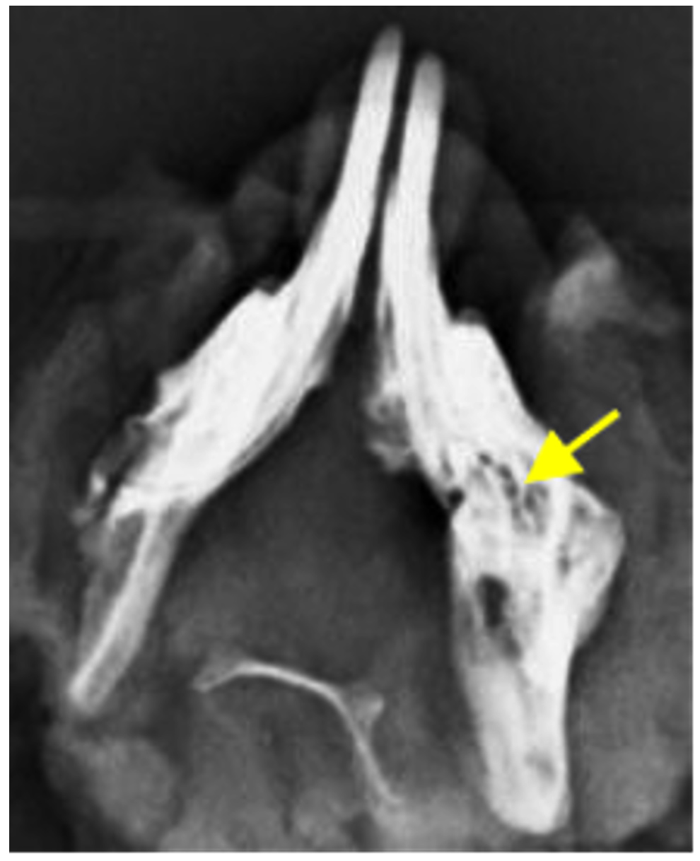
Figure 1. Radiographic bone destruction in the occlusal dimension
In the control group, the mean bone destruction values were 2.00 ± 2.49 on day 3, 5.00 ± 4.00 on day 7, 4.33 ± 3.61 on day 14, and 3.33 ± 3.44 on day 28. Conversely, the treatment group showed higher mean values of bone destruction on day 3 (5.67 ± 3.62), a reduction on day 7 (4.33 ± 3.50), followed by an increase on day 14 (5.83 ± 2.64), and a slight reduction on day 28 (5.50 ± 3.27). These values suggested that the treatment group inoculated with Staphylococcus aureus, exhibited greater bone destruction, particularly in the early and later phases of the study.
Bone destruction radiography features in the lateral dimension
Radiographic results showed bone destruction in the lateral dimension, including periosteal reaction, diaphyseal widening, osteolysis, bone deformation, and sequestrum formation (Figure 2).
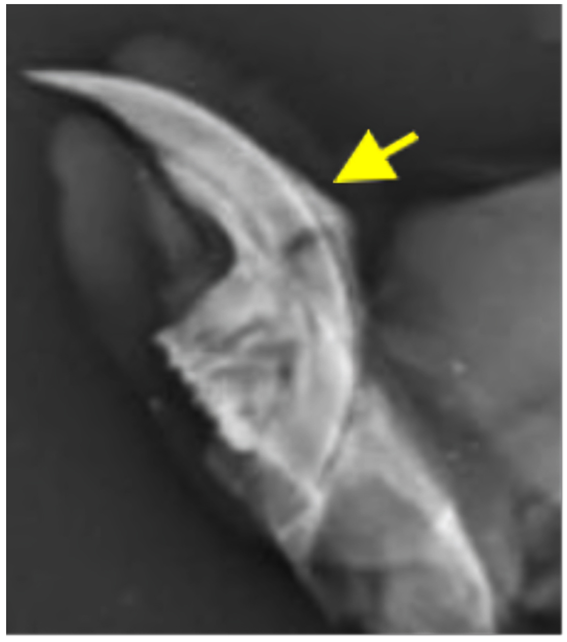
Figure 2. Radiographic bone destruction in the lateral dimension
The mean values of bone destruction after the injection of Staphylococcus aureus in the lateral dimension are shown in Table 2. The comparison of mean bone destruction values between the treatment and control groups is as follows: on day 3, both groups showed similar values (3.00 ± 2.68 in the treatment group vs. 3.00 ± 3.46 in the control group). On day 7, the control group had slightly higher bone destruction (3.83 ± 2.93) compared to the treatment group (2.50 ± 2.95). However, on day 14, the treatment group exhibited more bone destruction (3.00 ± 1.67) than the control group (2.17 ± 3.37). By day 28, the treatment group showed significantly higher bone destruction (5.67 ± 2.80) compared to the control group (4.00 ± 2.36), indicating a gradual progression of damage in the treatment group.
Table (2):
Radiographic bone destruction score in lateral dimension
| Bone destruction | Day | |||
|---|---|---|---|---|
| 3rd | 7th | 14th | 28th | |
| Treatment group | ||||
| Periosteum reaction | 0.00 | 0.00 | 0.00 | 0.45 |
| Diaphyseal widening | 0.41 | 0.00 | 0.55 | 0.71 |
| Osteolysis | 1.03 | 0.55 | 0.00 | 0.45 |
| Bone deformation | 0.41 | 0.00 | 0.55 | 0.84 |
| Sequestrum formation | 0.00 | 0.00 | 0.45 | 0.55 |
| Control group | ||||
| Periosteum reaction | 0.45 | 0.41 | 0.41 | 0.00 |
| Diaphyseal widening | 0.00 | 0.41 | 0.00 | 0.55 |
| Osteolysis | 0.89 | 0.00 | 0.00 | 1.14 |
| Bone deformation | 0.00 | 0.84 | 0.00 | 0.84 |
| Sequestrum formation | 0.00 | 0.00 | 0.00 | 0.00 |
Bone Destruction Levels in Mandibular Osteomyelitis post Staphylococcus aureus inoculation
Significant differences in the bone destruction levels in the occlusal dimensions between the treatment and control groups were observed on the 3rd day of observation
(z = -2.214; p = 0.027), the 14th day (z = -2.660; p = 0.008), and the 28th day (z = -2.034; p = 0.042). Significant differences in bone destruction levels in the occlusal dimension were also observed in diaphyseal widening and osteolysis.
The differences in diaphyseal widening were observed on the 3rd day (z = -2.449;
p = 0.014), the 14th day (z = -3.000; p = 0.003), and the 28th day (z = -2.390; p = 0.017). For osteolysis, significant differences were observed on the 3rd day (z = -2.300; p = 0.021), the 14th day (z = -2.835; p = 0.005), and the 28th day (z = -2.356;
p = 0.018; Table 3). No significant differences were observed in the appearance of bone destruction in the lateral dimensions between the treatment and control groups.
Table (3):
Differences in bone destruction in mandibular osteomyelitis after injection of Staphylococcus aureus
| Radiographic features | 3rd day | 7th day | 14th day | 28th day | ||||
|---|---|---|---|---|---|---|---|---|
| z | p | z | p | z | p | z | p | |
| Occlusal dimension | ||||||||
| Periosteum reaction | -0.956 | 0.339 | 0.000 | 1.000 | 0.000 | 1.000 | -1.800 | 0.072 |
| Diaphyseal widening | -2.449 | 0.014* | -1.500 | 0.134 | -3.000 | 0.003* | -2.390 | 0.017* |
| Osteolysis | -2.300 | 0.021* | -1.315 | 0.189 | -2.835 | 0.005* | -2.356 | 0.018* |
| Bone deformation | -1.964 | 0.050 | -1.017 | 0.309 | -1.678 | 0.093 | -1.386 | 0.166 |
| Sequestrum formation | 0.000 | 1.000 | 0.000 | 1.000 | -1.500 | 0.134 | -1.000 | 0.317 |
| Total score | -2.214 | 0.027* | -0.215 | 0.830 | -2.660 | 0.008* | -2.034 | 0.042* |
| Lateral dimension | ||||||||
| Periosteum reaction | -1.000 | 0.317 | -1.000 | 0.317 | -1.000 | 0.317 | -1.000 | 0.317 |
| Diaphyseal widening | -1.000 | 0.317 | -1.000 | 0.317 | -1.500 | 0.134 | -0.956 | 0.339 |
| Osteolysis | -1.491 | 0.136 | -1.500 | 0.134 | -1.491 | 0.136 | -1.346 | 0.178 |
| Bone deformation | -1.491 | 0.136 | -1.491 | 0.136 | -1.500 | 0.134 | 0.000 | 1.000 |
| Sequestrum formation | 0.000 | 1.000 | 0.000 | 1.000 | -1.000 | 0.317 | -1.964 | 0.050 |
| Total score | -1.890 | 0.059 | -1.890 | 0.059 | -0.900 | 0.368 | -1.596 | 0.110 |
Note: *significant <0.05
The treatment group showed a significant difference in the radiographic occlusal dimensions for the sequestrum formation criteria (p = 0.006). Several important differences were observed in the lateral dimensions of the treatment group, including diaphyseal widening (p = 0.022), osteolysis (p = 0.017), and sequestrum formation (p = 0.015; Table 4). No significant differences in occlusal or lateral radiographic dimensions were found in the control group.
Table (4):
Different bone destructions levels in treatment and control groups on the occlusal and lateral dimension
| Bone destruction | Group | |
|---|---|---|
| Treatment | Control | |
| Occlusal dimension | ||
| Periosteum reaction | 0.729 | 0.480 |
| Diaphyseal widening | 0.274 | – |
| Osteolysis | 0.178 | 0.241 |
| Bone deformation | 0.331 | 0.231 |
| Sequestrum formation | 0.006* | – |
| Lateral dimension | ||
| Periosteum reaction | 0.392 | 0.392 |
| Diaphyseal widening | 0.022* | 0.066 |
| Osteolysis | 0.017* | 0.101 |
| Bone deformation | 0.172 | 0.172 |
| Sequestrum formation | 0.015* | – |
Note: *Significant <0.05
Osteomyelitis is an inflammatory process involving the bone and its secondary structures, and is caused by infection by pyogenic organisms such as bacteria, fungi, and mycobacteria. Diagnosis must be performed quickly and accurately as this can influence the administration of antibiotics and the decision to perform surgery. Inadequate or prolonged diagnosis can significantly decrease the recovery rate and increase the incidence of complications and morbidity.3
Radiological examination is crucial for the secondary or adjunctive diagnosis of acute and chronic osteomyelitis. In contrast to acute osteomyelitis, which typically shows no visible abnormalities for two weeks or more, apart from soft tissue obstructions and obliteration of intermuscular planes, a periosteal reaction may be observed, which can persist for several months before merging with the underlying cortical bone.7
The occlusal observation in the treatment group revealed the formation of a sequestrum on the 14th day, which persisted on the 28th day. This finding is consistent with those of previous studies, which indicate that in the chronic stage, dead sclerotic bone fragments (sequestrum) are present at the original site or are extruded into the surrounding soft tissue. The repair process followed a classic pattern that included the removal of necrotic tissue, osteogenesis, and fibrosis.7 The formation of a sequestrum on day 14 was observed in the group with bacterial inoculation but was not present in any of the control group samples. Previous studies have shown that sequestra can be identified on plain radiographic imaging as focal sclerotic lesions with lucent margins in patients with chronic osteomyelitis. An involucrum may appear as thickened sclerotic bone surrounding the area. These findings may include cortical destruction, disorganized trabecular patterns, and indistinct bony lucencies.8
The hallmark of chronic osteomyelitis is the presence of necrotic bone, which typically forms over an average of 10 days. However, plain radiography may only reveal sequestration or sclerotic bone after the patient has experienced the condition for several weeks. Subperiosteal abscesses, which involve removal of the periosteum and formation of new bone and soft tissue fistulas, can lead to periostitis, involucrum formation, and sinus passage. These findings indicate a prolonged infectious process. One study identified common findings in patients with osteomyelitis, including soft tissue edema, osteopenia, osteolysis, bone destruction, and nonspecific periosteal reactions. Lytic lesions may be detectable by plain radiography only after 50-75% of the bone matrix has been lost, making this imaging technique inadequate for detecting early stage bone disease.9
Another review indicated that plain radiography may initially reveal soft tissue changes, muscle edema, and loss of soft tissue integrity. The first alterations in the pyogenically infected bone suggest that the infectious process has been present for 2-3 weeks or more. Generally, radiographs of osteomyelitis provide precise imaging when the lesion is ≥1 cm, and there is a 30-50% loss of bone mineral content. The initial findings may be ambiguous, with changes typically observed at approximately 5-7 days in children and 10-14 days in children and adults, respectively. Common early bone changes include periosteal thickening, lytic lesions, endosteal scalloping, osteopenia, loss of trabecular architecture, and new bone.3
Approximately 80% of patients undergo routine radiographic imaging during the first two weeks after the onset of infection. Bone marrow edema is the earliest pathological feature that cannot be detected on plain radiographs. Possible features of acute osteomyelitis include a periosteal reaction due to elevation of the periosteum, well-defined bony lucencies indicating the presence of an intraosseous abscess, and soft tissue edema.10
In occlusal observations, bone destruction in the mouse mandibular osteomyelitis defect model was evident in mice that received Staphylococcus aureus induction. These mice exhibited diaphyseal widening and osteolysis on days 3, 14, and 28, along with bone deformation on day 3 and sequestrum formation on days 14 and 28. In the lateral observations, sequestrum formation was also noted on days 14th and 28th days. Diaphyseal widening and osteolytic parameters were observed between days 7 and 28. A sequestrum in the mandibular osteomyelitis defect model was evident in the group of mice after Staphylococcus aureus infection on the 14th and 28th days of observation. Radiographic images of the mandibular defects in the induction group showed progressive changes that increased over the observation period.
ACKNOWLEDGMENTS
None.
CONFLICT OF INTEREST
The authors declare that there is no conflict of interest.
AUTHORS’ CONTRIBUTION
ADP, RAM and DM conceptualized the study. RAM, DM, ARS collected resources. ADP, RAM and BSL applied methodology. ADP and ARS performed data curation. ADP, RAM and ARS performed Investigation. ADP and BSL performed formal analysis. RAM, DM, ARS performed data validation. ARS and BSL performed supervision.ADP, ARS, BSL and DM wrote original draft. ADP and RAM wrote, reviewed and edited the manuscript. All authors read and approved the final manuscript for publication.
FUNDING
None.
DATA AVAILABILITY
All datasets generated or analyzed during this study are included in the manuscript.
ETHICS STATEMENT
This study was approved by the Institutional Ethics Committee, Universitas Airlangga, Faculty of Dental Medicine Health Research, Ethical Clearance Commission, Surabaya (No. 265/HRECC.FODM/V/2022).
- Lee YJ, Sadigh S, Mankad K, Kapse N, Rajeswaran G. The imaging of osteomyelitis. Quant Imaging Med Surg. 2016;6:184–98.
Crossref - Roux KM, Cobb LH, Seitz MA, Priddy LB. Innovations in osteomyelitis research: A review of animal models. Animal Model Exp Med. 2021;4:59-70.
Crossref - Pineda C, Espinosa R, Pena A. Radiographic Imaging in Osteomyelitis: The Role of Plain Radiography, Computed Tomography, Ultrasonography, Magnetic Resonance Imaging, and Scintigraphy. Semin Plast Surg. 2009;23:080-9.
Crossref - Desimpel J, Posadzy M, Vanhoenacker F. The Many Faces of Osteomyelitis: A Pictorial Review. J Belg Soc Radiol. 2017;101.
Crossref - Ridwan RD, Sidarningsih, Kusumaningsih T, Salim S. Effect of lipopolysaccharide derived from surabaya isolates of Actinobacillus actinomycetemcomitans on alveolar bone destruction. Vet World. 2018;11:161-6.
Crossref - Setiawan F, Wahjuningrum D, Yudianto A, Radhianto E, Sunariani J, Bhardwaj A. The inhibition effect of capsaicin extract against Staphylococcus aureus: An in vitro experimental study. Journal of International Oral Health. 2021;13:393.
Crossref - Aktekin CN, Ozturk AM, Tabak AY, Altay M, Korkusuz F. A different perspective for radiological evaluation of experimental osteomyelitis. Skeletal Radiol. 2007;36:945-50.
Crossref - Baur DA, Altay MA, Flores-Hidalgo A, Ort Y, Quereshy FA. Chronic Osteomyelitis of the Mandible: Diagnosis and Management-An Institution’s Experience Over 7 Years. Journal of Oral and Maxillofacial Surgery. 2015;73:655-65.
Crossref - Hatzenbuehler J, Pulling TJ. Diagnosis and management of osteomyelitis. Am Fam Physician. 2011;84:1027-33.
- Pugmire BS. Role of MRI in the diagnosis and treatment of osteomyelitis in pediatric patients. World J Radiol. 2014;6:530.
Crossref
© The Author(s) 2025. Open Access. This article is distributed under the terms of the Creative Commons Attribution 4.0 International License which permits unrestricted use, sharing, distribution, and reproduction in any medium, provided you give appropriate credit to the original author(s) and the source, provide a link to the Creative Commons license, and indicate if changes were made.