


ISSN: 0973-7510
E-ISSN: 2581-690X
Mucormycosis is a severe fungal infection which mainly caused by filamentous fungi of the Absidia sp., Rhizopus sp., Cunninghamella sp, Mucor sp., and Rhizomucor sp. Moreover, the pandemic of the SARS-CoV-2 virus expands the need to interfere with spread of the airborne respiratory infections. Accordingly, developing cutting-edge solutions to restrict and/or prevent air contamination by infectious microbes are very warranted. The current work aims to prepare biocidal La-nano complex treated filters and assess their anti-fungal capacity against 20 Rhizopus oryzae, 10 Candida albicans, and 11 Aspergillus fumigatus. These fungi were isolated from the inside parts of the air conditioning systems in the rehabilitation rooms for COVID-19 patients. The obtained results demonstrated that the prepared were able to significantly decrease the invading microbes and eradicate Rhizopus, Aspergillus, Mucor, Candida albicans isolates at 0.64 mg/ml concentration. DFT study compares the electronic properties and reactivity of a ligand in its uncoordinated form with its lanthanum complex. The ligand exhibits lower binding energy, ionization potential, electron affinity, absolute electronegativity, and chemical potential when coordinated with lanthanum. In contrast, the lanthanum complex has a smaller energy gap, absolute hardness, and global softness.
Mucormycosis, DFT, COVID-19, Biocide La-nano Complex Treated Filters
A new, opportunistic, life-threatening mycosis that affects both industrialized and developing countries worldwide is known as mucormycosis.1 The fungi of order Mucorales, class Zygomycetes, genus Mucor, Rhizopus, Cunninghamella, and Absidia are the causative agents of this illness.2-4 These fungi are distributed throughout the nature where they can be found in the ground, air, and water that has been tainted by pollutants, rotting fruits and vegetables, stored grains, groundnuts, bread, rice, wheat, and barley, as well as in dung and compost.3 The nose, sinuses, and eyes are the main targets of these fungi before their dissemination into the brain. In addition, the causative agents of mucormycosis favor the elastic lamina of both the big and small arteries leading to bleeding, infarction, and thrombosis.4 The illness can strike suddenly or spread rapidly causing epidemic, killing large numbers of vulnerable people, especially those with impaired immune systems.2,4
Of interest, Zygomycosis has been considered a new obstacle in the battle against the COVID-19 threat.5 A variety of conditions, including HIV/AIDS, continuous use of hydrocortisone and a wide range of antibacterial antibiotics, acidosis, malnutrition, debility, leukemia, stem cell recipient, severe burns, emaciation, diabetes mellitus, etc. are associated with Zygomycosis.6-9
Of note, the majority of people with mucormycosis were either chronic diabetics or had received unreasonable corticosteroid treatment.6-9 Humans being in close contact with pathogenic fungal species in the environment can develop mucormycosis. The skin infection might occur if a pathogenic fungal species comes into the skin via a cut, burn, or another type of skin injury.10
There is currently no vaccine available to mucormycosis, however, some therapies are in use to decrease the disease incidence risk in people with impaired immune systems.10
The typical person breathes about 16 kilograms of air each day.11 Influenza, common cold, bronchitis, headaches, eye pain, coughing, and dizziness all are associated with inhaling polluted air. The inhaled airborne spores can transmit to the lungs or nasal cavities, and toxins production including particularly aggressive substances can also take place.
Indoor activities produced moisture which supports the growth of molds whether it is cold, wet, or hot and muggy summer times. Mold grows on many different surfaces, including clothes, books, toys, CDs, and even walls, and has the power to turn treasured possessions into dingy relics. While mold infiltration cannot be controlled, exposure of the equipment to molds can be decreased by the use of air filtration systems, isolation procedures, and environmental protection measures.12 Since filters have the potential to act as a reservoir for contaminants in the indoor environment, the possibility of using silver nanoparticles in filter meshes has been investigated in several studies.13-15 Silver nanoparticles were incorporated into a nanofibrous air filter to provide an antimicrobial effect.16 Gram (+ve) and Gram (-ve) bacteria on the filters are less likely to survive when silver nanoparticles are present. No growth was seen over 24 hours, suggesting that the action was bacteriostatic rather than bactericidal.
In this study, we thought to follow the currently applied strategies of preparing biocidal filters. We examined the capacity of the prepared La nano-complex treated filters to restrict/prevent the spread of airborne microbes.
Materials
The purity of all substances used in the study was at the highest level. (BDH) produced pure organic solvents like C2H5OH and Dimethyl Formamide (DMF). Additionally, 5-bromo-salicylaldehyde (Sigma), o-phenylenediamine (Aldrich), and La (NO3)3.6H2O (Merck) were used.
Instruments
Using a 600 MHZ spectrometer, the 1H and 13C nuclear magnetic resonance spectra were captured in DMSO-d6. Dimethylformamide (DMF) was used as the solvent at room temperature, and the molar conductance was measured using a JEN WAY 4510 conductivity meter at Jouf University. A Perkin-Elmer 437 IR spectrometer was used to record the IR spectra of KBr discs between 400 and 4000 cm-1. The average nano complex diameter was calculated using ImageJ software by randomly selecting 100 photos from each sample’s Scan Electron Microscope images, (SEM; version 1.4.3).
Free ligand synthesis
A 2.01 g, 20 mmol, O-phenylenediamine (0.54 g, 10 mmol) heated solution was added to a heated and stirred 30 ml of 5-bromosalicylaldehyde in 100% C2H5OH. The stirred compounds were refluxed for two hours and then allowed to cool before the precipitate was filtered out and collected. The ligand was made using M. P; 240; yield (90.3 percent) as has been reported in an earlier study.17
Nano La complex synthesis
Free ligand (0.0025 mol) and (0.005 mol) of La(NO3)3.6H2O (1:2 molar ratio) were mixed to produce the nano La complex. The substances were crushed in a mortar at room temperature and were left some time to set up. The La-nano complex was crystallized as a light brown substance. The nano complex is a stable powder that dissolves in DMSO and DMF but not in other solvents.
Coating sterilized filter with La-nano-complex
A sterilized filter was cut into small pieces of 0.5 cm x 2 cm each, and equal volumes (100 ml) of 3% carboxymethylcellulose (CMC) and La-nano-complex (0.64 mg/ml) were mixed. The sterilized filters were then sprayed and coated with this mixture. After being prepared, the filter pieces were left overnight in an uncovered petri dish to dry at room temperature. The coating procedure was carried out in the dark at room temperature allowing the coating step to be carried out for 15, 30, and 60 minutes.18
Isolation and characterization of the growing fungal species
The filters of the air conditioners in the COVID-19 rehabilitation rooms were removed and cleaned 3 times with sterile type 3 water. Each filter was dried, and filter fragments of
(1 × 3 cm) were prepared. These fragments were allowed to be placed in 330 ml sanitized water bottles. The Potato Dextrose Agar (PDA) media was prepared by adding 10 ml of Rose Bengal with PDA to 5 ml sterile ddH2O. The filter fragments were individually placed on a sterile plate and the mixture was incubated at 26°C for five days.19
We followed described methods by Ainsworth et al. to grow and characterize the isolated grown microbes using the digital camera Motic microscope and the scanning electron microscope (the electronic microscopy unit, King Saud University, KSA).
Antifungal activity
The agar-well diffusion technique was used to assess the antifungal activity of the prepared La-nano complexes.6 Inoculum stock for each isolate was prepared in PDB. On the surfaces of separate agar plates, each isolate was inoculated and a cork borer was used to create wells with a 0.2 mm diameter. 100 µl of the prepared nano La complexes in four different concentrations (0.32, 0.64, 1.28, and 2.56 mg/ml) were poured into the wells. The petri dishes were incubated at 26°C for 5 days. The antifungal activity was calculated based on the diameter of the inhibitory zone. Every experiment was carried out twice.20-21
DFT studies
We performed cluster calculations using the DMoL3 program22 in the Materials Studio package,23 which is designed for the realization of large-scale density functional theory (DFT) calculations. DFT semi-core pseudopods calculations (dspp) were performed with the double numerical basis sets plus polarization functional (DNP). The DNP basis sets are of comparable quality to 6-31G Gaussian basis sets.24 Kessi and Delley showed that the DNP basis sets are more accurate than Gaussian basis sets of the same size.25 The RPBE functional26 is so far the best exchange–correlation functional,27 based on the generalized gradient approximation (GGA), and is employed to take account of the exchange and correlation effects of electrons. The geometric optimization was performed without any symmetry restriction.
In the current study, the growing microbes in the filters of the air conditioning systems were visually and microscopically investigated and purely isolated on PDA media. They were identified by analyzing the morphological characteristics (Figure 1) and nucleotide sequence of the ITS, calmodulin gene, and TEF-1a gene. Of the 5 different samples, 4 species were defined to belong to 5 genera. The fungi prevalence was Rhizopus oryze (37.35%), Mucor irregularis (1%), Aspergillus niger (20.4%), Aspergillus fumigatus (9.16%), and Candida glabrata (2.79%) (Table 1). This reflects similar risk ratios to those reported in COVID-19 coinfected patients. The morphological features of the isolated fungi (Aspergillus fumigatus, Aspergillus niger, Candida glabrata, Rhizopus oryze, and Mucor irregularis) are seen in Figure 1. SARS-CoV-2 and fungus infections have been documented among several nations like Germany, Brazil, the United States, India, China, France, Colombia, Argentina, Spain, Portugal, Austria, and Qatar.
Table (1):
Number and percentages of fungal isolates from different filters
| Samples | Fungal species | Numbers of grown isolates | Percentage (%) of grown isolates | Means |
|---|---|---|---|---|
| Sample 1 | Aspergillus fumigatus | 7.60 | 4.87 | 57.5% |
| Aspergillus niger | 17.90 | 11.08 | ||
| Candida glabrata | 17.98 | 11.17 | ||
| Rhizopus oryzae | 30.56 | 19.30 | ||
| Mucor irregularis | 19.89 | 12.01 | ||
| Sample 2 | Rhizopus oryzae | 16.80 | 10.07 | 20.9% |
| Mucor sp. | 17.33 | 10.77 | ||
| Sample 3 | Rhizopus oryzae | 18.93 | 12.00 | 21.5% |
| Mucor irregularis | 16.00 | 10 | ||
| Sample 4 | Aspergillus niger | 15.97 | 9.21 | 65.5% |
| Rhizopus oryzae | 20.00 | 11.66 | ||
| Mucor irregularis | 34.28 | 19.87 | ||
| Candida glabrata | 18.33 | 10.62 | ||
| Aspergillus fumigatus | 25.78 | 14.29 | ||
| Sample 5 | Aspergillus niger | 17.43 | 10.18 | 10.1% |
| Mucor irregularis | 16.83 | 10.42 | 24.4% | |
| Rhizopus oryzae | 25.06 | 14.12 |
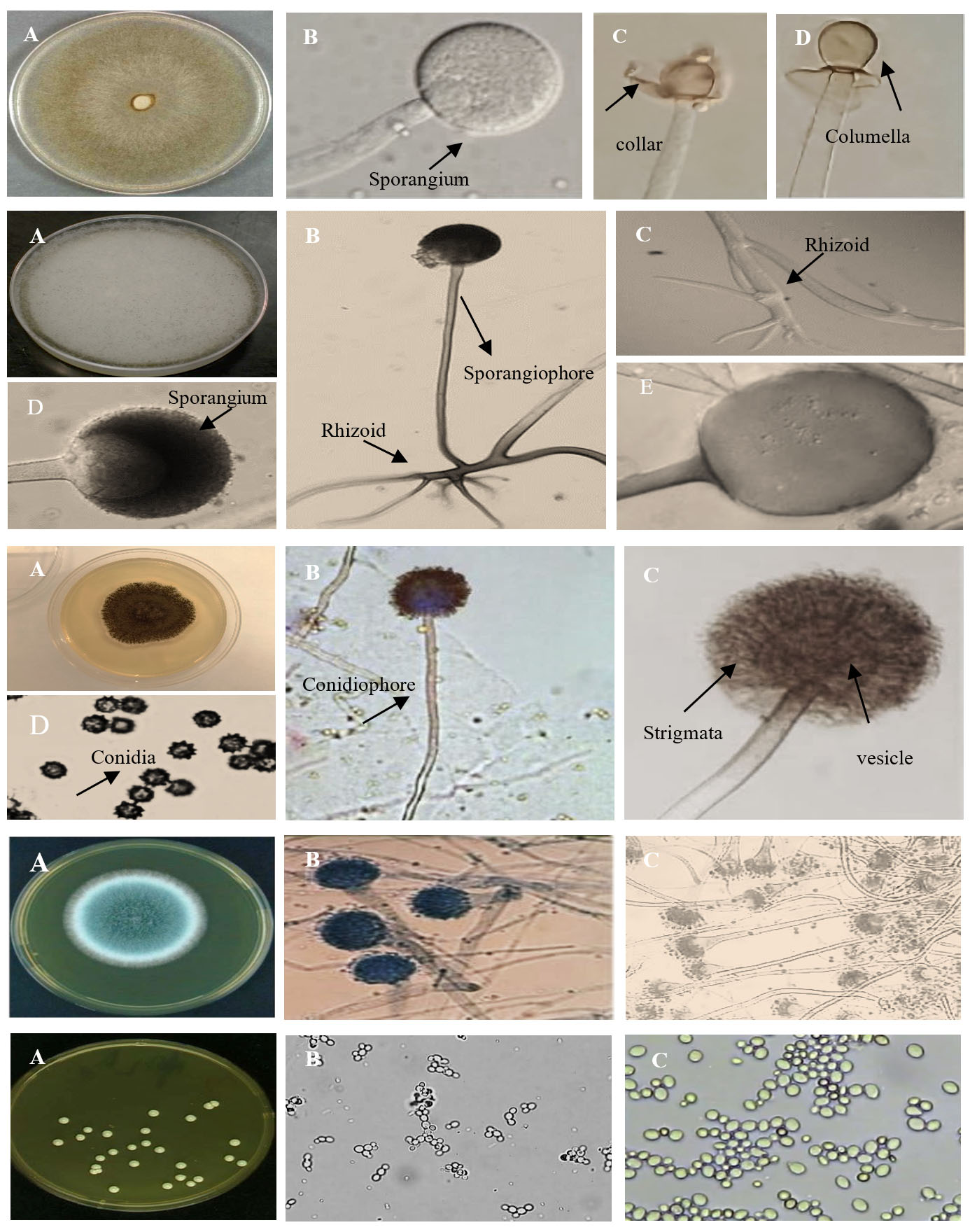
Figure 1. Morphological and microscopic characterization of isolated fungi. Microscopic photos of Mucor irregularis (A) on PDA medium, (B-D) Sporangiophores, sporangia, Sporangiospores and Columella with clear collar present (1). Microscopic photos of Rhizopus oryzae (A) on PDA medium, (B-E) Sporangiophores, Sporangia, Sporangiospores and Rhizoid present (2). Microscopic photos of Aspergillus niger (A) on PDA medium, (B-D) Conidia, conidiophore, vesicle present (3). Microscopic photos of Aspergillus fumigatus (A) on PDA medium, (B-C) Conidia, conidiophore, vesicle present (4). Microscopic photos of Candida glabrata (A) on PDA medium, (B-C) Budding yeast cells (5)
Previous reports indicated that COVID-19 patients who have fungal co-infections showed a greater possibility of death especially, if they have additional underlying illnesses like hypertension, diabetes mellitus, oncological illnesses, kidney, heart, and liver problems (Table 2).26,28,29
Table (2):
Fungal species related to COVID-19 infections
Fungal species in co-infection |
Site/place |
Country |
Year |
Author (s) |
|---|---|---|---|---|
Aspergillus fumigatus |
Pulmonary infection |
Germany |
2020 |
Koehler et al.30 |
Aspergillus fumigatus |
Respiratory infection |
EUA |
2020 |
Mitaka et al.31 |
Candida glabrata; Aspergillus fumigatus |
Pulmonary infection/Blood |
China |
2020 |
Lansbury et al.32 |
Candida glabrata |
Blood |
Colombia |
2021 |
Frías-De-León et al.33 |
Rhizopus oryzae |
Rhino-orbito-cerebral |
Brazil |
2021 |
Lacerda et al.34 |
Aspergillus fumigatus, A. niger |
Pulmonary infection |
Austria |
2022 |
Lackner et al.35 |
Mucormycoses (black fungi) |
Sino-nasal-orbital/ necroses |
India |
2022 |
Upadhayay et al.36 |
Characterization of the free ligand (L)C20H14Br2N2O2 and its La-L-(NO3)3C20H14Br2N5O11 nano complex
The displayed formula in Figure 2 is consistent with analytical findings of the used ligand in the current work. Analysis of C, H, N, and O has been investigated for both the ligand and it’s La-nano-complex. The M.P. was sharp, displaying purity of the investigated compounds. In addition, the results of the elemental analysis of the produced complexes are in match with the requirements given by the complex’s proposed formula.
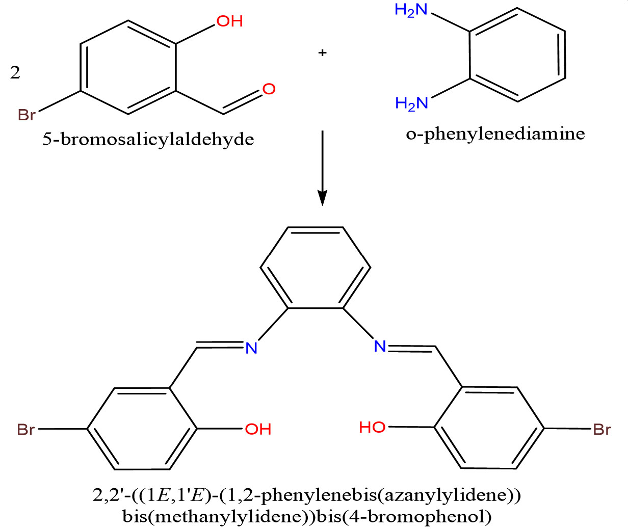
Figure 2. The proposed structure of the prepared free ligand
The single peaks on 9.34 ppm & 11.25 ppm in NMR spectra of the free ligand (Figure 3) are attributed to the HC=N and OH protons, respectively. Also visible are multiple peaks representing aromatic protons between 6.4 and 8.18 ppm.18,19 In contrast, the NMR data of the free Schiff base ligand and its La-nano complex are feasible to verify that the ligand and its metal ion have been complexed (Figure 4). The spectra of the La-nano complex show a noticeable shift in the signals attributable to the azomethine & phenolic protons after meticulous complexation. This observation implies that neutral tetradentate coordination via metal ions occurs without a need for deprotonation and both phenolic and azomethine groups are involved. The ligand also seems to act as an O2N2 coordination sphere.
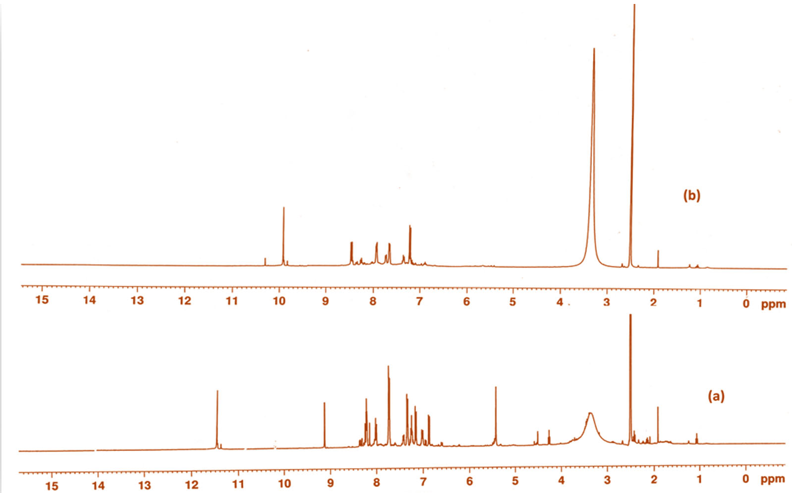
Figure 3. 1H NMR data for (a) Free ligand and (b)
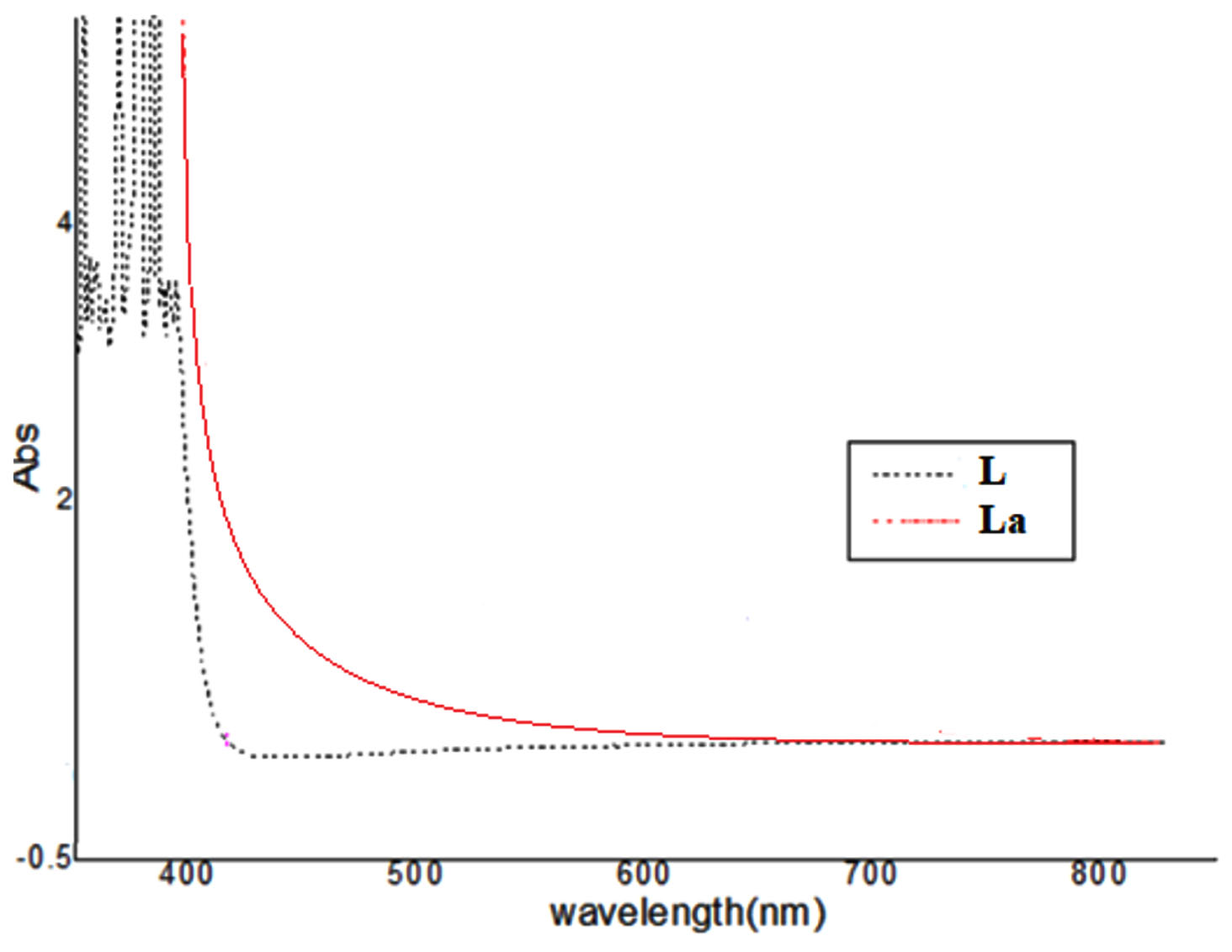
Figure 4. UV-vis spectra for the free ligand and its La complex
To establish the type of bonding, the IR spectra of the free ligand and its nano complex were compared. The group of phenol (OH) is responsible for a great absorption band in the ligand’s infrared spectra at 3456 cm-1.20,21 Due to the C-O (phenolic group), a medium band is also formed at 1290 cm-1 in the spectra. The azomethine group’s C=N resulted in the formation of a strong band at 1620 cm-1.17 When the spectra of La complex were compared to that of the free ligand, the band at 3456 cm-1 altered to a lesser rate at 3400 cm-1, demonstrating which OH group participated in complexation without being de-protonated. Strong bands associated with azomethine & C-O (azomethine & phenolic groups) at 1620 and 1290 cm-1 are stirred to lower wavenumbers (1530 cm-1 & 1250 cm-1, respectively) in complex, indicating N & O coordination to the metal ion. Three additional spectral bands at 1400 cm-1, 1370 cm-1, and 1025 cm-1 that are displayed by the complex are all compatible with the coordination mode of the NO3 group. The complete set of conductance data is supported by these findings. Novel bands in IR spectra of the metal nano complex in the low-frequency region at 545 cm-1 & 510 cm-1 may be assigned to these compounds because M-O & M-N, respectively, are not present in the spectrum of the free ligand, firmly proving bonding.21,22
The parent peak of the Schiff base ligand’s mass spectrum is located in m/e = 474.36 (48.78%), which is the same as the chemical formula C20H14Br2N2O2. The strength of the spectrum’s many peaks, which stand for various fragments and indicate the stability of those parts, is another feature. The suggested mechanism for the decomposition phases of the ligand is depicted in Figure 5.
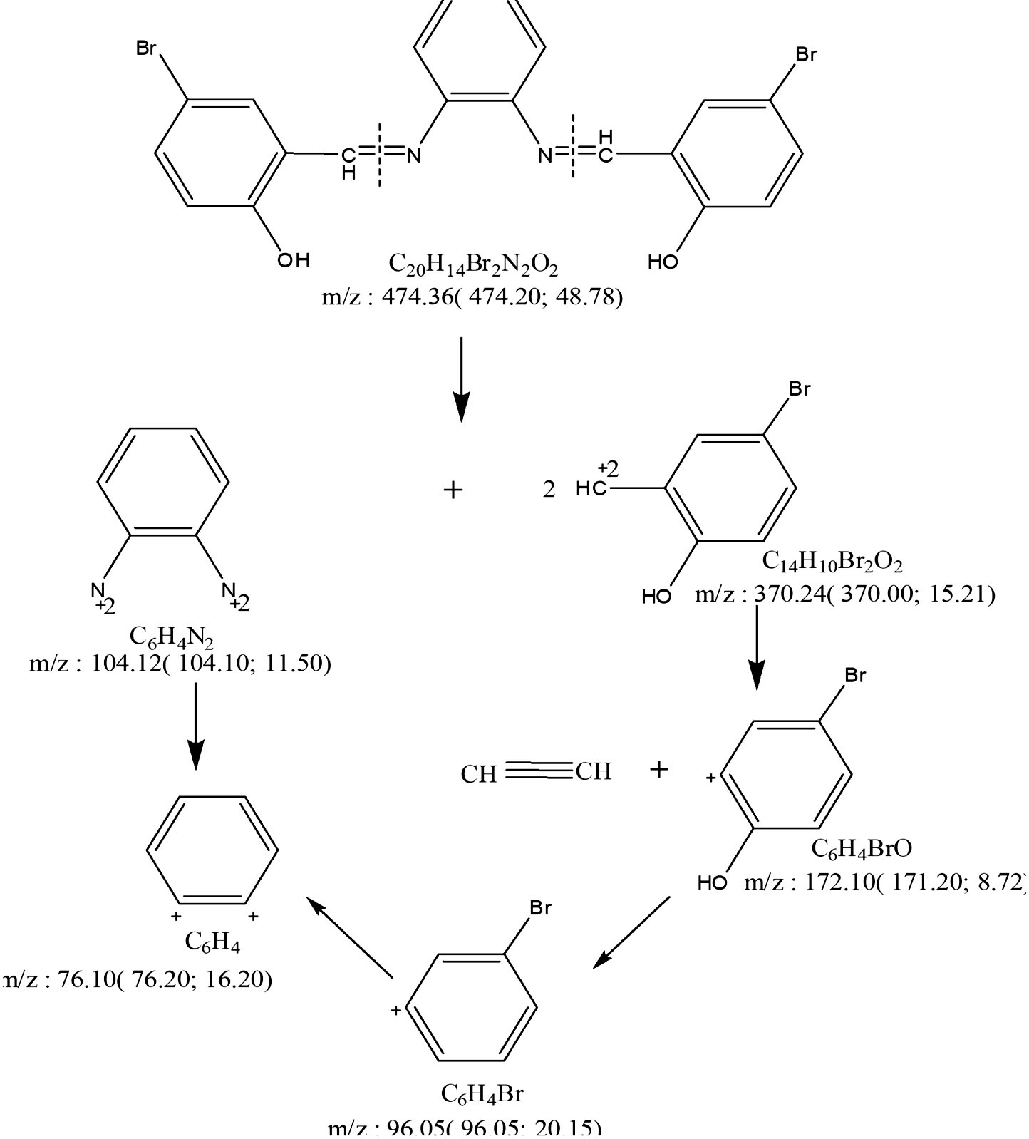
Figure 5. The pattern of mass fragmentation for the free ligand
The conductivity value of the La-nano complex can be calculated using the formula m = K/C, where K is the specific conductance and C is the molar concentration of the nanometal complex solution. Following the compound’s dissolution in (10-3M) DMF, the solution’s molar conductivity was calculated at 25°C. According to the results, the complex has a molar conductance of 50.50 Ohm-1mol-1cm2, which suggests that it is mono-electrolytic. Observations similarly show that nitrate ions are linked to metal cations.21,23
The Ultraviolet-vis spectra of the ligand & its metal complex in DMF were recorded at temperatures between 200 and 800 nm. Benzene & azomethine groups, respectively, were said to be responsible for two absorption peaks at 265 and 339 nm induced by p-p* and n-p* transitions in the ligand spectra.20,22
The coordination of the free ligand with the metal ion caused the bands related to p-p* and n-p* transitions in the spectrum of the e La nano complex to be shifted to a lower or higher frequency. No d-d transition exists in the La (III) complex. The octahedral structure may be suggested as a result of all of this knowledge.
SEM was used to investigate the size and shape of the nano complex (Figure 6). The particles seemed to be semi-spherical and/or Trapezoid in shape and to have some agglomerations. La(III) nanoparticles were stabilized by their interaction with the Schiff base ligand, as seen in SEM images.24 The complex’s structure can be inferred from the correlation of all these findings, as shown in Figure 7.
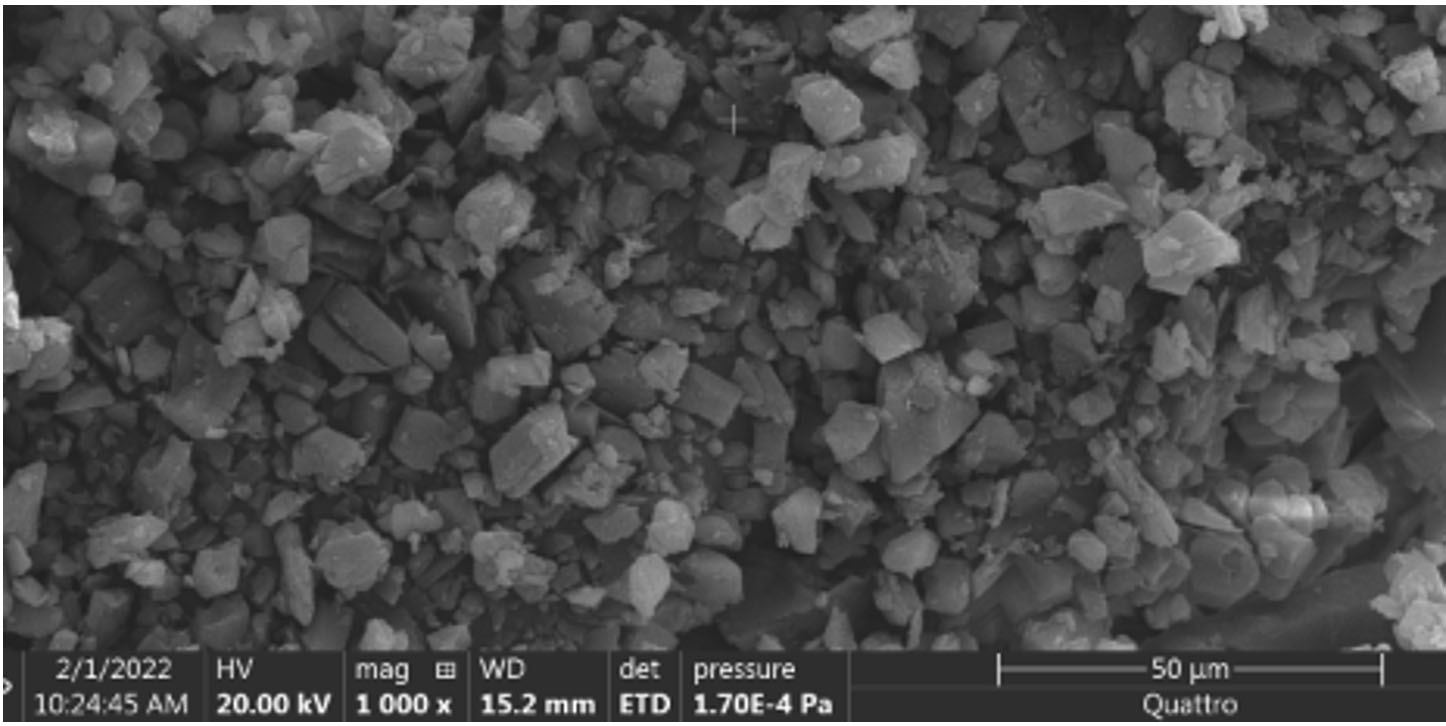
Figure 6. SEM image of nanoparticle complex
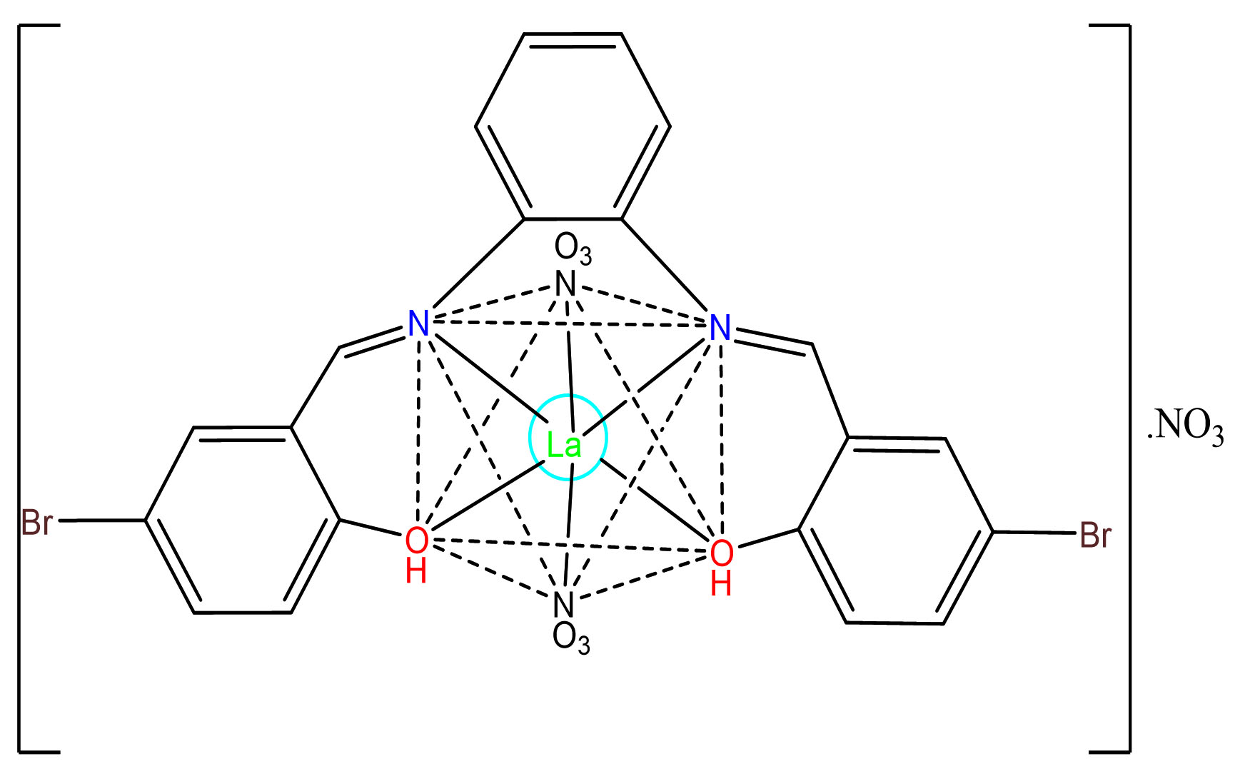
Figure 7. Expected structure of La nano complex
Antifungal activity
Limiting the growth of the mycelium of investigated fungi is considered the base of the antifungal activity in the current study. The results explored a concentration-dependent increase in biological activity, as indicated in Table 3,4 and Figure 8. The minimum inhibitory concentration (MIC) for the La-nano-complex was 0.64 mg/ml. Using an effective antifungal drug is crucial to save the patient’s life which must be coupled with treating the underlying illness. When used to treat mucormycosis, the La-nano complex has shown promising effects in terms of reducing the activity of fungi.37
Table (3):
Inhibition zones developed at different La-nano complex concentrations against isolated fungi
| Fungal strain | Inhibition zone diameter (mm) | ||||
|---|---|---|---|---|---|
| Control miconazole | 0.32 | 0.64 | 1.28 | 2.56 | |
| Aspergillus fumigatus | 13.4 ± 1.5 | 0 | 13.7± 1.8 | 17.9 ± 2.1 | 25 ± 0.8 |
| Aspergillus niger | 11.9 ± 0.01 | 0 | 15.9 ± 0.09 | 20.9 ± 0.8 | 28.7 ± 1.3 |
| Candida glabrata | 11.2 ± 2.02 | 0 | 15.2 ± 0.2 | 19.6 ± 1.00 | 28.6 ± 0.05 |
| Mucor irregularis | 12.1 ± 0.7 | 0 | 14.1 ± 1.4 | 18.3 ± 0.21 | 26.2 ± 0.13 |
| Rhizopus oryzae | 12.9 ± 1.7 | 0 | 15.5 ± 0.7 | 20.9 ± 1.13 | 27 ± 1.63 |
Table (4):
Inhibition zones developed by coated filter with La-nano-complex at 0.64 mg/ml against isolated fungi
| Fungal strain | Inhibition zone diameter (mm) | |||
|---|---|---|---|---|
| After 15 min | After 30 min | After 45 min | After 60 min | |
| Aspergillus fumigatus | 2.0 ± 2.01 | 3.2 ± 1.5 | 3.8 ± 0.09 | 5.1 ± 0.90 |
| Aspergillus niger | 2.8 ± 1.00 | 4.1 ± 0.5 | 4.6 ± 0.10 | 5.8 ± 0.01 |
| Candida glabrata | 2.5 ± 0.11 | 3.6 ± 1.7 | 4.0 ± 0.18 | 6.1 ± 0.08 |
| Mucor irregularis | 2.7 ± 0.06 | 3.5 ± 0.9 | 3.9 ± 2.00 | 4.8 ± 0.91 |
| Rhizopus oryzae | 2.5 ± 0.01 | 3.8 ± 1.2 | 4.2 ± 0.09 | 6.1 ± 0.21 |

Figure 8. Effect of La-nano complex with different concentration, 0.32 mg/ml (1), 0.64 mg/ml (2), 1.28 mg/ml (3), and 2.56 mg/ml (4) on the growth of tested isolated fungi, under dark condition at 30°C
Effective control/treatment of COVID-19 and the other respiratory viruses like influenza requires wider insights to interfere/prevent planning for the prevention, diagnosis, and treatment of these infections.
The coated filter with La-nano-complex proved the ability of the compound at 0.64 mg/ml concentration to inhibit all isolated fungi when applied for 60 min with an inhibition rate of up to 95% (Table 4). In conclusion, we were able to isolate different fungal species from the filters of the air-conditioning systems in COVID-19 rehabilitation rooms which increase the risk of co-infections. However, the prepared La-nano-complex filters were able to significantly decrease the invading microbes and eradicate Rhizopus, Aspergillus, Mucor, Candida albicans isolates at 0.64 mg/ml concentration Figure 9.
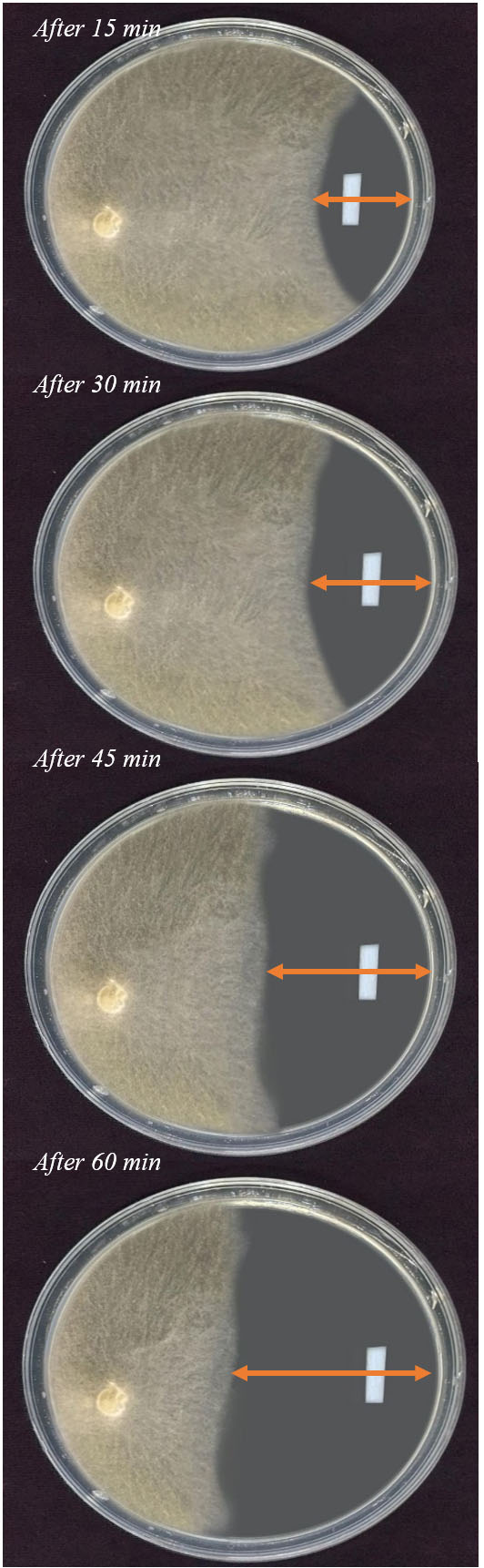
Figure 9. Effect of La-nano complex) 0.64 mg/ml) on the growth of R. oryze, under dark condition at 30°C, at different period
DFT study
The optimized structure of the ligand and its Lanthanum complex with labeling of the atoms are presented in Figure 10.
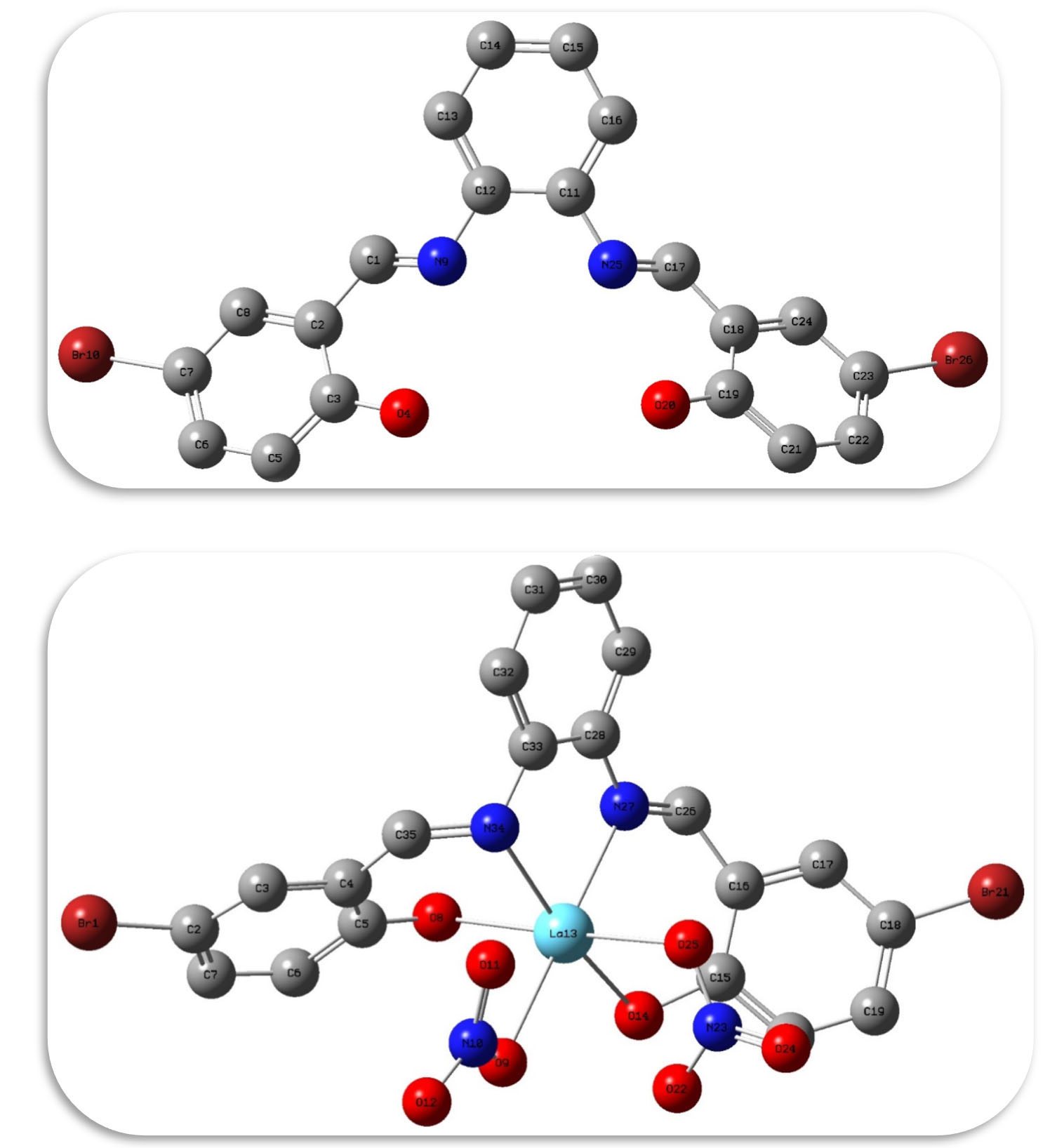
Figure 10. Optimized structure for Ligand and its La (III) complex with their labeling
Table 5 provides a comparison of various electronic properties between the ligand and its lanthanum complex. The ligand exhibits lower binding energy, ionization potential, electron affinity, absolute electronegativity, and chemical potential compared to the lanthanum complex. Meanwhile, the lanthanum complex displays smaller energy gap, absolute hardness, and global softness relative to the ligand. These findings indicate that coordination with lanthanum enhances molecular stability and electron-withdrawing nature. Specifically, the lanthanum complex features stronger bonding interactions, higher resistance to changes in electron density, and increased tendency to act as an electrophile in chemical reactions. Analysis of the HOMO-LUMO energy gap values observed reveals that the complex molecule possesses higher reactivity than the uncoordinated ligand molecule, as evidenced by its smaller energy gap (ִE = 2.08 eV for the complex, 2.36 eV for the ligand). This computational result corroborates well with the findings from biological activity assays, which demonstrated enhanced reactivity for the complex relative to the ligand alone. The decreased energy requirement for electron transition in the complex implies it may more readily undergo redox reactions involved in bioactivity mechanisms.
Table (5):
Theoretical parameters of the ligand and its Lanthanum complex
Ligand |
La (III) complex |
|
|---|---|---|
Binding Energy kcal/mol |
-4695.16 |
-4949.05 |
EHOMO (eV) |
-5.11 |
-5.68 |
ELUMO (eV) |
-2.75 |
-3.60 |
Gap, ∆E |
2.36 |
2.08 |
Ionization potential (eV) |
5.11 |
5.68 |
Electron affinity |
2.75 |
3.60 |
Absolute electronegativity |
3.93 |
4.64 |
Absolute hardness |
1.18 |
1.04 |
Chemical potential |
-3.93 |
-4.64 |
Global softness |
0.59 |
0.52 |
Global electrophilicity |
6.54 |
10.35 |
Additional electronic charge |
3.33 |
4.46 |
Dipole moment, (Debye) |
2.85 |
2.25 |
The atomic charges of the ligand and its Lanthanum complex as calculated by the Mulliken method26 are presented in Table 6. Mulliken charges have a direct correlation with a molecule’s vibrational properties. The distribution of positive and negative charges among atoms is a primary factor influencing increases or decreases in bond lengths. Specifically, polarization of charge away from bonding atomic centers tends to elongate bonds by weakening orbital overlap, while accumulation of charge between atoms serves to strengthen interactions and shorten distances. Computational analysis of Mulliken charges can provide useful insights into how coordination and charge transfer may impact fundamental vibrational characteristics by altering electrostatic forces along chemical bonds at the atomic scale. Higher electrophilic charge density is observed on the oxygen and nitrogen atoms. Notably, the charge on the N and o atoms atom in the ligand are more negative than other atoms, indicating that they have greater ability for metal ion coordination due to increased basicity.27 Upon complexation, the electrophilic charge densities increase relative to the uncoordinated ligand. This trend correlates well with the higher biological activity observed experimentally for the cobalt complex versus the ligand alone. The increased charge polarization upon complexation implies stronger binding interactions with biological targets, enhancing reactivity profiles in accordance with observed bioactivity outcomes.
Table (6):
The Mulliken atomic charges of the ligand and its Lanthanum complex
| Ligand | La (III) complex | ||||||
|---|---|---|---|---|---|---|---|
| Atom | charge | Atom | charge | Atom | charge | Atom | charge |
| C (1) | 0.031 | C (2) | 0.067 | Br (1) | 0.017 | C (7) | -0.018 |
| C (3) | 0.352 | O (4) | -0.493 | C (2) | -0.026 | O (8) | -0.425 |
| C (5) | -0.098 | C (6) | -0.043 | C (3) | -0.019 | O (9) | -0.407 |
| C (7) | 0.196 | C (8) | -0.088 | C (4) | -0.056 | N (10) | 0.728 |
| N (9) | -0.292 | Br (10) | -0.242 | C (5) | 0.312 | O (11) | -0.406 |
| C 11) | 0.119 | C (12) | 0.124 | C (6) | -0.067 | O (12) | -0.281 |
| C 13) | -0.068 | C (14) | -0.046 | C (35) | 0.109 | C (33) | 0.152 |
| C (15) | -0.047 | C (16) | -0.065 | N (34) | -0.255 | C (32) | -0.051 |
| C (17) | 0.037 | C (18) | 0.059 | C (31) | -0.041 | C (30) | -0.034 |
| C (19) | 0.338 | O (20) | -0.479 | C (29) | -0.050 | C (28) | 0.147 |
| C (21) | -0.086 | C (22) | -0.044 | N (27) | -0.254 | C (26) | 0.134 |
| C (23) | 0.184 | C (24) | -0.076 | O (25) | -0.409 | O (24) | -0.282 |
| N (25) | -0.283 | Br (26) | -0.235 | N (23) | 0.739 | O (22) | -0.400 |
| Br (21) | 0.016 | C (20) | -0.070 | ||||
| C (19) | -0.024 | C (18) | -0.032 | ||||
| C (17) | -0.010 | C (16) | -0.057 | ||||
| C (15) | 0.312 | O (14) | -0.431 | ||||
| La (13) | 1.340 | ||||||
Fungal co-infections are identified from air conditioning filters in the rooms of critically sick COVID-19 patients, with aspergillosis having the highest incidence, followed by candidemia. In light of these connections between fungi and infections, as well as between some treatments and fungi, contemporary medical practices are challenged to come up with creative ways to prevent subsequent fungus infections. The prognosis for COVID-19 patients may be improved in the interim, nevertheless, by quick detection of fungus co-infections and treatment with antifungal medication. The lanthanum complex exhibits enhanced stability and electron-withdrawing nature, as evidenced by its lower binding energy, ionization potential, electron affinity, absolute electronegativity, and chemical potential. Additionally, the lanthanum complex displays a smaller energy gap, absolute hardness, and global softness, indicating optimized structural features and reactivity profiles.
ACKNOWLEDGMENTS
The authors would like to thank the Deputyship for Research and Innovation, Ministry of Education in Saudi Arabia, for funding this research work through project number DSR-2021-03-03127.
CONFLICT OF INTEREST
The authors declare that there is no conflict of interest.
AUTHORS’ CONTRIBUTION
SMNM, RHT conceptualized and visualized the study. SMNM, RHT applied methodology. SMNM, TAY and RHT performed experiments. SMNM, TAY, RHT performed data curation. SMNM, TAY and RHT performed data analysis. SMNM, TAY and RHT drafted the manuscript. SMNM, TAY and RHT wrote the manuscript. SMNM and RHT reviewed the manuscript. All authors read and approved the final manuscript for publication.
FUNDING
This study was supported by the Deanship of Scientific Research at Jouf University through research grant No. (DSR-2021-03-03127).
DATA AVAILABILITY
The datasets generated and/or analysed during the current study are available from the corresponding author on reasonable request.
ETHICS STATEMENT
This article does not contain any studies on human participants or animals performed by any of the authors.
- Pal M. Zygomycosis: A highly infectious emerging opportunistic fungal disease of public health concern. OAJMMS. 2020;3(1):000122.
Crossref - Pal M, Lee CW. Pulmonary zygomycosis in chicks due to Absidia corymbifera. Korean J Vet Clin Med. 1994;11(1):339-341.
- Pal M. Veterinary and Medical Mycology. 1st (Edn.), ICAR, New Delhi, India. 2007.
- Eucker J, Sezer O, Graf B, Possinger K. Mucormycoses. Mycoses. 2001;44(7):253-260.
Crossref - Raut A, Huy NT. Rising incidence of mucormycosis in patients with COVID-19: another challenge for India amidst the second wave. Lancet Respir Med. 2021;9(8):e77.
Crossref - Sugar AM. Mucormycosis. Clin Infect Dis. 1992;14(1):126-129.
Crossref - Chayakulkeeree M, Ghannoum MA, Perfect JR. Zygomycosis: the re-emerging fungal infection. Eur J Clin Microbiol Infect Dis. 2006;25(4):215-229.
Crossref - Mantadakis E, Samonis G. Clinical presentation of zygomycosis. Clin Microbiol Infect. 2009;15(S5):15-20.
Crossref - Singh AK, Singh R, Joshi SR, Mishra A. Mucormycosis in COVID-19: A systematic review of cases reported worldwide and in India. Diabetes Metab Syndr. 2021;15(4):102146.
Crossref - Centers for Disease Control and Prevention Fungal diseases. 2019. https://www.cdc.gov/fungal/diseases/mucormycosis/risk-prevention.html
- Sutherland Ken. 5th ed. Filters and Filtration Handbook. Elsevier. 2008.
Crossref - Araujo R, Cabral JP. Fungal air quality in medical protected environments. Air Qual. 2010.
Crossref - Deshmukh SP, Patil SM, Mullani SB, Delekar SD. Silver nanoparticles as an effective disinfectant: A review. Mater Sci Eng C Mater Biol Appl. 2019;97:954-965.
Crossref - Balagna C, Francese R, Perero S, Lembo D, Ferraris M. Nanostructured composite coating endowed with antiviral activity against human respiratory viruses deposited on fibre-based air filters. Surf Coat Technol. 2021;409:126873.
Crossref - Park K, Kang S, Park J-W, Hwang J. Fabrication of silver nanowire coated fibrous air filter medium via a two-step process of electrospinning and electrospray for anti-bioaerosol treatment. J Hazard Mater. 2021;411:125043.
Crossref - Ju Y, Han T, Yin J, et al. Bumpy structured nanofibrous membrane as a highly efficient air filter with antibacterial and antiviral property. Sci Total Environ. 2021;777:145768.
Crossref - Taha RH. Preparation, Spectroscopic Characterization, DNA Cleavage, Antimicrobial and Antitumor Investigations of Nickel and Uranyl Schiff Base Complexes in Bulk and Nano size. Curr Sci Int. 2015;4(4):684-700.
- Fasina TM, Ogundele OO, Ayeni I. Synthesis and biological properties of N2O2 Schiff bases derived from o-phenylenediamine and substituted salicylaldehydes. J Chem Pharm Res. 2014;6(6):816-819.
- Taha RH, Almutlq NJ, Elnasr TAS, Alshammari MS, Mohamed SH, Moustafa SM. Ce and Fe complexes as potent antifungal agents for wallpapers. Bull. Chem. Soc. Ethiop, 2024; 38(2), 365-383.
Crossref - Moustafa SMN, Taha RH. Mycogenic Nano-Complex for Plant Growth Promotion and Bio-Control of Pythium aphanidermatumm. Plants. 2021;10(9):1858.
Crossref - Gomathi V, Selvameena R. Synthesis, Physico chemical and Antimicrobial Studies on Schiff Base Complexes of Sulfa Drug with Metals of Life. Indian J Appl Res. 2013;3(4):51-53.
- Karekal MR, Biradar V, Mathada MBH. Synthesis, Characterization, Antimicrobial, DNA Cleavage, and Antioxidant Studies of Some Metal Complexes Derived from Schiff Base Containing Indole and Quinoline Moieties. Bioinorg Chem Appl. 2013;2013:315972.
Crossref - Alsohaimi IH, Alotaibi NF, Albarkani AM, et al. Photocatalytic wastewater treatment and disinfection using green ZnO-NP synthesized via cera alba extract. Alexandria Engineering Journal, 2023;83, 113-121.
Crossref - Moustafa SMN, Taha RH. Biosynthesis of Zinc Nanocomplex Employing for Plant Growth Promotion and Bio-Control of Pythium ultimum. Intechopen. 2021.
Crossref - Taha RH, Alrabie A, Badr EE, Shymaa MN, Baker SA. Design, synthesis, characterization, spectral, chemical analysis, biological evaluation, and molecular docking studies of novel fatty Schiff base ligands and their nickel and mercury complexes. Inorganic Chemistry Communications, 2023;158:111382.
Crossref - Ahmed RM, Yousif EI, Hasan HA, Al-Jeboori MJ. Metal Complexes of Macrocyclic Schiff-Base Ligand: Preparation, Characterisation, and Biological Activity. Scientific World Journal. 2013;2013:289805.
Crossref - Wu X, Ray AK. Density-functional study of water adsorption on the PuO 2 (110) surface. Physical Review B. 2002;65(8):085403.
Crossref - Mohamed GG, Zayed MA, Abdallah SM. Metal Complexes of a Novel Schiff Base Derived from Sulphametrole and Varelaldehyde. Synthesis, Spectral, Thermal Characterization and Biological Activity. J Mol Struct. 2010;979(1-3):62-71.
Crossref - Seyedjavadi SS, Bagheri P, Nasiri MJ, Razzaghi-Abyaneh M, Goudarzi M. Fungal infection in co-infected patients with COVID-19: an overview of case reports/case series and systematic review. Front. Microbiol. 2022;13: 888452
- Mandegani Z, Mozaffar A, Zahra A, Afshan M, Nasser I, Akbar O. A Nano Tetraimine Pd(0) Complex: Synthesis, Characterization, Computational Studies and Catalytic Applications in the Heck-Mizoroki Reaction in Water. Green Chem. 2015;17:3326-3337.
Crossref - Koehler P, Cornely OA, Böttiger BW, et al. COVID-19 associated pulmonary aspergillosis. Mycoses, 2020; 63(6):528-534.
Crossref - Mitaka H, Perlman DC, Javaid W, Salomon N. Putative invasive pulmonary aspergillosis in critically ill patients with COVID-19: an observational study from New York City. Mycoses, 2020;63(12):1368-1372.
Crossref - Lansbury L, Lim B, Baskaran V, Lim WS. Co-infections in people with COVID-19: a systematic review and meta-analysis. Journal of infection, 2020;81(2):266-275.
Crossref - Frías-De-León MG, Hernández-Castro R, Conde-Cuevas E, et al. Candida glabrata antifungal resistance and virulence factors, a perfect pathogenic combination. Pharmaceutics, 2021;13(10):1529.
Crossref - Bonates P, João GAP, Cruz KS, et al. Fatal rhino-orbito-cerebral mucormycosis infection associated with diabetic ketoacidosis post-COVID-19. Rev Soc Bras Med Trop. 2021;54:e03582021.
Crossref - Lackner N, Thomé C, Öfner D, et al. COVID-19 associated pulmonary aspergillosis: diagnostic performance, fungal epidemiology and antifungal susceptibility. J. Fungi. 2022;8(2):93.
Crossref - Upadhayay P, Bansal K, Goyal A. Epidemiology, Risk Factors, Diagnosis and Treatment of Mucormycosis (Black Fungus): A Review. Current Pharmaceutical Biotechnology, 2023;24(13):1645-1656.
Crossref
© The Author(s) 2024. Open Access. This article is distributed under the terms of the Creative Commons Attribution 4.0 International License which permits unrestricted use, sharing, distribution, and reproduction in any medium, provided you give appropriate credit to the original author(s) and the source, provide a link to the Creative Commons license, and indicate if changes were made.