

ISSN: 0973-7510
E-ISSN: 2581-690X
 , Robin Gogoi1,3 , Deeba Kamil1, Rajesh Kumar4, Tapan Kumar Mondal5 and Gautam Chawla6
Copper nanoparticles are increasingly being explored as a sustainable alternative to conventional fungicides for controlling plant pathogens, offering enhanced efficacy and reduced environmental toxicity. This study investigated the antifungal mechanism of essential oil-grafted copper nanoparticles (EGCuNPs) using scanning electron microscopy against Sclerotium rolfsii, Fusarium verticillioides, and Rhizoctonia solani. Morphological and molecular characterization confirmed the identity of the pathogens, with ITS sequencing validating the identity of R. solani (GenBank accession OR251925). In vitro assays at ED50 concentrations of EGCuNPs revealed significant growth inhibition of all three fungi. Scanning electron microscopy (SEM) demonstrated disruption of hyphae and spores, confirming the potential antifungal activity of EGCuNPs. While S. rolfsii exhibited some tolerance, EGCuNPs caused significant damage to hyphae. The study demonstrates the antifungal mechanism of EGCuNPs, proving its potential as a next-generation fungicide. Further research should address molecular mechanisms through transcriptomics and long-term ecological impacts of repeated application of copper-based nano-formulations.
Copper Nanoparticles, Nanoformulations, Next Generation Fungicides, Antifungal Mechanism, Plant Pathogens
In recent times, copper nanoparticles (CuNPs) have shown considerable potential for managing phytopathogens, providing an effective alternative to conventional fungicides. The advantages of CuNPs over conventional fungicides include their enhanced efficacy, better penetration, better solubility, safety against non-target pathogens, and reduced environmental pollution by limiting the use of fungicides, etc.1-3 On the other hand, fungicides with their long residual activity can disrupt beneficial microbial communities and other unintended biota, including algae, aquatic fauna, invertebrates, and soil microorganisms. Fungicides widely used by Indian farmers such as carbendazim and chlorothalonil are implicated as carcinogenic, in addition to having been reported to cause birth defects and endocrine damage.4 Fungicides such as propiconazole, azoxystrobin, tebuconazole, and pyraclostrobin are found to have long residual activity with high toxicity to aquatic systems.4-6 The enhanced efficacy of CuNPs is primarily attributed to their considerable high surface area-to-volume ratio and small size, which facilitate easy penetration through the membrane, disrupting important cellular components, DNA, and other proteins by inducing reactive oxygen species (ROS). Numerous studies have demonstrated the remarkable efficacy of copper-based nanoparticles; for instance, biogenic CuNPs could inhibit Botrytis cinerea growth by 25% at 100 ppm.7 Similarly, CuNPs exhibited 100% inhibition at 30 ppm against Phytophthora capsici and F. oxysporum.8 In addition, excellent antimicrobial effects have been demonstrated against bacteria as well such as Micrococcus luteus, Escherichia coli, Staphylococcus aureus, Streptomyces parvulus, Xanthomonas axonopodis pv. citri, Bacillus subtilis, X. axonopodis pv. punicae, and Pseudomonas aeruginosa.9-11
Clove oil, extracted from Syzygium aromaticum is rich in eugenol, its main constituent, which exhibits strong antifungal activity. Clove oil has the ability to disrupt the cell walls and cellular membranes of microorganisms, thereby facilitating its penetration across the cytoplasmic membrane and entry into the cells. Consequently, it interferes with essential cellular processes, including the synthesis of DNA and proteins.12 Consistent with these properties, several studies have reported the broad-spectrum antimicrobial efficacy of clove oil against a range of fungal pathogens. For instance, a minimum inhibitory concentration (MIC) of 92.56 µL L-1 resulted in significant inhibition of Botrytis cinerea under both in vitro and in vivo conditions.13 Likewise, clove oil at 4% completely inhibited the growth of Fusarium oxysporum and Rhizoctonia solani and significantly reduced disease intensity by 72% and 81%, respectively, under in vivo conditions.14 More recently, clove oil at 5% have been demonstrated to cause 100% inhibition of F. equiseti growth and reduced wilt severity and incidence in bitter gourd by 66.99% with induction of defense genes in plant when applied to the soil.15 In contrast, another study reported a relatively low concentration of 1 µL mL-1 i.e. 1000 ppm of clove oil causing complete inhibition of three fungal pathogens, namely Fusarium redolens, F. commune, and F. oxysporum f.sp. radicis-lycopersici.16 Importantly, the antimicrobial potential of clove oil is not restricted to fungal pathogens; it has also demonstrated remarkable efficacy against several bacterial phytopathogens, including Gram-positive bacteria such as Streptomyces spp., and Rhodococcus fascians and Gram-negative bacteria such as Xanthomonas axonopodis pv. punicae, X. phaseoli pv. phaseoli, X. campestris pv. pelargonii, Erwinia carotovora pv. carotovora, Agrobacterium tumefaciens, Pseudomonas syringae pv. syringae, Ralstonia solanacearum, and Pectobacterium spp.17-20
However, comparatively limited reports are available on the synergistic effects of clove oil and CuNPs. Nevertheless, findings from few studies combining clove oil with other nanomaterials provide promising evidence of the potential of such nano-enabled formulations. For instance, clove oil exhibited enhanced efficacy against Gaeumannomyces graminis var. tritici when it was encapsulated in mesoporous silica nanoparticles.21 Also, combination of clove essential oil and chitosan nanoparticles demonstrated enhanced antifungal efficacy rendering complete growth inhibition of Aspergillus niger at 1.5 mg mL-1.22
Considering the remarkable antifungal efficacy of CuNPs and combination of CuNPs and clove oil, as established in our previous study from a low dose as 20 ppm,1,23-25 in the current study, the morphological alterations induced by CuNPs were examined using scanning electron microscopy (SEM) in the hyphae and spores of Fusarium verticillioides and the hyphae of two sclerotial-forming fungi viz., Rhizoctonia solani and Sclerotium rolfsii. We report the basis of the mode of action of EGCuNPs on fungal pathogens, advocating their potential role as a next generation fungicide.
Collection and isolation of fungi
The fungi Fusarium verticillioides and Sclerotium rolfsii were obtained from the Maize Pathology Lab, Division of Plant Pathology, ICAR-Indian Agricultural Research Institute, New Delhi. Rhizoctonia solani was isolated from a sample collected from an infected maize field, ICAR-IARI, New Delhi. For isolation and purification, the standard protocol of Tutte was followed.26 Leaf sheath from below 2nd internodes exhibiting water soaked lesions was collected for isolation. Symptomatic cut sheath pieces (~5 mm), including both diseased and healthy tissue, were surface-sterilized with 1% sodium hypochlorite (NaOCl) followed by rinsing three times with sterile distilled water. The pieces were dried on sterile filter paper using forceps and placed on solid media supplemented with streptomycin sulfate (100 µg mL-1). The resulting cultures were purified by hyphal tip method and maintained on PDA slants at 4 ± 1 °C for subsequent use. The CuNPs based nanoformulation referred as EGCuNPs employed in this study were synthesized following a previously standardized protocol.23
Morpho-molecular identification of fungi
The fungal isolates, R. solani, Sclerotium rolfsii, and Fusarium verticillioides were cultured on Potato Dextrose Agar (PDA). For microscopic observation, clean slides and coverslips were used and the prepared slide was examined under the microscope to determine its characteristic morphological structure using the appropriate magnification (Olympus microscope connected to CMOS HDMI Truechrome HD, Radical, Japan).
Rhizoctonia solani was cultivated in Potato Dextrose Broth (PDB) at 28 ± 2 °C with constant shaking at 100 rpm for 7 days to obtain a mycelial mat. The culture was passed through Whatman filter paper to separate the mycelia, which were then washed thoroughly with sterile distilled water to remove any remaining culture medium. The excess moisture in tissue was then removed using sterilized filter paper by squeezing. The mycelia were then folded in filter paper and preserved at -20 °C until future use. Genomic DNA was extracted following the extraction method given by Murray and Thompson.27 Further details of the fungal DNA extraction procedure are included as supplementary data. Following extraction, DNA was resuspended in 50 µl of 10 mM Tris-EDTA (pH 8) and its concentration and purity were analyzed using a Nanodrop 2000- UV-Vis spectrophotometer (Thermo Fisher Scientific, USA), by measuring the A260/A280 nm absorbance ratio. DNA quality was subsequently confirmed by agarose gel electrophoresis. The internal transcribed spacer (ITS) gene was amplified using the ITS1 (5′-TCCGTAGGTGAACCTGCCG-3′) and ITS4 (5′-TCCTCCGCTTATTGATATGC-3′) primers.28 The composition of the PCR reaction mixture and thermal cycling conditions are provided as supplementary data.
The amplified ITS fragments were subjected to sequencing with the ITS1 and ITS4 primers. The resulting sequences were initially processed for quality by trimming using FinchTV software. Post sequence trimming for quality control, sequence was subjected to BLAST analysis against the NCBI nucleotide database to check sequence similarity, query coverage, and confirm taxonomic identity. Upon verification, the sequence data was deposited in the NCBI to obtain accession number. Subsequently, phylogenetic analysis was performed by sequence alignment using the CLUSTAL W program, with some sequences retrieved from the NCBI nucleotide database and an appropriate out-group. The aligned sequences were then used to construct a phylogenetic tree.
Treatment of fungal culture with EGCuNPs for SEM sample preparation
The concentration of ED50 value determined by probit analysis (SPSS software version 16) in our previous study, i.e. 43.208 μg ml-1, 51.003 μg ml-1 and 55.096 μg ml-1 for R. solani, F. verticillioides, and S. rolfsii, respectively, were used to treat the fungi through poisoned food technique (Table 1).23 A stock solution of EGCuNPs at a concentration of 10,000 µg ml-1 was prepared. The previously determined ED₅₀ concentration was calculated using the formula C1V1 = C2V2 for a 60 ml of PDA medium. Accordingly, 0.259, 0.306, and 0.331 ml of EGCuNPs stock solution were added to 59.741, 59.694, and 59.669 ml of molten PDA (45-50 °C) for R. solani, F. verticillioides, and S. rolfsii, respectively, to obtain the desired ED50 concentrations. In addition, a control was also maintained. In total, three replication were maintained for each fungi and for the control and experimental design followed was Completely Randomized Design (CRD). Before the incorporation of EGCuNPs into the medium, streptomycin sulfate (SRL, India) was supplemented at 100 µg mL-1 to suppress bacterial contamination. After 5 days of incubation at 25 ± 1 °C and relative humidity of 60% under alternating 12 hrs light/12 hrs dark photoperiod, a 50 mm mycelial disc was cut out from the actively growing margin of the fungal colony using a sterile cork-borer for subsequent processing.
Table 1. ED50 values of essential oil-grafted CuNPs estimated by probit analysis23
| Target Fungi | ED50 (μg ml-1) | Fiducial limit (μg ml-1) | Chi-square (χ2) | Regression Equation | |
|---|---|---|---|---|---|
| Rhizoctonia solani | 43.208 | 32.267 | 53.265 | 27.572 | y=3.6 × x+(-6.143) |
| Fusarium verticillioides | 51.003 | 26.728 | 73.843 | 56.633 | y=2.9 × x+(-4.714) |
| Sclerotium rolfsii | 55.096 | 19.080 | 93.846 | 45.355 | y=1.2 × x+(-2.040) |
Sample preparation for SEM analysis
For the sample preparation, the routine protocol given by Alves et al.29 was employed with minor adjustments. The mycelial disc underwent fixation with 2.5% glutaraldehyde prepared in 0.1 M phosphate buffer (pH 7.2) for 24 hrs, subsequently subjected to three rinse with 0.1 M phosphate buffer. Then samples were subjected to 200 µl of 1% osmium tetroxide for post-fixation at room temperature in the dark conditions for 4 hrs. The samples were washed thoroughly with distilled water, then dehydrated gradually by sequential exposure to ethanol gradient solutions (25%, 50%, 75%, 90%, and 100%), and subsequently subjected to critical point drying. At concentrations of up to 90% ethanol, the treatment was given only once. However, at 100% ethanol concentration, the sample underwent dehydration twice to ensure the complete removal of water. The dehydrated specimens were affixed to aluminum stubs and gold-coated by sputter deposition to improve electrical conductivity. Finally, the sample was observed under SEM for the structural damage (VEGA3, Tescan, Brno, Czech Republic).
Morpho-cultural characterization of phytopathogens
In the plate, we observed compact and dark brownish macro sclerotia that were formed with a white-brownish cottony appearance for R. solani after 15 days of inoculation. However, initially, the culture was white and cottony. Under the microscope, septate hyaline hyphae with characteristic right-angled branching and foot cells with restriction were observed (Figure 1A).30,31 F. verticillioides culture appeared fluffy and cottony, and had a white-purplish colour upon culturing, covering a 7 cm Petriplate within 6-7 days. Under the microscope, hyaline single-celled microconidia in chains and 4-5 septate macroconidia with slightly curved shape with tapering end were observed, confirming its identity (Figure 1B).32-34 Sclerotium rolfsii grew fast on PDA media, covering the 7 cm plate within 5 days. The colour of the colony was white with radial spreading growth. In more than 15-day-old culture plates, roughly spherical dark brown, mustard seed-like sclerotia formed, which are characteristic features of Sclerotium rolfsii. Under the microscope, highly branched septate, hyaline hyphae were observed (Figure 1C).35-37
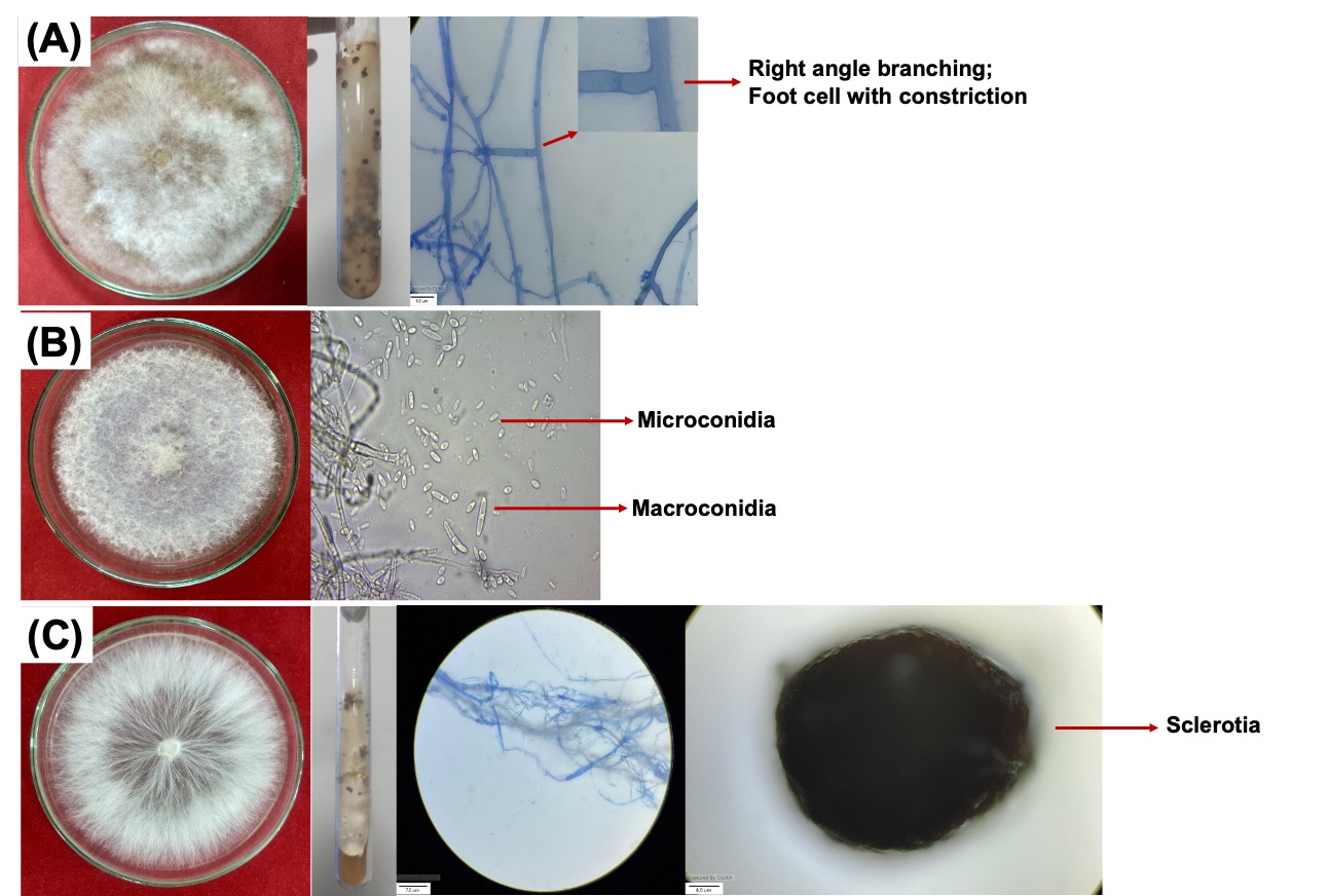
Figure 1. Morphological and cultural characteristics of Rhizoctonia solani (A), Fusarium verticillioides (B), and Sclerotium rolfsii (C)
Molecular characterization of Rhizoctonia solani
PCR amplification resulted in an amplicon size of 710 bp for R. solani (Figure 2A). BLAST analysis against the NCBI database showed 99% sequence identity for R. solani (Table 2), and the sequence was deposited in GenBank under accession number OR251925. A phylogenetic tree was constructed exhibiting the genetic relatedness of fungi with other ITS sequences from NCBI, as shown in Figure 2B. Furthermore, the culture was catalogued in the Indian Type Culture Collection (ITCC), ICAR-IARI, New Delhi, with accession number 9244.
Table 2. Molecular identity of the isolated fungus
Name |
Primer used |
Query length |
Query cover |
Percentage identity |
|---|---|---|---|---|
Rhizoctonia solani |
ITS1 and ITS 4 |
645 |
99% |
99% |
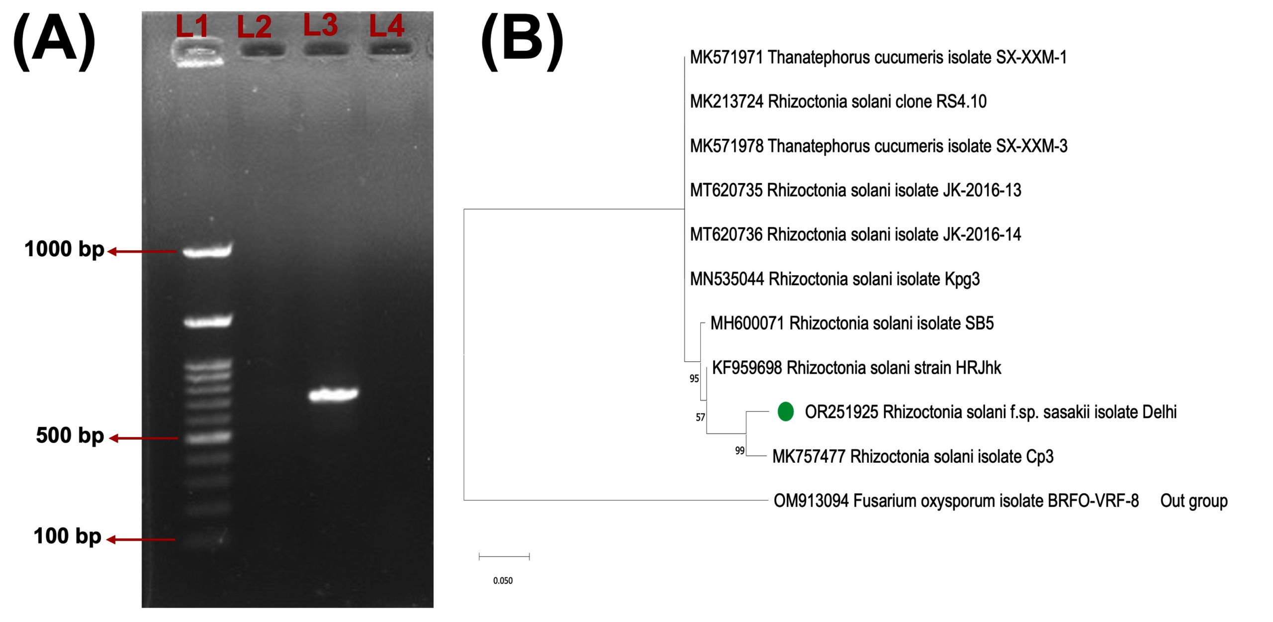
Figure 2. Agarose gel showing amplicon of ITS- Rhizoctonia solani of size 710 bp in third lane (L3) (A); the first lane (L1) is 100 bp ladder, second lane (L2) and fourth lane (L4) are blank. Phylogenetic analysis was conducted by Maximum Likelihood method based on Tamura-Nei model of Rhizoctonia solani (B).
Antifungal activity of EGCuNPs
The ED50 at 43.208 μg ml-1, 55.096 μg ml-1, and 51.003 μg ml-1 concentrations of EGCuNPs inhibited the growth of Rhizoctonia solani, Sclerotium rolfsii, and Fusarium verticillioides by 53.143%, 49.428%, and 54.714%, respectively (Figure 3A, B, and C). Such strong antifungal activity was observed in our previous study as well.23 The antifungal and antibacterial activity reported by numerous studies are largely associated with the ability to penetrate the membrane more efficiently due to their nanoscale dimension, promote reactive oxygen species (ROS) generation, and ultimately that disrupt intracellular organelles and genetic material. 38-43
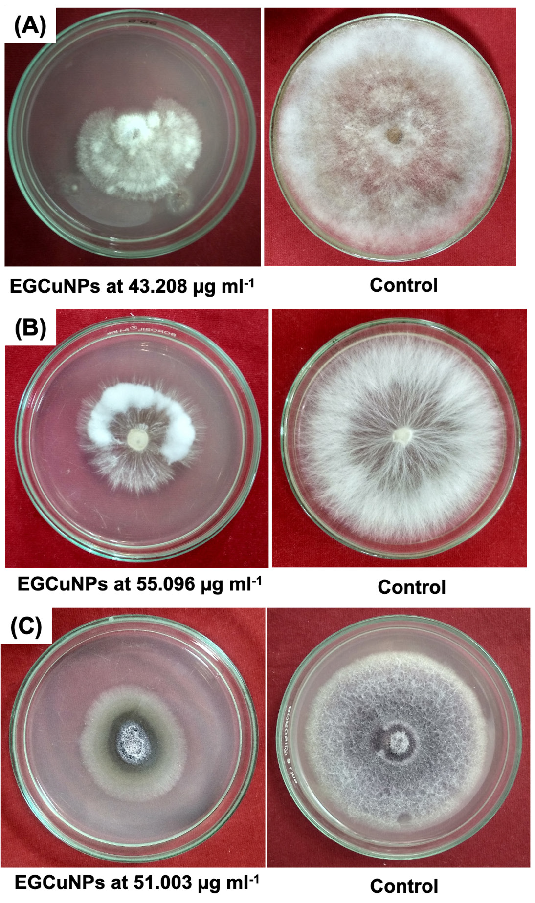
Figure 3. In vitro antifungal effect of EGCuNPs at ED50 concentrations on Rhizoctonia solani (A), Sclerotium rolfsii (B), and Fusarium verticillioides (C)
Scanning electron microscope analysis
Copper nanoparticles, when applied, first come in contact with the cell membrane. There, they could disrupt important cell wall components, leading to cell disruption, development, and eventually death of the pathogen. SEM analysis revealed pronounced structural alterations of fungal cell walls of all three fungi. In R. solani, treatment with EGCuNPs at 43.208 µg ml-1 caused twisting and structural damage of hyphae whereas untreated control hyphae retained their normal morphology (Figure 4). For F. verticillioides, the control sample showed intact and normal conidia, but the treated sample with 51.003 µg ml-1 of EGCuNPs, exhibited spores collapsed entirely, and also hyphae distortion occurred (Figure 5). Interestingly, in concurrence with the in vitro results, hyphae of S. rolfsii remained relatively intact with only minor distortions at a concentration of 55.096 µg ml-1 EGCuNPs (Figure 6). Additionally, uninternalized CuNPs were observed on the hyphae, suggesting the fungi’s ability to resist to some extent. The tolerance of S. rolfsii could be attributed to its fast-growing nature and its ability to effectively manage or counteract the toxic effects of these chemicals. The fungi could possess mechanisms that detoxify or sequester the fungicides, rendering them less harmful. We also believe high melanin content in the cell wall contributed to its tolerance.44 Nonetheless, exceptionally high antifungal activity of copper-based nanoformulations could be attributed to the Cu ions, enabling easy permeation the fungal cell owing to their small size and thereafter attach firmly to the membrane, causing disruption and damage.38 Furthermore, the observed hyphal deformation could be linked to the penetration of Cu ions into the cell interior, that further leads to the disruption of the cell’s metabolic functions by impairing DNA, proteins, and enzymes, also resulting in decreased conidia viability.45 Consistent with our observations, hyphal deformation was observed in Fusarium solani following treatment with CuNPs, with the severity of deformation increasing at higher CuNPs concentrations.41 Likewise, SEM analysis of Fusarium oxysporum treated with CuNPs at 0.25 mg L-1 revealed deformed and shrunk hyphae with granule-like structures, large vesicle-like tumours, and reduced conidial germination.46 In line with our work, several nanoparticles have been demonstrated to cause severe damage to hyphae and other morphological structures of fungi. For instance, SEM analysis and TEM analysis revealed disruption and shrinking of hyphae of Fusarium graminearum when treated with AgNPs.47 Similar damage to hyphae have also been reported on Aspergillus niger and A. flavus when treated with 200 mg L-1 h-MoO3 and a-MoO3 nanoparticles, suggesting high toxicity.48 Another study involving ZnONPs (9 mmol L-1) rendered considerable damage and distortion of the conidiophore, hyphae, and reduction of sporulation of A. niger.49 Also, treatment with AgNPs at concentrations ≤1.68 μg mL-1 induced extensive disruption of the cell wall and cytoplasmic membrane, along with leakage of intracellular contents in Candida spp. and Cryptococcus neoformans.50
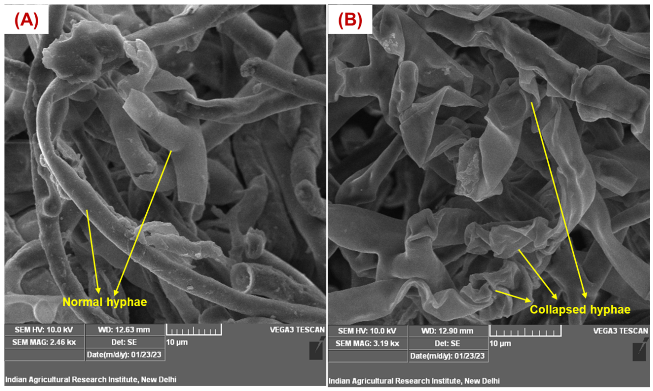
Figure 4. SEM image of Rhizoctonia solani, untreated (A) and treated with EGCuNPs at 43.208 µg ml-1 (B)
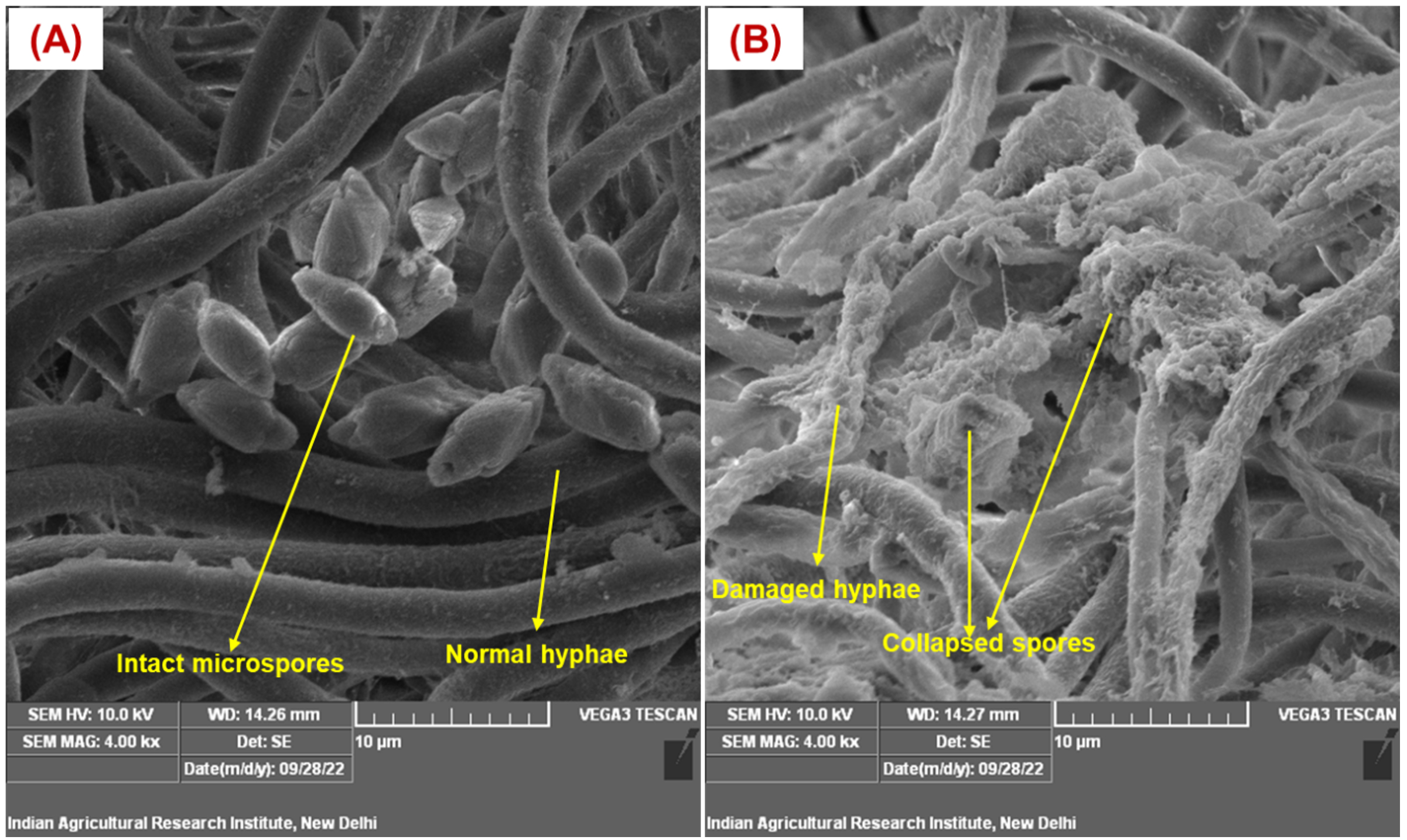
Figure 5. SEM image of Fusarium verticillioides, untreated (A) and treated with EGCuNPs at 51.003 µg ml-1 (B)
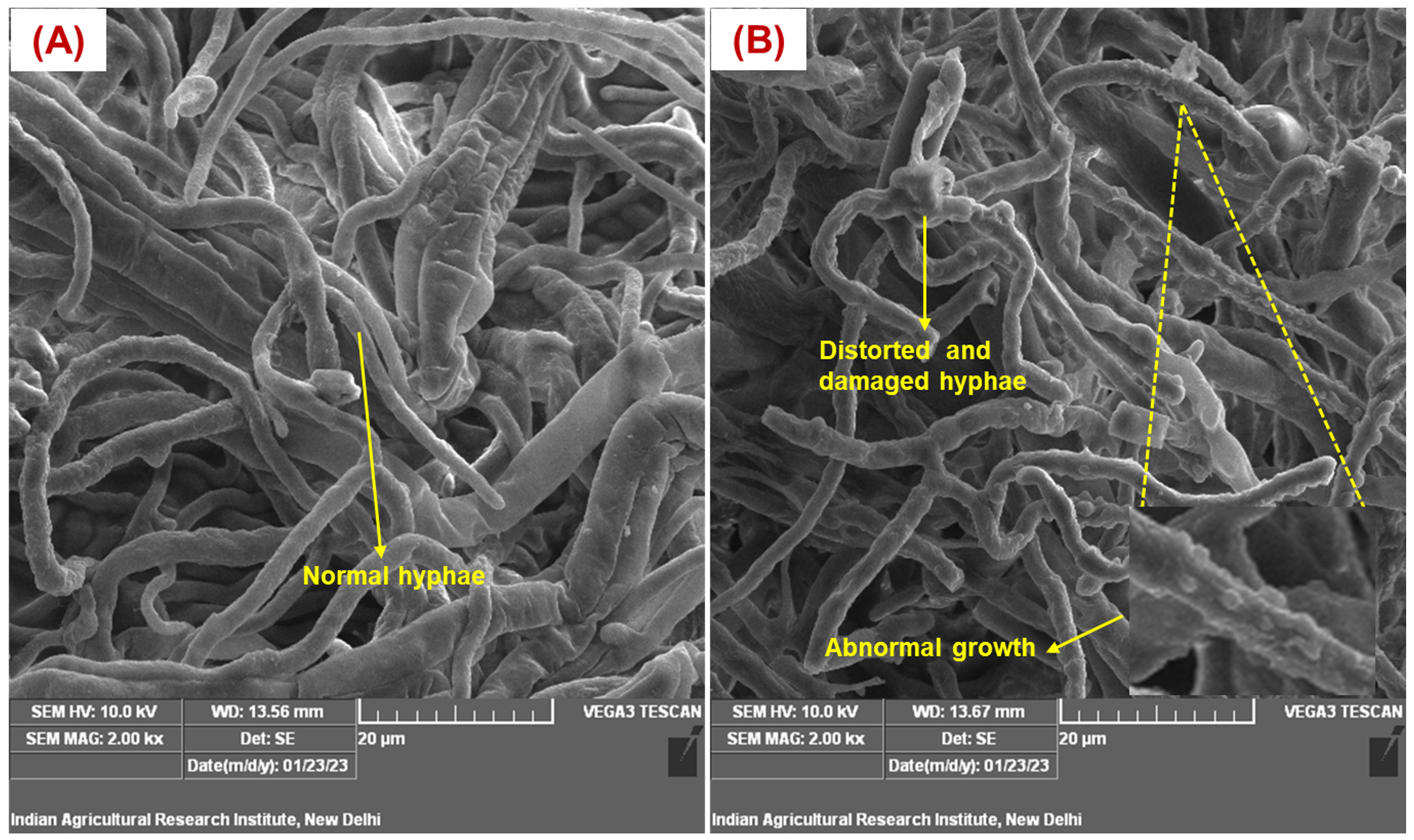
Figure 6. SEM image of Sclerotium rolfsii, untreated (A) and treated with EGCuNPs at 55.096 µg ml-1 (B)
Our formulation EGCuNPs contained clove oil, which also imparted antifungal activity, and along with the CuNPs, we achieved a synergistic effect.23 The inhibition mechanism of essential oils against pathogens is primarily associated with their bioactive functional groups and the interactions of these groups with cellular components.51 The antifungal constituent in clove oil is eugenol, which is characterized by a phenol functional group that has a hydroxyl moiety, which contributes to its bioactivity. It has been well established that the relative positioning of the hydroxyl group holds crucial importance for its effectiveness.52 Eugenol compromises cell membrane integrity, leading to membrane hyperpolarization, increased permeability, leakage of ions and essential intracellular constituents, and ultimately resulting in cell death.53
In conclusion, we identified Rhizoctonia solani based on both morphological and molecular characterization, with ITS sequencing confirming a 99% identity and GenBank accession number obtained being OR251925. The antifungal efficacy of essential oil grafted copper nanoparticles demonstrated significant inhibitory effects against R. solani, Fusarium verticillioides, and Sclerotium rolfsii at their respective ED50 concentrations. SEM analysis further revealed severe disruption of fungal hyphae, spore collapse, and membrane damage, which were consistent with the proposed mechanisms. While S. rolfsii exhibited partial tolerance, likely due to high melanin content and detoxification mechanisms, EGCuNPs still showed significant antifungal activity against it. Taken together, these findings strongly suggest that copper-based nano-formulations possess potent antifungal properties and could serve as a promising alternative to conventional fungicides for the management of phyto-pathogens. Detailed transcriptomic and proteomic studies need to be conducted to elucidate the molecular mechanisms of action of copper-based nanoparticles. However, emphasis should be given to evaluating the long-term impacts of repeated copper nanoparticle application on soil health, plant microbiomes, and ecological sustainability.
Additional file: Supplementary information.
ACKNOWLEDGMENTS
The authors would like to thank the Post Graduate School, ICAR-Indian Agricultural Research Institute, New Delhi, for the research support.
CONFLICT OF INTEREST
The authors declare that there is no conflict of interest.
AUTHORS’ CONTRIBUTION
RG and DK conceptualized and supervised the study. LD carried out the investigation, performed experiments. LD and TKM performed data analysis and interpretation. RK supervised the synthesis of Copper based nanoformulations. GC generated SEM images of fungus. LD wrote the manuscript. RG and DK and reviewed and revised the manuscript. All authors read and approved the final manuscript for publication.
FUNDING
None.
DATA AVAILABILITY
All datasets generated or analyzed during this study are included in the manuscript and/or in the supplementary files.
ETHICS STATEMENT
Not applicable.
- Dorjee L, Gogoi R, Kamil D, Kumar R, Verma A. Copper nanoparticles hold promise in the effective management of maize diseases without impairing environmental health. Phytoparasitica. 2023a;51(3):1-27.
Crossref - Truong HT, Nguyen LCT, Le LQ. Synthesis and antifungal activity of copper nanoparticles against Fusarium oxysporum pathogen of plants. Mater Res Express. 2023;10(2023):065001.
Crossref - Parada J, Tortella G, Seabra AB, Fincheira P, Rubilar O. Potential antifungal effect of copper oxide nanoparticles combined with fungicides against Botrytis cinerea and Fusarium oxysporum. Antibiotics. 2024;13(3):215.
Crossref - Zubrod JP, Bundschuh M, Arts G, et al. Fungicides: an overlooked pesticide class? Environ Sci Tech. 2019;53(7):3347-3365.
Crossref - Willming MM, Maul JD. Direct and indirect toxicity of the fungicide pyraclostrobin to Hyalella azteca and effects on leaf processing under realistic daily temperature regimes. Environ Pollut. 2016;211:435-442.
Crossref - Battaglin WA, Sandstrom MW, Kuivila KM, Kolpin DW, Meyer MT. Occurrence of azoxystrobin, propiconazole, and selected other fungicides in US streams, 2005–2006. Water Air Soil Pollut. 2011;218(1):307-322.
Crossref - Bayat M, Zargar M, Chudinova E, Astarkhanova T, Pakina E. In vitro evaluation of antibacterial and antifungal activity of biogenic silver and copper nanoparticles: The first report of applying biogenic nanoparticles against Pilidium concavum and Pestalotia sp. fungi. Molecules. 2021;26(17):5402.
Crossref - Pham L, Duong MM, Le MV, Hoang HA. Preparation and characterization of antifungal colloidal copper nanoparticles and their antifungal activity against Fusarium oxysporum and Phytophthora capsici. C R Chim. 2019;22(11-12):786-793.
Crossref - Essa AM, Khallaf MK. Antimicrobial potential of consolidation polymers loaded with biological copper nanoparticles. BMC Microbiol. 2016;16(1):144.
Crossref - Shende S, Gaikwad N, Bansod S. Synthesis and evaluation of antimicrobial potential of copper nanoparticle against agriculturally important phytopathogens. Synthesis. 2016;1(4):41-47.
- Usman MS, Zowalaty MEE, Shameli K, Zainuddin N, Salama M, Ibrahim NA. Synthesis, characterization, and antimicrobial properties of copper nanoparticles. Int J Nanomed. 2013;8(1):4467-4479.
Crossref - Xu JG, Liu T, Hu QP, Cao XM. Chemical composition, antibacterial properties, and mechanism of action of essential oil from clove buds against Staphylococcus aureus. Molecules. 2016;21(9):1194.
Crossref - Aguilar-Gonzalez AE, Palou E, Lopez-Malo A. Antifungal activity of essential oils of clove (Syzygium aromaticum) and/or mustard (Brassica nigra) in vapor phase against gray mold (Botrytis cinerea) in strawberries. Innov Food Sci Emerg Technol. 2015;32:181-185.
Crossref - Thabet M, Khalifa W. Antifungal activities of clove oil against root rot and wilt pathogens of tomato plants. Am – Eurasian J Agric Environ Sci. 2018;18(3):105-114.
Crossref - Ragul S, Vanitha S, Harish S, Johnson I, Irene V, Senthil A. Exploring the antifungal activity of clove oil (Syzygium aromaticum) against wilt disease caused by Fusarium equiseti in bitter gourd (Momordica charantia L.). Plant Sci Today. 2024;11(4):580-590.
Crossref - Hamini-Kadar N, Hamdane F, Boutoutaou R, Kihal, Henni JE. Antifungal activity of clove (Syzygium aromaticum L.) essential oil against phytopathogenic fungi of tomato (Solanum lycopersicum L) in Algeria. J Exp Biol Agric Sci. 2014;2(5):447-454.
- Huang Q, Lakshman DK. Effect of clove oil on plant pathogenic bacteria and bacterial wilt of tomato and geranium. J Plant Pathol. 2010;92(3):701-707.
- Kumar P, Lokesh V, Doddaraju P, et al. Greenhouse and field experiments revealed that clove oil can effectively reduce bacterial blight and increase yield in pomegranate. Food Energy Secur. 2021;10(4):e305.
Crossref - Imran M, Abo-Elyousr KA, Al-Harbi MS, Ali EF, Sallam NM, Bagy HMK. Antibacterial efficacy of clove essential oil against Xanthomonas phaseoli pv. phaseoli and its influence on pathogen responses in bean. Gesunde Pflanzen. 202375(2), 431-440.
Crossref - Bozik M, Novy P, Kloucek P. Chemical composition and antimicrobial activity of cinnamon, thyme, oregano and clove essential oils against plant pathogenic bacteria. Acta Univ Agric Silvic Mendel Brun. 2017;65(4):1129-1134.
Crossref - Sattary M, Amini J, Hallaj R. Antifungal activity of the lemongrass and clove oil encapsulated in mesoporous silica nanoparticles against wheat’s take-all disease. Pestic Biochem Physiol. 2020;170:104696.
Crossref - Hasheminejad N, Khodaiyan F, Safari M. Improving the antifungal activity of clove essential oil encapsulated by chitosan nanoparticles. Food Chem. 2019;275:113-122.
Crossref - Dorjee L, Gogoi R, Kamil D, et al. Essential oil-grafted copper nanoparticles as a potential next-generation fungicide for holistic disease management in maize. Front Microbiol. 2023b;14:1204512.
Crossref - Dorjee L, Kamil D, Kumar R, Khokhar M, Aggarwal R, Gogoi R. Synthesis of promising copper nanoparticles utilizing biocontrol agents, Trichoderma virens and Chaetomium globosum. Indian J Agric Sci. 2024;94(1):061-067.
Crossref - Dorjee L, Gogoi R, Kamil D, Kumar R. Biosynthesis of copper nanoparticles using Macrophomina phaseolina and evaluation of its antifungal activity against Fusarium verticillioides and Sclerotium rolfsii. Pharma Innov J. 2022;11(3):2212-20.
- Tutte J. Plant Pathological Methods: Fungi and Bacteria. Burgess Publishing Company.1969:220-239.
- Murray MG, Thompson W. Rapid isolation of high molecular weight plant DNA. Nucleic Acids Res. 1980;8(19):4321-4326.
Crossref - White TJ, Bruns TD, Lee SB, Taylor JW. Amplification and direct sequencing of fungal ribosomal RNA genes for phylogenetics. In: Innis MA, Gelfand DH, Sninsky JJ, White TJ, eds. PCR Protocols: A Guide to Methods and Applications. Academic Press; 1990:315-322.
Crossref - Alves E, Lucas GC, Pozza EA, de Carvalho Alves M. Scanning electron microscopy for fungal sample examination. In: Gupta VK, Tuohy M, Ayyachamy M, Turner K, O’Donovan A, eds. Laboratory Protocols in Fungal Biology: Current Methods in Fungal Biology. Springer. 2013:133-150.
Crossref - Madhavi M, Reddy PN, Reddy RR, Reddy SS. Morphological and molecular variability of Rhizoctonia solani isolates causing banded leaf and sheath blight in maize. Int J Bio-resour Stress Manag. 2015;6(3):375-385.
Crossref - Mishra PK, Gogoi R, Singh PK, Rai SN, Kumar A. Effect of Photo Period on Morpho-cultural Characteristic of Rhizoctonia solani f. sp. sasakii of Maize. Ann Biol. 2014;30(4):733-737.
- Kaur K, Kaur J, Puyam A, Singh K. Cultural, morphological, and molecular characterization of Fusarium verticillioides causing maize ear rot under Punjab condition. Int J Curr Microbiol Appl Sci. 2020;9(10):1698-1706.
Crossref - Ramirez-Camejo LA, Perez MG, Gordon-Mendoza R. Morphological, molecular and pathogenicity characteristics of Fusarium verticillioides causing Fusarium ear rot of corn in Panama. Microb Pathog. 2025;199:107237.
Crossref - Hirata T, Kimishima E, Aoki T, Nirenberg HI, O’Donnell K. Morphological and molecular characterization of Fusarium verticillioides from rotten banana imported into Japan. Mycoscience. 2001;42(2):155-166.
Crossref - Gunaseeli C, Muthukumar A. Isolation and Morphological Characteristics of Sclerotium rolfsii Inciting Collar Rot in Brinjal. J Adv Biol Biotechnol. 2025;28(5):1057-1063.
Crossref - Pandi VK, Gopalakrishnan C, Janahiraman V. Cultural and morphological variability in Sclerotium rolfsii causing stem rot disease. Int J Curr Microbiol App Sci. 2017;6(6):3090-3097.
Crossref - Najera JFD, Castellanos JS, Hernandez MV, et al. Diagnosis and integrated management of fruit rot in Cucurbita argyrosperma, caused by Sclerotium rolfsii. Plant Pathol J. 2018;34(3):171.
Crossref - Dayem AA, Hossain MK, Lee SB, et al. The role of reactive oxygen species (ROS) in the biological activities of metallic nanoparticles. Int J Mol Sci. 2017;18(1):120.
Crossref - Ramyadevi J, Jeyasubramanian K, Marikani A, Rajakumar G. Rahuman AA. Synthesis and antimicrobial activity of copper nanoparticles. Mater Lett. 2012;71:114-116.
Crossref - Wei L, Tang J, Zhang Z, Chen Y, Zhou G, Xi T. Investigation of the cytotoxicity mechanism of silver nanoparticles in vitro. Biomed Mater. 2010;5(4):044103.
Crossref - Pariona N, Mtz-Enriquez AI, Sanchez-Rangel D, Carrion G, Paraguay-Delgado F, Rosas-Saito, G. Green-synthesized copper nanoparticles as a potential antifungal against plant pathogens. RSC Adv. 2019;9(33):18835-18843.
Crossref - Kamel SM, Elgobashy SF, Omara RI, et al. Antifungal activity of copper oxide nanoparticles against root rot disease in cucumber. J Fungi. 2022;8(9):911.
Crossref - Viet PV, Nguyen HT, Cao TM, Hieu LV. Fusarium antifungal activities of copper nanoparticles synthesized by a chemical reduction method. J Nanomater. 2016;2016:1957612.
Crossref - Chet I, Henis Y. Effect of catechol and disodium EDTA on melanin content of hyphal and sclerotial walls of Sclerotium rolfsii sacc. and the role of melanin in the susceptibility of these walls to β-(1→3) glucanase and chitinase. Soil Biol Biochem. 1969;1(2):131-138.
Crossref - Rawashdeh R, Haik Y. Antibacterial mechanisms of metallic nanoparticles: a review. Dyn Biochem Process Biotech Mol Biol. 2009;3(2):12-20.
- Hermida-Montero LA, Pariona N, Mtz-Enriquez AI, Carrion G, Paraguay-Delgado F, Rosas-Saito G. Aqueous-phase synthesis of nanoparticles of copper/copper oxides and their antifungal effect against Fusarium oxysporum. J Hazard Mater. 2019;380:120850.
Crossref - Jian Y, Chen X, Ahmed T, et al. Toxicity and action mechanisms of silver nanoparticles against the mycotoxin-producing fungus Fusarium graminearum. J Adv Res. 2022;38:1-12.
Crossref - Chaves-Lopez C, Nguyen HN, Oliveira RC, Nadres ET, Paparella A, Rodrigues DF. A morphological, enzymatic, and metabolic approach to elucidate apoptotic-like cell death in fungi exposed to h-and a-molybdenum trioxide nanoparticles. Nanoscale. 2018;10(44):20702-20716.
Crossref - Erazo A, Mosquera SA, Rodriguez-Paez JE. Synthesis of ZnO nanoparticles with different morphology: study of their antifungal effect on strains of Aspergillus niger and Botrytis cinerea. Mater Chem Phys. 2019;234:172-184.
Crossref - Ishida K, Cipriano TF, Rocha GM, Weissmuller G, Gomes F, Miranda K, Rozental S. Silver nanoparticle production by the fungus Fusarium oxysporum: nanoparticle characterisation and analysis of antifungal activity against pathogenic yeasts. Mem Inst Oswaldo Cruz. 2013;109(2):220-228.
Crossref - Dorman HD, Deans SG. Antimicrobial agents from plants: antibacterial activity of plant volatile oils. J Appl Microbiol. 2000;88(2):308-316.
Crossref - Veldhuizen EJ, Tjeerdsma-van Bokhoven JL, Zweijtzer C, Burt SA, Haagsman HP. Structural requirements for the antimicrobial activity of carvacrol. J Agri Food Chem. 2006;54:1874–1879.
Crossref - Ulanowska M, Olas B. Biological properties and prospects for the application of eugenol—a review. In J Mol Sci. 2021;22(7):3671.
Crossref
© The Author(s) 2026. Open Access. This article is distributed under the terms of the Creative Commons Attribution 4.0 International License which permits unrestricted use, sharing, distribution, and reproduction in any medium, provided you give appropriate credit to the original author(s) and the source, provide a link to the Creative Commons license, and indicate if changes were made.