


ISSN: 0973-7510
E-ISSN: 2581-690X

Urinary tract infections (UTIs) are among the most prevalent bacterial infections globally, and their clinical management is increasingly complicated by the emergence of antimicrobial resistance in causative uropathogens. Resistance patterns are known to vary with patient age and clinical condition; however, comparative data stratified across distinct patient populations remain limited. This study aimed to isolate and identify uropathogens from three clinically defined patient groups: individuals below 25 years of age, individuals above 25 years, and hospitalized stroke patients. We also compared their antimicrobial resistance profiles to characterize the influence of age and clinical setting on resistance. A cross-sectional observational study was conducted at the Ayurgreen Scientifica Research Institute from October 2025 to March 2026. A total of eighteen midstream urine samples were collected from symptomatic patients at Ayurgreen Hospitals, Kerala, India, and Getwell Hospitals, Naduvattam, Kerala, India. Samples were cultured on Blood Agar and MacConkey Agar, and bacterial isolates were identified using Gram staining. Further biochemical characterization of the isolates was performed using the IMViC battery and the catalase, oxidase, coagulase, and motility tests. Antimicrobial susceptibility testing was performed using the Kirby–Bauer disc diffusion method on Mueller–Hinton Agar against 18 antibiotics, and the results were interpreted according to the Clinical and Laboratory Standards Institute (CLSI M100, 2023) guidelines. Pseudomonas spp. were the most frequently isolated organisms (44.4%), followed by Escherichia coli (22.2%) and Klebsiella spp. (16.7%). Isolates from patients below 25 years of age generally demonstrated higher susceptibility to the tested antibiotics, whereas isolates from patients above 25 years exhibited moderate susceptibility. The stroke patient group showed the highest frequency of multidrug-resistant isolates, including resistance to carbapenems and fluoroquinolones, among several Pseudomonas isolates. Earlier-generation β-lactams and fluoroquinolones demonstrated reduced activity across the study groups, whereas aminoglycosides and nitrofurantoin retained comparatively better antimicrobial activity. The findings indicate differences in antimicrobial susceptibility patterns among uropathogens isolated from the three groups, with multidrug-resistant organisms being more frequently observed in the hospitalized stroke cohort. These results support the importance of continuous local antimicrobial resistance surveillance and patient-specific empirical treatment strategies to promote appropriate antimicrobial use and strengthen antibiotic stewardship.
Escherichia coli, Pseudomonas aeruginosa, Kirby-Bauer Disc Diffusion, Stroke Patients
Urinary tract infections: Epidemiology and clinical significance
Urinary tract infections (UTIs) are among the most common bacterial infections worldwide and represent a major public health concern, accounting for millions of healthcare visits annually and imposing a substantial clinical and economic burden. They affect individuals across all age groups, with women experiencing a significantly higher lifetime risk because of anatomical and physiological factors.1 Recent global estimates indicate that approximately 150-400 million UTI cases occur each year, contributing considerably to antibiotic consumption in both community and hospital settings.2-4 The urinary tract, which comprises the kidneys, ureters, bladder, and urethra, is normally sterile; however, bacterial invasion, most commonly through the ascending urethral route, can establish infection and disrupt urinary tract homeostasis. While many infections remain confined to the lower urinary tract, untreated or inadequately managed infections may ascend to the kidneys, resulting in pyelonephritis, bacteremia, and other systemic complications.5
Clinical manifestations range from uncomplicated cystitis characterized by dysuria, urinary urgency, frequency, and suprapubic pain to complicated UTIs associated with structural abnormalities, urinary catheterization, neurological disorders, or immunosuppression.6 Older adults, individuals with diabetes mellitus, and hospitalized patients are particularly susceptible to recurrent and complicated infections. Beyond their clinical consequences, UTIs impose substantial economic costs through diagnostic investigations, antimicrobial treatment, prolonged hospitalization, and loss of productivity.7
Aetiological agents and common uropathogens
The microbiological aetiology of UTIs is dominated by Gram-negative facultative anaerobes, with Escherichia coli accounting for the majority of both community-acquired and hospital-associated cases. Their pathogenic success is attributable to a repertoire of virulence determinants, including type I and P fimbriae, hemolysin, and iron acquisition systems, which facilitate adhesion to and colonization of the uroepithelium.8 Other clinically significant uropathogens include Klebsiella spp., Proteus mirabilis, Pseudomonas aeruginosa, and, among Gram-positive organisms, Staphylococcus saprophyticus and Enterococcus faecalis. The relative prevalence of these organisms varies considerably according to clinical context. Pseudomonas aeruginosa, in particular, is predominantly associated with nosocomial and healthcare-associated infections, reflecting its intrinsic resistance mechanisms and capacity to persist in hospital environments.9
The distribution of uropathogens is further modulated by host factors including age, sex, immune competence, and prior antimicrobial exposure, as well as by extrinsic factors such as urinary catheterization, hospitalization, and the presence of urological abnormalities. Accurate pathogen identification through culture-based methods remains the cornerstone of microbiological diagnosis and is indispensable for directing targeted therapy, particularly in the context of escalating antimicrobial resistance.10,11
Antimicrobial therapy and emerging resistance
Antibiotic therapy is the primary treatment strategy for bacterial UTIs. Historically, agents such as trimethoprim-sulfamethoxazole, ampicillin, and first-generation fluoroquinolones formed the backbone of empirical treatment protocols for uncomplicated infection, offering broad Gram-negative coverage with favorable oral bioavailability. However, the therapeutic landscape has deteriorated markedly in recent decades, with widespread resistance to these first-line agents rendering them unreliable for empirical use in many clinical settings. Nitrofurantoin and fosfomycin have retained comparatively favourable susceptibility profiles for uncomplicated lower UTIs, while carbapenems represent a critical reserve option for infections caused by extended-spectrum β-lactamase (ESBL)-producing or multidrug-resistant organisms.12 Current international guidelines recommend that empirical therapy be guided by local susceptibility patterns because resistance rates vary considerably between geographic regions and healthcare settings.3
The emergence and global dissemination of antimicrobial resistance (AMR) among uropathogens have transformed UTI management from a straightforward clinical task into an increasingly complex therapeutic challenge. Resistance arises through multiple mechanisms, including enzymatic inactivation of antibiotics (particularly via β-lactamases), target site modification, reduced outer membrane permeability, and active drug efflux systems. The horizontal transfer of resistance determinants via plasmids and other mobile genetic elements facilitates the rapid interspecies and interinstitutional spread of resistance traits, further complicating containment efforts. The overprescription of antibiotics, non-adherence to prescribed courses, and availability of antimicrobials without prescription in many regions have collectively accelerated resistance selection, particularly in uropathogens, given the high volume of antibiotic use in the management of UTIs.13
Age-related variation in uropathogen susceptibility
Patient age is an important determinant of both UTI occurrence and antimicrobial susceptibility patterns. In younger individuals, uropathogens generally exhibit greater susceptibility to standard antimicrobial agents, a pattern attributable to relatively preserved immune function and limited cumulative antibiotic exposure. In contrast, older adults demonstrate a progressively higher burden of antibiotic-resistant infection, reflecting the interplay of age-related immunosenescence, increased healthcare contact, a higher prevalence of comorbid conditions, including diabetes mellitus and structural urological disease, and a greater likelihood of prior antibiotic exposure. These age-stratified differences in resistance have direct implications for empirical treatment selection and underscore the inadequacy of uniform treatment protocols across diverse patient populations.14
UTIs in stroke patients: A high-risk population
Stroke patients represent a clinically distinct and particularly vulnerable population with respect to UTI acquisition and antimicrobial resistance.15-17 Neurological sequelae of stroke, including impaired bladder function and reduced mobility, frequently necessitate urinary catheterization, which itself constitutes a major independent risk factor for catheter-associated UTI (CAUTI).15,17 Prolonged hospitalization exposes these patients to a nosocomial microbiological milieu enriched with multidrug-resistant organisms, whereas post-stroke immunosuppression further diminishes the host’s capacity to resist bacterial colonization.16 Empirical antibiotic use during inpatient rehabilitation reinforces selective pressure, favouring the emergence of resistant strains.15,18 As a consequence, the uropathogen spectrum and resistance profiles observed in stroke patients frequently diverge substantially from those encountered in community-acquired infection, with opportunistic organisms such as Pseudomonas aeruginosa assuming disproportionate clinical significance.15,18,19
Rationale and objectives of the present study
Despite the growing recognition that antimicrobial resistance in uropathogens is shaped by host characteristics, comparative analysis of resistance patterns across distinct clinical subgroups remains insufficiently characterized in the published literature. Most existing studies adopt a cross-sectional, non-stratified approach that obscures clinically meaningful inter-group differences in pathogen distribution and resistance profiles. An appreciation of these differences is essential both for optimizing individual patient management and for informing institution-specific empirical treatment guidelines.
The present study reports the isolation and identification of uropathogens from three clinically distinct patient groups – individuals below 25 years of age, individuals above 25 years of age, and hospitalized stroke patients – and compares their antimicrobial susceptibility profiles using standard microbiological methods. The findings are intended to provide locally relevant evidence on age- and condition-specific resistance patterns that may contribute to more rational empirical antibiotic selection and support institutional antimicrobial stewardship programs.
Study design and patient groups
A cross-sectional observational study was conducted at Ayurgreen Scientifica Research Institute (ASRI), Kerala, India, as part of an M.Sc. research project from October 2025 to March 2026. This study aimed to compare the microbiological characteristics and antimicrobial resistance patterns of uropathogens isolated from three clinically distinct patient groups.
Eligible participants were categorized into three groups: Group 1, patients <25 years of age; Group 2, hospitalized stroke patients; and Group 3, patients >25 years of age without stroke. Each group comprised six patients.
The study included all eligible patients who met the inclusion criteria during the study period; therefore, no formal sample size calculation was performed. Patient confidentiality was maintained throughout the study, and all microbiological data were analyzed anonymously for research purposes.
Ethics approval and consent to participate
This study involved only microbiological and biochemical analysis of urine specimens, with no direct participation of patients. This study was conducted at Ayurgreen Scientifica Research Institute (ASRI), Kerala, India, as part of an M.Sc. research project. No animals or human participants were directly involved in the study. The institute did not have an Institutional Ethics Committee during the study period; therefore, formal ethical approval was not obtained for this study. This study utilized routine clinical urine specimens collected for diagnostic purposes. Patient identifiers were removed before analysis, and confidentiality was maintained throughout the study.
Inclusion and exclusion criteria
Inclusion
Patients of either sex presenting with clinical features suggestive of UTI, including dysuria, urinary frequency, urgency, suprapubic pain, fever, or other urinary symptoms, were considered eligible for inclusion. Only urine specimens yielding significant bacteriuria (³105 colony-forming units [CFU]/mL) with a single predominant bacterial isolate were included for further analysis.
Exclusion
Urine samples yielding less than 105 CFU/mL were excluded. Samples were also excluded if they showed mixed growth of more than two bacterial species suggestive of contamination, or if there was inadequate specimen volume, improper collection, or delayed transport that could compromise microbiological analysis.
Urine specimens were obtained by the clean-catch midstream method to minimize periurethral contamination.20 Patients were instructed to cleanse the external urogenital area with a sterile wipe before collection. The initial urinary stream was discarded, and the midstream portion was collected directly into a sterile, wide-mouthed, screw-capped container without the container or lid contacting the patient. All specimens were labeled with patient identifiers, date, and time of collection, and transported to the microbiology laboratory within two hours of collection to preserve microbiological integrity. Where immediate processing was not feasible, samples were stored at 4 °C for no longer than four hours before inoculation.
Isolation of uropathogens
Each urine sample was inoculated onto Blood Agar (BA) and MacConkey Agar (MCA) using a sterile calibrated loop (0.001 mL) and incubated aerobically at 37 °C for 24 hours.21 Following incubation, plates were examined for the presence, morphology, and quantity of bacterial colonies. Significant bacteriuria was defined as bacterial growth of ³105 CFU/mL. On MacConkey agar, lactose-fermenting organisms produced pink colonies, whereas non-lactose fermenters appeared colourless, facilitating a preliminary differentiation of Gram-negative isolates. Haemolytic patterns on blood agar provided additional presumptive information regarding Gram-positive organisms. Pure colonies of each morphologically distinct type were sub-cultured onto Nutrient Agar for subsequent identification. Mueller–Hinton Agar (MHA), Simmons Citrate Agar, SIM agar, and MR-VP broth were additionally prepared for confirmatory biochemical testing.
Identification of uropathogens
Bacterial identification was performed using a systematic combination of colony morphology, Gram staining, and a panel of biochemical tests. Gram staining was carried out by the standard four-step method: crystal violet primary stain (60 seconds), Gram’s iodine mordant (60 seconds), decolourisation with Gram’s decolouriser (until runoff was clear), and safranin counterstain (45 seconds).22 Gram-positive organisms appeared violet and Gram-negative organisms appeared pink under oil-immersion microscopy, with cellular morphology (cocci versus bacilli) recorded to guide subsequent testing.
The IMViC battery (Indole, Methyl Red, Voges-Proskauer, and Citrate tests) was applied to Gram-negative isolates to differentiate members of the Enterobacteriaceae.23,24 Indole production was detected using Kovac’s reagent. The Methyl Red test assessed mixed-acid fermentation. Voges-Proskauer reactivity was determined with Barritt’s reagents A and B; and citrate utilization was assessed on Simmons Citrate Agar.23 Additional confirmatory tests included the catalase test (3% H2O2 applied to a fresh colony on a glass slide, with bubble formation indicating a positive result), the oxidase test (colony rubbed on an oxidase disc, with development of a dark purple colour within 10 seconds indicating a positive result), and the coagulase test (slide and tube methods using rabbit plasma) for differentiation of Staphylococcus aureus from coagulase-negative staphylococci.23,24 Motility was assessed using SIM (Sulphide-Indole-Motility) semi-solid agar by the stab inoculation technique, with diffuse growth radiating from the stab line indicating motility.
Bacterial identification was based exclusively on conventional phenotypic and biochemical methods. Automated identification systems and molecular identification techniques such as polymerase chain reaction (PCR) or gene sequencing were not performed.
Antimicrobial susceptibility testing
Antimicrobial susceptibility testing was performed using the Kirby–Bauer disc diffusion method on Mueller–Hinton Agar following the Clinical and Laboratory Standards Institute (CLSI) M100, 33rd Edition (2023) guidelines. A standardised bacterial suspension was prepared in sterile normal saline from overnight cultures and adjusted to a turbidity equivalent to a 0.5 McFarland standard, corresponding to approximately 1-2 × 10⁸ CFU/mL. Within 15 minutes of preparation, the suspension was inoculated onto the surface of MHA plates using a sterile cotton swab in three directions to achieve a uniform bacterial lawn. Antibiotic-impregnated discs were dispensed onto the inoculated surface with a minimum inter-disc distance of 24 mm and a disc-to-plate edge distance of at least 10 mm to prevent overlapping of inhibition zones. Plates were then inverted and incubated at 37 °C for 24 hours. The diameters of zones of inhibition were measured to the nearest millimetre using a calibrated ruler and interpreted as sensitive (S), intermediate (I), or resistant (R) according to CLSI breakpoint criteria.25
Routine quality-control procedures were performed in accordance with CLSI recommendations to ensure the reliability of antimicrobial susceptibility testing. However, the specific quality-control reference strains used during routine laboratory quality assurance were not documented in the study records.
The antibiotics tested were selected to represent all four major mechanistic classes: cell wall synthesis inhibitors (cefuroxime, cefixime, cefoperazone-sulbactam, cefotaxime, ceftriaxone, cephalexin, cefepime, piperacillin-tazobactam), protein synthesis inhibitors (amikacin, azithromycin, gentamicin, colistin), nucleic acid synthesis inhibitors (ciprofloxacin, cotrimoxazole, levofloxacin, norfloxacin, ofloxacin), and metabolic pathway inhibitors (nitrofurantoin). The full panel is presented in Table 1.
Table 1. Antibiotic disc panel used for susceptibility testing, classified by mechanism of action
Cell Wall Synthesis Inhibitors |
Protein Synthesis Inhibitors |
Nucleic Acid Synthesis Inhibitors |
Metabolic Pathway Inhibitors |
|---|---|---|---|
Cefuroxime (CB) |
Amikacin (AK) |
Ciprofloxacin (RC) |
Nitrofurantoin (FD) |
Cefixime (SF) |
Azithromycin (AZK) |
Cotrimoxazole (BA) |
|
Cefoperazone-sulbactam (CM) |
Gentamicin (GM) |
Levofloxacin (QB) |
|
Cefotaxime (CF) |
Colistin (CL) |
Norfloxacin (NX) |
|
Ceftriaxone (RP) |
Ofloxacin (ZN) |
||
Cephalexin (PR) |
|||
Cefepime (ZX) |
|||
Piperacillin-tazobactam (PT) |
Data recording and analysis
All microbiological findings, including organism identity, patient group allocation, and susceptibility results for each antibiotic, were recorded on standardized data collection forms and entered into a spreadsheet for analysis. Resistance rates for each organism were calculated as the percentage of isolates classified as resistant (R) for a given antibiotic. Inter-group comparisons of resistance profiles were conducted descriptively, with patterns stratified by patient group (Group 1: below 25 years; Group 2: stroke patients; Group 3: above 25 years) and by antibiotic class. Intermediate results were recorded separately and not collapsed into the resistant category to preserve the granularity of the susceptibility data.
Uropathogen isolation and distribution
A total of 18 bacterial isolates were recovered from urine samples across the three patient groups, with six isolates obtained from each cohort. Pseudomonas spp. were the most frequently isolated organisms overall, accounting for eight of eighteen isolates (44.4%), with a disproportionately high representation in the stroke patient group (Group 2, n = 4). Escherichia coli was the second most common organism (n = 4, 22.2%), and was distributed across all three groups. Klebsiella spp. were recovered from each group (n = 3, 16.7%), whereas Enterobacter spp., Staphylococcus spp., and Streptococcus spp. were each isolated as single representatives (5.6%), confined to Groups 3, 2, and 1, respectively. The full distribution of isolates across the patient groups is presented in Table 2 and illustrated in Figure 1.
Table 2. Distribution of uropathogens across the three patient groups
Organism |
Group 1 (
| Group 2 (Stroke) |
Group 3 (>25 yrs) |
Total |
|---|---|---|---|---|
Escherichia coli |
2 |
1 |
1 |
4 |
Klebsiella spp. |
1 |
1 |
1 |
3 |
Pseudomonas spp. |
2 |
4 |
2 |
8 |
Enterobacter spp. |
0 |
0 |
1 |
1 |
Staphylococcus spp. |
0 |
0 |
1 |
1 |
Streptococcus spp. |
1 |
0 |
0 |
1 |
Total |
6 |
6 |
6 |
18 |
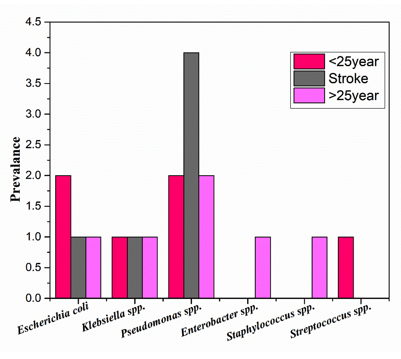
Figure 1. Distribution of uropathogens by patient group. Each bar represents the proportional contribution of each species within the group
Biochemical identification
Gram staining established the preliminary identity of all isolates: Gram-negative bacilli predominated, whereas Gram-positive cocci (Staphylococcus spp. and Streptococcus spp.) were confined to Groups 1 and 3. Confirmatory biochemical profiling was consistent with expected species-specific patterns. E. coli isolates were indole-positive, methyl red-positive, Voges-Proskauer-negative, and citrate-negative (IMViC: + + – -), and demonstrated motility and lactose fermentation. Klebsiella spp. displayed the reciprocal IMViC pattern (- – + +) with characteristic mucoid colonies and lactose fermentation, but were non-motile. Pseudomonas spp. were oxidase-positive, non-lactose fermenters with demonstrable motility, consistent with their established biochemical profile.22 Staphylococcus spp. were catalase-positive and coagulase-positive, confirming their identity as S. aureus, while Streptococcus spp. were catalase-negative.26
Representative IMViC test results for the three principal Gram-negative organisms are illustrated in Figure 2, and the characteristic IMViC profiles used for identification are summarised in Table 3.
Table 3. IMViC reaction profiles for Gram-negative uropathogens
Organism |
Indole |
Methyl Red |
VP |
Citrate |
|---|---|---|---|---|
Escherichia coli |
+ |
+ |
– |
– |
Klebsiella spp. |
– |
– |
+ |
+ |
Enterobacter spp. |
– |
– |
+ |
+ |
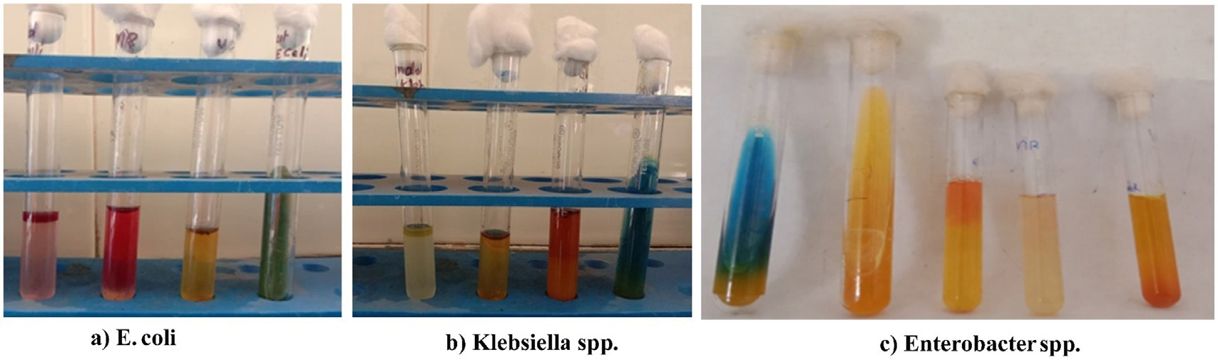
Figure 2. IMViC test results. (a) E. coli; (b) Klebsiella spp.; (c) Enterobacter spp.
The catalase test distinguished Staphylococcus spp. (positive; effervescence with H2O2) from Streptococcus spp. (negative). The oxidase test identified Pseudomonas spp. (positive; dark purple colour within 10 seconds, Figure 3), distinguishing them from the oxidase-negative Enterobacteriaceae. The tube coagulase test confirmed the presence of coagulase-positive Staphylococcus aureus in the relevant isolates. Motility testing in SIM agar demonstrated motility in E. coli, Enterobacter spp., and Pseudomonas spp., while Klebsiella spp. were consistently non-motile. Results are summarised in Table 4.
Table 4. Summary biochemical characterisation of isolated uropathogens
Organism |
Catalase |
Oxidase |
Coagulase |
Motility |
Lactose Ferm. |
H2S |
|---|---|---|---|---|---|---|
Escherichia coli |
+ |
– |
– |
+ |
+ |
– |
Klebsiella spp. |
+ |
– |
– |
– |
+ |
– |
Pseudomonas spp. |
+ |
+ |
– |
+ |
– |
– |
Enterobacter spp. |
+ |
– |
– |
+ |
+ |
– |
Staphylococcus spp. |
+ |
– |
+ |
– |
V |
– |
Streptococcus spp. |
– |
– |
– |
– |
– |
– |
V = variable.
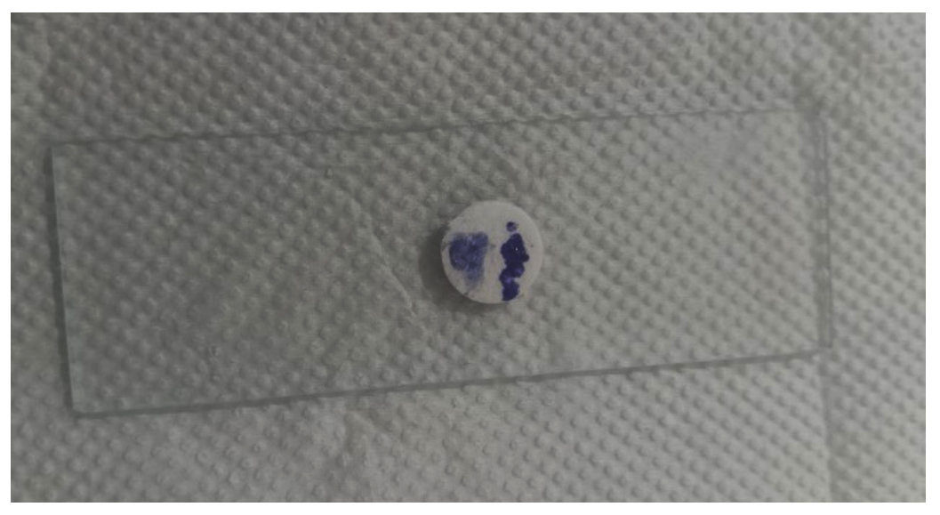
Figure 3. Oxidase test result
Antibiotic susceptibility
Antibiotic susceptibility testing was performed on all isolates using 18 discs spanning four mechanistic classes. Results are presented in Table 5, and the antibiotic susceptibility patterns of the three groups are shown in Figure 4. The isolate-wise antimicrobial susceptibility profiles, including sensitive, intermediate, and resistant responses across the tested antibiotics, are visualised in the heatmap presented in Figure 5.
Table 5. Antibiotic resistance profiles of uropathogens isolated from all three patient groups. Resistant (R) results are highlighted in red, Intermediate (I) in yellow, and Sensitive (S) in green to facilitate visual comparison
| Group | Organism | FD | ZX | CB | CF | CM | PT | SF | RP | PR | AK | AZK | CL | GM | RC | BA | QB | NX | ZN |
|---|---|---|---|---|---|---|---|---|---|---|---|---|---|---|---|---|---|---|---|
| Group 1 (Below 25 Years) | Pseudomonas spp. | S | S | I | S | S | S | S | S | I | S | I | I | S | S | S | S | S | S |
| E. coli | I | S | I | S | S | S | S | I | I | S | I | I | I | S | S | S | S | S | |
| Pseudomonas spp. | S | S | R | I | S | S | I | I | R | S | I | I | S | S | R | I | S | S | |
| Klebsiella spp. | I | I | R | I | S | S | R | R | I | I | I | I | I | R | S | I | R | I | |
| Streptococcus spp. | S | I | I | S | S | S | R | I | I | S | R | I | S | I | S | S | I | I | |
| E. coli | I | I | I | I | I | S | I | S | I | I | I | I | I | S | S | S | I | S | |
| Group 2 (Stroke Patients) | Pseudomonas spp. | S | R | R | R | S | S | R | R | R | S | S | R | I | R | S | I | R | I |
| E. coli | S | S | R | I | S | S | I | I | I | S | I | I | S | I | R | I | R | I | |
| Pseudomonas spp. | R | I | R | I | S | S | I | I | R | S | I | I | S | I | R | I | I | I | |
| Klebsiella spp. | I | S | S | S | S | S | S | S | S | S | I | I | S | R | S | I | R | R | |
| Pseudomonas spp. | I | I | R | R | I | I | R | I | R | R | R | I | R | R | R | R | R | R | |
| Pseudomonas spp. | S | S | R | I | S | S | I | I | I | S | I | I | S | I | R | S | R | I | |
| Group 3 (Above 25 Years) | E. coli | I | S | I | S | S | S | S | I | I | I | S | I | I | S | S | S | S | I |
| Klebsiella spp. | S | I | R | I | S | S | I | I | I | I | S | I | I | R | I | I | R | I | |
| Staphylococcus spp. | S | I | I | I | S | S | R | I | I | I | R | I | R | I | S | I | R | I | |
| Pseudomonas spp. | I | I | I | I | S | S | I | I | I | S | S | I | S | S | S | S | S | S | |
| Enterobacter spp. | S | I | R | R | I | S | R | I | R | I | I | R | I | R | S | I | R | R | |
| Pseudomonas spp. | I | I | R | S | I | I | S | I | I | I | I | I | R | R | R | I | R | R |
*Abbreviation key: S = Sensitive; I = Intermediate; R = Resistant
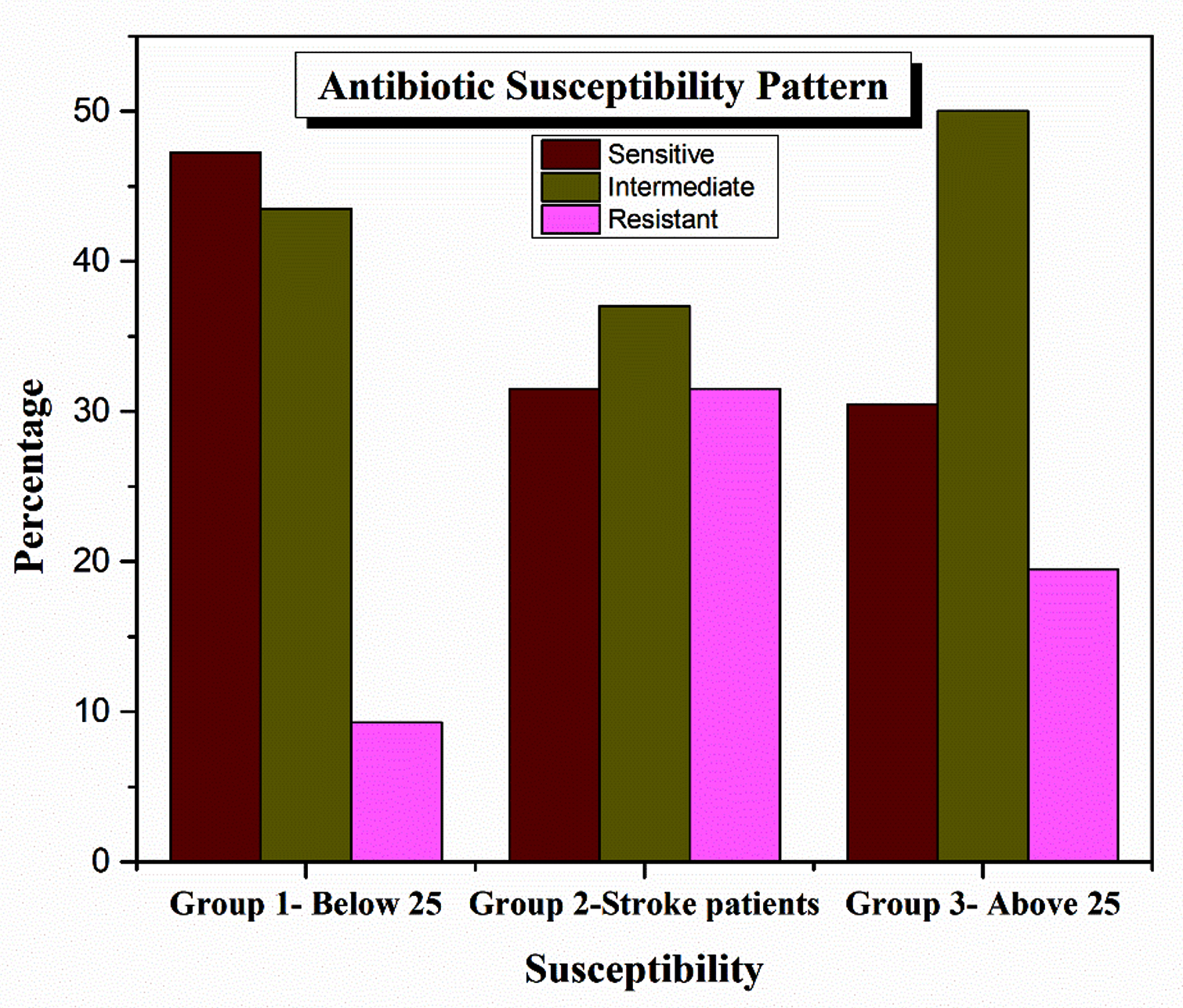
Figure 4. Comparative antibiotic susceptibility pattern across the three patient groups. Group 1: below 25 years; Group 2: stroke patients; Group 3: above 25 years
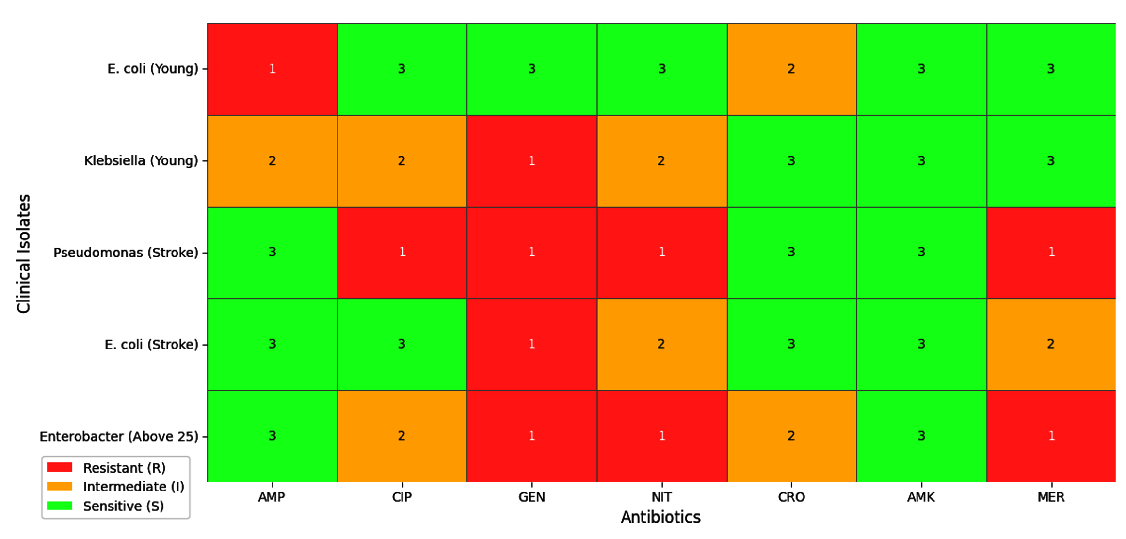
Figure 5. Heatmap of antimicrobial susceptibility profiles for all 18 isolates. S = sensitive (green); I = intermediate (amber); R = resistant (red)
Group 1 (Below 25 years)
Isolates from Group 1 demonstrated the most favorable overall susceptibility profile across all antibiotic classes. Pseudomonas spp. in this group were sensitive to most tested agents, with only isolated intermediate and resistant responses noted against select β-lactams and nucleic acid synthesis inhibitors. E. coli isolates were broadly susceptible, with intermediate responses to certain cephalosporins and fluoroquinolones, indicating the early emergence of resistance. Klebsiella spp. exhibited comparatively greater resistance, particularly to cephalosporins and cotrimoxazole, consistent with previously reported resistance trends in this genus. Streptococcus spp. showed mixed susceptibility, with resistance observed to certain cell wall synthesis inhibitors. Taken together, the resistance burden in Group 1 was low, suggesting that infections in this cohort are largely amenable to standard therapeutic regimens.
Group 2 (stroke patients)
The most complex and clinically concerning resistance patterns were observed in Group 2. Pseudomonas spp. dominated this cohort and demonstrated extensive multidrug resistance, with resistant phenotypes recorded across β-lactams, fluoroquinolones, and protein synthesis inhibitors in several isolates. One Pseudomonas isolate exhibited resistance to most agents in the panel, consistent with a pan-resistant or near-pan-resistant phenotype attributable to the synergistic action of efflux pumps, porin loss, and β-lactamase production.27 E. coli and Klebsiella spp. in this group also showed elevated resistance compared to equivalent isolates in Groups 1 and 3, particularly to carbapenems and fluoroquinolones, suggesting the possible presence of ESBL-producing or carbapenem-resistant strains driven by prolonged antibiotic exposure during hospitalisation.28 Nitrofurantoin and aminoglycosides (amikacin, gentamicin) retained partial activity, representing the most reliable agents within the panel for this group.
Group 3 (above 25 years)
Group 3 displayed an intermediate resistance profile between the relatively susceptible Group 1 and the highly resistant Group 2 cohort. Pathogen diversity was greatest in this group, with isolates of E. coli, Klebsiella spp., Pseudomonas spp., Enterobacter spp., and Staphylococcus spp. all represented. Enterobacter spp. exhibited pronounced resistance across multiple antibiotic classes, consistent with the inducible AmpC β-lactamase production characteristic of this genus.29 Pseudomonas spp. isolates showed variable resistance, with some demonstrating intermediate profiles and others displaying frank resistance to carbapenems and fluoroquinolones. Staphylococcus spp. showed resistance to several β-lactam and nucleic acid synthesis inhibitors, warranting consideration of methicillin-resistant phenotypes. The elevated resistance relative to Group 1 likely reflects cumulative antibiotic exposure and age-related immune decline in this cohort.30
Antibiotic class-wise resistance comparison
Analysis stratified by antibiotic class revealed a consistent gradient of resistance across the three groups, as depicted in Figure 6. Cell wall synthesis inhibitors, particularly earlier-generation cephalosporins and ampicillin, showed markedly reduced efficacy in Groups 2 and 3 relative to Group 1, consistent with widespread β-lactamase dissemination among Gram-negative uropathogens.25 Fluoroquinolones demonstrated substantially diminished activity across all groups, with the highest resistance rates in stroke patient isolates, corroborating global trends of fluoroquinolone resistance in Pseudomonas and Enterobacteriaceae.27 Protein synthesis inhibitors, particularly aminoglycosides, retained comparatively better activity, though intermediate responses were increasingly common in Group 2. Metabolic pathway inhibitors, principally nitrofurantoin, maintained a broadly active profile, particularly against Gram-negative enteric organisms in Groups 1 and 3, though its activity against Pseudomonas spp. was expectedly limited. The summarised class-wise resistance trends are presented in Table 6.
Table 6. Summary of antibiotic resistance trends by class across patient groups
Antibiotic (Class) |
Group 1 % R |
Group 2 % R |
Group 3 % R |
Overall Trend |
|---|---|---|---|---|
β-lactams (Cell Wall) |
Low – Mod |
High |
Moderate |
Increasing with healthcare exposure |
Carbapenems (Cell Wall) |
Low |
Moderate |
Low |
Reservoir; resistance emerging in stroke |
Fluoroquinolones (Nucleic Acid) |
Low – Mod |
High |
Moderate |
Substantially reduced efficacy |
Aminoglycosides (Protein) |
Low |
Moderate |
Low-Mod |
Relatively preserved activity |
Nitrofurantoin (Metabolic) |
Low |
Low – Mod |
Low |
Retained; variable in stroke group |
Linezolid (Protein) |
Low |
Moderate |
Low-Mod |
Active vs. Gram-positives |
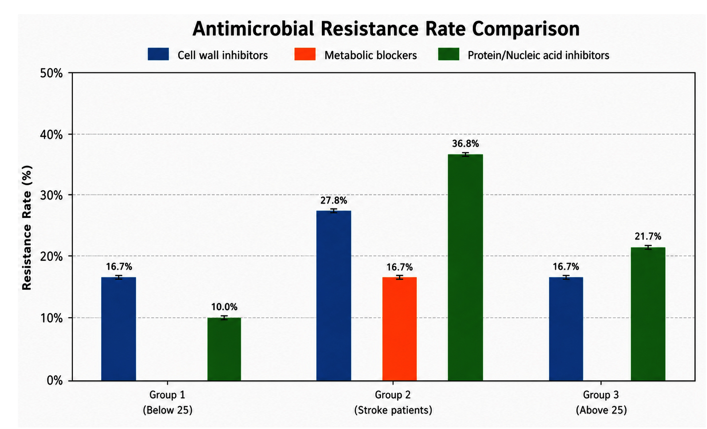
Figure 6. Comparative antibiotic resistance rates (%) by mechanistic class across the three patient groups. Group 1: below 25 years; Group 2: stroke patients; Group 3: above 25 years
Uropathogen spectrum and Its clinical determinants
The microbiological findings of this study are consistent with established global patterns of uropathogen prevalence, while simultaneously revealing clinically meaningful inter-group differences that reflect the influence of patient characteristics on infection ecology. The overall predominance of Gram-negative organisms, particularly E. coli, Klebsiella spp., and Pseudomonas spp., aligns with the published literature across diverse geographical settings.15,31 However, the disproportionate isolation of Pseudomonas spp. in the stroke patient cohort is a notable and clinically significant departure from the E. coli dominant pattern typically observed in community-acquired UTIs.32,33 This finding reflects the well-recognised capacity of Pseudomonas spp. to colonise catheterised and immunocompromised patients in nosocomial environments, where selective antibiotic pressure, biofilm formation on catheter surfaces, and reduced host immune surveillance collectively facilitate their establishment and persistence.34,35
The absence of Enterobacter spp. and Staphylococcus spp. from the below 25 cohort, and their occurrence exclusively in older patients, is consistent with observations that these organisms, often associated with healthcare exposure and comorbid conditions, are uncommon causes of primary community-acquired UTIs in younger, otherwise healthy individuals.22,23 Similarly, the isolation of Streptococcus spp. only in Group 1 may reflect its recognized association with UTIs in younger patients, particularly in the absence of the structural and physiological risk factors that predispose older individuals to Gram-negative nosocomial pathogens.
Resistance in the below 25 Cohort: Early signals
Although the below 25 group demonstrated the most favourable susceptibility profile overall, the presence of intermediate and resistant phenotypes, particularly in Klebsiella spp. and Streptococcus spp., is clinically relevant and should not be dismissed. In populations with limited prior antibiotic exposure, such resistance signals often reflect community-level dissemination of resistant clones rather than individual selective pressure, and may represent the early stages of a trajectory that, without intervention, will mirror the more pronounced resistance patterns observed in older cohorts.36,37 The findings from this group reinforce the importance of culture-directed therapy even in younger patients presenting with apparently uncomplicated infection, and underscore the need for community-level antibiotic stewardship to preserve the efficacy of first-line agents.38
Multidrug resistance in stroke patients: Mechanistic and clinical implications
The pronounced multidrug-resistance observed in the stroke patient cohort warrants detailed consideration. The clinical circumstances of stroke patients create a convergence of risk factors that are individually significant and collectively synergistic in promoting resistant infection. Prolonged urinary catheterization provides both a direct portal of bacterial entry and a substrate for biofilm formation, within which bacteria are phenotypically and genotypically distinct from their planktonic counterparts and substantially more tolerant of antibiotic exposure.34 Extended inpatient stays increase the probability of cross-acquisition of resistant strains circulating within the hospital environment, while repeated or prophylactic antibiotic courses impose sustained selective pressure that enriches for resistant populations.27,28 Post-stroke neurological changes, including stroke-induced immunodepression syndrome (SIDS), further compromise the host’s capacity to limit bacterial proliferation and clear early infection.
The resistance profile of Pseudomonas spp. in this cohort is consistent with the organism’s well-characterized intrinsic and acquired resistance mechanisms. Intrinsic low outer membrane permeability, constitutive expression of efflux pumps (notably MexAB-OprM), and the capacity for rapid mutational and horizontal gene transfer-mediated resistance acquisition collectively render P. aeruginosa one of the most therapeutically challenging uropathogens in hospitalised patients.27 The elevated resistance of E. coli and Klebsiella spp. in this group, relative to their behaviour in community-acquired settings, may indicate ESBL-producing or carbapenem-resistant phenotypes; however, molecular confirmation was not performed in this study.29,39
Age-related resistance gradient and its drivers
The progressive increase in antimicrobial resistance across Groups 1, 3, and 2, from lowest to highest, suggests an age-related trend in antimicrobial resistance that is driven by the interplay of biological and environmental factors.40 The accumulation of prior antibiotic courses over a lifetime imposes a cumulative selective pressure that progressively enriches for resistant strains within an individual’s microbiome. Age-associated immunosenescence reduces the immunological barrier to infection by less virulent but more resistant opportunistic organisms. The increasing burden of comorbidities in older adults, particularly diabetes mellitus, which impairs phagocytic function and creates a glucose-rich urinary environment favourable to bacterial growth, further compounds this risk. These observations align with published multicentre data demonstrating that resistance rates in UTI isolates increase significantly with patient age and highlight the importance of considering patient characteristics, rather than applying uniform protocols without age stratification, when selecting empirical therapy.36,40
Antibiotic class performance and therapeutic implications
The antimicrobial susceptibility profiles observed in this study provide useful information for local empirical therapy while recognizing the limitations of the dataset. Earlier-generation β-lactams, including ampicillin and first-generation cephalosporins, demonstrated reduced activity against several isolates, suggesting that their empirical use may require confirmation by local susceptibility data.36 Fluoroquinolones also exhibited reduced activity, particularly among isolates recovered from hospitalized stroke patients, consistent with recent reports describing increasing fluoroquinolone resistance among uropathogens.27
Aminoglycosides, particularly amikacin and gentamicin, retained comparatively better activity against many isolates and may remain useful options for the treatment of moderate-to-severe infections when clinically appropriate.25,36 Nitrofurantoin also demonstrated favourable activity against many community-associated Gram-negative isolates, supporting current guideline recommendations for uncomplicated lower urinary tract infections. However, its limited activity against Pseudomonas spp. and poor tissue penetration restrict its use in complicated or upper urinary tract infections.36,37 Carbapenems and β-lactam/β-lactamase inhibitor combinations remained active against several resistant isolates, although their use should continue to be guided by antimicrobial stewardship principles to minimise the emergence of further resistance.39
Limitations and future directions
Several limitations should be considered when interpreting the findings of this study. First, the study included only 18 bacterial isolates (six per patient group), limiting statistical power and reducing the generalisability of the results. Second, this was a single-center investigation, and the observed resistance patterns may not represent those of other healthcare institutions or geographic regions. Third, patient-specific clinical information, including prior antibiotic use, duration of hospitalization, catheterization history, diabetes mellitus, recurrent UTIs, renal disease, and other comorbidities, was not systematically collected; consequently, these potential confounding factors could not be evaluated. Fourth, antimicrobial susceptibility was assessed using the Kirby–Bauer disc diffusion method only, and molecular characterization of resistance mechanisms, including extended-spectrum β-lactamase (ESBL) production and carbapenemase genes, was not performed. Finally, no statistical analyses were undertaken to determine whether differences observed between study groups were statistically significant. Therefore, the findings should be regarded as descriptive and hypothesis-generating rather than definitive.
Future investigations should include larger multicentre cohorts with adequate statistical power to evaluate differences among patient populations while controlling for potential confounding variables through multivariable analyses. The incorporation of molecular techniques for detecting resistance determinants, biofilm formation assays, and longitudinal surveillance would provide greater insight into resistance evolution and transmission.33 Such studies would contribute to the development of institution-specific empirical treatment guidelines and strengthen antimicrobial stewardship programs based on local epidemiological evidence.38,36
This study describes the distribution of uropathogens and their antimicrobial susceptibility profiles among three clinically distinct patient groups: individuals below 25 years of age, individuals above 25 years of age, and hospitalized stroke patients. Pseudomonas spp. were the most frequently isolated organisms, particularly in the stroke patient group, whereas Escherichia coli and Klebsiella spp. were identified across all study groups. The susceptibility patterns observed in this study suggest variation among the patient groups, with isolates from younger individuals generally exhibiting greater susceptibility and isolates from hospitalized stroke patients demonstrating more extensive multidrug resistance.
The higher frequency of resistant isolates in the stroke patient group may be associated with factors such as hospitalization, urinary catheterization, prior antimicrobial exposure, underlying comorbidities, and post-stroke immune dysfunction, although these factors were not specifically evaluated in the present study. Earlier-generation β-lactams and fluoroquinolones demonstrated reduced activity against several isolates, whereas aminoglycosides and nitrofurantoin retained comparatively favourable activity against many isolates, particularly those associated with community-acquired infections. These findings may help inform empirical treatment decisions when interpreted alongside local antimicrobial susceptibility data and current clinical guidelines.
Given the limited sample size, single-center study design, and absence of molecular characterization and statistical analysis, the findings should be considered descriptive and interpreted with appropriate caution. Nevertheless, this study provides locally relevant baseline data that may contribute to antimicrobial stewardship initiatives and supports the need for continued surveillance, larger multicentre studies, and patient-specific approaches to empirical antimicrobial therapy.
ACKNOWLEDGMENTS
The authors sincerely acknowledge the support and facilities provided by Ayurgreen Scientifica Research Institute, Kerala, India, for enabling this research work. The authors also thank all individuals who contributed to sample collection and laboratory assistance.
CONFLICT OF INTEREST
The authors declare that there is no conflict of interest.
AUTHORS’ CONTRIBUTION
KNS conceptualized the study. AKA performed sample collection and experimental analysis. KPSH performed data validation. JD supervised the study. AKA wrote the manuscript.
KPSH reviewed and edited the manuscript. All authors read and approved the final manuscript for publication.
FUNDING
None.
DATA AVAILABILITY
The datasets generated and/or analysed during the current study are available from the corresponding author on reasonable request.
ETHICS STATEMENT
This study involved a retrospective analysis of 18 urine samples collected during routine diagnostic procedures. No additional samples or procedures were performed. Patient confidentiality was maintained, and all data were anonymized. Therefore, ethical approval was not required.
- Glover EK, Sheerin NS. Urinary tract infection. Medicine. 2023;51(4):239-243.
Crossref - Conover MS, Flores-mireles AL, Hibbing ME, Dodson K, Hultgren SJ. Establishment and Characterization of UTI and CAUTI in a Mouse Model. 2015;100:1-12.
Crossref - G. Bonkat (Chair), R. Bartoletti, F. Bruyère, T. Cai, S.E. Geerlings, B. Köves, J. Kranz, S. Schubert, A. Pilatz, R. Veeratterapillay, F. Wagenlehner, EAU Guidelines On Urological Infections, Urological Infections, 2026, https://www.srm.ro/media/2024/12/7-eau-pocket-on-urological-infections-2024.pdf
- Yang X, Chen H, Zheng Y, Qu S, Wang H, Yi F. Disease burden and long-term trends of urinary tract infections: A worldwide report. Front Public Heal. 2022;10:888205.
Crossref - Walsh C, Collyns T. Pathophysiology of urinary tract infections. Surg. 2023;41(5):272-277.
Crossref - Bilsen MP, Jongeneel RMH, Schneeberger C, et al. Definitions of urinary tract infection in current research: a systematic review. Open Forum Infect Dis. 2023;10(7):ofad332.
Crossref - Flores-Mireles AL, Walker JN, Caparon M, Hultgren SJ. Urinary tract infections: epidemiology, mechanisms of infection and treatment options. Nat Rev Microbiol. 2015;13(5):269-284.
Crossref - Terlizzi ME, Gribaudo G, Maffei ME. UroPathogenic Escherichia coli (UPEC) infections: virulence factors, bladder responses, antibiotic, and non-antibiotic antimicrobial strategies. Front Microbiol. 2017;8:1566.
Crossref - Wanke-Rytt M, Sobierajski T, Lachowicz D, Seliga-Gasior D, Podsiadly E. Analysis of Etiology of Community-Acquired and Nosocomial Urinary Tract Infections and Antibiotic Resistance of Isolated Strains: Results of a 3-Year Surveillance (2020-2022) at the Pediatric Teaching Hospital in Warsaw. Microorganisms. 2023;11(6):1438.
Crossref - Zhou Y, Zhou Z, Zheng L, et al. Urinary Tract Infections Caused by Uropathogenic Escherichia coli: Mechanisms of Infection and Treatment Options. Int J Mol Sci. 2023;24(13):10537.
Crossref - Whelan S, Lucey B, Finn K. Uropathogenic Escherichia coli ( UPEC ) -Associated Urinary Tract Infections: The Molecular Basis for Challenges to Effective Treatment. Microorganisms. 2023;11(9):2169.
Crossref - Ari MM, Dashtbin S, Ghasemi F, et al. Nitrofurantoin: properties and potential in treatment of urinary tract infection: a narrative review. Front Cell Infect Microbiol. 2023;13:1148603.
Crossref - Nasrollahian S, Graham JP, Halaji M. A review of the mechanisms that confer antibiotic resistance in pathotypes of E . coli. Front Cell Infect Microbiol. 2024:14:1387497.
Crossref - Rodriguez-Mañas L. Urinary tract infections in the elderly: a review of disease characteristics and current treatment options. Drugs Context. 2020:9:2020-4-13.
Crossref eCollection 2020 - El Husseini N, Carter JA, Lee VT. Urinary tract infections and catheter-associated urinary tract infections caused by Pseudomonas aeruginosa. Microbiol Mol Biol Rev. 2024;88(4):e00066-22.
Crossref - Ascaso-Vidal L, Simats A, Brea D. Beyond the usual suspects: rethinking post-stroke immunosuppression. Front Immunol. 2026;17:1824573.
Crossref - Fluck D, Fry CH, Robin J, et al. Impact of healthcare-associated infections within 7-days of acute stroke on health outcomes and risk of care-dependency: a multi-centre registry-based cohort study. Intern Emerg Med. 2024;19(4):919-929.
Crossref - Awere-Duodu A, Darkwah S, Osman AH, Donkor ES. A systematic review and meta-analysis show a decreasing prevalence of post-stroke infections. BMC Neurol. 2024;24(1):479.
Crossref - Al Qahtani M, El Deen Mohamed Naghib M, Alshamrani AMM. The incidence, clinical features and outcome of urinary tract infections in geriatric patients: A prospective longitudinal study. IJID Reg. 2024;13:100469.
Crossref - Gupta K, Grigoryan L, Trautner B. Urinary tract infection. Ann Intern Med. 2017;167(7):ITC49-ITC64.
Crossref - Khan S. Uropathogens and Their Susceptibility Pattern-a Retrospective Analysis. Journal-Pakistan Medical Association. 2001.
- Behzadi P, Behzadi E, Yazdanbod H, Aghapour R, Akbari Cheshmeh M, Salehian Omran D. A survey on urinary tract infections associated with the three most common uropathogenic bacteria. Maedica. 2010;5(2):111-5
- Tille PM. Bailey & Scott’s Diagnostic Microbiology. 13th ed. Mosby; 2013.
- Carroll KC, Pfaller MA, Karlowsky J, et al. Manual of Clinical Microbiology, 4 Volume Set. John Wiley & Sons. 2024.
- Mamuye Y. Antibiotic resistance patterns of common gram-negative uropathogens in St. Paul’s Hospital Millennium Medical College. Ethiop J Health Sci. 2016;26(2):93-100.
Crossref - Tabibian JH, Gornbein J, Heidari A, et al. Uropathogens and Host Characteristics. J Clin Microbiol. 2008;46(12):3980-3986.
Crossref - Hrbacek J, Cermak P, Zachoval R. Current antibiotic resistance patterns of rare uropathogens: survey from Central European Urology Department 2011 – 2019. BMC Urol. 2021;21(1):61.
Crossref - Hrbacek J, Cermak P, Zachoval R. Current Antibiotic Resistance Trends of Uropathogens in Central Europe: Survey from a Tertiary Hospital Urology Department 2011-2019. Antibiotics. 2020;9(9):630.
Crossref - Kot B, Gruzewska A, Szweda P, Wicha J, Parulska U. Antibiotic Resistance of Uropathogens Isolated from Patients Hospitalized in District Hospital in Central Poland in 2020. Antibiotics. 2021;10(4):447.
Crossref - Mcgregor JC, Elman MR, Bearden DT, Smith DH. Sex- and age-specific trends in antibiotic resistance patterns of Escherichia coli urinary isolates from outpatients. BMC Fam Pract. 2013:14:25.
Crossref - Mohamed AH, Omar NMS, Osman MM, Mohamud HA, Eraslan A, Gur M. Antimicrobial resistance and predisposing factors associated with catheter-associated UTI caused by uropathogens exhibiting multidrug-resistant patterns: a 3-year retrospective study at a tertiary hospital in Mogadishu, Somalia. Trop Med Infect Dis. 2022;7(3):42.
Crossref - Donkor ES, Darkwah S, Akpalu A. Post-stroke bacteriuria: a longitudinal study among stroke outpatients and inpatients at the Korle-Bu teaching hospital in Ghana. Med Sci. 2017;5(2):11.
Crossref - Kim JH, Lee JH, Sim YJ, Kim GC, Jeong HJ. Uropathogenic distribution and antibiotic resistance patterns according to multidrug-resistant bacteria colonization status in patients with stroke. Medicine. 2024;103(4):e36980.
Crossref - Pramodhini S, Niveditha S, Umadevi S, Kumar S. Antiobiotic resistance pattern of biofilm- forming uropathogens isolated from catheterised patients in Pondicherry, India. Australas Med J. 2012;5(7):344-348.
Crossref - Cole SJ, Records AR, Orr MW, Linden SB, Lee VT. Catheter-associated urinary tract infection by Pseudomonas aeruginosa is mediated by exopolysaccharide-independent biofilms. Infect Immun. 2014;82(5):2048-2058.
Crossref - Bhargava K, Nath G, Bhargava A, Kumari R, Aseri GK, Jain N. Bacterial profile and antibiotic susceptibility pattern of uropathogens causing urinary tract infection in the eastern part of Northern India. Front Microbiol. 2022;13:965053.
Crossref - Kot B. Antibiotic Resistance Among Uropathogenic Escherichia coli. 2019;68(4):403-415.
Crossref - Sheerin NS. Urinary tract infection. Medicine. 2011;39(7):384-389.
Crossref - Muhammad A, Khan SN, Ali N, Rehman MU, Ali I. Prevalence and antibiotic susceptibility pattern of uropathogens in outpatients at a tertiary care hospital. New Microbes New Infect. 2020;36:100716.
Crossref - Huang L, Huang C, Yan Y, Sun L, Li H. Urinary Tract Infection Etiological Profiles and Antibiotic Resistance Patterns Varied Among Different Age Categories: A Retrospective Study From a Tertiary General Hospital During a 12-Year Period. Front Microbiol. 2022:12:813145.
Crossref
© The Author(s) 2026. Open Access. This article is distributed under the terms of the Creative Commons Attribution 4.0 International License which permits unrestricted use, sharing, distribution, and reproduction in any medium, provided you give appropriate credit to the original author(s) and the source, provide a link to the Creative Commons license, and indicate if changes were made.