

ISSN: 0973-7510
E-ISSN: 2581-690X
 , Tran Dinh Binh1, Nguyen Thi Minh Nga2, Truong Ha Minh Nhat3, Nguyen Thi Khanh Linh1, Nguyen Thi Tuyen1, Duong Thi Ngoc Mai1, Dinh Thi Hai1, Le Van An1 and Nguyen Duc Huy2
 Micrococcus is a genus of Gram-positive cocci capable of producing various classes of bioactive compounds, including bacteriocins. Here, we performed whole-genome sequencing of a Micrococcus isolate recovered from healthy human skin in Vietnam and evaluated its antibacterial activity using a cell-free supernatant assay. The hybrid assembly of Illumina short reads and Oxford Nanopore long reads generated a single circular chromosome of 2,791,667 bp with 71.52% GC content, 98.52% estimated completeness, and low contamination (0.46%). Genome annotation predicted 2,490 coding sequences, 53 tRNAs, and 6 rRNA genes; 81% of the coding sequences were assigned to clusters of orthologous groups functional categories. Genus-level classification using Kraken2 identified Micrococcus; however, average nucleotide identity analysis against the available complete Micrococcus reference genomes yielded a maximum identity of 88.8% for Micrococcus lylae, suggesting that the isolate was genomically distinct from the currently available M. lylae references. Core-genome phylogenetic analysis placed the isolate closest to M. lylae. BAGEL4 detected four putative open reading frames with post-translationally modified peptides/bacteriocin-associated signatures, whereas antiSMASH predicted a lanthipeptide class III-associated biosynthetic gene cluster in both the study isolate and M. lylae and an arylpolyene-associated cluster only in the study isolate among the genomes examined. Phenotypically, the cell-free supernatant inhibited Staphylococcus aureus ATCC 25923 and other Gram-positive reference strains, including methicillin-resistant S. aureus ATCC 43300, but showed no inhibition of Escherichia coli ATCC 25922 or Pseudomonas aeruginosa ATCC 27853. Together, these results highlight the antimicrobial potential of the skin-derived Micrococcus isolate and support follow-up studies to identify and validate the active molecule(s).
Antimicrobial Activity, Bacteriocin, Micrococcus, Skin Microbiome, Whole-Genome Sequencing
Bacteriocins are short peptides or proteins produced by bacteria that inhibit the growth of closely or distantly related bacterial strains. These molecules are of significant interest owing to their potential applications in food preservation, clinical therapy, and microbiome modulation.1-3 Among the diverse bacterial genera known to produce bacteriocins, Micrococcus, a genus of Gram-positive cocci widely distributed in the environment and human skin microbiota, has received comparatively less attention.
The genus Micrococcus belongs to the phylum Actinobacteria and family Micrococcaceae. These bacteria are ubiquitous in the terrestrial, marine, and aquatic environments. Micrococcus species show considerable biosynthetic potential and are promising sources for drug discovery. Marine Micrococcus spp. are considered invaluable resources for providing bioactive compounds that play significant roles in biomedical research and the pharmaceutical industry. Pigments are secondary metabolites produced by Micrococcus spp. A Micrococcus sp. isolated from the Persian Gulf produced a yellow pigment with antibacterial effects against pathogenic bacteria.4 Crude pigment extracts from M. luteus exhibit antagonistic activity against Escherichia sp., Klebsiella sp., Staphylococcus sp., Pseudomonas sp., Salmonella typhi, and other multidrug-resistant strains.5 Several other Micrococcus species produce bioactive compounds with diverse therapeutic potential. Pigments are common byproducts of Micrococcus, including canthaxanthin from M. roseus,6 echinenone from M. lylae,7 and a yellow cytotoxic pigment (MY3) from M. terreus,8 all of which demonstrate antioxidant, antibacterial, anticancer, and anti-inflammatory properties. Micrococcus species also produce notable nonpigmented bioactive compounds, including micrococcin,9 neoberninamycin, triclosan, kocurin, microluside A (antibacterial), KLUF-13 (wound healing), and diketopiperazines (anticancer), highlighting their pharmaceutical potential. Thus, Micrococcus is a potential natural antimicrobial reservoir, offering new strategies for addressing antibiotic resistance and improving microbial management in clinical and industrial settings.
While nonpathogenic Micrococcus have commonly been associated with the human skin microbiota, there is limited knowledge about the antimicrobial potential of these species. Recent bioinformatics-based studies have further highlighted human skin microbiota as a potential reservoir of bacteriocin-producing bacteria and antimicrobial candidates.10 In this study, we aimed to provide a genome-wide characterization of a novel Micrococcus isolated from Vietnamese human skin, which could provide further insight into the potential of this strain to produce bioactive compounds.
Isolation and identification of Micrococcus spp. from human skin
Human skin swab samples were transported to the Department of Microbiology, Hue University of Medicine and Pharmacy Hospital, and processed within 15 min of collection. Each swab was streaked onto Brain Heart Infusion Agar (BHA) (Merck, Germany) and incubated at 37 °C for 18-24 hrs. Representative colonies were selected based on morphological characteristics such as color, shape, and size.
The isolated colonies were first examined by Gram staining, and the Gram-positive cocci were further tested for catalase activity. Coccal isolates were subjected to coagulase test, as well as oxidase testing, to differentiate between coagulase-negative staphylococci (CoNS), coagulase-positive staphylococci (CoPS) and Micrococcus spp.; the latter were coagulase-negative and oxidase-positive.11 A single isolate with a phenotype consistent with Micrococcus was selected for antimicrobial screening.
The study was approved by the Biomedical Research Ethics Committee of Hue University of Medicine and Pharmacy, Hue University (Approval No. H2024/116; May 23, 2024). Written informed consent was obtained from all participants before skin swab sample collection.
Screening for bacteriocin and antimicrobial compound production
The selected Micrococcus sp. isolate was subcultured in 5 mL of Brain Heart Infusion (BHI) broth (Merck, Germany) at 37 °C for 24 hrs. The cultures were then centrifuged at 10,000 rpm for 10 min to remove the bacterial cells. The resulting supernatant was adjusted to pH 6.9-7.2 with 1 M NaOH, stored at 4 °C, and used within 24 hrs for antimicrobial assays.
Antimicrobial activity assay
Antimicrobial activity was evaluated against the indicator bacteria listed below. For each assay, 50 µL of the cell-free supernatant was added to wells on agar plates that had been pre-inoculated with the indicator bacteria. The plates were incubated at 4 °C for 1 hrs to allow diffusion and then further incubated at 37 °C for 24 hrs. The antibacterial activity was determined by measuring the diameter of the inhibition zones around the wells. During the initial screening, the selected Micrococcus isolate was screened for bacteriocin and antimicrobial compound production by assessing their inhibitory effects against Pseudomonas aeruginosa ATCC 27853 (Gram-negative non-fermenting quality-control strain), Staphylococcus aureus ATCC 25923 (methicillin-susceptible reference strain), and Escherichia coli ATCC 25922 (Gram-negative quality-control strain). To further evaluate the antibacterial spectrum against clinically relevant Gram-positive bacteria, additional indicator strains were tested, including S. aureus ATCC 29213 (methicillin-susceptible Staphylococcus aureus (MSSA)), S. aureus ATCC 43300 (methicillin-resistant S. aureus (MRSA)), S. saprophyticus ATCC BAA-750 (CoNS), Enterococcus faecalis ATCC 29212 (vancomycin-susceptible), E. faecalis ATCC 51299 (vancomycin-resistant enterococci (VRE)), and E. casseliflavus ATCC 700327 (intrinsic low-level vancomycin resistance). The Micrococcus sp. isolate that showed antibacterial activity was selected for downstream whole-genome sequencing.
Genome sequencing and assembly
DNA extraction
Genomic DNA was isolated using the Monarch HMW DNA Extraction Kit (NEB, USA) following the manufacturer’s recommendations. The concentration and purity of the extracted genomic DNA were assessed using a Qubit DNA assay, a NanoDrop spectrophotometer, and electrophoresis on a 1% agarose gel. Only samples with concentrations ≥50.00 ng/μL and OD260/OD280 ratios ≥1.50 were considered suitable for downstream applications, while DNA fragments shorter than 1000 bp were carefully evaluated before further experiments.
Library preparation and sequencing
Paired-end short-read libraries (150 bp) were constructed using NEBNext dsDNA Fragmentase and NEBNext Ultra II DNA Library Prep Kit for Illumina (NEB, USA), and subsequent sequencing was performed using the NovaSeq system. Long-read libraries (~6000 bp) were prepared using a Ligation Sequencing Kit (Oxford Nanopore Technologies, USA), and sequencing data were generated using the Flongle platform.
Genome assembly
Raw sequencing data were subjected to quality control and filtering to ensure reliability. Low-quality bases and ambiguous nucleotides were removed based on Phred quality scores, and long reads were additionally filtered by length; the short and long reads were processed using fastp v0.23.112 and Filtlong v0.2.1 (https://github.com/rrwick/Filtlong), respectively. The cleaned datasets were subsequently assembled using a pipeline comprising Flye v2.9, Medaka v1.4.3, and POLCA v4.0.9.13,14 The assembly was first generated with Flye using Nanopore reads, followed by error correction of contigs with Medaka and POLCA. The assembly quality was evaluated using QUAST v5.2.015 for statistical metrics, and CheckM v1.2.1 for genome completeness.16 Finally, the chromosome start positions were standardized using the Dnaapler software.
Genomics-based species identification
For genomics-based identification, we employed the K-mer-based method using Kraken2 for classification up to the genus level,17 and the Average Nucleotide Identity (ANI) method calculated by FastANI18 for species-level identification by computing the value of each assembled genome in comparison to each of the reference genomes. For FastANI, the reference set comprised the published complete genome of each of the following Micrococcus species: GCF_900475555.1 (Micrococcus luteus NCTC 2665), GCF_020097155.1 (Micrococcus porci), GCF_044872765.1 (Micrococcus antarcticus), GCF_014138885.1 (Micrococcus yunnanensis), GCF_022664405.1 (Micrococcus aloeverae), GCF_014205115.1 (Micrococcus endophyticus), GCF_014204815.1 (Micrococcus flavus), GCF_014205175.1 (Micrococcus cohnii), GCF_900116905.1 (Micrococcus terreus), GCF_900163825.1 (Micrococcus lylae), GCF_023155805.1 (Micrococcus lacusdianchii). For Kraken2, the Prebuilt Refseq Standard-16 database was used to infer the taxon of the isolate.
Gene content analysis
Annotation of the assembly was performed using Prokka v1.14.6,19 with a genus-specific option to detect coding sequences and other genomic elements.
The predicted protein sequences were subsequently analyzed using COGclassifier (https://github.com/moshi4/COGclassifier) and GhostKOALA20 to assign them to functional categories. The classification outputs from GhostKOALA were visualized in R using the ggplot2 and ggpubr packages.21,22
Phylogenetic analysis
Pan-genome analysis of the representative genomes and the novel Micrococcus isolate was performed using Roary v3.13,23 based on the annotations generated by Prokka v1.14.6.19 Based on the resulting core genome, the maximum likelihood (ML) method and best-fit substitution model were implemented to build a phylogenetic tree using IQ-TREE v2.2.2.4.24 The ML tree was visualized and annotated using iTOL software.25
Bacteriocin-encoding gene identification and analysis
BLASTp26 was used to screen for potential bacteriocins using a custom reference database derived from BAGEL4 with an E-value threshold of 0.01.27 The most significant protein matches based on the bit score, E-value, and sequence similarity were regarded as putative bacteriocins and selected for subsequent analyses. Other biosynthetic loci encoding secondary metabolite compound classes were identified using antiSMASH with the default parameters.28
Antimicrobial activity of Micrococcus isolate against indicator bacteria
The Micrococcus isolate was initially screened for antimicrobial activity against three indicator strains (P. aeruginosa ATCC 27853, E. coli ATCC 25922, and S. aureus ATCC 25923) (Figure 1). Figure 1 and Table 1 show that the Micrococcus isolate exhibited antimicrobial activity against S. aureus ATCC 25923, with an inhibition-zone diameter of 8.23 ± 0.45 mm, whereas no inhibition zone was observed against E. coli ATCC 25922 or P. aeruginosa ATCC 27853.
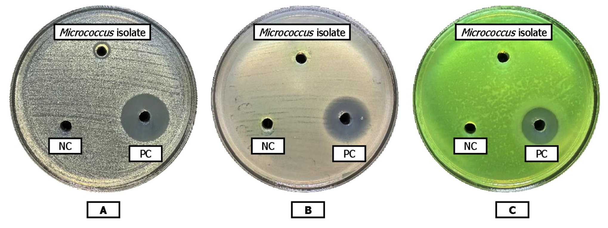
Figure 1. Antimicrobial activity of Micrococcus isolate with the different indicators bacteria: (A) S. aureus ATCC 25923, (B) E. coli ATCC 25922, (C) P. aeruginosa ATCC 27853. NC: Negative control, PC: Positive control
Table 1. Antibacterial activity of the cell-free culture supernatant against selected indicator strains
Indicator strain |
Relevant characteristic |
Inhibition-zone diameter (mm), Mean ± SD |
|---|---|---|
S. aureus ATCC 25923 |
Methicillin-susceptible |
8.23 ± 0.45 |
S. aureus ATCC 29213 |
Methicillin-susceptible |
7.07 ± 0.31 |
S. aureus ATCC 43300 |
Methicillin-resistant |
7.30 ± 0.75 |
S. saprophyticus ATCC BAA-750 |
Coagulase-negative Staphylococcus |
9.07 ± 0.51 |
E. faecalis ATCC 29212 |
Vancomycin-susceptible |
7.83 ± 0.29 |
E. faecalis ATCC 51299 |
Vancomycin-resistant |
No inhibition zone |
E. casseliflavus ATCC 700327 |
Intrinsic low-level vancomycin resistance |
No inhibition zone |
E. coli ATCC 25922 |
Gram-negative quality-control |
No inhibition zone |
P. aeruginosa ATCC 27853 |
Gram-negative non-fermenting quality-control |
No inhibition zone |
To further examine its antibacterial activity against Gram-positive bacteria, the cell-free culture supernatant was tested against additional indicator strains. The Micrococcus isolate exhibited antibacterial activity against S. aureus ATCC 29213 (7.07 ± 0.31 mm), MRSA S. aureus ATCC 43300 (7.30 ± 0.75 mm), S. saprophyticus ATCC BAA-750 (9.07 ± 0.51 mm), and E. faecalis ATCC 29212 (7.83 ± 0.29 mm) (Table 1). No inhibition zone was observed against E. faecalis ATCC 51299 or E. casseliflavus ATCC 700327 (Figure 2).
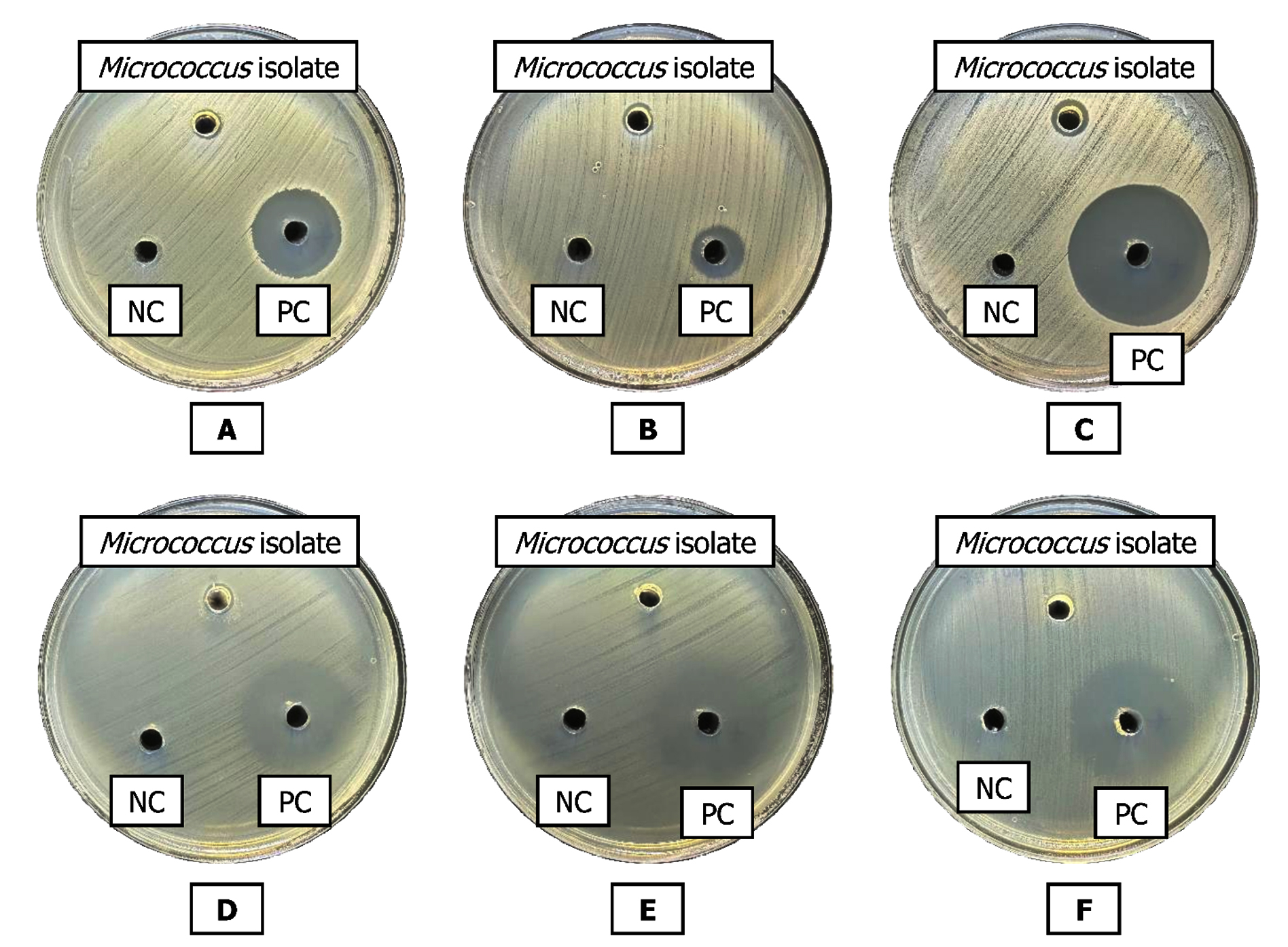
Figure 2. Antimicrobial activity of Micrococcus isolate with the additional Gram-positive indicator strains. (A) S. aureus ATCC 29213, (B) S. aureus ATCC 43300, (C) S. saprophyticus ATCC BAA-750, (D) E. faecalis ATCC 29212, (E) E. faecalis ATCC 51299, (F) E. casseliflavus ATCC 700327. NC: Negative control, PC: Positive control
Genome sequencing of the sampled Micrococcus
For the short-read sequencing data (Illumina), a total of 3,376,387 raw reads with a %Q30 of 96.5% were obtained. For the long-read data (Nanopore), the total number of raw reads reached 19,565, providing sufficient volume for the de novo assembly step. De novo genome assembly was carried out using both the Illumina and the Nanopore data, and the resulting genome was a single circular contig of 2,791,667 bp in length with a GC content of 71.52% (Figure 3). Genome completeness was 98.52%, with no contamination from the same species and only 0.46% contamination from other species.
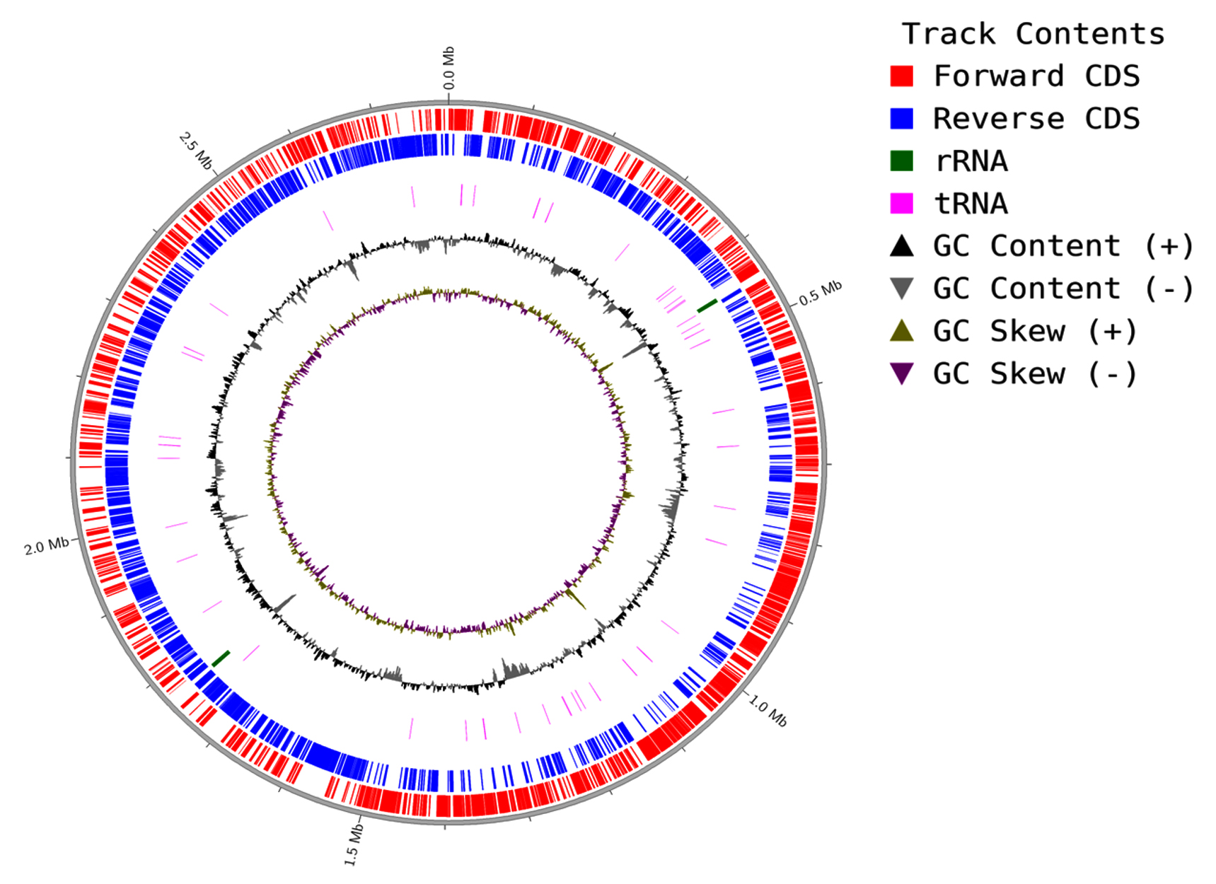
Figure 3. Circular genome map of the skin-derived Micrococcus isolate
Species identification of the Micrococcus
Based on the result from Kraken2, the isolate was mapped 100% to Micrococcus. However, the ANI results showed a low identity percentage of <90% against every Micrococcus reference genome, with the highest ANI value belonging to M. lylae (88.8%) (Table 2). Thus, the isolate may represent a novel Micrococcus strain most closely related to M. lylae.
Table 2. ANI value between the reference Micrococcus spp. and the isolated Micrococcus
Reference genome |
ANI (%) |
|---|---|
Micrococcus lylae |
88.8 |
Micrococcus porci |
83.7 |
Micrococcus endophyticus |
83.7 |
Micrococcus yuuanaensis |
83.7 |
Micrococcus aloeverae |
83.7 |
Micrococcus luteus |
83.2 |
Micrococcus flavus |
83.2 |
Micrococcus lacusdianchii |
83.0 |
Micrococcus antarcticus |
82.8 |
Micrococcus cohnii |
82.0 |
Micrococcus terreus |
80.5 |
Genomic comparison with representative genomes
Based on the phylogenetic analysis of the novel Micrococcus and representative Micrococcus genomes used in the ANI step, the phylogenetic tree of Micrococcus showed a close relationship between the novel Micrococcus and M. lylae (Figure 4). The presence of putative metabolites also suggested a similar pattern between the two genomes, with both harboring lanthipeptide class III-related genes. However, the study genome contained a gene encoding an arylpolyene that was not detected in any of the other Micrococcus genomes examined, indicating a potential new metabolite product.
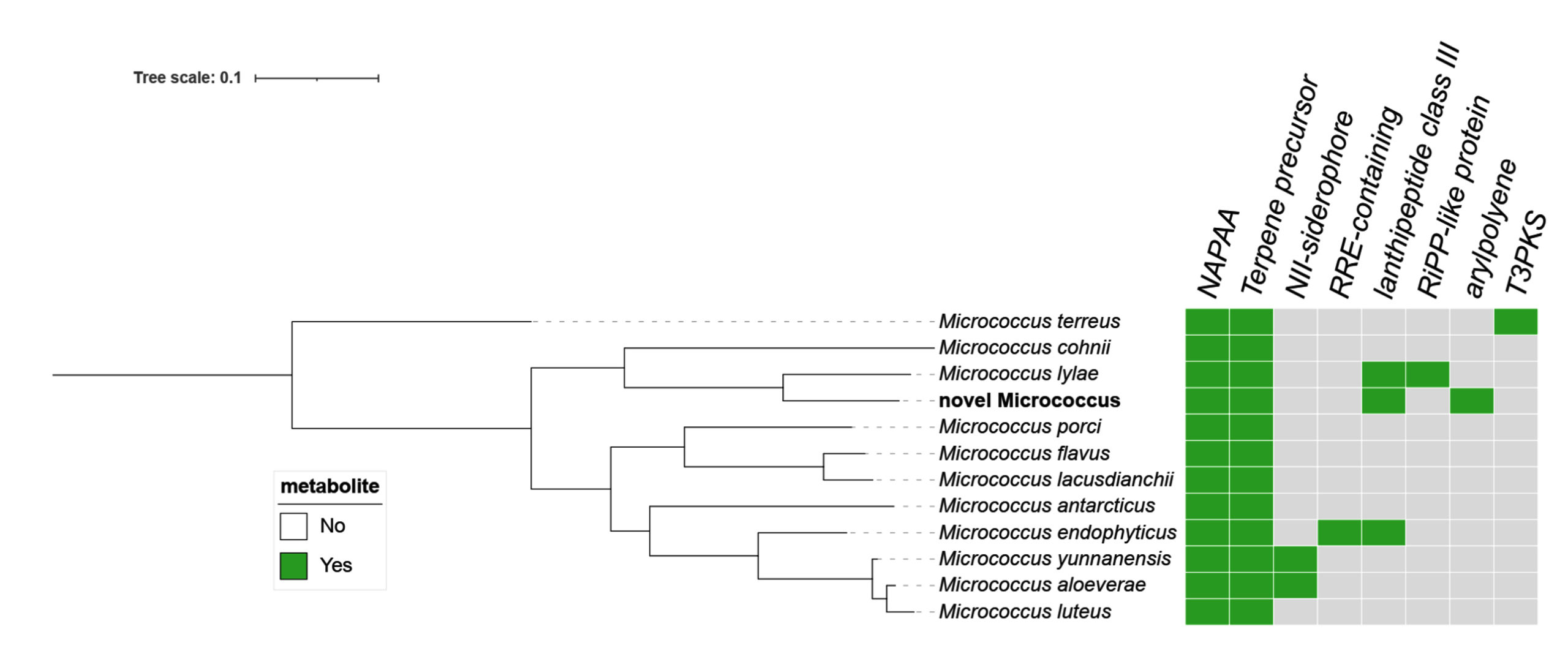
Figure 4. Phylogenetic trees of Micrococcus isolate in comparison to other Micrococcus species Gene content analysis result
The assembled genome was annotated using Prokka v1.14.6; the annotation identified 2,490 coding sequences (CDSs), 53 tRNA genes, and 6 rRNA genes. Functional annotation with COGclassifier assigned COG (clusters of orthologous groups) categories to 81% (2017/2490) of the CDSs (Figure 5). Apart from genes for essential functions of transcription and translation, there were a large number of genes (over 100 sequences) contributing to different metabolic processes, including coenzyme (H), lipid (I), and inorganic ion (P) metabolism, highlighting the potential of this strain to produce secondary metabolites. However, more than 120 genes had unknown functions, which may affect the overall distribution of COG categories. Thus, to accurately detect potential bacteriocins, the Micrococcus genome was functionally annotated using the bacteriocin detection web tool BAGEL4. Four putative open reading frames (ORFs) were detected; of these, orf000005 and orf000006 showed high identity with the membrane protein insertase YidC of Streptomyces avermitilis (89%) and the membrane protein insertion efficiency factor YidD of Pasteurella multocida (96%), respectively (Figure 6). The presence of ORFs with high similarity to bacteriocins may hinder the antimicrobial capability of this strain, and further bacteriocin assays should be performed to confirm this.
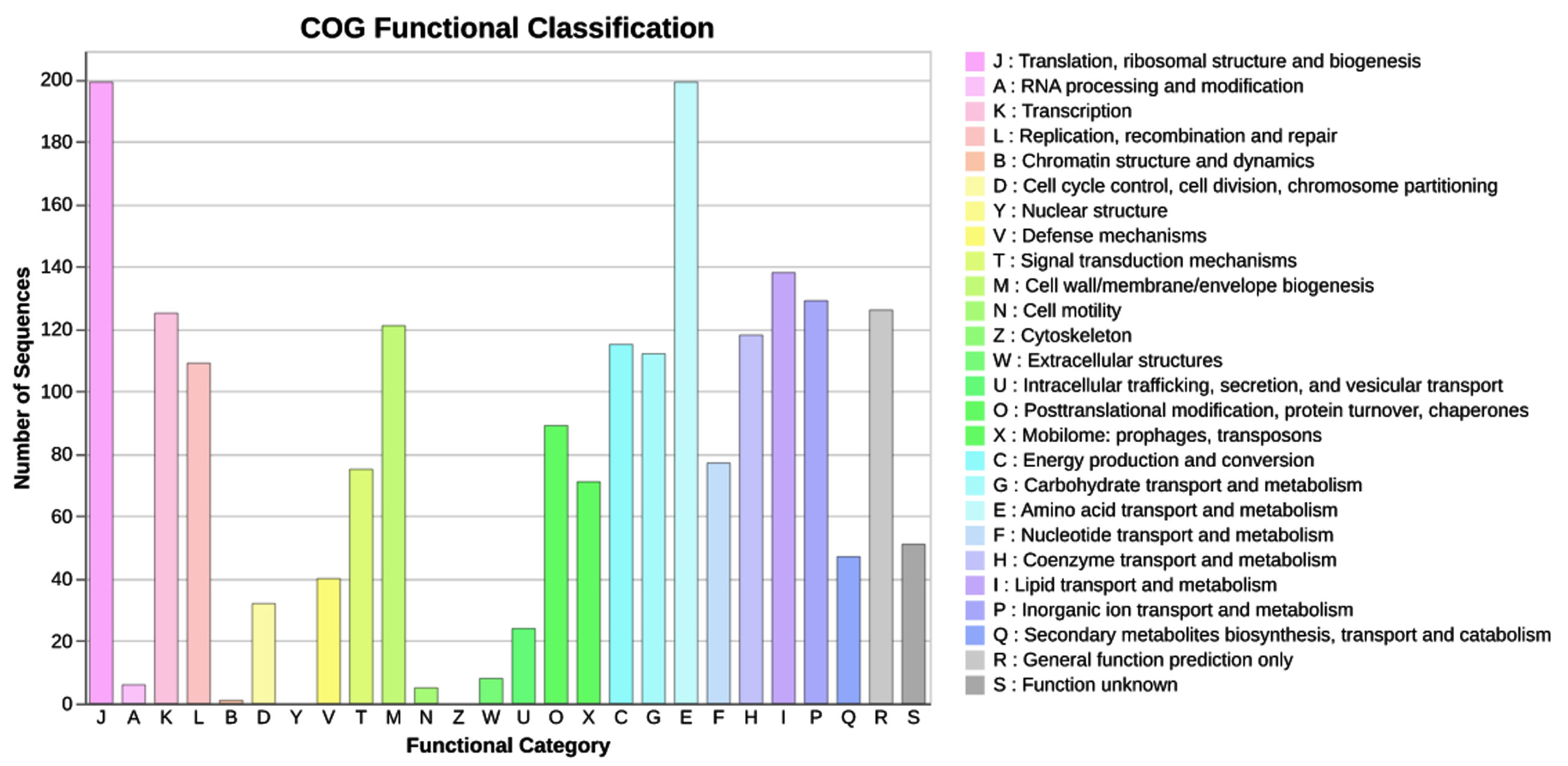
Figure 5. Functional annotation of the Micrococcus genome
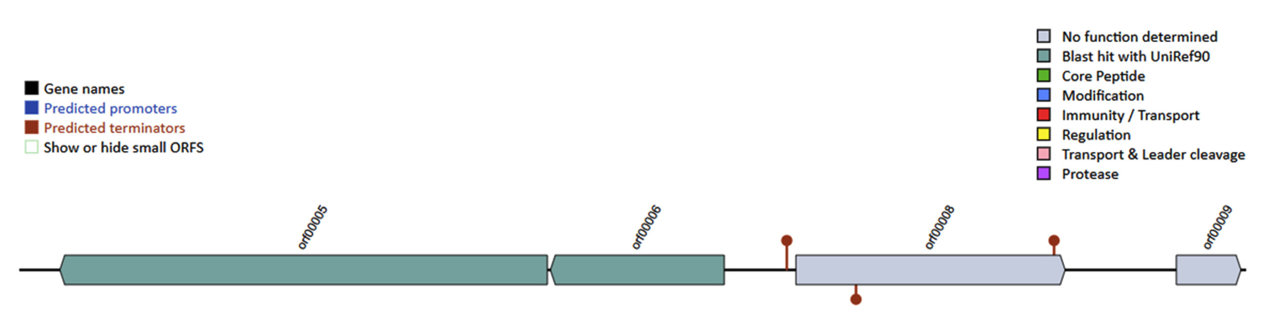
Figure 6. Putative bacteriocin encoding genes detection based on the Micrococcus genome
Micrococcus spp. are common members of healthy human skin microbiota and have been reported to produce antimicrobial substances that may help suppress opportunistic pathogens. Furthermore, skin commensals are being explored as potential microbiome-based therapeutics to reduce harmful bacteria on the skin, while potentially limiting the selective pressure associated with conventional antibiotics.29 In our initial screening of bacteria isolated from healthy human skin, we identified a Micrococcus isolate with strong antagonistic activity. Notably, the cell-free supernatant of the tested strain inhibited S. aureus but not E. coli or P. aeruginosa, indicating a pattern of selective activity against Gram-positive bacteria. In addition to the initial inhibition observed against S. aureus ATCC 25923, expanded testing demonstrated antibacterial activity against S. aureus ATCC 29213 (7.07 ± 0.31 mm), MRSA S. aureus ATCC 43300 (7.30 ± 0.75 mm), S. saprophyticus ATCC BAA-750 (9.07 ± 0.51 mm), and E. faecalis ATCC 29212 (7.83 ± 0.29 mm). In contrast, no inhibitory activity was observed against E. faecalis ATCC 51299 or E. casseliflavus ATCC 700327. These findings indicated that the antibacterial activity of the isolate was not uniform across all Gram-positive indicator strains. The observed activity against MRSA is particularly noteworthy and supports further investigation of the antimicrobial compound(s) produced by this isolate. Targeted antibacterial activity of bacteriocins has attracted interest as a potential strategy for controlling clinically relevant bacterial pathogens.30 This profile is typical of many bacteriocins and thiopeptide/Ribosomally synthesized and post-translationally modified peptides (RiPP) molecules that preferentially target Gram-positive bacteria, owing to differences in the cell envelope structure and target accessibility.27 This result is in accordance to that of earlier studies, where pigments or crude extracts from Micrococcus luteus and marine Micrococcus strains showed strong activity against Staphylococcus spp. and other Gram-positive pathogens.4,5 Because Micrococcus spp. can produce both peptides and pigments with antimicrobial activity, the observed anti-Staphylococcus effects may involve multiple metabolite classes.4,5 Discriminating between these possibilities requires protease-sensitivity assays, stability testing, and chemical analysis of the active fractions.27,31,32
Genome assembly produced a single circular chromosome with high estimated completeness and low contamination, supporting robust downstream analyses.13-16 Kraken2 assigned the isolate to the genus Micrococcus, but ANI analysis against available reference genomes revealed a maximum identity of only ~88.8% to M. lylae, clearly below the ~95% ANI threshold generally used for species delineation.18 Thus, the isolate might represent a novel Micrococcus taxon distinct from the currently identified Micrococcus spp., suggesting that human skin could harbor an under-characterized Micrococcus community with an unexplored biosynthetic potential.4,8,20
To provide insights into the genomic content of the novel Micrococcus, Prokka annotation was performed, which predicted 2,490 CDSs, 53 tRNAs, and 6 rRNA genes. These counts are comparable to those of other Micrococcus genomes and are consistent with a compact yet metabolically diverse gene repertoire.6,7,19 Functional classification showed that approximately 81% of the CDSs could be assigned to COG categories, including many genes involved in coenzyme, lipid, and inorganic ion metabolism. These functional categories suggest the potential for diverse secondary metabolism.6,7,20 Previous studies have reported that the bioactive pigments and metabolites in Micrococcus spp., including carotenoids, have antioxidant and antimicrobial activities.4,6-8 In this strain, the enrichment of metabolic genes was consistent with its biosynthetic potential. At the same time, this Micrococcus isolate harbored more than 120 genes of unknown function, consistent with the view that many biosynthetic pathways in Micrococcus remain uncharacterized and warrant further investigation.19,20 The novel Micrococcus strain was also investigated for the presence of potential bacteriocins, and two putative ORFs with RiPP/bacteriocin-associated signatures were predicted, which may contribute to the antimicrobial capability of the isolate. Although no canonical micrococcin cluster was detected in the tested strain, the BAGEL4 output suggested that this strain may encode noncanonical or novel antimicrobial peptides that warrant further investigation.27,28 This study has some limitations. Bacteriocin-related genes were predicted in silico but were not validated using genetic or biochemical approaches.
Although the cell-free culture supernatant showed antibacterial activity against several Gram-positive reference strains, including MRSA S. aureus ATCC 43300, the antimicrobial spectrum was evaluated using only a limited number of ATCC strains and therefore may not fully reflect the activity against diverse multidrug-resistant clinical isolates.5,8,9 Nevertheless, future evaluation of the tested strain and its metabolites in skin-relevant models would clarify their potential role in modulating skin microbiota and controlling S. aureus colonization.
In conclusion, our data show that a skin-derived Micrococcus isolate that is genomically distinct from the currently available Micrococcus reference genomes possesses both genetic and phenotypic hallmarks of antimicrobial potential. Notably, the cell-free culture supernatant showed antibacterial activity against several Gram-positive reference strains, including MRSA S. aureus ATCC 43300. Together with previous evidence of bioactive pigments and bacteriocin production in Micrococcus spp., these findings support the further exploration of this genus as a source of new antimicrobial agents and a possible contributor to the protective functions of the human skin microbiome.
ACKNOWLEDGMENTS
This work was supported by the Hue University-level Science and Technology Project (2024), code DHH2024-04-224.
CONFLICT OF INTEREST
The authors declare that there is no conflict of interest.
AUTHORS’ CONTRIBUTION
All authors listed have made a substantial, direct and intellectual contribution to the work, and approved it for publication.
FUNDING
None.
DATA AVAILABILITY
The datasets generated and analysed during the current study are available from the corresponding author on reasonable request.
ETHICS STATEMENT
This study was approved by the Biomedical Research Ethics Committee, Hue University of Medicine and Pharmacy, Hue University (Approval No. H2024/116; May 23, 2024).
INFORMED CONSENT
Written informed consent was obtained from the participants before enrolling in the study.
- Simons A, Alhanout K, Duval RE. Bacteriocins, antimicrobial peptides from bacterial origin: overview of their biology and their impact against multidrug-resistant bacteria. Microorganisms. 2020;8(5):639.
Crossref - Sugrue I, Ross RP, Hill C. Bacteriocin diversity, function, discovery and application as antimicrobials. Nat Rev Microbiol. 2024;22(9):556-571.
Crossref - Reuben RC, Torres C. Bacteriocins: potentials and prospects in health and agrifood systems. Arch Microbiol. 2024;206(5):233.
Crossref - Karbalaei-Heidari HR, Partovifar M, Memarpoor-Yazdi M. Evaluation of the bioactive potential of secondary metabolites produced by a new marine Micrococcus species isolated from the Persian Gulf. Avicenna J Med Biotechnol. 2020;12(1):61-65.
- Umadevi K, Krishnaveni M. Antibacterial activity of pigment produced from Micrococcus luteus KF532949. Int J Chem Anal Sci. 2013;4(3):149-152.
Crossref - Cooney JJ, Marks HW, Smith AM. Isolation and identification of canthaxanthin from Micrococcus roseus. J Bacteriol. 1966;92(2):342-345.
Crossref - Shahin YH, Elwakil BH, Ghareeb DA, Olama ZA. Micrococcus lylae MW407006 pigment: production, optimization, nano-pigment synthesis, and biological activities. Biology. 2022;11(8):1171.
Crossref - Shukla M, Nadumane VK. Yellow pigment from a novel bacteria, Micrococcus terreus, activates caspases and leads to apoptosis of cervical and liver cancer cell lines. J Appl Pharm Sci. 2021;11(8):77-84.
Crossref - Su TL. Micrococcin, an antibacterial substance formed by a strain of Micrococcus. Br J Exp Pathol. 1948;29(5):473-481.
- Munir A, Janbey A, Sajjad B, Perumal KV, Qayyum H, Bakhtiar M. Bioinformatics-driven discovery of skin microbiota bacteriocins as potential antibiotics and probiotics. J Antibiot. 2025;78(10):606-620.
Crossref - Baker JS. Comparison of various methods for differentiation of staphylococci and micrococci. J Clin Microbiol. 1984;19(6):875-879.
Crossref - Chen S, Zhou Y, Chen Y, Gu J. fastp: an ultra-fast all-in-one FASTQ preprocessor. Bioinformatics. 2018;34(17):i884-i890.
Crossref - Zimin AV, Salzberg SL. The genome polishing tool POLCA makes fast and accurate corrections in genome assemblies. PLoS Comput Biol. 2020;16(6):e1007981.
Crossref - Krasnov GS, Pushkova EN, Novakovskiy RO, et al. High-quality genome assembly of Fusarium oxysporum f. sp. lini. Front Genet. 2020;11:959.
Crossref - Gurevich A, Saveliev V, Vyahhi N, Tesler G. QUAST: quality assessment tool for genome assemblies. Bioinformatics. 2013;29(8):1072-1075.
Crossref - Parks DH, Imelfort M, Skennerton CT, Hugenholtz P, Tyson GW. CheckM: assessing the quality of microbial genomes recovered from isolates, single cells, and metagenomes. Genome Res. 2015;25(7):1043-1055.
Crossref - Wood DE, Lu J, Langmead B. Improved metagenomic analysis with Kraken 2. Genome Biol. 2019;20:257.
Crossref - Jain C, Rodriguez-R LM, Phillippy AM, Konstantinidis KT, Aluru S. High throughput ANI analysis of 90K prokaryotic genomes reveals clear species boundaries. Nat Commun. 2018;9(1):5114.
Crossref - Seemann T. Prokka: rapid prokaryotic genome annotation. Bioinformatics. 2014;30(14):2068-2069.
Crossref - Kanehisa M, Sato Y, Morishima K. BlastKOALA and GhostKOALA: KEGG tools for functional characterization of genome and metagenome sequences. J Mol Biol. 2016;428(4):726-731.
Crossref - Wickham H, Chang W, Henry L, et al. ggplot2: create elegant data visualisations using the grammar of graphics. R package. Accessed June 16, 2024. https://cran.r-project.org/package=ggplot2
- Kassambara A. ggpubr: ‘ggplot2’ based publication ready plots. R package. Accessed October 31, 2024. https://rpkgs.datanovia.com/ggpubr/
- Page AJ, Cummins CA, Hunt M, et al. Roary: rapid large-scale prokaryote pan genome analysis. Bioinformatics. 2015;31(22):3691-3693.
Crossref - Nguyen LT, Schmidt HA, von Haeseler A, Minh BQ. IQ-TREE: a fast and effective stochastic algorithm for estimating maximum-likelihood phylogenies. Mol Biol Evol. 2015;32(1):268-274.
Crossref - Letunic I, Bork P. Interactive Tree Of Life (iTOL) v5: an online tool for phylogenetic tree display and annotation. Nucleic Acids Res. 2021;49(W1):W293-W296.
Crossref - Camacho C, Coulouris G, Avagyan V, et al. BLAST+: architecture and applications. BMC Bioinformatics. 2009;10(1):421.
Crossref - van Heel AJ, de Jong A, Song C, Viel JH, Kok J, Kuipers OP. BAGEL4: a user-friendly web server to thoroughly mine RiPPs and bacteriocins. Nucleic Acids Res. 2018;46(W1):W278-W281.
Crossref - Medema MH, Blin K, Cimermancic P, et al. antiSMASH: rapid identification, annotation and analysis of secondary metabolite biosynthesis gene clusters in bacterial and fungal genome sequences. Nucleic Acids Res. 2011;39(suppl 2):W339-W346.
Crossref - Oh J, Voigt AY. The human skin microbiome: from metagenomes to therapeutics. Nat Rev Microbiol. 2025;23(12):771-787.
Crossref - Rutter JW, Dekker L, Clare C, et al. A bacteriocin expression platform for targeting pathogenic bacterial species. Nat Commun. 2024;15(1):6332.
Crossref - Van der Veken D, Hollanders C, Verce M, et al. Genome-based characterization of a plasmid-associated micrococcin P1 biosynthetic gene cluster and virulence factors in Mammaliicoccus sciuri IMDO-S72. Appl Environ Microbiol. 2022;88(4):e0208821.
Crossref - Lean SS, Shang AZ, Booth T, Lacey E, Chooi YH. The genome of Micrococcus luteus MST-118984C isolated from Australian soil harbours a bacteriocin biosynthetic gene cluster and biocide/multidrug efflux genes. Int J Antimicrob Agents. 2021;58(Suppl 1):21002727.
Crossref
© The Author(s) 2026. Open Access. This article is distributed under the terms of the Creative Commons Attribution 4.0 International License which permits unrestricted use, sharing, distribution, and reproduction in any medium, provided you give appropriate credit to the original author(s) and the source, provide a link to the Creative Commons license, and indicate if changes were made.