


ISSN: 0973-7510
E-ISSN: 2581-690X
 , Dwi Kusuma Wahyuni2, Teguh Hari Sucipto3, Arif Nur Muhammad Ansori4, Rollando Rollando5 and Hery Purnobasuki2
 Dengue virus (DENV) infection is caused by Aedes sp. mosquitoes such as A. aegypti, A. albopictus, and A. scutellaris. DENV has been reported to possess a cell tropism consisting of mast cells, fibroblasts, dendritic cells, macrophages, langerhans, and keratinocytes. Viral replication consists of various stages such as attachment, fusion, replication of genetic material, assembly, and virus release. Fusion inhibitors often act to interfere with the endocytosis process mediated by parts on the virus and host receptor. However, few previous studies have used in silico approaches to screen candidate DENV fusion inhibitors with bioactive compounds. Several volatile compounds such as flavonoids contained in Sonchus arvensis L. leaf extracts are predicted to have potential for treatment. Therefore, this study aims to determine the activity of volatile compounds from Sonchus arvensis L. to inhibit DENV fusion through in silico approach. The in silico methods used were ligand collection, protein retrieval, druglikeness prediction, virtual screening, molecular interaction analysis, molecular dynamics, toxicity profiling, and antiviral probability. The results showed that volatile compounds from Sonchus arvensis L. consisting of Hexadecanoic acid, ethyl ester, Phytol, 1,2-benzenedicarboxylic acid, bis(2-methylpropyl)ester, and Benzoic acid, 2-ethylhexyl ester could inhibit DENV fusion through binding to DENV proteins.
Antiviral, Dengue, Fusion Inhibitors, Sonchus arvensis, Viroinformatics
Dengue virus (DENV) infection is transmitted through the bite of Aedes sp. mosquitoes such as A. aegypti, A. albopictus, and A. scutellaris which are commonly found in subtropical and tropical regions. DENV has been reported to have 4 strains consisting of DENV-1, DENV-2, DENV-3, and DENV-4. This virus has an envelope and is composed of single-stranded RNA (ssRNA) with regions encoding capsid (C), membrane (M), and envelope (E). It also possesses structural proteins and 7 nonstructural proteins such as NS1, NS2A, NS3, NS4A, NS4B, and NS5.1 DENV replication is often stimulated through viral attachment to host receptors and then endocytosis through fusion. Viral replication occurs in RE, where the RNA produces polyproteins and new viruses undergo exocytosis through vesicles to infect new host cells.2 At present, vaccines and antiviral drugs for dengue fever are not commercially available due to ongoing investigations. DENV can infect a wide variety of cell types (permissive cells) in the epidermal and dermal layers of human skin consisting of mast cells, fibroblasts, dendritic cells, macrophages, langerhans, and keratinocytes.3 Therefore, some of these permissive cells are also targets for therapies to inhibit viral replication.4
Accounting to previous studies, viral replication consists of various stages such as attachment, fusion, replication of genetic material, assembly, and virus release. Fusion inhibitors act to interfere with the endocytosis process mediated by parts on the virus and host receptor. The viral attachment protein E stage of DENV interacts with host cell receptors such as DC-SIGN, heparan sulfate, and mannose receptor.5 Fusion of viral particles occurs through interaction on receptors for regulation of clathrin-mediated endocytosis to form endosomal membranes under low pH conditions.6 The release of genomic RNA from DENV is initiated by the acidic pH of the endosomal environment, which triggers capsid uncoating to process viral proteins and replication. The mRNA translation process occurs to trigger the production of new viral proteins.7 Host receptors that facilitate DENV fusion consist of langerin/CD207 and Type Lectin Domain Containing 5A (CLEC5A) found on the surface.8,9 Capsid proteins (Cprotein) are involved as facilitators of DENV fusion to maintain stable interactions with the envelope (Eprotein) and viral genome.10,11
Previous studies using pocket binding on the DENV Eprotein domain to design candidate fusion inhibitors were confirmed by in vitro analysis resulting in an IC50 of 6 μM with a viral load reduction of 9.8 μM.12 Several candidate fusion inhibitors consisting of 1OAN1, DN57opt, DN59, rolitetracycline, doxycycline, NITD448, A5, and SA-17 interact with the hydrophobic pocket on the envelope of DENV-1, DENV-2, and DENV-3.13 Screening of DENV antiviral candidates is carried out based on bioactive compounds from natural ingredients such as triterpenoids with an in silico approach by targeting non-structural 4B (NS4B).14 Geraniin from Nephelium lappaceum L. and Phyllanthus myrtifolius were identified as having DENV antiviral activity through inhibition of transcription and regulation of viral translation.15 However, few previous studies have used in silico approaches to screen candidate DENV fusion inhibitors with bioactive compounds.
Antiviral activity is demonstrated by various extracts from natural materials such as green tea, mangosteen, turmeric, and others.16,17 Natural compounds have antiviral mechanisms through direct and indirect mechanisms, such as inhibiting target activity in the virus and then suppressing excessive inflammatory responses or initiating apoptosis in infected cells.18 Sonchus arvensis L. is one of the natural ingredients used in Indonesia as an alternative medicine with antioxidant, antiplasmodial, and anti-inflammatory potential.19,20 Antiviral activity of Sonchus arvensis L. extract as a potential inhibitor of key proteins of SARS-CoV-2 was shown by a previous study.21 The use of Sonchus arvensis L. for the treatment of dengue fever has never been carried out, but allows for further exploration of natural compounds. Several volatile compounds such as flavonoids contained in the leaf extracts are predicted to have potential for treatment.20 Therefore, this study aims to determine the activity of volatile compounds from Sonchus arvensis L. with dual mechanism to inhibit the fusion mechanism in DENV entry phase through in silico approach.
Ligand collection
Volatile compounds from the leaves of Sonchus arvensis L. consisted of Decanal, 2,4-Decadienal, Tetradecane, Pentadecane, Hexadecane, Benzoic acid, 2-ethylhexyl ester, 3-Tetradecene, Tetradecanoic acid, Octadecene, Tetradecanal, and 1,2-benzenedicarboxylic acid. Others included bis(2-methylpropyl)ester, 1-Hexadecanol, Nonadecane, Hexadecanoic acid, ethyl ester, Eicosane, 10-Heneicosene, Heneicosene, Phytol, 9, 12, 15-octadecatrienoic acid, ethyl ester, Dokozan, 9-Tricosene, Z, Triclosan, Tetracosane, Pentacosane, and Hexacosane.20 PubChem (https://pubchem.ncbi.nlm.nih.gov/) was used for data retrieval of volatile compounds from Sonchus arvensis L., which consisted of name, CID, formula, canonical SMILES, and structure data format (SDF). Minimization of SDF files into Protein Data Bank format (PDB) was performed on the Open Babel plugin through PyRx – Python Prescription Virtual Screening Tool v1.1.0 with academic license for ligand optimization.22
Protein retrieval
This study used DENV Envelope (Eprotein), DENV Capsid (Cprotein), Langerin/CD207, and C-Type Lectin Domain Containing 5A (CLEC5A) as target inhibitors. DENV fusion included several components on the virion such as Eprotein and Cprotein in the replication process.10,11 Langerin and CLEC5A were identified on the host as receptors to facilitate viral entry.8,9 The 3-dimensional structures of the 4 targets were obtained from RCSB PDB (https://www.rcsb.org/), and the water molecules and native ligands in the target PDB files were removed using PyMOL v2.5.0 with academic license.23
Druglikeness prediction
Assessment of druglikeness in this study using rules such as Lipinski, Ghose, Veber, Egan, and Muegge, which was performed through SwissADME (http://www.swissadme.ch/).24 Another parameter used in druglikeness was the probability of bioavailability and absorption of the query compound in the Homo sapiens was also predicted. Positive predictions in druglikeness were drug-like molecules by following all the parameters used.25
Virtual screening
Prediction of ligand binding ability of Sonchus arvensis L. volatile compounds on Eprotein, Cprotein, Langerin, and CLEC5A was conducted through ligand-based virtual screening (LBVS). The method used was docking screening by directing the autogrid on target surface entire to identify ligand binding affinity (kcal/mol).26 Autogrid played a major role to direct ligand binding according to the XYZ center position and dimensions on the target. Molecular docking was performed through the VinaWizard plugin in PyRx – Python Prescription Virtual Screening Tool v1.1.0 with academic license.27 Visualization of the 3-dimensional structure from docking results was performed through PyMOL v2.5.0 with academic license.28
Molecular interaction
Molecular interactions in this study were identified through Discovery Studio v2016 BIOVIA using 2D plot.29 Weak bonds such as hydrogen, van der Waals, pi, as well as alkyl, and electrostatic were present in the ligand-protein complex. These bonds contributed to the initiation of biological responses in targets such as inhibitor activity.30
Dynamic simulation
Stability of ligand-protein complexes was identified through molecular dynamics simulations on CABS-flex v2.0 (https://biocomp.chem.uw.edu.pl/CABSflex2). This study used molecular dynamics simulations based on structural flexibility through root mean square fluctuation (RMSF) (Å) in molecular complexes.31,32 Average RMSF value used for stability parameter assessment must be <3 (Å) with simulation parameters consisting of restraints, rigidity, restraints, number of cycles, trajectory, and temperature range.33
Toxicity prediction and antiviral probability
Toxicity prediction in this study was performed using ProTox v3.0 (https://tox.charite.de/protox3/). The data consisted of LD50 (mg/kg), toxicity class, average similarity, accuracy, and organ toxicity. This server worked with random forest and neural networks algorithms to predict the toxicity properties of a query compound based on similarity and coverage to identify toxicity class, LD50, toxicity type, target, and clinical toxicity. The query compound was predicted to have non-toxic characteristics for its safe use.34 Antiviral probability prediction was performed through PASSOnline (https://way2drug.com/PassOnline/predict.php) based on the probability activation (Pa) and inhibition (Pi) values, the Pa > Pi value was selected to predict the probability.35
Data collection from compounds and target database
A total of 25 volatile compounds from Sonchus arvensis L. leaves were obtained from PubChem with information consisting of CID, formula, and SMILE (Table 1). Volatile compound groups from Sonchus arvensis L. leaves consisted of fatty acid, aliphatic hydrocarbon, ester, aldehyde, and fatty alcohol with antidiarrheal, antioxidant, antibacterial, and anticancer activities.36,37 Subsequently, the selection process was performed on volatile compounds through the prediction of druglikeness to identify activity similarity with commercial drugs, bioavailability characteristics, and absorption effectiveness in the human body.
Table 1. Data collection of volatile compounds of Sonchus arvensis L. from database
Label |
Compounds |
CID |
Formula |
Canonical SMILES |
|---|---|---|---|---|
1. |
Decanal |
8175 |
C10H20O |
CCCCCCCCCC=O |
2. |
2,4-Decadienal |
5283349 |
C10H16O |
CCCCC/C=C/C=C/C=O |
3. |
Tetradecane |
12389 |
C14H30 |
CCCCCCCCCCCCCC |
4. |
Pentadecane |
12391 |
C15H32 |
CCCCCCCCCCCCCCC |
5. |
Hexadecane |
11006 |
C16H34 |
CCCCCCCCCCCCCCCC |
6. |
Benzoic acid, 2- ethylhexyl ester |
94310 |
C15H22O2 |
CCCCC(CC)COC(=O)C1=CC=CC=C1 |
7. |
3-Tetradecene |
5352802 |
C14H28 |
CCCCCCCCCC/C=C/CC |
8. |
Tetradecanoic acid |
11005 |
C14H28O2 |
CCCCCCCCCCCCCC(=O)O |
9. |
Octadecene |
8217 |
C18H36 |
CCCCCCCCCCCCCCCCC=C |
10. |
Tetradecanal |
31291 |
C14H28O |
CCCCCCCCCCCCCC=O |
11. |
1,2-benzenedicar- boxylic acid, bis(2-methylpropyl)ester |
1017 |
C8H6O4 |
C1=CC=C(C(=C1)C(=O)O)C(=O)O |
12. |
1-Hexadecanol |
2682 |
C16H34O |
CCCCCCCCCCCCCCCCO |
13. |
Nonadecane |
12401 |
C19H40 |
CCCCCCCCCCCCCCCCCCC |
14. |
Hexadecanoic acid, ethyl ester |
12366 |
C18H36O2 |
CCCCCCCCCCCCCCCC(=O)OCC |
15. |
Eicosane |
8222 |
C20H42 |
CCCCCCCCCCCCCCCCCCCC |
16. |
10-Heneicosene |
5364553 |
C21H42 |
CCCCCCCCCC/C=C/CCCCCCCCC |
17. |
Heneicosene |
3015374 |
C21H42 |
CCCCCCCCCCCCCCCCCCCC=C |
18. |
Phytol |
5280435 |
C20H40O |
C[C@@H](CCC[C@@H](C)CCC/C(=C/CO)/C) |
19. |
9, 12, 15-octadecatrienoic acid, ethyl ester |
5367460 |
C20H34O2 |
CC/C=C\C/C=C\C/C=C\CCCCCCCC(=O)OCC |
20. |
Dokozan |
12620 |
C22H46O |
CCCCCCCCCCCCCCCCCCCCCCO |
21. |
9-Tricosene, Z |
5365075 |
C23H46 |
CCCCCCCCCCCCC/C=C\CCCCCCCC |
22. |
Triclosan |
5564 |
C12H7Cl3O2 |
C1=CC(=C(C=C1Cl)O)OC2=C(C=C(C=C2)Cl)Cl |
23. |
Tetracosane |
12592 |
C24H50 |
CCCCCCCCCCCCCCCCCCCCCCCC |
24. |
Pentacosane |
12406 |
C25H52 |
CCCCCCCCCCCCCCCCCCCCCCCCC |
25. |
Hexacosane |
12407 |
C26H54 |
CCCCCCCCCCCCCCCCCCCCCCCCCC |
Inhibition targets of DENV fusion process in this study used DENV Eprotein (ID: 1TG8), DENV Cprotein (ID: 6VG5), Langerin (ID: 3KQG), and CLEC5A (ID: 2YHF) with 3-dimensional structures obtained from RCSB PDB (Figure 1). Viral proteins that had a significant role in membrane attachment and fusion, such as Eprotein, targeting DENV inhibitor candidates by previous studies was performed on this protein.38 Eprotein in DENV had a size of 53 kDa with dimer and trimer structures.39 The fusion mechanism in DENV was facilitated by cell receptors, intracellular factors, and viral proteins, while Cprotein is included in fusion to facilitate the preparation of viral genome transport vesicles to target organelles in host cells.40 Host receptors such as CLEC5A interacted with DENV Eprotein to trigger increased production of proinflammatory cytokines and viral fusion stage.41,42 Langerin (CD207) on host cells were identified to interact with DENV Eprotein in the process of virus fusion initiation.8
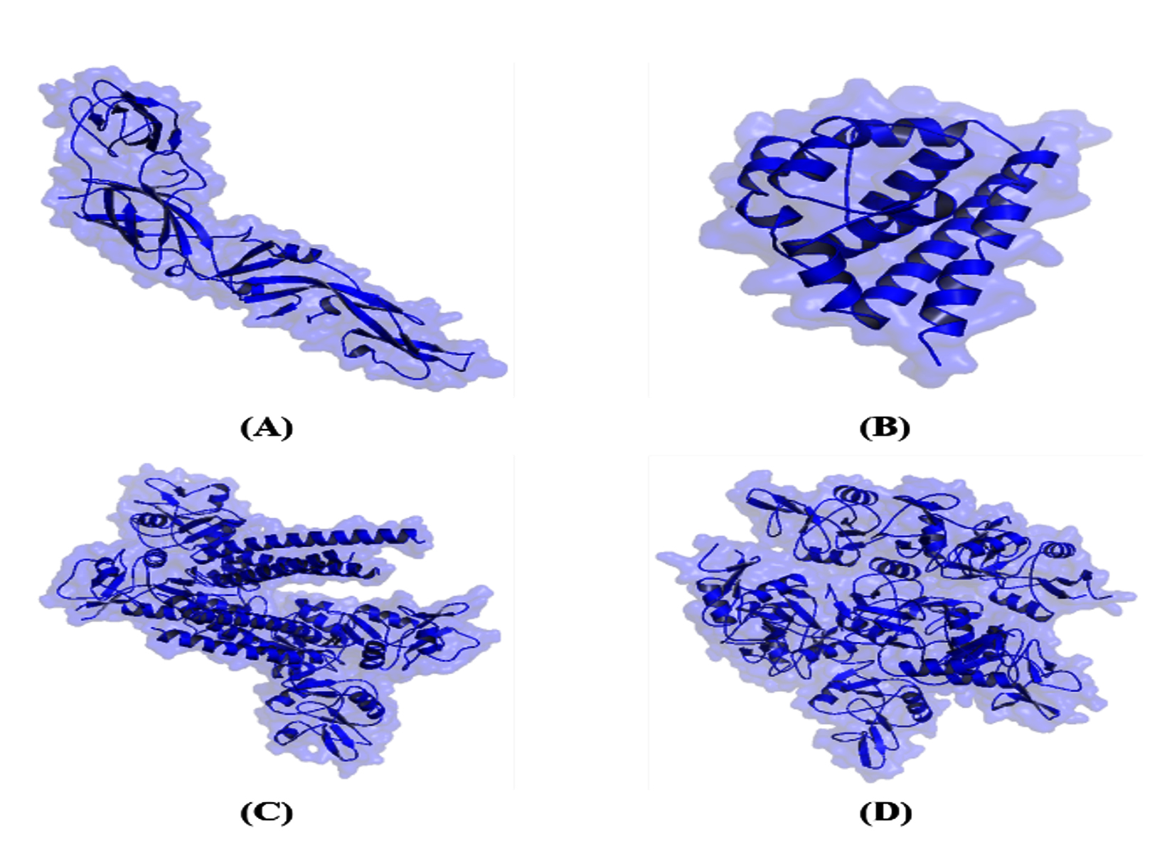
Figure 1. Three-dimensional structure of the target protein: (A) DENV Eprotein (B) DENV Cprotein (C) Langerin (D) CLEC5A
Drug-like molecule for Sonchus arvensis L. volatile compounds
Druglikeness prediction evaluated query compounds that had the potential to become good drug candidates by following the rules consisting of Lipinski, Ghose, Veber, Egan, and Muegge, then bioavailability and absorption predictions were used for support on druglikeness data.43 Compounds with labels 1-18 and 20-23 were included in the drug-like molecule that could be used for further analysis. Meanwhile, compounds with label 19 were not used because of errors when predicted and did not qualify as a drug-like molecule (Table 2). Compounds with drug-like molecule properties had a bioavailability value of 0.55 with diverse GIabsorption.
Table 2. Drug-like molecules from volatile compounds of Sonchus arvensis L. leaves
Compounds |
Lipinski |
Ghose |
Veber |
Egan |
Muegge |
Bioavailability |
GIabsorption |
Probable |
|---|---|---|---|---|---|---|---|---|
1. |
Yes |
No |
Yes |
Yes |
No |
0.55 |
High |
Drug-like molecule |
2. |
Yes |
No |
Yes |
Yes |
No |
0.55 |
High |
Drug-like molecule |
3. |
Yes |
No |
No |
Yes |
No |
0.55 |
Low |
Drug-like molecule |
4. |
Yes |
No |
No |
No |
No |
0.55 |
Low |
Drug-like molecule |
5. |
Yes |
No |
No |
No |
No |
0.55 |
Low |
Drug-like molecule |
6. |
Yes |
Yes |
Yes |
Yes |
Yes |
0.55 |
High |
Drug-like molecule |
7. |
Yes |
Yes |
Yes |
Yes |
No |
0.55 |
Low |
Drug-like molecule |
8. |
Yes |
Yes |
No |
Yes |
No |
0.85 |
High |
Drug-like molecule |
9. |
Yes |
No |
No |
No |
No |
0.55 |
Low |
Drug-like molecule |
10. |
Yes |
Yes |
No |
Yes |
No |
0.55 |
High |
Drug-like molecule |
11. |
Yes |
No |
Yes |
Yes |
No |
0.85 |
High |
Drug-like molecule |
12. |
Yes |
Yes |
No |
Yes |
No |
0.55 |
High |
Drug-like molecule |
13. |
Yes |
No |
No |
No |
No |
0.55 |
Low |
Drug-like molecule |
14. |
Yes |
No |
No |
No |
No |
0.55 |
High |
Drug-like molecule |
15. |
Yes |
No |
No |
No |
No |
0.55 |
Low |
Drug-like molecule |
16. |
Yes |
No |
No |
No |
No |
0.55 |
Low |
Drug-like molecule |
17. |
Yes |
No |
No |
No |
No |
0.55 |
Low |
Drug-like molecule |
18. |
Yes |
No |
No |
No |
No |
0.55 |
Low |
Drug-like molecule |
19. |
No Data |
No Data |
No Data |
No Data |
NoData |
No Data |
No Data |
– |
20. |
Yes |
No |
No |
No |
No |
0.55 |
Low |
Drug-like molecule |
21. |
Yes |
No |
No |
No |
No |
0.55 |
Low |
Drug-like molecule |
22. |
Yes |
Yes |
Yes |
Yes |
Yes |
0.55 |
High |
Drug-like molecule |
23. |
Yes |
No |
No |
No |
No |
0.55 |
Low |
Drug-like molecule |
24. |
Yes |
No |
No |
No |
No |
0.55 |
Low |
Drug-like molecule |
25. |
Yes |
No |
No |
No |
No |
0.55 |
Low |
Drug-like molecule |
Binding affinity of volatile compounds from Sonchus arvensis L. as inhibitor candidate
Simulation of ligand binding to the target for identification of inhibitory activity in this study was performed through molecular docking.44 Grid setting aimed to direct ligand binding to the target domain, which was the entire target domain used in this study for ligand activity screening. The docking grid settings for the binding of volatile compounds from the leaves of Sonchus arvensis L. with AutoGrid were performed on DENV Eprotein Center (Å) X: 9.804 Y: 20.820 Z: 15.317 Dimensions (Å) X: 60.462 Y: 52.009 Z: 128.993, DENV Cprotein Center (Å) X: 13.889 Y: 11.560 Z: 13.369 Dimensions (Å) X: 68.759 Y: 68.865 Z: 137.056, Langerin Center (Å) X: 20.840 Y: 61.121 Z: 99.689 Dimensions (Å) X: 99.940 Y: 99.583 Z: 107.969, and CLEC5A Center (Å) X: 20.782 Y: -36.661 Z: 12.824 Dimensions (Å) X: 96.695 Y: 118.944 Z: 116.002.
The docking results showed several ligands with more negative binding affinity values on each target consisting of compound with label 14 on DENV Eprotein (-5.9 kcal/mol), label 18 on DENV Cprotein (-6.8 kcal/mol), label 11 on langerin (-6.3 kcal/mol), and label 6 on CLC5A (-6.6 kcal/mol) (Table 3). Ligands with more negative binding affinity values were predicted to have good activity for inhibitor candidates on the target.45 Ligands with the most negative binding affinity values were used for docking validation analysis through molecular interactions and dynamics.46 Ligand-target complexes were displayed with a stick structure with colors based on the constituent atoms C (green), H (gray), O (red), and blue cartoons on transparent surfaces (Figure 2).
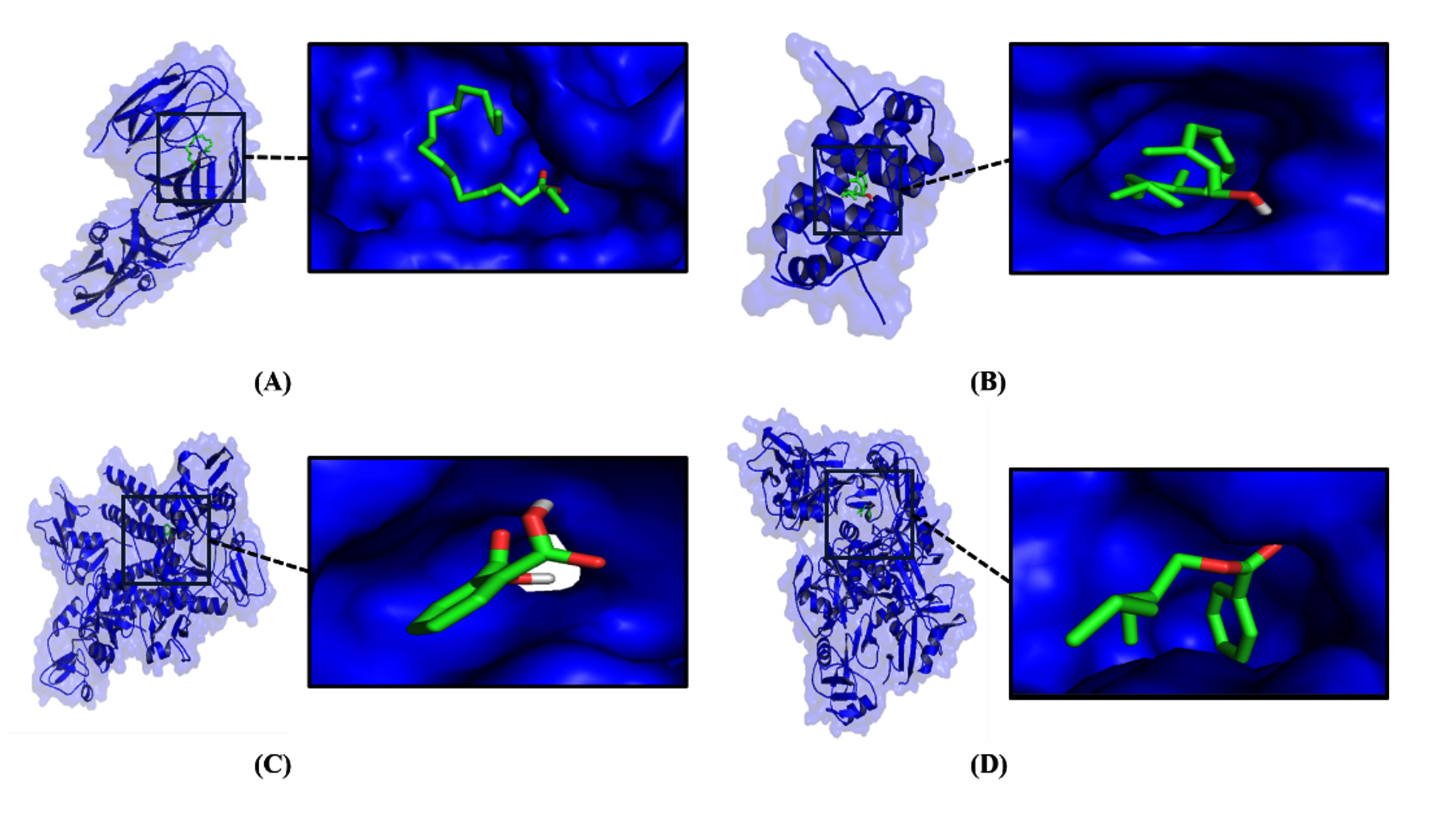
Figure 2. Ligand-protein molecular complex from docking simulation results: (A) Compound 14_DENV Eprotein (B) Compound 18_DENV Cprotein (C) Compound 11_Langerin (D) Compound 6_CLEC5A
Table 3. Comparison of binding affinity values of ligand-protein complexes
| Compounds | CID | Weight (g/mol) | Binding Affinity (kcal/mol) | |||
|---|---|---|---|---|---|---|
| DENV Eprotein | DENV Cprotein | Langerin | CLEC5A | |||
| 1. | 8175 | 156.26 | -3.7 | -5.2 | -3.9 | -4.4 |
| 2. | 5283349 | 152.23 | -4.0 | -5.5 | -4.8 | -4.8 |
| 3. | 12389 | 198.39 | -3.7 | -5.8 | -4.3 | -4.8 |
| 4. | 12391 | 212.41 | -4.0 | -5.8 | -4.0 | -4.8 |
| 5. | 11006 | 226.44 | -4.0 | -5.6 | -4.2 | -4.6 |
| 6. | 94310 | 234.33 | -5.2 | -6.3 | -6.2 | -6.6 |
| 7. | 5352802 | 196.37 | -3.8 | -6.0 | -4.2 | -5.3 |
| 8. | 11005 | 228.37 | -4.2 | -5.9 | -4.6 | -5.3 |
| 9. | 8217 | 252.5 | -3.9 | -5.8 | -3.8 | -4.5 |
| 10. | 31291 | 212.37 | -3.9 | -5.8 | -4.1 | -4.4 |
| 11. | 1017 | 166.13 | -5.4 | -5.9 | -6.3 | -6.4 |
| 12. | 2682 | 242.44 | -4.2 | -5.9 | -4.5 | -4.7 |
| 13. | 12401 | 268.5 | -4.8 | -6.1 | -4.4 | -4.8 |
| 14. | 12366 | 284.5 | -5.9 | -5.9 | -4.5 | -4.9 |
| 15. | 8222 | 282.5 | -4.9 | -6.0 | -4.6 | -4.8 |
| 16. | 5364553 | 294.6 | -4.9 | -6.1 | -4.6 | -5.2 |
| 17. | 3015374 | 294.6 | -5.2 | -6.2 | -4.7 | -5.0 |
| 18. | 5280435 | 296.5 | -5.5 | -6.8 | -5.5 | -5.3 |
| 20. | 12620 | 326.6 | -5.5 | -6.2 | -5.0 | -4.7 |
| 21. | 5365075 | 322.6 | -4.5 | -5.9 | -4.2 | -5.0 |
| 22. | 5564 | 289.5 | -5.4 | -6.5 | -6.0 | -5.9 |
| 23. | 12592 | 338.7 | -5.2 | -5.7 | -3.9 | -4.8 |
| 24. | 12406 | 352.7 | -4.1 | -6.0 | -4.5 | -4.6 |
| 25. | 12407 | 366.7 | -4.0 | -6.3 | -4.4 | -4.7 |
Interactions stability of ligand-protein complexes
All ligands consisting of compounds 14, 18, 11, and 6 were able to bind to each target with weak bond interactions such as hydrogen, van der Waals, pi, and alkyl. Compound with label 14 (Hexadecanoic acid, ethyl ester) interacted with DENV Eprotein through hydrogen bonds at residues Ala35 and Lys36, while van der Waals bonds through residues Glu13, Met34, Arg350, Leu351, Asn37, Leu294, Met297, and Asn355, then alkyl bonds through residue Lys36. Compound with label 18 (Phytol) interacted with DENV Cprotein through hydrogen bonds at residue Thr30, van der Waals bonds through residues Thr30, Ser34, Leu35, Leu50, and Met37, pi-sigma bonds at Phe53, then pi/alkyl bonds at Leu38, Phe33, Leu50, Met37 (Table 4).
Table 4. Position and type of interaction in ligand-protein complexes
| Ligand-protein | Molecular Interaction |
|---|---|
| Compound 14_DENV Eprotein | Hydrogen: Ala35, Lys36 |
| van der Waals: Glu13, Met34, Arg350, Leu351, Asn37, Leu294, Met297, Asn355 | |
| Alkyl: Lys36 | |
| Compound 18_DENV Cprotein | Hydrogen: Thr30 |
| van der Waals: Thr30, Ser34, Leu35, Leu50, Met37 | |
| Pi-Sigma: Phe53 | |
| Pi/Alkyl: Leu38, Phe33, Leu50, Met37 | |
| Compound 11_Langerin | Hydrogen: Trp199, Arg226, Gln197 |
| van der Waals: Gly198, Tyr208, Phe241 | |
| Pi-Alkyl: Lys244 | |
| Compound 6_CLEC5A | Hydrogen: Gln155, Val152 |
| van der Waals: Glu76, Phe77, Gly150, Asn151, Trp140, Lys138 | |
| Pi-Pi T-shaped: Phe148 | |
| Pi/Alkyl: Arg139, Phe117, Ile121, Tyr78 |
Compound with label 11 (1,2-benzene-dicarboxylic acid, bis(2-methylpropyl)ester) interacted with langerin through hydrogen bonds at residues Trp199, Arg226, and Gln197, van der Waals bonds through residues Gly198, Tyr208, Phe241, then pi/alkyl bonds at Lys244. Compound label 6 (Benzoic acid, 2-ethylhexyl ester) interacted with CLEC5A through hydrogen bonds at residues Gln155 and Val152, van der Waals bonds through residues Glu76, Phe77, Gly150, Asn151, Trp140, and Lys138, then T-shaped pi-pi bonds at Phe148 and pi/alkyl bonds at Arg139, Phe117, Ile121, Tyr78.
Molecular dynamic simulations showed that all ligand-protein complexes consisting of Compound 14_DENV Eprotein, Compound 18_DENV Cprotein, Compound 11_Langerin, and Compound 6_CLEC5A were stable with an average RMSF value of <3 (Å), and the stable complex was displayed through 2-dimensional plot data (Figure 3). The stable interaction formed in the ligand-protein complex showed high affinity to trigger antiviral activity.47
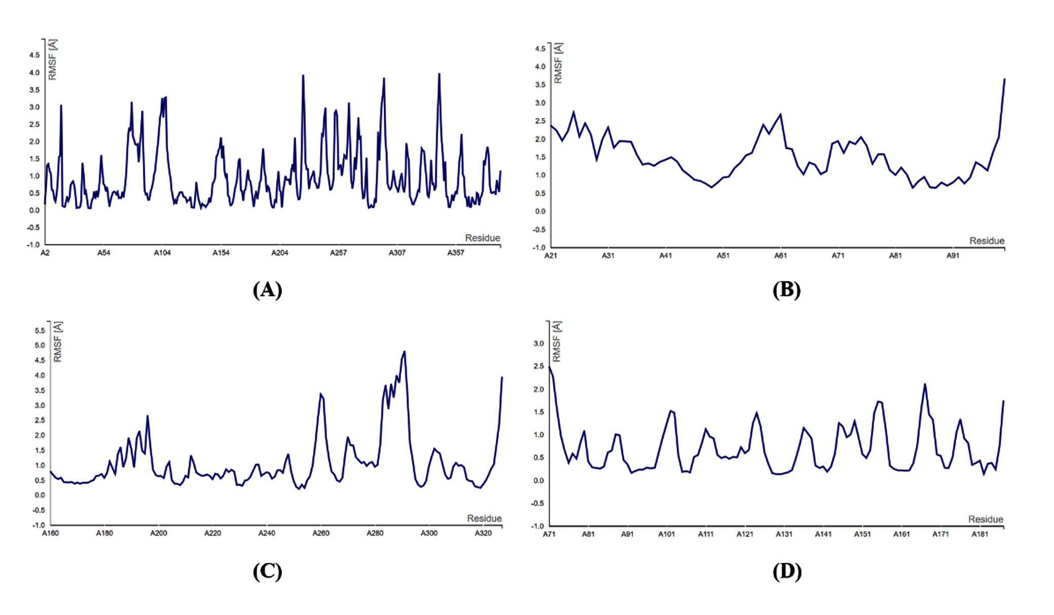
Figure 3. RMSF plot from the results of molecular dynamic simulation: (A) Compound 14_DENV Eprotein (B) Compound 18_DENV Cprotein (C) Compound 11_Langerin (D) Compound 6_CLEC5A
Toxicity profile and antiviral probability
Compound 14 (Hexadecanoic acid, ethyl ester) had an LD50 of 5000 mg/kg with 100% similarity and 100% prediction accuracy, the toxicity class of the compound was included in class 5 and was predicted to be non-toxic to organs. Compound 18 (Phytol) had an LD50 of 5000 mg/kg with 100% similarity and 100% prediction accuracy, while the toxicity class of the compound was included in class 5 and was predicted to be non-toxic to organs. Compound 11 (1,2-benzenedicarboxylic acid, bis(2-methylpropyl)ester) had an LD50 of 2530 mg/kg with 100% similarity and 100% prediction accuracy, the toxicity class of the compound was included in class 5 and was predicted to be non-toxic to organs. Compound 6 (Benzoic acid, 2-ethylhexyl ester) had an LD50 of 12300 mg/kg with 97.92% similarity and 72.9% prediction accuracy, the toxicity class was 6 and was predicted to be non-toxic to organs, all query compounds were good antiviral candidates with Pa > Pi values (Table 5).
Table 5. Toxicity profile and antiviral probability of candidate compounds
| Compounds | Predicted (mg/kg) | Toxicity LD50 Class | Average Similarity | Prediction Accuracy | Organ Toxicity | Probability Antiviral | |
|---|---|---|---|---|---|---|---|
| Pa | Pi | ||||||
| Compound 14 | 5000 | 5 | 100% | 100% | Hepatotoxicity: 0.76 (Inactive) | ||
| Neurotoxicity: 0.87 (Inactive) | 0.695 | 0.006 | |||||
| Nephrotoxicity: 0.57 (Inactive) | |||||||
| Cardiotoxicity: 0.79 (Inactive) | |||||||
| Compound 18 | 5000 | 5 | 100% | 100% | Hepatotoxicity: 0.79 (Inactive) | ||
| Neurotoxicity: 0.78 (Inactive) | 0.710 | 0.003 | |||||
| Nephrotoxicity: 0.74 (Inactive) | |||||||
| Cardiotoxicity: 0.84 (Inactive) | |||||||
| Compound 11 | 2530 | 5 | 100% | 100% | Hepatotoxicity: 0.65 (Inactive) | ||
| Neurotoxicity: 0.85 (Inactive) | 0.659 | 0.010 | |||||
| Nephrotoxicity: 0.65 (Inactive) | |||||||
| Cardiotoxicity: 0.79 (Inactive) | |||||||
| Compound 6 | 12300 | 6 | 97.92% | 72.9% | Hepatotoxicity: 0.74 (Inactive) | ||
| Neurotoxicity: 0.71 (Inactive) | 0.588 | 0.007 | |||||
| Nephrotoxicity: 0.56 (Inactive) | |||||||
| Cardiotoxicity: 0.68 (Inactive) | |||||||
Sonchus arvensis L. referred a medicinal plant from the Asteraceae family with benefits for traditional medicine and culinary spices in Indonesia. Sonchus arvensis L. had benefits such as antioxidants and antiplasmodium. Anti-inflammatory activity was also demonstrated by Sonchus arvensis L. through an in vivo study with gouty arthritis rats by decreasing the inflammatory response.48,49 Previous studies also showed that Sonchus arvensis L. leaf extract was predicted to be antiviral through the mechanism of inhibiting strategic proteins from the virus. Volatile compound of Sonchus arvensis L. allowed it to be a therapeutic candidate because it had characteristics as a drug-like molecule. Natural compounds as drug-like molecules that fulfilled the Lipinski, Ghose, Veber, Egan, and Muegge rules in druglikeness assessment were good therapeutic candidates for further testing. Bioavailability value of a query compound at 0.55 showed a high probability to be circulated and absorbed in the body.50 This was indicated by the bioavailability value of 0.55 in the druglikeness prediction results of all Sonchus arvensis L. volatile compounds in the study.
Fusion inhibitors played a major role in disrupting the viral entry stage in host cells, which could be developed in the treatment of DENV infections.51 Previous studies showed that fusion inhibition could be done by targeting proteins in the virus and viral receptor activity in host cells. Volatile compounds from Sonchus arvensis L. were predicted to have inhibitor activity on viral fusion consisting of compounds labels 14, 18, 11, and 6, which had more negative binding affinity values. The 4 compounds bind to targets from DENV such as Eprotein and Cprotein, then the compounds also targeted viral receptors on host cells, namely langerin and CLEC5A which played an important role for DENV fusion. The query compound with the best inhibitor activity as an antiviral candidate must have a more negative binding affinity value than others. Therefore, volatile compounds labels 14, 18, 11, and 6 from Sonchus arvensis L. consisting of Hexadecanoic acid, ethyl ester, Phytol, 1,2-benzenedicarboxylic acid, bis(2-methylpropyl)ester, and Benzoic acid, 2-ethylhexyl ester can be good candidates for DENV fusion inhibitors.
The molecular interactions formed from ligand-protein complexes consisted of weak bonds such as hydrogen, van der Waals, pi, alkyl, and electrostatic.52 The presence of weak bonds could be used by the ligand to affect the inhibitory activity of the target protein.53-55 Volatile compounds labels 14, 18, 11, and 6 from Sonchus arvensis L. were predicted to affect the inhibitory activity of the target through weak bonds to specific domains. Inhibition of targets on DENV consisting of Eprotein and Cprotein, then on host cell receptors namely langerin and CLEC5A allowed the 4 query ligands to interfere with the DENV fusion process. Ligand-protein molecular complexes formed from all docking results were stable with RMSF values <3 (Å) that were predicted to trigger specific biological responses such as disrupts viral fusion.56-60 The stable ligand-protein complex referred to the RMSF value formed at the interaction hotspot residues.61-64 Stable bonds allowed the formation of inhibitory activity on the target by candidate inhibitor compound.65
Toxicity prediction of query compounds aimed to ensure safety in their use.34 Compounds labels 14, 18, 11, and 6 were identified as good antiviral candidates and non-toxic for organ toxicity, having low or 5 and 6 categories, an average similarity to the template in the database of 100%, with an accuracy prediction of >50% accurate, and an LD50 value (mg/kg) of >1000. The toxicity profile was used for consideration for in vitro and in vivo analysis. However, further tests were needed to reveal the potential of the 4 volatile compounds from Sonchus arvensis L. as DENV fusion inhibitors to provide additional scientific evidence.
Development of a DENV antiviral based on volatile compounds from Sonchus arvensis L. through the inhibition of several proteins involved in viral entry through fusion was conducted in this study. Antiviral activity was identified in volatile compounds with the most negative binding affinity values for targets composed of the DENV Eprotein, DENV Cprotein, Langerin, and CLEC5A. Hydrogen and hydrophobic interactions were formed by volatile compounds consisting of hexadecanoic acid ethyl ester, phytol, 1,2-benzenedicarboxylic acid bis(2-methylpropyl) ester, and benzoic acid 2-ethylhexyl ester within specific binding pockets, which had the potential to inhibit viral entry. Volatile compounds could inhibit the activity of DENV proteins C and E (directly), and also that of Langerin and CLEC5A (indirectly) in host cells through stable binding interactions. Volatile compounds from Sonchus arvensis L. had the potential to exhibit dual mechanism against viral fusion as a DENV antiviral agent with low toxicity. However, additional studies using a wet lab approach were needed to provide further scientific evidence supporting the antiviral activity recommended in this study.
In conclusion, volatile compounds from Sonchus arvensis L. consisting of Hexadecanoic acid, ethyl ester, Phytol, 1,2-benzenedicarboxylic acid, bis(2-methylpropyl)ester, and Benzoic acid, 2-ethylhexyl ester can inhibit DENV fusion through binding to DENV proteins such as Eprotein, Cprotein and host cell receptors consisting of Langerin and CLEC5A. The 4 compounds have the most negative binding affinity value, non-toxin, and stable interaction on the target with lower bonding such as hydrogen, van der Waals, pi, and akyl to inhibit the target activity. These four compounds were recommended as antiviral candidates that act through a dual mechanism to prevent DENV from entering host cells.
ACKNOWLEDGMENTS
The authors are thankful to LPPM Universitas Airlangga and KEMENDIKBUTRISTEK Indonesia, for supporting this research.
CONFLICT OF INTEREST
The authors declare that there is no conflict of interest.
AUTHORS’ CONTRIBUTION
VDK, DWK, and HP conceptualized the study. HP performed supervision. VDK, DKW, ANMA, and RR applied the methodology. VDK, DKW, THS, and ANMA performed investigation. VDK and ANMA performed data curation and visualization. VDK and ANMA wrote the original draft. VDK and ANMA wrote, reviewed and edited the manuscript. All authors read and approved the final manuscript for publication.
FUNDING
This study was funded by DRTPM skema Penelitian Disertasi Doktor (PDD) (Contract No. 1735/B/UN3.LPPM/PT.01.03/2024).
DATA AVAILABILITY
All datasets generated or analyzed during this study are included in the manuscript.
ETHICS STATEMENT
Not applicable.
- Bhatt P, Sabeena SP, Varma M, Arunkumar G. Current Understanding of the Pathogenesis of Dengue Virus Infection. Curr Microbiol. 2021;78(1):17-32.
Crossref - Cloherty APM, Rader AG, Patel KS, et al. Dengue virus exploits autophagy vesicles and secretory pathways to promote transmission by human dendritic cells. Front Immunol. 2024;15:1260439.
Crossref - Praneechit H, Thiemmeca S, Prayongkul D, et al. Whole-blood model reveals granulocytes as key sites of dengue virus propagation, expanding understanding of disease pathogenesis. mBio. 2024;15(12):e0150524.
Crossref - Martí MM, Castanha PMS, Barratt-Boyes SM. The Dynamic Relationship between Dengue Virus and the Human Cutaneous Innate Immune Response. Viruses. 2024;16(5):727.
Crossref - Cruz-Oliveira C, Freire JM, Conceicao TM, Higa LM, Castanho MA, Da Poian AT. Receptors and routes of dengue virus entry into the host cells. FEMS Microbiol Rev. 2015;39(2):155-70.
Crossref - Kato F, Hishiki T. Dengue Virus Reporter Replicon is a Valuable Tool for Antiviral Drug Discovery and Analysis of Virus Replication Mechanisms. Viruses. 2016;8(5):122.
Crossref - Slon Campos JL, Mongkolsapaya J, Screaton GR. The immune response against flaviviruses. Nat Immunol. 2018;19(11):1189-1198.
Crossref - Begum F, Das S, Mukherjee D, Ray U. Hijacking the Host Immune Cells by Dengue Virus: Molecular Interplay of Receptors and Dengue Virus Envelope. Microorganisms. 2019;7(9):323.
Crossref - Sung PS, Hsieh SL. CLEC2 and CLEC5A: Pathogenic Host Factors in Acute Viral Infections. Front Immunol. 2019;10:2867.
Crossref - Ambroggio EE, Costa Navarro GS, Perez Socas LB, Bagatolli LA, Gamarnik AV. Dengue and Zika virus capsid proteins bind to membranes and self-assemble into liquid droplets with nucleic acids. J Biol Chem. 2021;297(3):101059.
Crossref - Villalain J. Membrane fusion by dengue virus: The first step. Biochim Biophys Acta Biomembr. 2025;1867(1):184400.
Crossref - Poh MK, Yip A, Zhang S, et al. A small molecule fusion inhibitor of dengue virus. Antiviral Res. 2009;84(3):260-6.
Crossref - De La Guardia C, Lleonart R. Progress in the identification of dengue virus entry/fusion inhibitors. Biomed Res Int. 2014;2014:825039.
Crossref - Ali S, Ali U, Safi K, et al. In silico homology modeling of dengue virus non-structural 4B (NS4B) protein and its molecular docking studies using triterpenoids. BMC Infect Dis. 2024;24(1):688.
Crossref - Loaiza-Cano V, Monsalve-Escudero LM, Filho CDSMB, Martinez-Gutierrez M, Sousa DP. Antiviral Role of Phenolic Compounds against Dengue Virus: A Review. Biomolecules. 2020;11(1):11.
Crossref - Wang YQ, Li QS, Zheng XQ, Lu JL, Liang YR. Antiviral Effects of Green Tea EGCG and Its Potential Application against COVID-19. Molecules. 2021;26(13):3962.
Crossref - Elizabeth E, Rohmawaty E, Bashari MH. The Effects of Turmeric and Mangosteen Pericarp Ethanol Extract on Eosinophil Count, TNF-a and TGF-β1 Gene Expression in Asthmatic Rat Model. J Exp Pharmacol. 2024;16:397-411.
Crossref - Nicoliche T, Bartolomeo CS, Lemes RMR, et al. Antiviral, anti-inflammatory and antioxidant effects of curcumin and curcuminoids in SH-SY5Y cells infected by SARS-CoV-2. Sci Rep. 2024;14(1):10696.
Crossref - Wahyuni DK, Purnobasuki H, Kuncoro EP, Ekasari W. CALLUS INDUCTION OF Sonchus arvensis L. AND ITS ANTIPLASMODIAL ACTIVITY. Afr J Infect Dis. 2020;14(1):1-7.
Crossref - Nita P, Muhammad TK, Masagus IS, Ernawati S, Radiyati UP, Irfannuddin I. Flavonoids as Antioxidants: A Review on Tempuyung Plant (Sonchus Arvensis), Iraq. J Med Chem Sci. 2023; 6:(10) 2310-2318.
Crossref - Wahyuni DK, Wacharasindhu S, Bankeeree W, et al. Molecular simulation of compounds from n-hexane fraction of Sonchus arvensis L. leaves as SARS-CoV-2 antiviral through inhibitor activity targeting strategic viral protein. J Pharm Pharmacog Res. 2020; 10(6):1126-1138.
Crossref - Mun CS, Hui LY, Sing LC, Karunakaran R, Ravichandran V. Multi-targeted molecular docking, pharmacokinetics, and drug-likeness evaluation of coumarin based compounds targeting proteins involved in development of COVID-19. Saudi J Biol Sci. 2022;29(12):103458.
Crossref - Abbasi M, Sadeghi-Aliabadi H. An In-silico Screening Strategy to the Prediction of New Inhibitors of COVID-19 Mpro Protein. Iran J Pharm Res. 2021;20(4):125-136.
Crossref - Duran-Iturbide NA, Diaz-Eufracio BI, Medina-Franco JL. In Silico ADME/Tox Profiling of Natural Products: A Focus on BIOFACQUIM. ACS Omega. 2020;5(26):16076-16084.
Crossref - Ononamadu CJ, Ibrahim A. Molecular docking and prediction of ADME/drug-likeness properties of potentially active antidiabetic compounds isolated from aqueous-methanol extracts of Gymnema sylvestre and Combretum micranthum. BioTechnologia. 2021;102(1):85-99.
Crossref - Hosseini M, Chen W, Xiao D, Wang C. Computational molecular docking and virtual screening revealed promising SARS-CoV-2 drugs. Precis Clin Med. 2021;4(1):1-16.
Crossref - Chandel V, Tripathi G, Nayar SA, Rathi B, Kumar A, Kumar D. In silico identification and validation of triarylchromones as potential inhibitor against main protease of severe acute respiratory syndrome coronavirus 2. J Biomol Struct Dyn. 2022;40(19):8850-8865.
Crossref - Kagami LP, das Neves GM, Timmers LFSM, Caceres RA, Eifler-Lima VL. Geo-Measures: A PyMOL plugin for protein structure ensembles analysis. Comput Biol Chem. 2020;87:107322.
Crossref - Iqbal D, Alsaweed M, Jamal QMS, et al. Pharmacophore-Based Screening, Molecular Docking, and Dynamic Simulation of Fungal Metabolites as Inhibitors of Multi-Targets in Neurodegenerative Disorders. Biomolecules. 2023;13(11):1613.
Crossref - Khan SA, Wu Y, Li AS, Fu XQ, Yu ZL. Network pharmacology and molecular docking-based prediction of active compounds and mechanisms of action of Cnidii Fructus in treating atopic dermatitis. BMC Complement Med Ther. 2022;22(1):275.
Crossref - Ghahremanian S, Rashidi MM, Raeisi K, Toghraie D. Molecular dynamics simulation approach for discovering potential inhibitors against SARS-CoV-2: A structural review. J Mol Liq. 2022;354:118901.
Crossref - Kakhar Umar A, Zothantluanga JH, Luckanagul JA, Limpikirati P, Sriwidodo S. Structure-based computational screening of 470 natural quercetin derivatives for identification of SARS-CoV-2 Mpro inhibitor. PeerJ. 2023;11:e14915.
Crossref - Wijaya RM, Hafidzhah MA, Kharisma VD, Ansori ANM, Parikesit AP. COVID-19 In Silico Drug with Zingiber officinale Natural Product Compound Library Targeting the Mpro Protein. Makara J Sci. 2021;25(3): 162−171.
- Banerjee P, Kemmler E, Dunkel M, Preissner R. ProTox 3.0: a webserver for the prediction of toxicity of chemicals. Nucleic Acids Res. 2024;52(W1):W513-W520.
Crossref - Gupta A, Sahu N, Singh AP, et al. Exploration of Novel Lichen Compounds as Inhibitors of SARS-CoV-2 Mpro: Ligand-Based Design, Molecular Dynamics, and ADMET Analyses. Appl Biochem Biotechnol. 2022;194(12):6386-6406.
Crossref - Delyan E. Analisys of composition of volatile compounds of field sow thistle (Sonchus arvensis L.) leaves using the method of gas chromatography with mass-detection. Pharm Innov. 2016;5(10):118–121.
- Sukandar EY, Safitri D. Evaluation of teratogenic effect of tempuyung (Sonchus arvensis) extract on Wistar Rats. Int J Farmakog Phyto Res. 2016;8(5):761–766.
- Anasir MI, Ramanathan B, Poh CL. Structure-Based Design of Antivirals against Envelope Glycoprotein of Dengue Virus. Viruses. 2020;12(4):367.
Crossref - Lee MF, Wu YS, Poh CL. Molecular Mechanisms of Antiviral Agents against Dengue Virus. Viruses. 2023;15(3):705.
Crossref - Boon PLS, Martins AS, Lim XN, et al. Dengue Virus Capsid Protein Facilitates Genome Compaction and Packaging. Int J Mol Sci. 2023;24(9):8158.
Crossref - Watson AA, Lebedev AA, Hall BA, et al. Structural flexibility of the macrophage dengue virus receptor CLEC5A: implications for ligand binding and signaling. J Biol Chem. 2011;286(27):24208-18.
Crossref - Sung PS, Chang WC, Hsieh SL. CLEC5A: a promiscuous pattern recognition receptor to microbes and beyond. In: Hsieh SL, ed. Lectin in Host Defense Against Microbial Infections. Advances in Experimental Medicine and Biology. Vol 1204. Springer; 2020:57-73.
Crossref - Daina A, Michielin O, Zoete V. SwissADME: a free web tool to evaluate pharmacokinetics, drug-likeness and medicinal chemistry friendliness of small molecules. Sci Rep. 2017;7:42717.
Crossref - Mahmud S, Hasan MR, Biswas S, et al. Screening of Potent Phytochemical Inhibitors Against SARS-CoV-2 Main Protease: An Integrative Computational Approach. Front Bioinform. 2021;1:717141.
Crossref - Shallangwa GA, Adeniji SE. Binding profile of protein-ligand inhibitor complex and structure based design of new potent compounds via computer-aided virtual screening. J Clin Tuberc Other Mycobact Dis. 2021;24:100256.
Crossref - Terefe EM, Ghosh A. Molecular Docking, Validation, Dynamics Simulations, and Pharmacokinetic Prediction of Phytochemicals Isolated From Croton dichogamus Against the HIV-1 Reverse Transcriptase. Bioinform Biol Insights. 2022;16:11779322221125605.
Crossref - Dey D, Paul PK, Al Azad S, et al. Molecular optimization, docking, and dynamic simulation profiling of selective aromatic phytochemical ligands in blocking the SARS-CoV-2 S protein attachment to ACE2 receptor: an in silico approach of targeted drug designing. J Adv Vet Anim Res. 2021;8(1):24-35.
Crossref - Rahardjanto A, Ikhtira DA, Nuryady MM, Pantiwati Y, Widodo N, Husamah H. The medicinal plant potential parts and species diversity as antipyretic: Ethnobotany study at Senduro Lumajang. AIP Conf Proc. 2021;2353:030018.
Crossref - Parisa N, Hidayat R, Maritska Z, Prananjaya BA. Evaluation of the anti-gout effect of Sonchus Arvensis on monosodium urate crystal-induced gout arthritis via anti-inflammatory action – an in vivo study. Med Pharm Rep. 2021;94(3):358-365.
Crossref - Ramya S, Manivannan HP, Veeraraghavan VP, Francis AP. In Silico Analysis of Selective Bioactive Compounds from Acronychia Pedunculata as a Potential Inhibitor of HER2 in Colorectal Cancer. J Pharm Bioallied Sci. 2024;16(Suppl 2):S1281-S1286.
Crossref - Ajmal A, Shahab M, Waqas M, et al. In silico design of peptide inhibitors for Dengue virus to treat Dengue virus-associated infections. Sci Rep. 2024;14(1):13130.
Crossref - Elkaeed EB, Youssef FS, Eissa IH, et al. Multi-Step In Silico Discovery of Natural Drugs against COVID-19 Targeting Main Protease. Int J Mol Sci. 2022;23(13):6912.
Crossref - Oner E, Demirhan I, Miraloglu M, Yalin S, Kurutas EB. Investigation of antiviral substances in Covid 19 by Molecular Docking: In Silico Study. Afr Health Sci. 2023;23(1):23-36.
Crossref - Nada H, Elkamhawy A, Lee K. Identification of 1H-purine-2,6-dione derivative as a potential SARS-CoV-2 main protease inhibitor: molecular docking, dynamic simulations, and energy calculations. Peer J. 2022;10:e14120.
Crossref - Chauhan N, Gaur KK, Asuru TR, Guchhait P. Dengue virus: pathogenesis and potential for small molecule inhibitors. Biosci Rep. 2024;44(8):BSR20240134.
Crossref - Wahyuni DK, Rahmawati CT, Syukriya AJ, et al. Metabolite characterization and in vitro antiplasmodial potential assessment of Sonchus arvensis L. callus under glutamine treatment. Sci Rep. 2025;15(1):35956.
Crossref - Maslahat M, Mardinata D, Surur SM, et al. Untargeted Metabolomics Analysis Using FTIR and LC-HRMS for Differentiating Sonchus arvensis Plant Parts and Evaluating Their Biological Activity. Chem Biodivers. 2025;22(2):e202401537.
Crossref - Aldeeb MME, Wilar G, Suhandi C, et al. Formulation and Characterization of Sonchus arvensis L. Nanosuspension for Enhanced Antioxidant and Lipid-Lowering Activities. Int J Nanomedicine. 2025;20:5457-5473.
Crossref - Simsek S, Akyol E, Kucuk I. A Review on In Silico Evaluation of Flavonoids as Potential Pharmaceutical Ingredients. J Med Food. 2026;29(8):517-530.
Crossref - Khalaf HS, El-Sayed AF, Sediek AA, Radwan MAA. Synthesis, molecular docking, ADMET studies and biological evaluation of fused pyrazolopyridopyrimidine derivatives as antioxidant and antimicrobial agents. Sci Rep. 2025;15(1):43654.
Crossref - Sankar DS, Selvam SP, Krishnasamy N, Pandiar D. Ethnomedicine-derived Approach of Astilbe rivularis Phytometabolites as Natural SAP5 Inhibitors against Candida albicans Biofilms. J Pure Appl Microbiol. 2026;20(2):1529-1539.
Crossref - Rauf A, Naz S, Ahmad Z, et al. Aldose Reductase Inhibition, Molecular Docking, and Density Functional Theory Studies of Dinaphthodiospyrol H from Diospyros kaki (Japanese Persimmon). J Pure Appl Microbiol. 2025;19(1):428-437.
Crossref - Kousar SM, Krishnaveni V, Audipudi AV. In vitro Antimicrobial Activity, Optimization, GC-MS, and In silico analysis for Anticancer Potency of Schizophyllum commune (AVNK2), An Endophyte Isolated from Nigella sativa Seeds. J Pure Appl Microbiol. 2025;19(2):1297-1311.
Crossref - Kharisma VD, Sahadewa S, Durry FD,et al. In silico identification of a Lactobacillus plantarum metabolic product that potentially inhibits three essential human immunodeficiency virus-2 enzymes. Community Acquir Infect. 2025;12.
Crossref - Alanzi AR, Alhaidhal BA, Aloatibi RM. Computational investigation of natural compounds as inhibitors against macrolide-resistant protein using virtual screening, molecular docking and MD simulations. Sci Rep. 2025;15(1):16385.
Crossref
© The Author(s) 2026. Open Access. This article is distributed under the terms of the Creative Commons Attribution 4.0 International License which permits unrestricted use, sharing, distribution, and reproduction in any medium, provided you give appropriate credit to the original author(s) and the source, provide a link to the Creative Commons license, and indicate if changes were made.