


 and Yandiswa Yolanda Yako
 Type 2 diabetes (T2D) is a multifactorial metabolic disease that arises from the intricate interplay between genetic predisposition and environmental factors. Beyond traditional risk factors, substantial evidence points to the gut microbiota (GM) as a critical regulator of host metabolism through epigenetic processes, specifically DNA methylation. Microbial metabolites, including short-chain fatty acids (SCFAs), methyl-donor precursors, and inflammatory mediators, modulate DNA methyltransferase activity, chromatin accessibility, and metabolic gene expression. These microbiota-derived signals modulate gene expression programs involved in insulin secretion, insulin sensitivity, inflammation, and energy homeostasis. This review summarizes current knowledge on the interplay between GM dysbiosis and DNA methylation in the development and progression of T2D. We emphasize the role of SCFAs, one-carbon metabolism, inflammatory signaling, and metabolic memory in shaping the diabetic epigenome. Furthermore, we critically examine microbiota-targeted interventions, including prebiotics, probiotics, fecal microbiota transplantation, antibiotics and dietary modulation, as potential strategies to restore epigenetic balance and improve metabolic outcomes. Understanding the microbiota-epigenome axis may redefine T2D as not only a metabolic disease but also a disorder of maladaptive epigenetic programming.
Gut Microbiota, DNA Methylation, Type 2 Diabetes, Short-Chain Fatty Acids, Microbiotherapy, Epigenetics
Type 2 diabetes (T2D) represents a multifaceted metabolic disorder defined by chronic hyperglycemia and a progressive decline in β-cell function, showing a significant correlation with gut microbial composition.1 Approximately 10.5% (537 million people) of the global population is affected, and this number is projected to rise to reach 783 million by 2045.2 This silent disease develops gradually under the influence of environmental factors,3 which affect the regulation and function of genes, sometimes disrupting essential processes, notably the regulation of glycemic balance and systemic inflammatory modulations.4 Although we have long recognized the impact of genetics and lifestyle on T2D, the focus of recent studies has shifted toward the hidden influence of the gut microbiome and epigenetic pathways in the disease’s development.5,6 Within this pathological framework, DNA methylation (DNAM) and gut microbial dynamics emerge as two pivotal forces shaping the metabolic landscape. Rather than being mere biological bystanders, these factors appear to actively steer the patient’s journey, fundamentally impacting both the initial triggering of T2D and the subsequent complexity of its clinical progression.
In mammals, DNAM functions by covalently modifying cytosine at its C5 position by adding a methyl group, forming 5-methylcytosine.7 DNAM influences organism physiology by recruiting proteins that repress genes or by blocking transcription factors, so affecting gene expression without altering the genetic sequence itself.8 Impaired insulin secretion in diabetic patients is commonly attributed to hypermethylation of the promoters of the INS, PDX1, and GLP1R genes, which encode insulin, a transcription factor crucial for pancreatic cell development and β-cell maturation, and the GLP1 receptor, which enhances insulin secretion and safeguards β-cells.9-11 Interestingly, DNAM is strongly modulated by the microbiome, sparking growing interest in developing solutions to improve the health of patients with diabetes, especially those with T2D.
The intestinal ecosystem is populated by a complex microbial community, known as the “gut microbiota”. It consists of various microbial communities whose composition is continuously influenced by both intrinsic and extrinsic factors of the host. It is now well-established that the microbiota of healthy individuals differs significantly from that of those with T2D, with an increased Firmicutes/Bacteroidetes ratio.11 Fluctuations in the intestinal flora modify the production and release of microbial metabolites, including bile acids, lipopolysaccharides (LPS), trimethylamine-N-oxide, and short-chain fatty acids (SCFAs). These active molecules serve as biochemical signals that modulate DNAM patterns and transcriptional activity within the host. Due to their promising effects, SCFAs (butyrate, propionate, acetate) are now being targeted for the development of new treatments for T2D.12-14 Thus, a bidirectional, triangular communication exists between DNAM, the microbiota, and T2D development.
This review examines the contribution of the intestinal flora to DNAM within the pathology of T2D. By analysing the interplay between the gut microbiota and DNAM, we can shed light on how they influence the pathophysiological mechanisms of T2D and the therapeutic perspectives emerging from these interactions.
DNA methylation and type 2 diabetes
DNA methylation
DNA methylation is a reversible biochemical reaction that typically occurs in CpG islands, where a methyl group is transferred from the universal methyl donor, S-adenosyl-L-methionine (SAM), to the 5-position of cytosine residues.15 DNAM reactions are catalyzed by a class of enzymes known as DNA methyltransferases (DNMTs), of which four forms exist in humans: DNMT3L, DNMT3B, DNMT3A and DNMT1. Often associated with gene expression repression, DNAM can occur at thousands of sites within the genome, sometimes leading to conformational changes that reduce DNA accessibility to the transcriptional machinery.16,17 It is generally a response to environmental stimuli, and is essential for normal development and cellular differentiation through repression of gene expression and regulation of metabolism as well as in diseases such as cancer, neurodegenerative disorders, and T2D.15,18,19
The activity of DNMTs is partly controlled by SAM, which is synthesized from several nutrients such as riboflavin, biotin, cobalamin, folates, choline, methionine, and betaine, through an intermediate, 6-methyltetrahydrofolate.20 It’s important to recognize that the majority of these nutrients mainly originate from the diet. For instance, research conducted by Wainfan and Poirier21 found that a SAM-deficient diet reduces hepatic DNAM levels in rat models. Although most of the nutrients used in SAM synthesis cannot be produced by humans, they can be replenished through diet or through the intestinal microbiota.20 Epigenetic marks carried by histones, such as acetylation or methylation, can influence the availability of CpG sites for methylation, thus creating a regulatory network between histone modifications and DNA.22 Under specific circumstances, DNAM self-regulates. Indeed, the methylation of lysine residues on histone 3 (H3) is involved in the formation and repression of DNAM, while the methylation of lysine 9 on histone H3 (H3K9) is often associated with highly methylated heterochromatic DNA regions. This self-regulation is catalyzed by histone methyltransferases, which recruit repressive complexes that facilitate DNAM and inhibit the expression of associated genes.23 Furthermore, histone acetylation, generally associated with chromatin decondensation, facilitates the access of transcription factors and methyltransferases to DNA. The histone deacetylase HDAC1 interacts with DNMT1, allowing this methyltransferase to maintain DNAM patterns during cellular replication. Thus, modifications that increase chromatin condensation, such as deacetylation, may promote the establishment or maintenance of DNAM.24,25
Changes in DNA methylation profiles associated with type 2 diabetes
T2D is a result of the complex interaction between environmental and predisposing polymorphisms in multiple genes. Evidence indicates that environmental exposures are associated with the development and progression of T2D through their influence on DNAM, although the mechanisms underlying this relationship remain unclear because published findings are not entirely consistent. Both targeted analyses and genome-wide association studies have reported changes in DNA methylation profile in skeletal muscle, liver, pancreatic islets and adipose tissue in individuals with T2D compared with non-diabetic controls.26 A recent systematic review identified 130 differentially methylated genes/loci, as reported by 32 genome-wide association studies (only one conducted in an African population).11 Four of these genes (ABCG1, TXNIP, PPARGC1A and PTPRN2) with well-known T2D-susceptibility single nucleotide polymorphisms have emerged across multiple independent studies as reliable markers of differential methylation. Some of these genes (ABCG1, TXNIP, and PPARGC1A) were confirmed by studies that conducted a candidate-gene DNAM approach. A large proportion of published data comes from European countries, necessitating studies in other ethnic groups to validate these reports and better represent the genetic diversity of T2DM. The paucity of DNAM data in the context of T2D in African countries is of concern, considering that the prevalence of diabetes is increasing at an alarming rate. A recent GWAS conducted in two ethnic population groups identified four novel DNA-methylated genes in Africans and one in Caucasians.27 Differential DNAM in the thioredoxin-interacting protein and ATP-binding cassette sub-family G member 1 genes were also replicated in a Caucasian (German) population study recently published, and this was in addition to fifteen novel sites.28 There are a few known T2D-associated DNAM sites that continue to be replicated in different studies, while more novel sites are reported. This indicates that population-specific GWAS is necessary to understand the nature of T2D and determine the clinical relevance of DNAM in diabetes. This is important because DNAM changes are influenced by environmental factors that may be unique in each region and population. Interestingly, the study also identified several CpG sites, including TXNIP (cg19693031) and CPT1A (cg00574958), among others, that align with known genetic regions linked to T2D. Beyond just showing consistent effect directions across the board, three of these markers (specifically at PLCB2, SREBF1, VPS25, and ABCG1) successfully met the epigenome-wide threshold when replicated in black adult cohorts.
However, the molecular mechanism governing this population-specific replication relies heavily on epistatic interactions. Genetic variations, such as single nucleotide polymorphisms, frequently function as methylation quantitative trait loci (mQTLs) that dictate the binding efficiency of DNMTs at specific CpG sites. Because baseline genetic architectures vary significantly across global ethnic groups, an environmental stimulus will not yield an identical epigenetic response across different populations. Extrapolating Western-derived epigenome-wide association studies models globally without accounting for these localized mQTL networks introduces severe analytical bias.
Despite the recurring identification of loci like ABCG1 and TXNIP, the overall reproducibility of genome-wide DNA methylation signatures in T2D remains low across distinct ethnic cohorts.29 Population-specific epigenome-wide association studies are essential to confirm the diagnostic and therapeutic relevance of these markers without overgeneralizing western cohort data to globally diverse patient groups because epigenetic marks are highly sensitive to regional environmental exposures and unique population backgrounds.30
Gut microbiota and type 2 diabetes
Gut microbiota
The GM, composed of approximately 100 billion diverse and dynamic microorganisms, predominantly resides in the colon, with its composition being highly variable due to both endogenous and exogenous factors.31 It is crucial in regulating various physiological processes, including inflammation, immune function, and energy metabolism.32 The contribution of the gut microbiome to the physiology of healthy individuals implies that microbial communities are sensitive to changes in the host’s internal and external environments. The specific mechanisms that involve GM underlying the development of diseases are not yet known and, therefore, require focused research to generate knowledge that can be used to guide the development of intervention programs for better diagnose and manage the associated diseases. One emerging mechanism involves an imbalance in the gut microbial community, termed dysbiosis, shifting towards the predominance of pathobionts. This paradigm shift may be influenced by host-specific factors such as consumption of specific food and medication (antibiotics), exposure to infectious and other environmental agents, genetics, biomolecules produced by intestinal epithelial cells, and age.
Dysbiosis and type 2 diabetes
Dysbiosis is characterized by an increased proportion of facultative anaerobic bacteria (such as Firmicutes) and a reduction in the number of beneficial obligate anaerobes (such as Bacteroidetes).33 It has been linked to several diseases, including T2D, and may have diverse compositional and functional characteristics in different pathophysiological conditions.14,34 Specifically regarding T2D, there is still not enough convincing evidence of the clinical relevance of the findings published to date. For example, the measures of alpha and beta diversities show inconsistent findings in relation to T2D, and this could be due to ethnic, antidiabetic agent use, and individual biological factor variations. This inconsistency highlights the limitation of relying on the Firmicutes to Bacteroidetes ratio as a universal biomarker.35 Beyond structural shifts, a major methodological challenge in human cohorts is dissecting whether dysbiosis is an upstream etiological driver or a secondary physiological fingerprint.35 Chronic hyperglycemia alters intestinal transit time and luminal pH, which selectively suppresses obligate anaerobes.36 Furthermore, first-line antidiabetic therapies, particularly metformin, actively reshape the taxonomic landscape by expanding specific taxa like Akkermansia muciniphila. Consequently, cross-sectional metagenomic profiles often capture a secondary signature of metabolic distress and pharmacological treatment rather than a primary causal factor, necessitating longitudinal tracking of treatment-naive cohorts to establish true chronological causality.35 While most research indicates that α-diversity remains largely unchanged, some studies have noted conflicting shifts toward either higher or lower diversity levels.34,37-40 Recent reviews have given insightful perspectives about the relationship between changes in gut microbiota and T2D,34,39 suggesting that the presence of Roseburia, Bifidobacterium, Akkermansia, Faecalibacterium, and Bacteroides species have a protective metabolic effect, specifically demonstrating a negative association with T2D. One of the underlying protective mechanisms has been suggested to be that of improving glucose tolerance when the Bifidobacterium dominates the gut. These results have led to testing some of these bacteria as probiotics for metabolic disease mostly in mouse models.
On the other hand, a dominant presence of Lactobacillus, Escherichia-Shigella, Enterococcus, Subdoligranulum, Fusobacteria, Ruminococcus, Fusobacterium, Senegalimassilia, Allisonella, Collinsella, Anaerostipes, Intestinibacter, and Blautia genera has been positively associated with T2D, suggesting that these microorganisms have a detrimental metabolic effect.39,41,42 Dysbiosis in individuals with T2D has been thought to induce overexpression of histone deacetylase 3 (HDAC3), which affects epigenetic activities and systemic inflammation, thereby increasing peripheral insulin resistance, T2D, and related complications.43 These metabolic disturbances are thought to result from increased permeability which allows gut-derived microorganisms and their byproducts to infiltrate the circulatory system.
Increased intestinal membrane permeability due to dysbiosis promotes the diffusion of microbial metabolites such as LPS into the gut, which has been shown to stimulate metabolic endotoxemia, triggering low-grade systemic inflammation and related diseases such as T2D.44-46 Lipopolysaccharides and peptidoglycans are among the biomolecules produced by opportunistic pathogens such as the Escherichia-Shigella genus. Indeed, the abundance of the Escherichia-Shigella genus has been linked to T2D-related traits such as β-cell function, insulin resistance and obesity. Another metabolite that has attracted researchers’ attention in this field is butyrate. In contrast to LPS, butyrate exerts a beneficial effect on metabolic regulation, primarily by reducing triglyceride accumulation in adipocytes and inhibiting inflammatory responses. This has been supported by evidence showing that destabilization of intestinal membrane integrity is closely associated with decreased levels of butyrate.47
The importance of butyrate in humans is evident in individuals with T2D, with numerous studies consistently demonstrating a decrease in butyrate-producing bacteria belonging to the genera Faecalibacterium, Akkermansia and Clostridium in individuals with prediabetes and those with T2D who were not receiving antidiabetic treatment.14 This opened an opportunity for investigating therapeutic strategies to combat diseases by targeting pathobionts. For example, it has been demonstrated that gut microbiota can be changed temporarily or permanently back to its normal composition and function using prebiotics, probiotics and fecal microbiota transplantation (FMT).48
Effect of gut microbiota on DNA methylation
Emerging research has shown that the GM directly influences metabolism and immune reactions and affects these biological processes through epigenetics.49 Changes in the composition and function of GM affect the types and concentrations of their metabolites, which, in turn, influence changes in the DNAM profile. The availability and concentration of metabolites such as riboflavin, biotin, cobalamin, folates, choline, methionine, and betaine, which are needed as methyl donors are influenced by specific gut microbial communities such as Bifidobacteria and Lactobacillus that are known for folate production.50,51 The intestinal microbiota exerts its epigenetic influence mainly through the production of metabolites such as SCFAs that are synthesized in the gut by the fermentation of nondigestible carbohydrates by certain microbes, and these include butyrate, acetate, and propionate.49 These metabolites influence the host’s physiology in various ways, such as regulating metabolism, modulating immunity and inflammation, and their abnormalities are associated with a heightened risk of T2D.52
Among SCFAs, butyrate, largely synthesized by the Firmicutes phylum, has garnered the most extensive research attention for its diverse contributions to metabolic reprogramming. Butyrate inhibits histone deacetylases (HDACs) and methyltransferases. It also down-regulates DNMT1 through the alteration of extracellular signal-regulated kinases (ERK) or classical MAP kinase-1, thus removing the methyl groups from certain tumor suppressor genes (RARB2, p21, and p16).53 Direct mechanistic translation from in vitro and animal discoveries to human pathology, however, faces significant barriers. While rodent models fed high-fat diets display rapid, clear-cut variations in DNA methylation profiles following SCFA exposure, human clinical validation is scarce.54 In humans, the epigenetic response to microbial metabolites is highly subtle, tissue-specific (primarily occurring within the liver, skeletal muscle, or visceral adipose tissue rather than peripheral blood mononuclear cells), and heavily confounded by host genetic backgrounds, age, and lifestyle factors. Consequently, caution must be exercised when extrapolating preclinical microbiodata-driven epigenetic reprogramming to actual clinical settings.55 By enhancing the expression of peroxisome proliferator-activated receptor gamma, butyrate actively supports a metabolic regulator that orchestrates everything from how fat cells develop to how the body manages lipids and insulin response.56 Because of the roles involved by butyrate, Qin et al.57 concluded that a reduction in butyrate production by the intestinal microbiota is associated with T2D. In the intestine, butyrate production is linked to a specific microbiota community, and an imbalance in this community leads to reduced butyrate levels, thus affecting expression levels of genes that are implicated in metabolism regulation. A metagenomic study of the fecal flora of T2D patients revealed a significant decrease in Roseburia, Faecalibacterium prausnitzii, and Anaerobutyricum soehngenii (the primary butyrate-producing species), this depletion was positively associated with T2D risk.43 Little has been published regarding the effect of butyrate and other SCFA on DNAM profiles in relation to T2D. However, a study conducted in obese mice models demonstrated the effect of propionate butyrate and acetate on alterations in DNAM of adiponectin and resistin genes which influenced changes in the expression levels of these genes.58 The SCFAs also reduced the expressions of DNMT3b, DNMT3a and DNMT1, the methyl-CpG-binding domain protein 2, thus suppressing the binding of these enzymes to the promoters of adiponectin and resistin. Propionate has been shown to induce significantly increased DNAM in the DAB1 promoter, leading to inhibition of DAB1 gene expression both in vitro and in vivo.59,60 In a GWAS in both Punjabi Sikh and Hispanics populations, DAB1 was associated with the concentration of vitamin D; This is significant because clinical vitamin D deficiency has been linked to the development, progression, and severity of T2D,61,62 suggesting that any changes in the concentration levels of propionate may affect the risk of T2D in obese individuals. Propionate is an HDAC inhibitor and a recognised potent activator of GPR43 and GPR41, which, when cleaved by microbiota from inulin-propionate ester in the colon, increases postprandial GLP-1 and PYY levels while reducing energy intake, resulting in significant long-term weight management.63,64 Table 1 provides a snapshot of the probiotic-driven mechanisms that turn the tide on metabolic inflammation at the epigenetic level.
Table 1. Summary of the effects of probiotics, their epigenetic mechanisms, and clinical impact on type 2 diabetes pathophysiology
Probiotic strains/ microbial communities |
Study Model |
Action on microbiota and metabolite production |
Effect on gene regulation |
Effect on diabetes |
Ref. |
|---|---|---|---|---|---|
Bifidobacterium and Lactobacillus |
Human |
Produce B-vitamins (methyl donors) |
Provide substrates for DNA and histone methylation |
Better glucose tolerance and insulin sensitivity |
65,49 |
Akkermansia muciniphila |
Human |
Restores gut microbial balance |
Improves liver and inflammatory markers |
Lower insulin/fat/cholesterol; ↑sensitivity |
14,66 |
Butyrate Producers (Faecalibacterium prausnitzii, Roseburia, Anaerobutyricum soehngenii) |
Ex vivo (HT-29) and human |
Produce butyrate; lower endotoxemia |
Inhibit HDACs/ DNMT1; ↑Pparg; ¯Hypermethylation (INS, PDX1) |
↓Adipocyte triglycerides and systemic inflammation |
56,60,67 |
Propionate Producers (Functional communities stimulated by fiber/inulin) |
Ex vivo (neutrophils) |
Produce propionate; activate GPR41/43 |
Inhibit HDACs/DNMTs; induce DAB1 methylation |
↑GLP-1/PYY; ↓Calories and weight loss |
63,64 |
Bifidobacterium longum, Lactococcus, Pediococcus, Streptococcus |
C57BL/6J male mice |
↓Oxidative stress and SCFA production |
Modulate epigenetic complications |
Lower glucose and T2D |
68 |
↑: Increase; ↓: Decrease
In addition to the aforementioned SCFAs, choline and vitamins act as methyl group donors and cofactors or substrates for enzymes involved in DNAM.4 The intestinal microbiota is also capable of synthesizing important compounds with acetyl or methyl groups, vitamins B (folate, niacin, riboflavin, pantothenic acid and pyridoxine), as well as various enzymes (acetyltransferases, deacetylases, phosphotransferases, methyltransferases and Escherichia coli biotin ligase), all of which are involved in the biochemical pathways leading to histone modifications and DNAM.65 Figure summarizes the microbiota’s role in improving clinical parameters in diabetics and how its imbalance promotes physiological disturbances that culminate in insulin resistance.
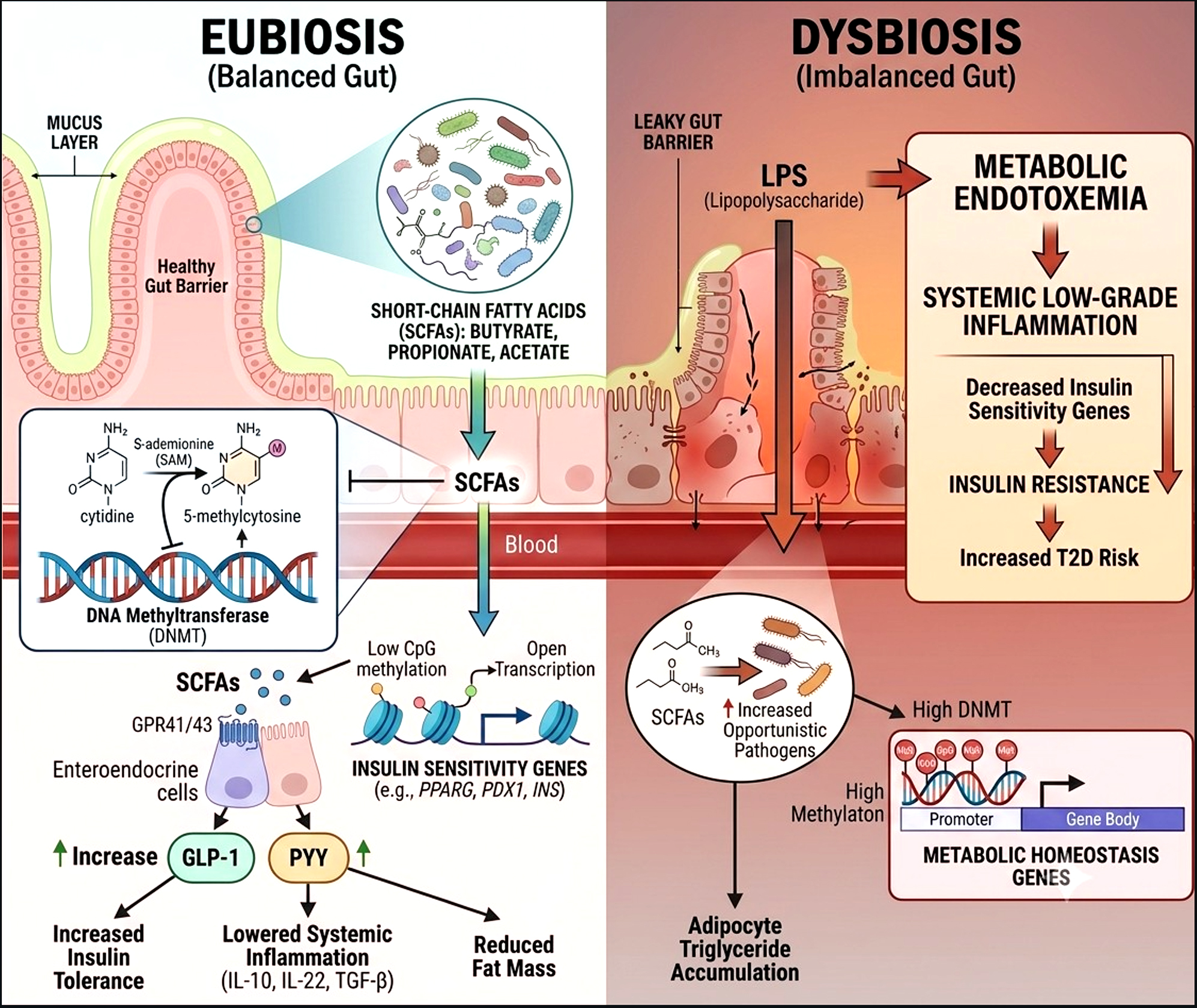
Figure. Impact of gut microbiota in regulating insulin tolerance and the development of insulin resistance. The intestinal microbiota is normally associated with the proper functioning of the organism
Targeting DNA methylation through microbiota modulation
Recent research suggests that modulating DNAM by specifically targeting the intestinal microbiota could be a promising therapeutic strategy for the treatment or prevention T2D. Therefore, the focus should be on understanding the nature of the microbiota and the factors that can promote the development of beneficial microbial communities that impact energy metabolism.
Nutritional interventions
Fiber-rich diets, which are highly recommended for individuals with diabetes, are well known for promoting beneficial microbial diversity and stimulating the production of SCFAs. The gut microbiota, by metabolizing nutrients provided by the diet, particularly fibers into SCFAs, serves as an intermediary between the food and its host.60 Thus, it is possible to promote the growth of one or more beneficial microbial communities (notably beneficial Firmicutes) by increasing fiber intake, which produces butyrate, a significant modulator of DNAM in patients with T2D.57 Furthermore, inulin supplementation effectively reshapes the gut landscape by favouring the growth of Bacteroides and Cyanobacteria at the expense of Tenericutes, Ruminiclostridium and Deferribacteres. This indicates that dietary inulin helps alleviate T2D by reducing inflammation and improving gastrointestinal function. Polyphenols are also converted by the intestinal microbiota into more active compounds. For example, epigallocatechin-3-gallate, by cleavage of the O-heterocycle, generates phenolic acids through dihydroxylation, which inhibit up to 80% of DNMT activity.69 A reduction in the Firmicutes/Bacteroidetes ratio and decreased expression of IL-6 mRNA and DNMT1 levels in the liver were observed when rats’ diets were supplemented with epigallocatechin-3-gallate.68 These studies illustrate that specific dietary interventions could help restore normal gene methylation associated with inflammation and insulin sensitivity, thus reducing the risk or impact of T2D.
Probiotics and prebiotics
The use of prebiotics and probiotics to restore a balanced and functional microbiota could also have a favorable impact on DNAM. When consumed as supplements or nutraceuticals, these probiotics can influence the production of SCFAs and epigenetic regulation once they reach the intestine. Several probiotic bacterial species are commonly used in commercial supplements, Lactobacillus, Bifidobacterium longum subsp. infantis, Lactococcus, Pediococcus, and Streptococcus are the most common.70-72 Evidence of the functional effects of probiotics is varied; they exert antioxidant and immunomodulatory effects by reducing oxidative stress and pro-inflammatory molecule levels, which leads to a reduction in blood glucose levels through improved insulin sensitivity, thereby reducing the risk of complications in patients with T2D. A randomized clinical trial evaluating the administration of Akkermansia muciniphila in overweight or obese individuals with insulin resistance demonstrated a reduction in insulin levels, as well as improvements in insulin sensitivity, fat mass, plasma cholesterol, hip circumference, and biomarkers associated with liver function and inflammation.66 However, adverse effects from probiotic administration are relatively rare, but a few cases of invasive disease have been reported,73,74 and they are important to acknowledge.
Targeted modification of the microbiota by antibiotics
A more radical approach involves the selective elimination of undesirable microbiota communities through the administration of antibiotics. Although this approach is still under-researched, attention is being focused on narrow-spectrum antibiotics, which offer the advantage of preserving beneficial microbiota.75 In a study conducted on an animal model, the administration of antibiotics from days 7-42 in piglets resulted in differential alterations to the microbial composition in both the small and large intestines. Most alterations were observed in the small intestine, including a reduction in Sharpea, Bacillus and Clostridium in the digesta of the stomach, duodenum, and jejunum, alongside a minor reduction in Prevotella in the colon digesta.76 Nevertheless, additional research is required to evaluate the efficacy and safety of employing antibiotics for the modulation of microbiota in the management of T2D.
Fecal transplantation
FMT has emerged as a promising strategy for the management of diabetes by reversing dysbiosis and enhancing the population of potentially symbiotic beneficial bacteria.77 Over the past decade, FMT has demonstrated effectiveness in treating Clostridium difficile infections and has exhibited positive outcomes in conditions such as ulcerative colitis and metabolic disorders, including T2D.78,79 Notably, FMT from lean donors to obese individuals with metabolic syndrome has been shown to improve insulin sensitivity. Significant findings include an increase in the diversity of butyrate-producing microbiota.80,81 Additionally, correction of glucose metabolism disorders, enhanced production of SCFAs, and induction of GLP-1 secretion were observed in diabetic mice receiving fecal transplants from healthy human donors.82,83 Another study in individuals with metabolic diseases found that FMT resulted in changes in the intestinal microbial community, modulation of blood metabolome, and epigenetic changes in the DNAM of the AFAP1 gene in mononuclear blood cells. Remarkably, the Prevotella genus was shown to affect mitochondrial function and insulin sensitivity through DNAM.84 However, a clinical trial revealed that fecal microbiota transplantation had no significant effect in some patients, and undesirable side effects such as infections, diarrhea, abdominal pain, and constipation were reported.85 Despite its potential benefits, fecal microbiota transplantation continues to encounter challenges, including the need for standardization of the procedure to minimize risks, such as the transmission of viral diseases or poorly understood pathogens. Consequently, the preparation, method of administering fecal matter, and treatment protocols must be standardized and meticulously regulated to guarantee the safety and efficacy of this approach (Table 2).
Table 2. Overview of microbiota-targeted modulation strategies
Technique |
Advantage |
Limitation/Risk |
Ref. |
|---|---|---|---|
Fibres and Inulin |
↑ SCFAs (butyrate) and reduces inflammation |
Efficacy depends on the existing baseline flora |
68 |
Probiotics |
↑ insulin sensitivity and ↓ blood glucose levels |
Rare risk of infection/translocation in immunocompromised subjects. |
66 |
Antibiotics |
Targeted elimination of undesirable bacteria |
Insufficient data for the treatment of diabetes |
75,76 |
Transplantation |
Restores microbial diversity and reverses dysbiosis |
Infectious risks and lack of procedural standardization |
79,85 |
↑: Increase; ↓ : Decrease
Beyond these transient adverse effects, the clinical translation of FMT is hindered by severe procedural and biological bottlenecks. Unlike the targeted displacement required in acute infectious diseases, reversing a chronic metabolic syndrome requires long-term functional engraftment.86 Metabolic FMT trials often show a rapid decay in efficacy within 3 to 6 months due to the host’s resilient baseline diet and genetic constraints.86,87 This variation is driven by the lack of standardized definitions for metabolic ‘super-donors’ and a lack of anaerobic harvesting protocols, which are crucial for preserving oxygen-sensitive butyrate-producing strains. Additionally, long-term biosafety concerns include the theoretical risk of horizontally transferring subclinical donor phenotypes, such as genetic predispositions to low-grade inflammation or specific adiposity pathways, through crude fecal material.86
As our understanding of the gut-genome axis deepens, recent insights have clarified the mechanisms through which the GM communicates with host DNA. It is now well established that this dialogue is driven by bioactive compounds, most notably SCFAs, which interact with DNA methyltransferases and histone deacetylases to regulate DNA methylation. This epigenetic regulation significantly impacts glucose metabolism by improving insulin sensitivity, reducing systemic inflammation, alleviating oxidative stress, and decreasing fat mass. These mechanisms contribute to limiting the progression and complications associated with T2D. Promising research findings support the potential of utilizing specific microbiotic communities or probiotics to modulate the microbiota composition in cases of dysbiosis induced by T2D. Investigating the symbiotic interactions between beneficial species could lead to the development of effective microbial consortia that reprogram the host’s transcriptional machinery to better manage metabolic dysfunctions.
ACKNOWLEDGMENTS
The authors thank their institution for providing the academic environment and resources that supported the preparation of this review.
CONFLICT OF INTEREST
The authors declare that there is no conflict of interest.
AUTHORS’ CONTRIBUTION
Both authors listed have made a substantial, direct and intellectual contribution to the work, and approved it for publication.
FUNDING
None.
DATA AVAILABILITY
Not applicable.
ETHICS STATEMENT
Not applicable.
- Zhou H, Li T, Li J, Zhuang X, Yang J. The association between visceral adiposity index and risk of type 2 diabetes mellitus. Sci Rep. 2024;14(1):16634. 10.1038/s41598-024-67430-x
- Ogurtsova K, Guariguata L, Barengo NC, et al. IDF diabetes Atlas: Global estimates of undiagnosed diabetes in adults for 2021. Diabetes Res Clin Pract. 2022;183:109118.
Crossref - Kumar A, Bharti SK, Kumar A. Type 2 diabetes mellitus: the concerned complications and target organs. Apollo Medicine. 2014;11(3):161-166.
Crossref - Miro-Blanch J, Yanes O. Epigenetic regulation at the interplay between gut microbiota and host metabolism. Front Genet. 2019;10:638.
Crossref - Hazar N, Jokar M, Namavari N, Hosseini S, Rahmanian V. An updated systematic review and Meta-analysis of the prevalence of type 2 diabetes in Iran, 1996–2023. Front Public Health. 2024;12:1322072.
Crossref - Wang S, Shi J, Peng Y, et al. Stronger association of triglyceride glucose index than the HOMA-IR with arterial stiffness in patients with type 2 diabetes: a real-world single-centre study. Cardiovasc Diabetol. 2021;20(1):82.
Crossref - Coppede F. Genes and the environment in cancer: focus on environmentally induced DNA methylation changes. Cancers. 2023;15(4):1019.
Crossref - Moore LD, Le T, Fan G. DNA methylation and its basic function. Neuropsychopharmacol. 2013;38(1):23-38.
Crossref - Yang BT, Dayeh T, Kirkpatrick C, et al. Insulin promoter DNA methylation correlates negatively with insulin gene expression and positively with HbA1c levels in human pancreatic islets. Diabetologia. 2011;54(2):360-367.
Crossref - Hall E, Dayeh T, Kirkpatrick CL, Wollheim CB, Dekker Nitert M, Ling C. DNA methylation of the glucagon-like peptide 1 receptor (GLP1R) in human pancreatic islets. BMC Med Genet. 2013;14(1):76.
Crossref - Nadiger N, Veed JK, Chinya Nataraj P, Mukhopadhyay A. DNA methylation and type 2 diabetes: a systematic review. Clin Epigenetics. 2024;16(1):67.
Crossref - Wang Y, Wen L, Tang H, Qu J, Rao B. Probiotics and prebiotics as dietary supplements for the adjunctive treatment of type 2 diabetes. Pol J Microbiol. 2023;72(1):3.
Crossref - Zhou Z, Sun B, Yu D, Zhu C. Gut microbiota: an important player in type 2 diabetes mellitus. Front Cell Infect Microbiol. 2022;12:834485.
Crossref - Baars DP, Fondevila MF, Meijnikman AS, Nieuwdorp M. The central role of the gut microbiota in the pathophysiology and management of type 2 diabetes. Cell Host Microbe. 2024;32(8):1280-1300.
Crossref - Romano KA, Rey FE. Is maternal microbial metabolism an early-life determinant of health? Lab Animal. 2018;47(9):239-243.
Crossref - Smith ZD, Meissner A. DNA methylation: roles in mammalian development. Nat Rev Genet. 2013;14(3):204-220.
Crossref - Smith ZD, Hetzel S, Meissner A. DNA methylation in mammalian development and disease. Nat Rev Genet. 2025;26(1):7-30.
Crossref - Huda MN, Kim M, Bennett BJ. Modulating the microbiota as a therapeutic intervention for type 2 diabetes. Front Endocrinol. 2021;12:632335.
Crossref - Mbali BB, Piame LT, Sehole S, Musarurwa HT, Yako YY. DNA methylation and exosomes in relation to type 2 diabetes in Black South Africans: A pilot study. Glob Med Genet. 2026;13(2):100109.
Crossref - Menezo Y, Clement P, Clement A, Elder K. Methylation: an ineluctable biochemical and physiological process essential to the transmission of life. Int J Mol Sci. 2020;21(23):9311.
Crossref - Wainfan E, Poirier LA. Methyl groups in carcinogenesis: effects on DNA methylation and gene expression. Cancer Res. 1992;52(7_Suppl):2071s-2077s.
- Zhou W, Dinh HQ, Ramjan Z, et al. DNA methylation loss in late-replicating domains is linked to mitotic cell division. Nature genetics. 2018;50(4):591-602.
Crossref - Ehrlich M, Lacey M. DNA methylation and differentiation: silencing, upregulation and modulation of gene expression. Epigenomics. 2013;5(5):553-568.
Crossref - Gopalan S, Carja O, Fagny M, et al. Trends in DNA methylation with age replicate across diverse human populations. Genetics. 2017;206(3):1659-1674.
Crossref - English DM, Lee SN, Sabat KA, et al. Rapid degradation of histone deacetylase 1 (HDAC1) reveals essential roles in both gene repression and active transcription. Nucleic Acids Res. 2025;53(4):gkae1223.
Crossref - Kim S-B, Heo J-I, Kim H, Kim KS. Acetylation of PGC1ב by histone deacetylase 1 downregulation is implicated in radiation-induced senescence of brain endothelial cells. J Gerontol A Biol Sci Med Sci. 2019;74(6):787-793.
Crossref - Venkataraghavan S, Pankow JS, Boerwinkle E, Fornage M, Selvin E, Ray D. Epigenome-wide association study of incident type 2 diabetes in Black and White participants from the Atherosclerosis Risk in Communities Study. Diabetologia. 2025;68(4):1-20.
Crossref - Lai L, Juntilla DL, Del C Gomez-Alonso M, et al. Longitudinal association between DNA methylation and type 2 diabetes: findings from the KORA F4/FF4 study. Cardiovasc Diabetol. 2025;24(1):19.
Crossref - Fraszczyk E, Spijkerman AMW, Zhang Y, et al. Epigenome-wide association study of incident type 2 diabetes: a meta-analysis of five prospective European cohorts. Diabetologia. 2022;65(5):763-776.
Crossref - Chilunga FP, Meeks KAC, Henneman P, et al. An epigenome-wide association study of insulin resistance in African Americans. Clin Epigenet. 2022;14(1):88.
Crossref - Cunningham A, Stephens JW, Harris DA. Gut microbiota influence in type 2 diabetes mellitus (T2DM). Gut Pathog. 2021;13(1):50.
Crossref - Cunningham AL, Stephens JW, Harris DA. A review on gut microbiota: a central factor in the pathophysiology of obesity. Lipids in Health Dis. 2021;20(1):65.
Crossref - Mashal R, Al-Muhanna A, Khader S, Khudair A, Khudair A, Butler AE. The Role of the Gut Microbiome in Type 2 Diabetes Mellitus. Int J Mol Sci. 2025;26(23):11412.
Crossref - Chong S, Lin M, Chong D, Jensen S, Lau NS. A systematic review on gut microbiota in type 2 diabetes mellitus. Front Endocrinol. 2025;15:1486793.
Crossref - Nez-Castro AT, Gonzalez-Olivares LG, Olvera-Rosales LB, et al. Firmicutes/Bacteroidetes Ratio as an Insufficient Indicator of Metabolic Status in Mexican Young Adults. Nutrients. Mar 28 2026;18(7):nu18071084.
Crossref - Bravard A, Gerard C, Defois C, et al. Metformin treatment for 8 days impacts multiple intestinal parameters in high-fat high-sucrose fed mice. Sci Rep. 2021;11(1):16684.
Crossref - Candela M, Biagi E, Soverini M, et al. Modulation of gut microbiota dysbioses in type 2 diabetic patients by macrobiotic Ma-Pi 2 diet. Br J Nutr. 2016;116(1):80-93.
Crossref - Zhang Y, Zhang L, Gu E, Zhu B, Zhao X, Chen J. Long-term insulin treatment restores cardioprotection induced by sufentanil postconditioning in diabetic rat heart. Exp Biol Med. 2016;241(6):650-657.
Crossref - Gurung M, Li Z, You H, et al. Role of gut microbiota in type 2 diabetes pathophysiology. EBioMedicine. 2020;51:102590.
Crossref - Shi L, Wang Y, Guan Y, Men L, Sun J, Yuan G. To establish a new quality assessment method based on the regulation of intestinal microbiota in type 2 diabetes by lignans of Schisandra chinensis (Turcz.) Baill. J Ethnopharmacol. 2025;348:119822.
Crossref - Neri-Rosario D, Martinez-Lopez YE, Esquivel-Hernandez DA, et al. Dysbiosis signatures of gut microbiota and the progression of type 2 diabetes: a machine learning approach in a Mexican cohort. Front Endocrinol. 2023;14:1170459.
Crossref - Salamon D, Sroka-Oleksiak A, Kapusta P, et al. Characteristics of gut microbiota in adult patients with type 1 and type 2 diabetes based on next-generation sequencing of the 16S rRNA gene fragment. Pol Arch Intern Med. 2018;128(6):336-343.
Crossref - Arora T, Tremaroli V. Therapeutic potential of butyrate for treatment of type 2 diabetes. Front Endocrinol. 2021;12:761834.
Crossref - Lee CB, Chae SU, Jo SJ, Jerng UM, Bae SK. The relationship between the gut microbiome and metformin as a key for treating type 2 diabetes mellitus. Int J Mol Sci. 2021;22(7):3566.
Crossref - Wang J, Gu X, Yang J, Wei Y, Zhao Y. Gut microbiota dysbiosis and increased plasma LPS and TMAO levels in patients with preeclampsia. Front Cell Infect Microbiol. 2019;9:409.
Crossref - Mohammadi S, Eslami M, Pourghazi F, et al. Gut Microbiota Derived Trimethylamine N Oxide and the Risk of Diabetes: An Updated Systematic Review and Meta Analysis. Obes Rev. 2025;26(11):e13963.
Crossref - Peng J, Narasimhan S, Marchesi JR, Benson A, Wong FS, Wen L. Long term effect of gut microbiota transfer on diabetes development. J Autoimmun. 2014;53:85-94.
Crossref - Hasan N, Yang H. Factors affecting the composition of the gut microbiota, and its modulation. PeerJ. 2019;7:e7502.
Crossref - Koh A, Backhed F. From association to causality: the role of the gut microbiota and its functional products on host metabolism. Mol cell. 2020;78(4):584-596.
Crossref - Strozzi GP, Mogna L. Quantification of folic acid in human feces after administration of Bifidobacterium probiotic strains. J Clin Gastroenterol. 2008;42:S179-S184.
Crossref - Rossi M, Amaretti A, Raimondi S. Folate production by probiotic bacteria. Nutrients. 2011;3(1):118-134.
Crossref - Sanna S, van Zuydam NR, Mahajan A, et al. Causal relationships among the gut microbiome, short-chain fatty acids and metabolic diseases. Nat Genet. 2019;51(4):600-605.
Crossref - Sarkar S, Abujamra AL, Loew JE, Forman LW, Perrine SP, Faller DV. Histone deacetylase inhibitors reverse CpG methylation by regulating DNMT1 through ERK signaling. Anticancer Res. 2011;31(9):2723-2732.
- Kopczyסska J, Kowalczyk M. The potential of short-chain fatty acid epigenetic regulation in chronic low-grade inflammation and obesity. Front Immunol. 2024;15:1380476.
Crossref - Krautkramer KA, Dhillon RS, Denu JM, Carey HV. Metabolic programming of the epigenome: host and gut microbial metabolite interactions with host chromatin. Transl Res. 2017;189:30-50.
Crossref - Kinoshita M, Suzuki Y, Saito Y. Butyrate reduces colonic paracellular permeability by enhancing PPARg activation. Biochem Biophys Res Commun. 2002;293(2):827-831.
Crossref - Qin J, Li Y, Cai Z, et al. A metagenome-wide association study of gut microbiota in type 2 diabetes. Nature. 2012;490(7418):55-60.
Crossref - Lu Y, Fan C, Liang A, et al. Effects of SCFA on the DNA methylation pattern of adiponectin and resistin in high-fat-diet-induced obese male mice. Br J Nutrit. 2018;120(4):385-392.
Crossref - Guo W, Zhang Z, Li L, et al. Gut microbiota induces DNA methylation via SCFAs predisposing obesity-prone individuals to diabetes. Pharmacol Res. 2022;182:106355.
Crossref - D’Aquila P, Lynn Carelli L, De Rango F, Passarino G, Bellizzi D. Gut microbiota as important mediator between diet and DNA methylation and histone modifications in the host. Nutrients. 2020;12(3):597.
Crossref - Braun TR, Been LF, Singhal A, et al. A replication study of GWAS-derived lipid genes in Asian Indians: the chromosomal region 11q23. 3 harbors loci contributing to triglycerides. PloS one. 2012;7(5):e37056.
Crossref - Mohammadi S, Hajhashemy Z, Saneei P. Serum vitamin D levels in relation to type-2 diabetes and prediabetes in adults: a systematic review and dose–response meta-analysis of epidemiologic studies. Crit Rev Food Sci Nutr. 2022;62(29):8178-8198.
Crossref - Aoyama M, Kotani J, Usami M. Butyrate and propionate induced activated or non-activated neutrophil apoptosis via HDAC inhibitor activity but without activating GPR-41/GPR-43 pathways. Nutrition. 2010;26(6):653-661.
Crossref - Chambers ES, Morrison DJ, Frost G. Control of appetite and energy intake by SCFA: what are the potential underlying mechanisms? Proc Nutr Soc. 2015;74(3):328-336.
Crossref - Magnusdottir S, Ravcheev D, de Crecy-Lagard V, Thiele I. Systematic genome assessment of B-vitamin biosynthesis suggests co-operation among gut microbes. Front Genet. 2015;6:148.
Crossref - Depommier C, Everard A, Druart C, et al. Supplementation with Akkermansia muciniphila in overweight and obese human volunteers: a proof-of-concept exploratory study. Nat Med. 2019;25(7):1096-1103.
Crossref - Guo H, Xiang X, Lin X, et al. Oropharyngeal probiotic ENT-K12 as an effective dietary intervention for children with recurrent respiratory tract infections during cold season. Front Nutr. 2022;9:900448.
Crossref - Remely M, Ferk F, Sterneder S, et al. EGCG prevents high fat diet induced changes in gut microbiota, decreases of DNA strand breaks, and changes in expression and DNA methylation of Dnmt1 and MLH1 in C57BL/6J male mice. Oxid Med Cell Longev. 2017;2017(1):3079148.
Crossref - Paluszczak J, Krajka-Kuzniak V, Baer-Dubowska W. The effect of dietary polyphenols on the epigenetic regulation of gene expression in MCF7 breast cancer cells. Toxicol Lett. 2010;192(2):119-125.
Crossref - Rao V, Rao L. Probiotics and prebiotics in human nutrition and health. BoD–Books on Demand. 2016.
- Piame LT. Potential of Non-Dairy Probiotic Beverages to Modulate Gut Microbiota and Improve Management of Non-Communicable Diseases. Review. Nov Res Microbiol J. 2025;2025:15.
Crossref - Tchamani Piame L. Bioactive Metabolites from Yeasts Presumptively Qualified as Safe as Functional Agents in the Management of Type 2 Diabetes. Appl Microbiol. 2025;5(3):84.
Crossref - Rayes N, Seehofer D, Theruvath T, et al. Supply of pre and probiotics reduces bacterial infection rates after liver transplantation—a randomized, double blind trial. Am J Transplant. 2005;5(1):125-130.
Crossref - Sotoudegan F, Daniali M, Hassani S, Nikfar S, Abdollahi M. Reappraisal of probiotics’ safety in human. Food Chem Toxicol. 2019;129:22-29.
Crossref - Avis T, Wilson FX, Khan N, Mason CS, Powell DJ. Targeted microbiome-sparing antibiotics. Drug Discov Today. 2021;26(9):2198-2203.
Crossref - Mu C, Yang Y, Su Y, Zoetendal EG, Zhu W. Differences in microbiota membership along the gastrointestinal tract of piglets and their differential alterations following an early-life antibiotic intervention. Front Microbiol. 2017;8:797.
Crossref - Sadagopan A, Mahmoud A, Begg M, et al. Understanding the role of the gut microbiome in diabetes and therapeutics targeting leaky gut: a systematic review. Cureus. 2023;15(7):e41559.
Crossref - Wang Y, Zhang S, Borody TJ, Zhang F. Encyclopedia of fecal microbiota transplantation: a review of effectiveness in the treatment of 85 diseases. Chin Med J. 2022;135(16):1927-1939.
Crossref - Wu J, Yang K, Fan H, Wei M, Xiong Q. Targeting the gut microbiota and its metabolites for type 2 diabetes mellitus. Front Endocrinol. 2023;14:1114424.
Crossref - Scheithauer TP, Rampanelli E, Nieuwdorp M, et al. Gut microbiota as a trigger for metabolic inflammation in obesity and type 2 diabetes. Front Immunol. 2020;11:571731.
Crossref - Su L, Hong Z, Zhou T, et al. Health improvements of type 2 diabetic patients through diet and diet plus fecal microbiota transplantation. Sci Rep. 2022;12(1):1152.
Crossref - Han X, Wang Y, Zhang P, et al. Kazak faecal microbiota transplantation induces short-chain fatty acids that promote glucagon-like peptide-1 secretion by regulating gut microbiota in db/db mice. Pharm Biol. 2021;59(1):1075-1085.
Crossref - Zhang J, Wang H, Liu Y, et al. Advances in fecal microbiota transplantation for the treatment of diabetes mellitus. Front Cell Infect Microbiol. 2024;14:1370999.
Crossref - van der Vossen EW, Bastos D, Stols-Gonחalves D, et al. Effects of fecal microbiota transplant on DNA methylation in subjects with metabolic syndrome. Gut Microbes. 2021;13(1):1993513.
Crossref - Yu EW, Gao L, Stastka P, et al. Fecal microbiota transplantation for the improvement of metabolism in obesity: The FMT-TRIM double-blind placebo-controlled pilot trial. PLoS Med. 2020;17(3):e1003051.
Crossref - Wang H, Tian J, Mi J. Clinical effectiveness of fecal microbial transplantation for metabolic syndrome: Advances in clinical efficacy and multi-omics research. Curr Res Microb Sci. 2025;9:100415.
Crossref - Qiu B, Liang J, Li C. Effects of fecal microbiota transplantation in metabolic syndrome: A meta-analysis of randomized controlled trials. PLOS ONE. 2023;18(7):e0288718.
Crossref
© The Author(s) 2026. Open Access. This article is distributed under the terms of the Creative Commons Attribution 4.0 International License which permits unrestricted use, sharing, distribution, and reproduction in any medium, provided you give appropriate credit to the original author(s) and the source, provide a link to the Creative Commons license, and indicate if changes were made.