

ISSN: 0973-7510
E-ISSN: 2581-690X
 , Vijayanand Selvaraj2, Reshma Girirajan1, Rajalakshmi Balaji1 and Hemapriya Janarthanam1
 Swim bladders from the freshwater fish Piaractus brachypomus are a rich source of Type I collagen, representing a sustainable alternative for collagen extraction and the valorization of fish-processing by-products. The present study aimed to isolate Acid-Soluble Swim Bladder Collagen (ASSBC), characterize its structural and functional properties, produce low-molecular-weight collagen peptides through papain hydrolysis, and evaluate their antioxidant, antimicrobial, cytocompatibility, and wound-healing activities. ASSBC was extracted using acetic acid and characterized by Lowry protein estimation, SDS-PAGE, UV-visible spectroscopy, FTIR spectroscopy, SEM, and EDAX analyses. Bioactive collagen peptides were generated by papain hydrolysis and evaluated using DPPH radical scavenging, antimicrobial, MTT cytotoxicity, and in vitro scratch wound-healing assays. The extraction process yielded 62.0 ± 2.3% collagen (wet-weight basis) and 13.3 ± 0.6% collagen (dry-weight basis). The extracted collagen exhibited a protein concentration of 74.0 ± 2.1 µg/mL. SDS-PAGE analysis confirmed the presence of Type I collagen with characteristic α₁/α₃ (114 kDa), α₂ (98 kDa), β (193 kDa), and γ (272 kDa) chains. Papain hydrolysis successfully generated low-molecular-weight collagen peptides with an approximate molecular weight of 6 kDa. The hydrolyzed collagen peptides exhibited 59.80 ± 1.7% DPPH radical scavenging activity and demonstrated antibacterial activity against Salmonella spp. (14.0 ± 0.8 mm) and Staphylococcus spp. (7.0 ± 0.5 mm), together with antifungal activity against Penicillium spp. (20.0 ± 0.9 mm) and Aspergillus niger (8.0 ± 0.6 mm). MTT assay demonstrated high cytocompatibility toward NIH-3T3 fibroblast cells up to 50 µg/mL, with cell viability remaining above 90%. In the scratch wound-healing assay, collagen peptide-treated cells exhibited accelerated wound closure, reducing the wound gap from 100% to 5.8 ± 0.6% after 48 hrs, whereas the untreated control retained 24.6 ± 1.3% of the wound gap. These findings demonstrate that the swim bladder of Piaractus brachypomus is an excellent source of structurally intact Type I collagen and bioactive low-molecular-weight collagen peptides with potential antioxidant, antimicrobial, cytocompatibility, and wound-healing properties. The study highlights the potential application of this underutilized fish-processing by-product in the development of sustainable biomaterials for regenerative medicine and wound-healing applications.
Swim Bladder, Collagen Peptides, Piaractus brachypomus, Papain, MTT, Scratch Assay
The extracellular matrix is mostly composed of collagen, the most common structural protein in vertebrates, which accounts for around 25%-35% of the total protein composition in the body. Connective tissues, such as skin, bones, cartilage, tendons, ligaments, and blood vessels, rely on it to keep them structurally sound, mechanically strong, and elastic. Approximately 28 types of collagen have been identified, among which Type I collagen is the most abundant and widely distributed.1,2 Owing to its unique triple-helical structure and characteristic amino acid composition, particularly glycine, proline, and hydroxyproline, collagen serves as an excellent precursor for the production of bioactive peptides with diverse biological functions following enzymatic hydrolysis.3
Collagen has found extensive use in many different fields, including food, cosmetics, pharmaceuticals, nutraceuticals, and biomedicine, due to its exceptional biocompatibility, biodegradability, low immunogenicity, and bioactivity. Because of their capacity to stimulate cell adhesion, proliferation, extracellular matrix remodeling, and tissue regeneration, collagen-based biomaterials find widespread use in wound dressings, drug delivery systems, regenerative medicine, and cosmetic formulations. In addition, collagen exhibits antioxidant, antimicrobial, coagulation-promoting, and anti-inflammatory properties, making it one of the most valuable natural biomaterials for therapeutic applications.4
The increasing global demand for natural and sustainable collagen has considerably expanded the collagen market, driving intensive research toward identifying alternative and environmentally friendly collagen sources.5 Conventionally, commercial collagen is obtained from bovine, porcine, and avian tissues. But worries about zoonotic disease transmission, immunogenic responses, and cultural or religious constraints have prompted research into safer and more long-term options.
Marine-derived collagen has emerged as an attractive substitute because of its excellent biocompatibility, lower molecular weight, superior bioavailability, and reduced risk of disease transmission. Furthermore, marine collagen can be efficiently recovered from fish-processing by-products, thereby promoting environmental sustainability through waste valorization. India is one of the world’s leading fish-producing countries, generating substantial quantities of fish-processing waste, including skins, scales, bones, fins, heads, viscera, and swim bladders. These by-products are rich in collagen and other valuable biomolecules but are frequently discarded, resulting in environmental pollution and economic loss.6,7 Efficient utilization of these underexploited resources provides an opportunity to produce high-value biomaterials while supporting sustainable fisheries and circular bioeconomy initiatives.
Among the various fish-processing by-products, the swim bladder is recognized as an excellent source of highly purified Type I collagen because of its high collagen content, low lipid content, and relatively simple extracellular matrix composition. Recent investigations have demonstrated successful extraction and characterization of collagen from the swim bladders of several fish species, including bighead carp, grass carp, grouper, monkfish, rohu, and Amur sturgeon.8 These studies have shown that swim bladder-derived collagen possesses desirable physicochemical characteristics, excellent fibril-forming ability, and promising biological properties, suggesting its potential for biomedical, pharmaceutical, and tissue engineering applications.9-12 Moreover, enzymatic hydrolysis of collagen has been reported to generate low-molecular-weight bioactive peptides exhibiting antioxidant, antimicrobial, antihypertensive, anti-inflammatory, and wound-healing activities, thereby further expanding the biomedical significance of fish-derived collagen.10
Despite these advances, information regarding the extraction, structural characterization, and biofunctional properties of collagen derived from the swim bladder of Piaractus brachypomus (Red-bellied Pomfret/Yeri Vaval) remains unavailable. To date, no comprehensive study has investigated the isolation of acid-soluble Type I collagen from this freshwater fish species, followed by the production of low-molecular-weight collagen peptides and systematic evaluation of their antioxidant, antimicrobial, cytocompatibility, and wound-healing properties. Considering the widespread cultivation of P. brachypomus and the substantial quantity of processing by-products generated, scientific evaluation of its swim bladder collagen is essential to facilitate the sustainable utilization of this underexploited biological resource.
Therefore, the present study aimed to isolate acid-soluble swim bladder collagen (ASSBC) from Piaractus brachypomus, characterize its structural and functional properties using biochemical and spectroscopic techniques, generate low-molecular-weight collagen peptides through papain-mediated enzymatic hydrolysis, and evaluate their antioxidant, antimicrobial, cytocompatibility, and in vitro wound-healing activities. To the best of our knowledge, this is the first comprehensive investigation describing the isolation, structural characterization, and biofunctional evaluation of collagen peptides derived from the swim bladder of Piaractus brachypomus, thereby providing a scientific basis for the development of sustainable collagen-based biomaterials for biomedical and regenerative applications.
Sample collection and experimental design
Fresh swim bladders of Piaractus brachypomus (Red-bellied pacu; locally known as Yeri Vaval) were obtained from a local fish market in Vellore, Tamil Nadu, India. The fish were taxonomically identified based on the FishBase database and confirmed prior to experimentation. Healthy adult fish weighing approximately 1-2 kg with an average swim bladder length of 10-15 cm was selected for the study to minimize biological variation. Immediately after slaughter, swim bladders were aseptically excised, transferred into sterile polyethylene bags, transported to the laboratory under chilled conditions (4 °C), thoroughly washed with distilled water to remove blood and adhering tissues, and stored at -20 °C until further processing.13
Extraction and Recovery of Acid-Soluble Swim Bladder Collagen (ASSBC)
Acid-soluble swim bladder collagen (ASSBC) was extracted following the method described by Xiao et al.13 with minor modifications. To summarize, new swim bladders were sliced into pieces about 0.5 × 0.5 cm and then pretreated to remove lipids and non-collagenous proteins. To achieve deproteinization, the tissue pieces were submerged in 0.1 M sodium hydroxide (NaOH) with a sample-to-solution ratio of 1:10 (w/v). The mixture was gently stirred for 48 hours at 4 °C. Every 12 hours, the alkali solution was changed to guarantee that any non-collagenous proteins were removed. The tissues were then rinsed extensively with cold distilled water until the pH was neutralized. The samples were defatted by exposing them to 10% (v/v) butanol for 24 hours while being constantly stirred. Washing with cooled distilled water was repeated after every 6 hours, after which the butanol solution was replenished.
The pretreated swim bladder tissues were then immersed in 0.5 M acetic acid at a ratio of 1:20 (w/v) and extracted for 48-72 hrs at 4 °C under continuous stirring. The suspension was centrifuged at 10,000 × g for 30 min at 4 °C to remove insoluble residues. Collagen present in the supernatant was precipitated by gradual addition of sodium chloride to final concentrations of 1.2 M and subsequently 2.0 M, followed by incubation for 6 hrs at 4 °C. Redissolved in 0.5 M acetic acid, the precipitated collagen was placed into dialysis tubing {HiMedia LA393; molecular weight cut-off 12-14 kilodalton (kDa)} after being recovered by centrifugation at 10,000 × g for 20 minutes. The dialysis procedure was initiated with 24 hours of 0.1 M acetic acid dialysis, followed by 48 hours of deionized water dialysis, with the dialysate being replaced every 12 hours. The purified collagen solution was finally freeze-dried using a laboratory lyophilizer to obtain acid-soluble swim bladder collagen (ASSBC), which was stored at -20 °C until further characterization.
Determination of collagen recovery yield
The extraction efficiency of ASSBC was determined by calculating the collagen recovery based on both wet-weight and dry-weight measurements according to the methods described by Toniasso et al.14 and Hashim et al.15 The collagen recovery (%) was calculated using the following equations:
Yield on wet weight basis = Weight of the lyophilized collagen (g) / Wet weight of the sample (g) × 100
Yield on dry weight basis = Weight of the lyophilized collagen (g) / Wet weight of the sample (g)-Moisture content of the sample × 100
All measurements were performed in triplicate, and the results were expressed as mean ± standard deviation (SD).
Protein Quantification of Acid-Soluble Swim Bladder Collagen (ASSBC)
The total protein concentration of ASSBC was determined by the Lowry protein assay using bovine serum albumin (BSA) as the calibration standard.16 Appropriate dilutions of collagen samples were reacted with alkaline copper reagent followed by Folin–Ciocalteu phenol reagent according to the standard protocol. After incubation for 30 min at room temperature, absorbance was measured at 600 nm using a UV–Visible spectrophotometer (Shimadzu UV-1900, Japan). Protein concentration was calculated from the BSA standard calibration curve and expressed as µg/mL. Each sample was analyzed in triplicate, and the results were expressed as mean ± SD.
SDS-Polyacrylamide Gel Electrophoresis (SDS-PAGE)
The molecular profile of ASSBC and papain-hydrolyzed collagen peptides was evaluated by sodium dodecyl sulfate–polyacrylamide gel electrophoresis (SDS-PAGE) following the method described by Nurhayati and Astiana17 with minor modifications.
Collagen samples were mixed with loading buffer containing 60 mM Tris–HCl (pH 6.8), 2% SDS, 25% glycerol, and 10% β-mercaptoethanol in a 1:1 (v/v) ratio and heated at 100 °C for 2 min to denature the proteins. Approximately 50 µL of each sample was loaded onto 8% stacking and 12% resolving polyacrylamide gels together with a pre-stained molecular weight marker (270-6.5 kDa). Electrophoresis was initially carried out at 50 V until the samples entered the resolving gel and subsequently at 100 V for approximately 2 hrs. Following electrophoresis, the gels were stained with 0.05% Coomassie Brilliant Blue R-250 for 30 min and destained overnight using a solution containing 30% methanol and 10% acetic acid. Protein bands corresponding to α1/α3, α2, β, γ, and collagen peptides were identified by comparison with the molecular weight marker. Representative electrophoretic images from three independent experiments were used for analysis
Structural Characterization of Acid-Soluble Swim Bladder Collagen (ASSBC)
Ultraviolet-Visible (UV-Vis) spectroscopic analysis
The ultraviolet absorption spectrum of acid-soluble swim bladder collagen (ASSBC) was recorded to evaluate its characteristic absorption profile according to the method described by Zhang et al.18 Lyophilized collagen was dissolved in 0.5 M acetic acid to obtain a final concentration of 0.5 mg/mL. Prior to analysis, the UV-Visible spectrophotometer (UV-1900, Shimadzu, Japan) was calibrated using 0.5 M acetic acid as the blank. The absorbance spectrum was recorded over the wavelength range of 190-400 nm with a scanning interval of 1 nm and slit width of 1 nm. The obtained spectra were processed using UVProbe software to identify the characteristic absorption peak of collagen.
Fourier Transform Infrared (FTIR) spectroscopic analysis
The functional groups and structural integrity of ASSBC were analyzed by Fourier Transform Infrared (FTIR) spectroscopy according to the method described by Ge et al.19 Lyophilized collagen samples were analyzed using a Shimadzu IRSpirit FTIR spectrometer equipped with a QATR-S single reflection ATR accessory. Spectra were recorded over the spectral range of 4000-400 cm-1 at a resolution of 4 cm-1 under ambient laboratory conditions (25 °C). The characteristic amide bands (Amide A, B, I, II, and III) were identified to evaluate the preservation of the native triple-helical collagen structure. Spectral analysis was performed using Shimadzu Spectrum software.
Scanning Electron Microscopy (SEM)
The surface morphology and microstructural characteristics of ASSBC were examined by scanning electron microscopy following the procedure described by Ge et al.19 Lyophilized collagen samples were sectioned into small fragments using sterile surgical blades and mounted onto aluminum specimen stubs with double-sided conductive carbon tape. The mounted samples were sputter-coated with carbon to improve electrical conductivity before microscopic examination.
Scanning electron micrographs were acquired using a Carl Zeiss Sigma 300 Field Emission Scanning Electron Microscope (FE-SEM, Gemini-1) operated at an accelerating voltage recommended by the manufacturer. Representative images were captured at magnifications of 250×, 500×, 2500×, and 5000× to evaluate the fibrillar arrangement, porosity, and surface architecture of the extracted collagen.
Energy-Dispersive X-ray Analysis (EDAX)
The elemental composition of ASSBC was determined using an integrated Energy-Dispersive X-ray (EDAX) detector attached to the FE-SEM system. Representative regions of the collagen samples were analyzed to determine the qualitative and quantitative distribution of the major constituent elements. Elemental spectra were recorded and analyzed using the instrument software to confirm the organic composition and purity of the extracted collagen.
Functional Properties of Acid-Soluble Swim Bladder Collagen (ASSBC)
Determination of foaming capacity and foaming stability
The foaming properties of ASSBC were evaluated according to the procedure described by Shaik et al.20 Collagen solutions were prepared at concentrations of 1%, 3%, and 5% (w/v) using distilled water and homogenized for 5 min at room temperature. Immediately after homogenization, the total foam volume was recorded using graduated cylinders.
Foaming capacity (FC) was calculated using the following equation:
Foaming capacity (%) = [A-B / B] × 100
where: A = Volume after whipping (mL), B = Initial volume before whipping (mL)
Foaming stability (FS) was determined by recording the remaining foam volume after 30 min at room temperature.
Foaming capacity (%) = [A / B] × 100
where: A = Foam volume after 30 min, B = Initial foam volume immediately after whipping
All experiments were carried out in triplicate and expressed as mean ± standard deviation (SD).
Determination of emulsifying activity and emulsion stability
The emulsifying properties of ASSBC were determined according to the modified method of Wang and Kinsella.21 Collagen solutions at concentrations of 1%, 3%, and 5% (w/v) were prepared in distilled water. Equal volumes (5 mL) of collagen solution and sunflower oil were mixed and homogenized for 1 min to obtain a stable emulsion. The emulsions were centrifuged at 4000 rpm for 6 min, and emulsifying activity (EA) was calculated as:
Emulsify activity (%) = [A / B] × 100
where: A = Height of emulsified layer (mL), B = Total height of the emulsion (mL)
For emulsion stability (ES), the prepared emulsions were heated at 80 °C for 30 min, cooled to 15 °C, and centrifuged again at 4000 rpm for 6 min. Emulsion stability was calculated using the following equation:
Emulsify Stability (%) = [A / B] × 100
where: A = Height of emulsified layer after heating, B = Initial emulsion height. Each experiment was performed in triplicate, and the results were expressed as mean ± SD.
Production of bioactive collagen peptides by enzymatic hydrolysis
Extraction and purification of papain
Papain was extracted and purified from dried latex of Carica papaya following the method described by Khatun et al.22 with slight modifications. Approximately 10 g of dried papaya latex was separately suspended in 190 mL of distilled water and 190 mL of 0.1 M citrate-phosphate buffer (pH 6.5). To prevent enzyme oxidation and preserve proteolytic activity, 5 mM ethylenediaminetetraacetic acid (EDTA) and 5 mM L-cysteine were added to each extraction mixture. The suspensions were incubated for 10 min at room temperature and subsequently filtered through Whatman No. 1 filter paper.
The filtrates were centrifuged at 4,000 × g for 60 min at 4 °C, and the resulting supernatants were collected for enzyme purification. Purification was performed by adding 40 mM L-cysteine to the supernatant at a ratio of 3:1 (v/v), followed by adjustment of the pH to 5.6 using 6 M hydrochloric acid. The solution was stirred continuously for 15 min at 4 °C and filtered to remove insoluble impurities.
The filtrate was subsequently adjusted to pH 9.0 using 6 M sodium hydroxide and centrifuged at 4,000 × g for 30 min at 4 °C. The recovered supernatant was subjected to protein precipitation using 70% ammonium sulfate saturation. The precipitated enzyme was collected by centrifugation, dissolved in 20 mM L-cysteine, supplemented with 10% sodium chloride, and stirred for 30 min at 4 °C. The solution was centrifuged again, and the resulting precipitate was dissolved in citrate buffer before dialysis against distilled water overnight at 4 °C, replacing the dialysate periodically every 12 hrs. The purified papain solution was stored at -20 °C until further use.
Qualitative confirmation of papain proteolytic activity
Proteolytic activity of the purified papain was qualitatively confirmed according to the method of Dhivya et al.23 A 20% (w/v) skim milk solution was prepared and adjusted to pH 5.5. Three drops of purified papain solution (1 mg/mL) were added to 10 mL of the skim milk solution and incubated at 37 °C. Proteolytic degradation of casein was confirmed by the appearance of visible casein precipitation. The assay was performed in triplicate to confirm reproducibility.
Enzymatic Hydrolysis of Acid-Soluble Swim Bladder Collagen (ASSBC)
Bioactive collagen peptides were produced following the procedure described by Cruz-Lopez et al.24 with slight modifications. Lyophilized acid-soluble swim bladder collagen (ASSBC) was dissolved in ultrapure water at a ratio of 1:30 (w/v) and denatured by heating at 50 °C for 10 min. Enzymatic hydrolysis was initiated by adding purified papain under optimized conditions of pH 7.0 and 50 °C. The reaction mixture was incubated in a thermostatically controlled water bath for 5 hrs with gentle agitation. Following hydrolysis, papain was inactivated by heating the reaction mixture at 95 °C for 10 min. Insoluble residues were removed by centrifugation at 10,000 × g for 10 min at 4 °C, and the clear supernatant containing low-molecular-weight collagen peptides was collected and stored at -20 °C until further analysis.
Qualitative detection of hydroxyproline
Hydroxyproline, the characteristic amino acid of collagen, was qualitatively detected using the ninhydrin assay, as described by DeMeglio and Svanberg.25 A 2% (w/v) ninhydrin reagent was prepared by dissolving 0.2 g of ninhydrin in 10 mL of ethanol. A 1% collagen hydrolysate solution was prepared in distilled water, and a few drops of the freshly prepared ninhydrin reagent were added. The reaction mixture was incubated in a boiling water bath for approximately 5 min. Development of a yellow-colored complex indicated the presence of hydroxyproline in the collagen hydrolysate. All analyses were performed in triplicate.
Thin-Layer Chromatographic (TLC) Analysis of Proline
The presence of proline in the collagen hydrolysate was qualitatively determined by thin-layer chromatography (TLC) following the procedure reported by Mohite and Pardesi.26 Standard L-proline was prepared at a concentration of 5 mg/mL in distilled water. The mobile phase consisted of n-butanol:acetone:acetic acid:distilled water (14:14:4:8, v/v/v/v). Precoated silica gel 60 F254 TLC plates (Merck, Germany) were used as the stationary phase. Approximately 5 µL of the standard and collagen hydrolysate samples were carefully spotted onto the TLC plates at a distance of 2.5 cm from the lower edge. The plates were air-dried and equilibrated for 1 hrs before chromatographic development. The chromatograms were developed for approximately 8 hrs, air-dried, sprayed with 0.25% ninhydrin solution prepared in 95% ethanol, and heated at 105 °C for 5 min to visualize amino acid spots.
The retention factor (Rf) was calculated using the following equation:
Rf = (Distance moved by the substance from the origin) / (Distance moved by the solvent from the origin)
Rf = distance moved by the substance from the origin / distance moved by the solvent from the origin
The Rf values of the hydrolysate were compared with those of the authentic proline standard to confirm the presence of proline. All chromatographic analyses were performed in triplicate.
Evaluation of bioactive properties of hydrolyzed collagen peptides
Antimicrobial activity of hydrolyzed collagen peptides
The antimicrobial activity of papain-hydrolyzed collagen peptides was evaluated against selected Gram-positive bacteria (Staphylococcus spp. and Bacillus spp.), Gram-negative bacteria (Escherichia coli, Salmonella spp., and Klebsiella spp.), and fungal pathogens (Aspergillus niger and Penicillium spp.) using the agar well diffusion method.
Fresh microbial cultures were adjusted to 0.5 McFarland standard (approximately 1-3 × 10⁸ CFU/mL) and uniformly spread onto Mueller–Hinton Agar (MHA) plates for bacterial strains and Sabouraud Dextrose Agar (SDA) plates for fungal strains. Sterile cork borers were used to prepare wells of 6 mm diameter. Hydrolyzed collagen peptide solution (5 µg/mL) was loaded into each well at volumes of 25, 50, 75, and 100 µL. Commercial antibiotic discs served as the positive control, while sterile distilled water was used as the negative control. Plates were incubated at 37 °C for 24 hrs for bacterial cultures and 28 °C for 48-72 hrs for fungal cultures.27 Antimicrobial activity was expressed as the diameter of the inhibition zone (mm). Each assay was performed in triplicate, and the results were expressed as mean ± standard deviation (SD).
Determination of antioxidant activity by DPPH radical scavenging assay
The antioxidant activity of hydrolyzed collagen peptides was evaluated using the 2,2-diphenyl-1-picrylhydrazyl (DPPH) free radical scavenging assay following the method of Song et al.28 A 0.1 mM DPPH solution was prepared by dissolving 0.004 g of DPPH in 100 mL of analytical-grade ethanol and stored in the dark at 4 °C until use 5 milligrams of hydrolyzed collagen peptides were dissolved in 10 milliliters of ethanol to create a stock solution with a concentration of 500 micrograms per milliliter. Ratios of 15.625, 31.25, 62.5, 125, 250, and 500 µg/mL were achieved by preparing serial dilutions. After combining 1 mL of each sample solution with 3 mL of DPPH reagent, the mixture was left to incubate at room temperature for 30 minutes in the dark. A UV-Visible spectrophotometer was used to detect the absorbance at 517 nm. One group was given ascorbic acid to act as a positive control, whereas the other group received ethanol with DPPH but no sample.
The percentage radical scavenging activity was calculated using the following equation:
DPPH % Inhibition = [Control-sample / Control] × 100
The IC50 value was calculated from the dose-response curve. All experiments were performed in triplicate, and the results were expressed as mean ± SD.
Cytocompatibility assessment by MTT assay
The cytocompatibility of hydrolyzed collagen peptides was evaluated using the MTT assay on NIH-3T3 mouse fibroblast cells according to the procedure described by Ghasemi et al.29 Cultured NIH-3T3 fibroblasts were kept at 37 °C in a humidified environment with 5% CO₂, in a medium that also included 10% fetal bovine serum, 100 U/mL penicillin, and 100 µg/mL streptomycin. Before being treated, cells were incubated for 48 hours after being seeded onto 96-well culture plates at a density of 5 × 10³ cells/well. Various amounts of hydrolyzed collagen peptides (3.125-50 µg/mL) were added and left to incubate for another 48 hours. Twenty microliters of MTT solution (5 mg/mL) were added to every well after treatment and left to incubate for four hours. After delicately removing the media, 100 µL of acidified isopropanol was added to dissolve the purple formazan crystals, which were gently shaken for 20 minutes. A microplate reader was used to measure the absorbance at 570 nm.
Cell viability was calculated using the following equation:
Cell Viability % = [OD (Treated) / OD (control)] × 100
All experiments were performed in triplicate, and results were expressed as mean ± SD.
In vitro wound-healing activity by scratch assay
The wound-healing potential of hydrolyzed collagen peptides was evaluated using an in vitro scratch migration assay according to Yang et al.30 NIH-3T3 fibroblast cells were seeded into 96-well culture plates at a density of 4 × 10⁴ cells/well and cultured until approximately 90% confluency. A sterile 10 μL pipette tip was used to make a consistent linear scratch. Dissolved cells were delicately removed by rinsing with sterile PBS. The cells were then incubated with DMEM containing 1% FBS supplemented with hydrolyzed collagen peptides (50 μg/mL). Untreated cells cultured in DMEM containing 1% FBS served as the negative control. A reversed phase-contrast microscope was used to take pictures at 0, 24, and 48 hours. In order to determine the percentage of wound closure, the program ImageJ was used to compare the original wound width with the remaining wound area. All experiments were conducted three times separately, and the quantitative data was presented as the mean plus or minus the standard deviation.
Statistical analysis
There were a total of three biological replicates used in each experiment, with three technical replicates for each measurement. The mean plus or minus the standard deviation (SD) was used to represent the quantitative data. We used GraphPad Prism 10.0 (GraphPad Software, San Diego, CA, USA) to conduct the statistical analysis. A one-way ANOVA and a Tukey’s multiple comparison test were used to examine the differences between the experimental groups. It was deemed statistically significant when the value of P was less than 0.05.
Sample collection and morphometric characteristics
Fresh swim bladders of Piaractus brachypomus (freshwater red pomfret; locally known as Yeri Vaval) were successfully collected and taxonomically authenticated using FishBase identification. The isolated swim bladders were creamy white, translucent, and free from visible tissue damage. The average swim bladder weight ranged from 1.5 ± 0.2 g, while the average length measured 12.3 ± 1.4 cm (n = 10), indicating adequate size and quality for collagen extraction (Figure 1).
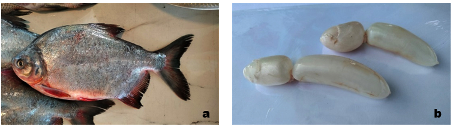
Figure 1. (a) Freshwater pomfret (Piaractus brachypomus), locally known as Yeri Vaval. (b) Isolated swim bladder of Piaractus brachypomus used for collagen extraction
Yield of acid-soluble swim bladder collagen
Acid-soluble swim bladder collagen (ASSBC) was successfully extracted from P. brachypomus using acetic acid extraction followed by dialysis and lyophilization. The collagen recovery based on wet tissue weight was 62.0 ± 2.3%, whereas the recovery based on dry weight after lyophilization was 13.3 ± 0.6% (mean ± SD, n = 3), demonstrating efficient extraction of collagen from fish-processing by-products (Figure 2).
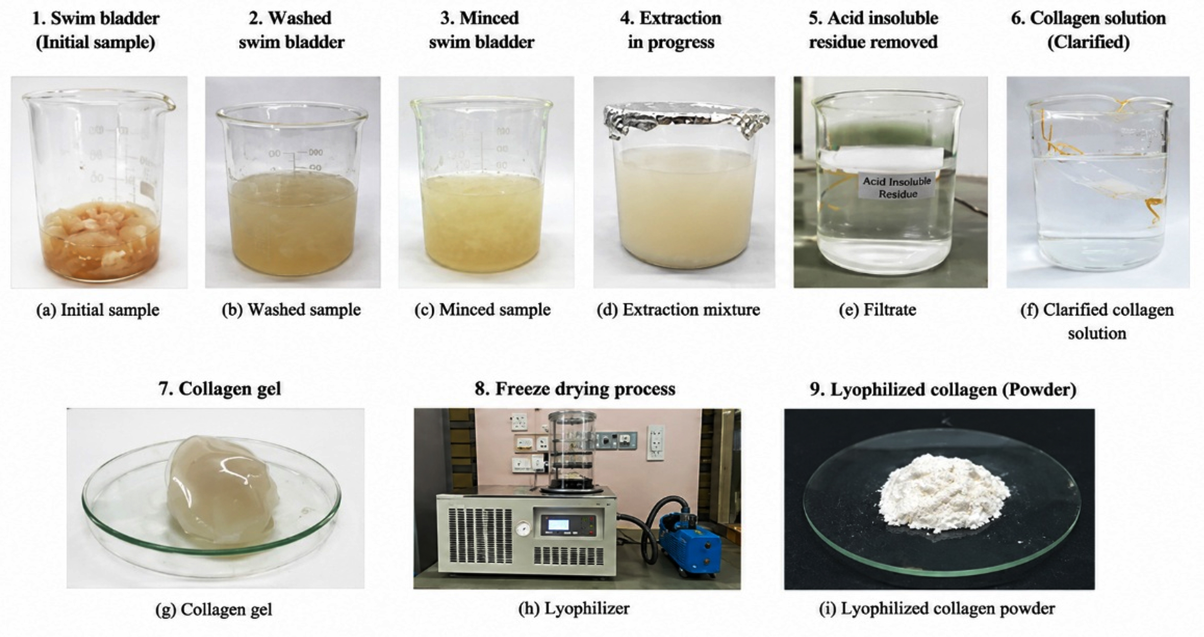
Figure 2. (a-f) Schematic overview of the pretreatment, (g-i) acid-soluble extraction and the recovery yield of collagen from the swim bladder of Piaractus brachypomus
Protein quantification and electrophoretic profiling of ASSBC
Protein estimation using the Lowry method demonstrated that the extracted collagen possessed a protein concentration of 74.0 ± 2.1 μg/mL (mean ± SD, n = 3) based on the bovine serum albumin standard curve (Figure 3a). Electrophoretic analysis by SDS–PAGE revealed the characteristic molecular profile of Type I collagen. Distinct protein bands corresponding to the γ-chain (272 kDa), β-chain (193 kDa), α1-chain (114 kDa), and α2-chain (98 kDa) were clearly observed (Figure 3b). The presence of these characteristic α, β and γ chains confirmed the preservation of the native triple-helical collagen structure following extraction.
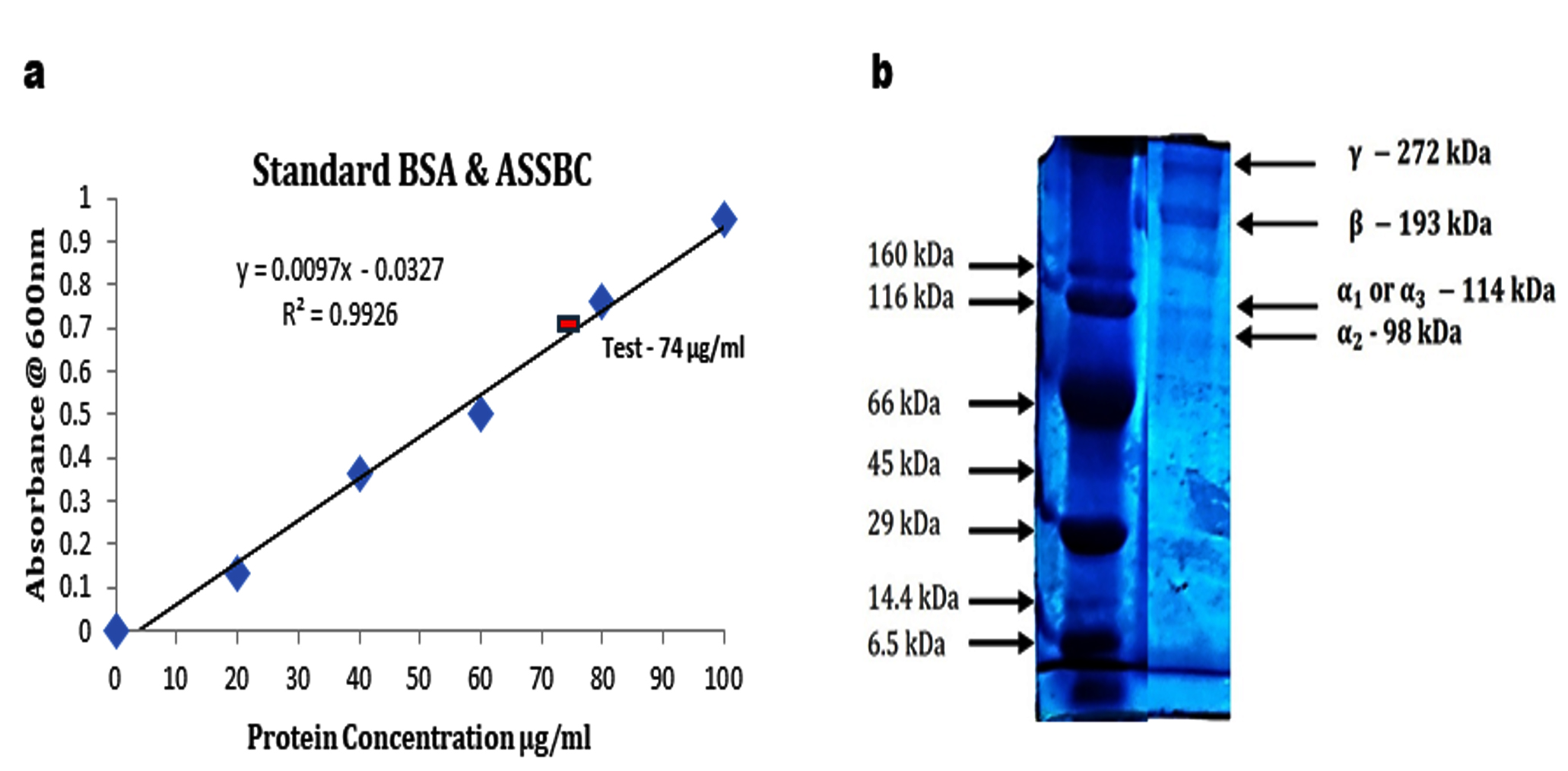
Figure 3. (a) Quantification of ASSBC by the Lowry method, showing a protein concentration of 74 µg/mL based on the BSA standard curve. (b) SDS-PAGE profile of ASSBC showing a typical type I collagen pattern with α, β, and ϒ chains
Structural characterization of acid-soluble swim bladder collagen
UV-visible spectroscopy
The UV-visible absorption spectrum of ASSBC exhibited a characteristic absorption maximum at 218 nm, which is consistent with the typical absorption profile of collagen molecules. No significant absorption was observed between 260 and 280 nm, indicating minimal contamination by aromatic amino acids and non-collagenous proteins (Figure 4).
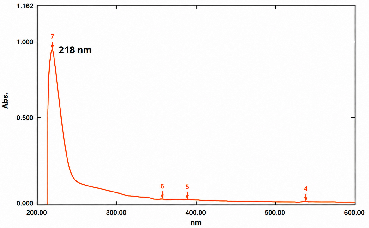
Figure 4. UV-visible absorption spectrum of ASSBC showing a characteristic absorption peak at 218 nm
Fourier transform infrared spectroscopy (FTIR)
FTIR analysis confirmed the structural integrity of ASSBC through the presence of characteristic collagen amide bands. The amide A, amide B, amide I, amide II, and amide III bands were detected at 3328, 2938, 1655, 1545, and 1232 cm-1, respectively. The absorbance ratio of the amide III band to the 1451 cm-1 band was approximately 0.90, indicating preservation of the native triple-helical collagen structure (Figure 5).
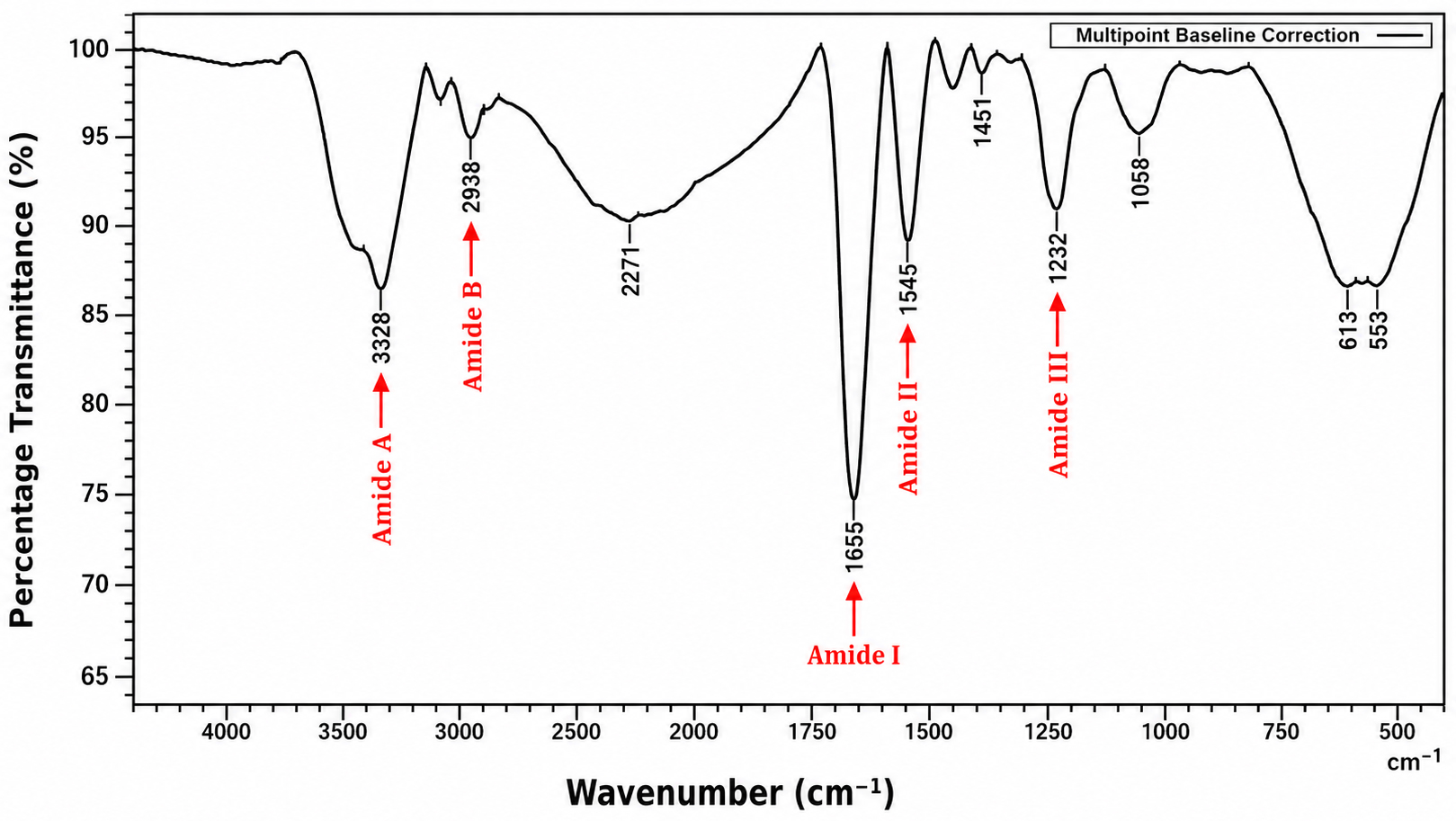
Figure 5. FTIR spectrum of acid-soluble swim bladder collagen (ASSBC) showing characteristic amide bands (A, B, I, II, and III)
Surface morphology and elemental composition – scanning electron microscopy (SEM)
SEM analysis revealed that lyophilized ASSBC exhibited a multilayered, porous, sponge-like fibrous architecture. At lower magnifications (250× and 500×), densely packed collagen fibrils were observed, whereas higher magnifications (2500× and 5000×) demonstrated an interconnected porous collagen network with loosely arranged fibrillar structures, which are characteristic features of native collagen (Figure 6).
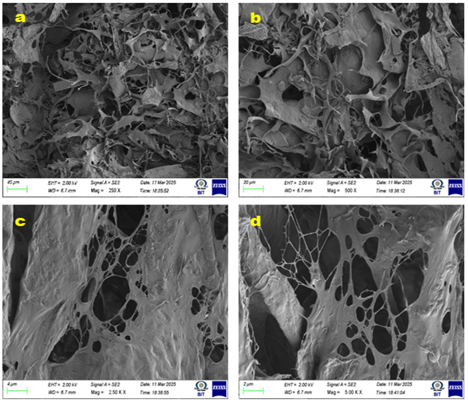
Figure 6. SEM micrographs of lyophilized acid-soluble swim bladder collagen showing surface morphology at different magnifications: (a) 250x, (b) 500x, (c) 2500x, and (d) 5000x
Energy Dispersive X-ray Analysis (EDAX)
EDAX analysis confirmed that carbon was the predominant element within ASSBC, followed by oxygen and trace quantities of nitrogen, sodium, phosphorus, sulfur, chlorine, and calcium. This elemental composition verified the organic proteinaceous nature of the extracted collagen and supported its high purity (Figure 7 and Table).
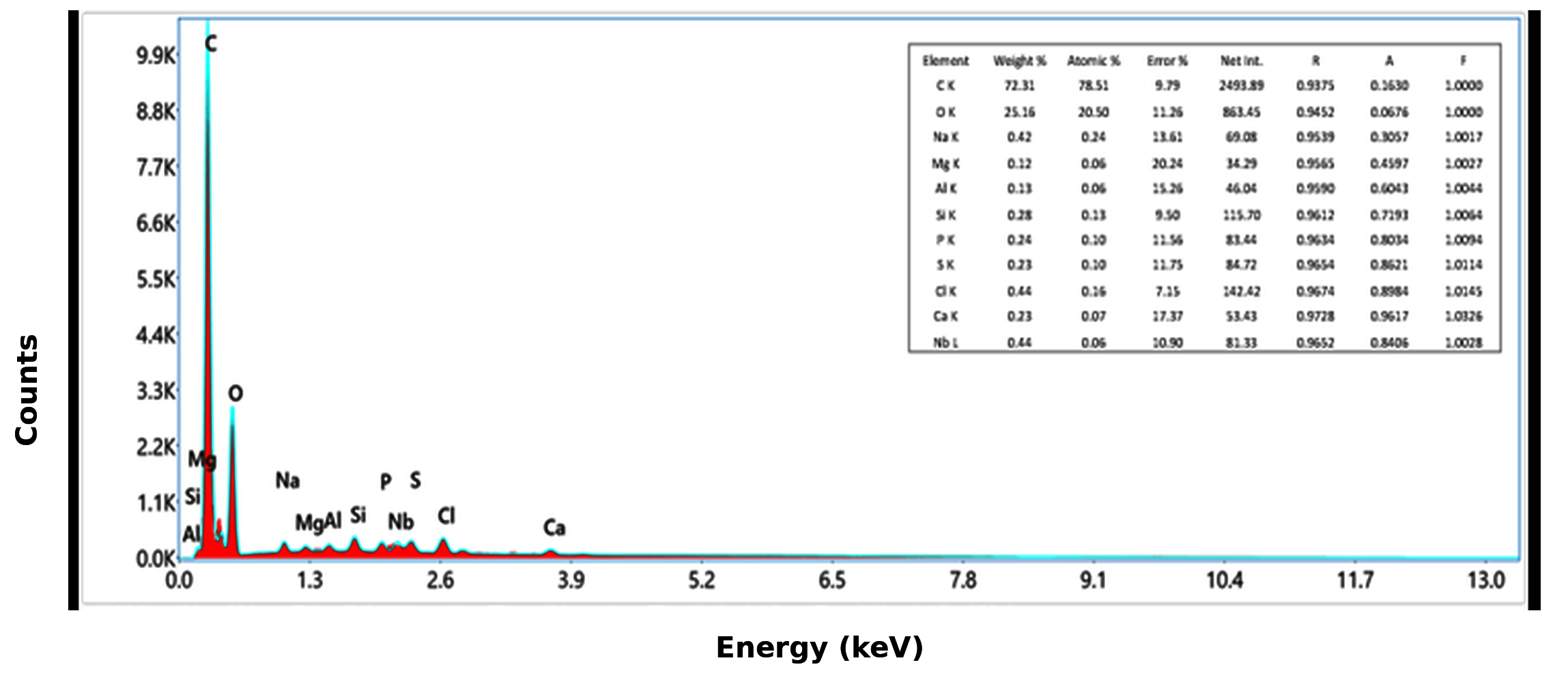
Figure 7. EDAX spectrum of acid-soluble swim bladder collagen showing elemental composition, with the corresponding quantitative elemental distribution was represented in the table
Table. Element Composition Analysis of acid soluble swim bladder collagen
Element |
Weight % |
Atomic % |
Error % |
Net Int. |
R |
A |
F |
|---|---|---|---|---|---|---|---|
C K |
72.31 |
78.51 |
9.79 |
2493.89 |
0.9375 |
0.1630 |
1.0000 |
O K |
25.16 |
20.50 |
11.26 |
863.45 |
0.9452 |
0.0676 |
1.0000 |
Na K |
0.42 |
0.24 |
13.61 |
69.08 |
0.9539 |
0.3057 |
1.0017 |
Mg K |
0.12 |
0.06 |
20.24 |
34.29 |
0.9565 |
0.4597 |
1.0027 |
Al K |
0.13 |
0.06 |
15.26 |
46.04 |
0.9590 |
0.6043 |
1.0044 |
Si K |
0.28 |
0.13 |
9.50 |
115.70 |
0.9612 |
0.7193 |
1.0064 |
P K |
0.24 |
0.10 |
11.56 |
83.44 |
0.9634 |
0.8034 |
1.0094 |
S K |
0.23 |
0.10 |
11.75 |
84.72 |
0.9654 |
0.8621 |
1.0114 |
Cl K |
0.44 |
0.16 |
7.15 |
142.42 |
0.9674 |
0.8984 |
1.0145 |
Ca K |
0.23 |
0.07 |
17.37 |
53.43 |
0.9728 |
0.9617 |
1.0326 |
Nb L |
0.44 |
0.06 |
10.90 |
81.33 |
0.9652 |
0.8406 |
1.0028 |
Production and characterization of collagen hydrolysates
Papain extracted from Carica papaya latex was successfully purified and used for the enzymatic hydrolysis of acid-soluble swim bladder collagen (ASSBC). The extraction process yielded 75.0 ± 2.4% papain (mean ± SD, n = 3). Proteolytic activity was confirmed by the casein precipitation assay, where visible casein coagulation at pH 5.5 indicated active enzymatic degradation (Figure 8a). SDS–PAGE analysis of purified papain revealed a distinct protein band corresponding to 23 kDa, confirming the successful isolation of active papain enzyme (Figure 8b). Following enzymatic hydrolysis, ASSBC was effectively converted into low-molecular-weight collagen peptides’. SDS–PAGE demonstrated a predominant peptide band at approximately 6 kDa, indicating efficient cleavage of native collagen into bioactive peptides (Figure 9). Qualitative characterization further confirmed the successful hydrolysis of collagen. The ninhydrin assay produced a characteristic yellow coloration, indicating the presence of hydroxyproline, while thin-layer chromatography demonstrated an Rf value of 0.24, which closely corresponded with the proline standard (Rf = 0.22). These findings verified the generation of collagen-derived bioactive peptides following papain hydrolysis.
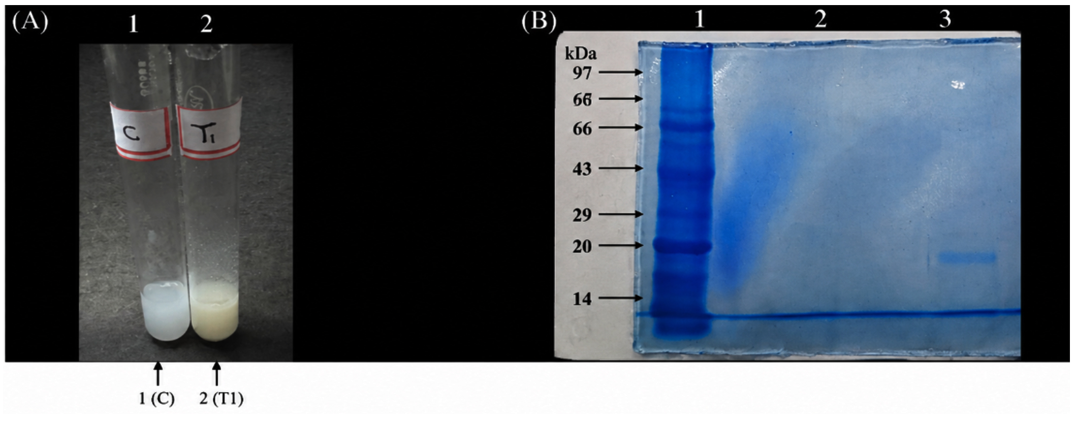
Figure 8. (A) Photograph of ASSBC (C; test tube 1) and papain-hydrolysed collagen peptides (T1; test tube 2). (B) SDS-PAGE profile showing Lane 1, molecular weight marker (270–6.5 kDa); Lane 2, ASSBC; and Lane 3, papain-hydrolysed collagen peptides. The bands were identified based on their migration relative to the molecular weight marker
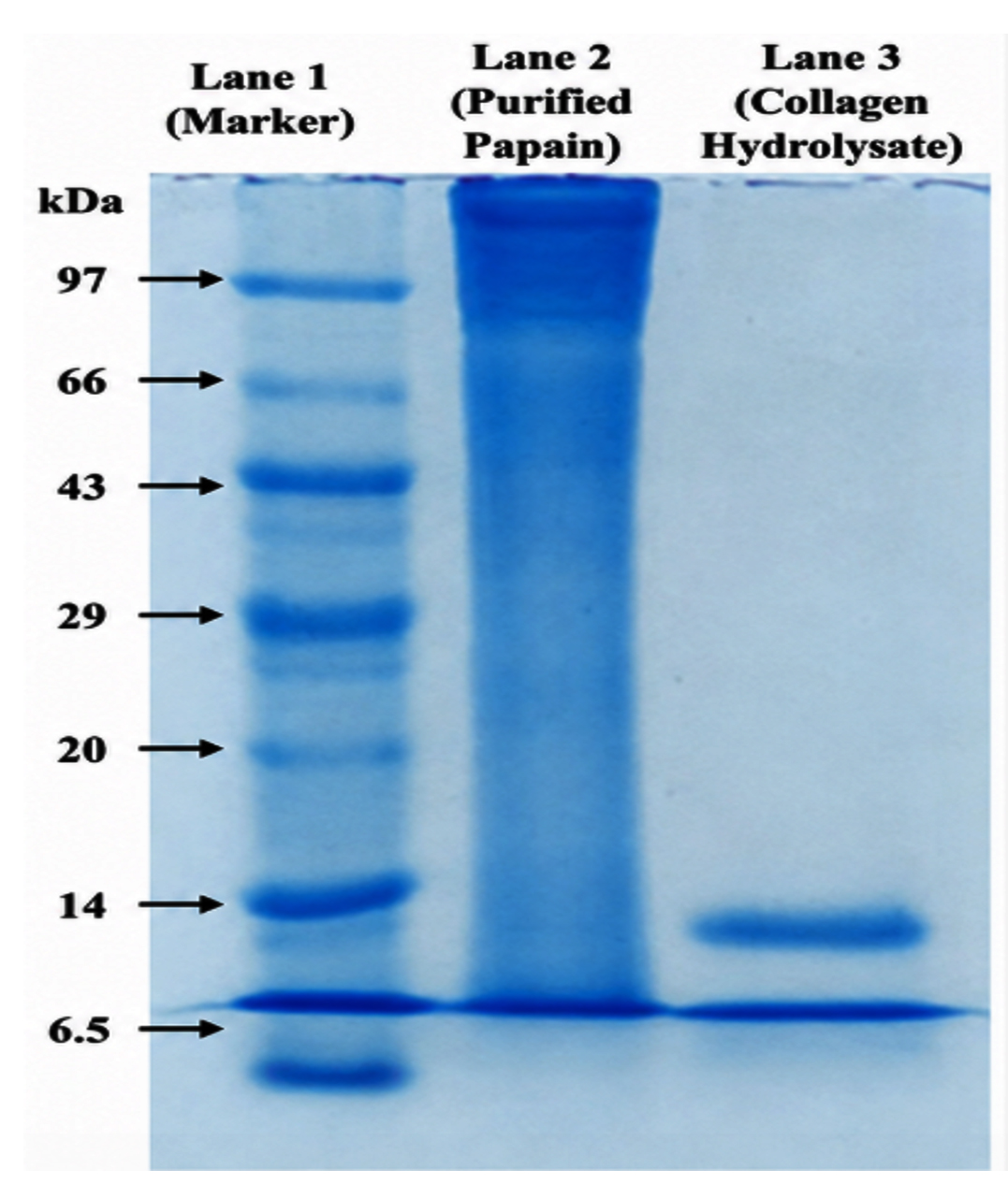
Figure 9. SDS–PAGE profile of papain-hydrolyzed ASSBC showing the formation of low-molecular-weight collagen peptides with a predominant 6 kDa band
Bioactive properties of hydrolyzed collagen peptides
Antimicrobial activity
The agar diffusion experiment was used to assess the antibacterial efficacy of hydrolyzed collagen peptides against certain bacterial and fungal infections. The collagen peptides exhibited selective antibacterial activity against Salmonella spp. and Staphylococcus spp., producing maximum inhibition zones of 14.0 ± 0.8 mm and 7.0 ± 0.5 mm, respectively (mean ± SD, n = 3). No measurable inhibitory activity was observed against Escherichia coli, Klebsiella spp., or Bacillus spp.
Among the fungal pathogens, collagen peptides demonstrated appreciable antifungal activity against Aspergillus niger and Penicillium spp., with maximum inhibition zones of 8.0 ± 0.6 mm and 20.0 ± 0.9 mm, respectively (Figure 10). These findings indicate that papain-derived collagen peptides possess selective antimicrobial properties, particularly against Gram-positive bacteria and filamentous fungi.
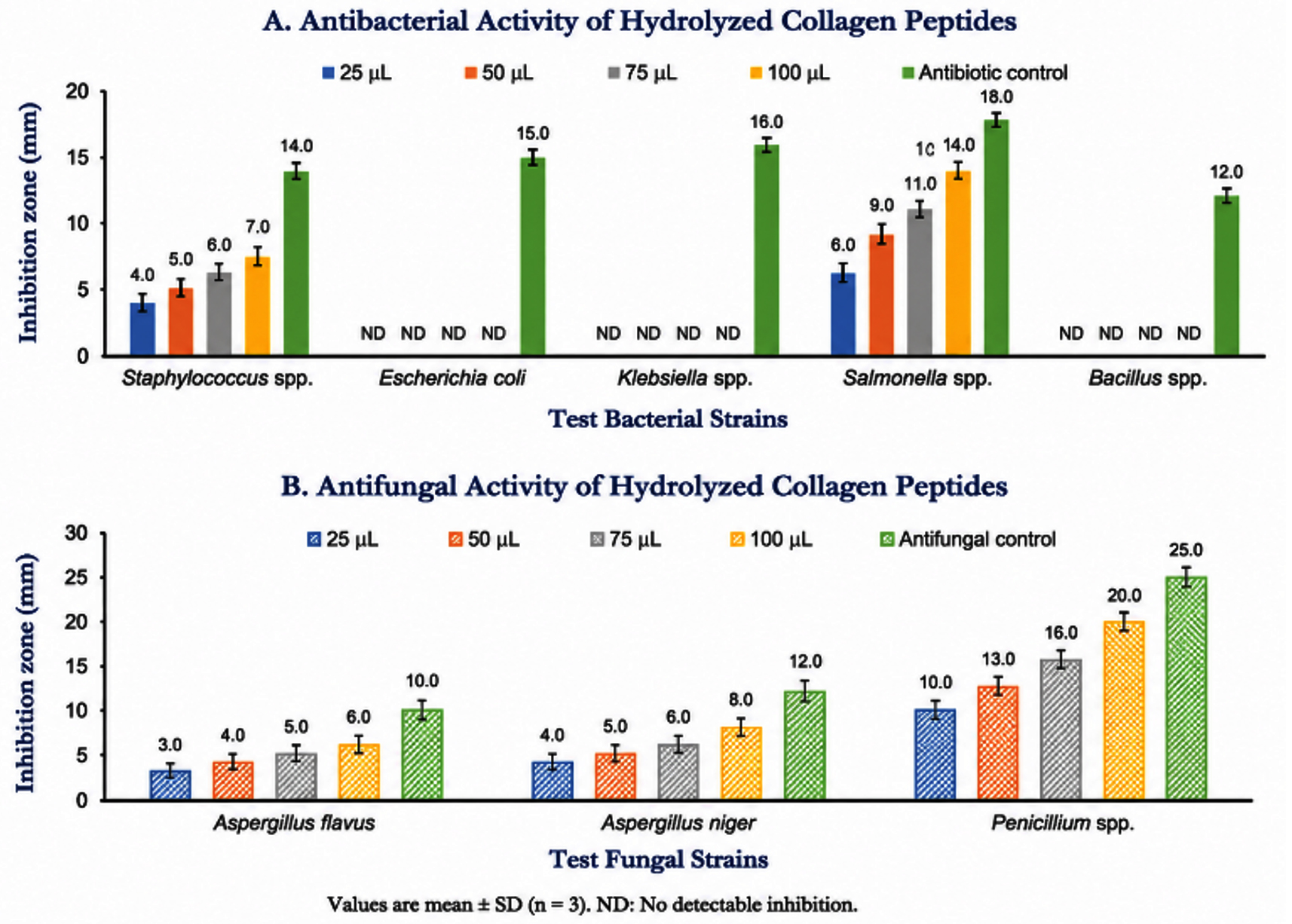
Figure 10. Antimicrobial activity of hydrolyzed collagen peptides evaluated by agar diffusion assay. (a) Graph showing the antibacterial activity against Salmonella spp. and Staphylococcus spp. (b) Graphical representation of antifungal activity against Aspergillus niger and Penicillium spp.
Antioxidant activity (DPPH radical scavenging assay)
We used the DPPH free radical scavenging experiment to evaluate the antioxidant capacity of hydrolyzed collagen peptides. Figure 11 shows that the radical scavenging activity rose as a function of concentration, with the greatest peptide concentration resulting in a maximum inhibition of 59.8 ± 1.7% (mean ± SD, n = 3). The observed increase in free radical scavenging activity demonstrated the effective antioxidant potential of the low-molecular-weight collagen peptides generated by papain hydrolysis.
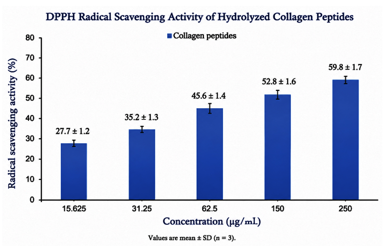
Figure 11. DPPH radical scavenging assay showing the free radical–quenching potential of the collagen peptides at different concentrations
Cytocompatibility of hydrolyzed collagen peptides (MTT assay)
The cytocompatibility of hydrolyzed collagen peptides was investigated using NIH-3T3 fibroblast cells. Cell viability remained above 90% at all tested concentrations (3.125-50 µg/mL), indicating excellent biocompatibility of the collagen peptides. Even at the highest concentration (50 µg/mL), cell viability remained 92.4 ± 2.1% (mean ± SD, n = 3), demonstrating only minimal cytotoxicity (Figure 12). Microscopic examination showed normal fibroblast morphology, intact cell membranes, and dense cellular distribution comparable to the untreated control, further confirming the non-cytotoxic nature of the collagen peptides.
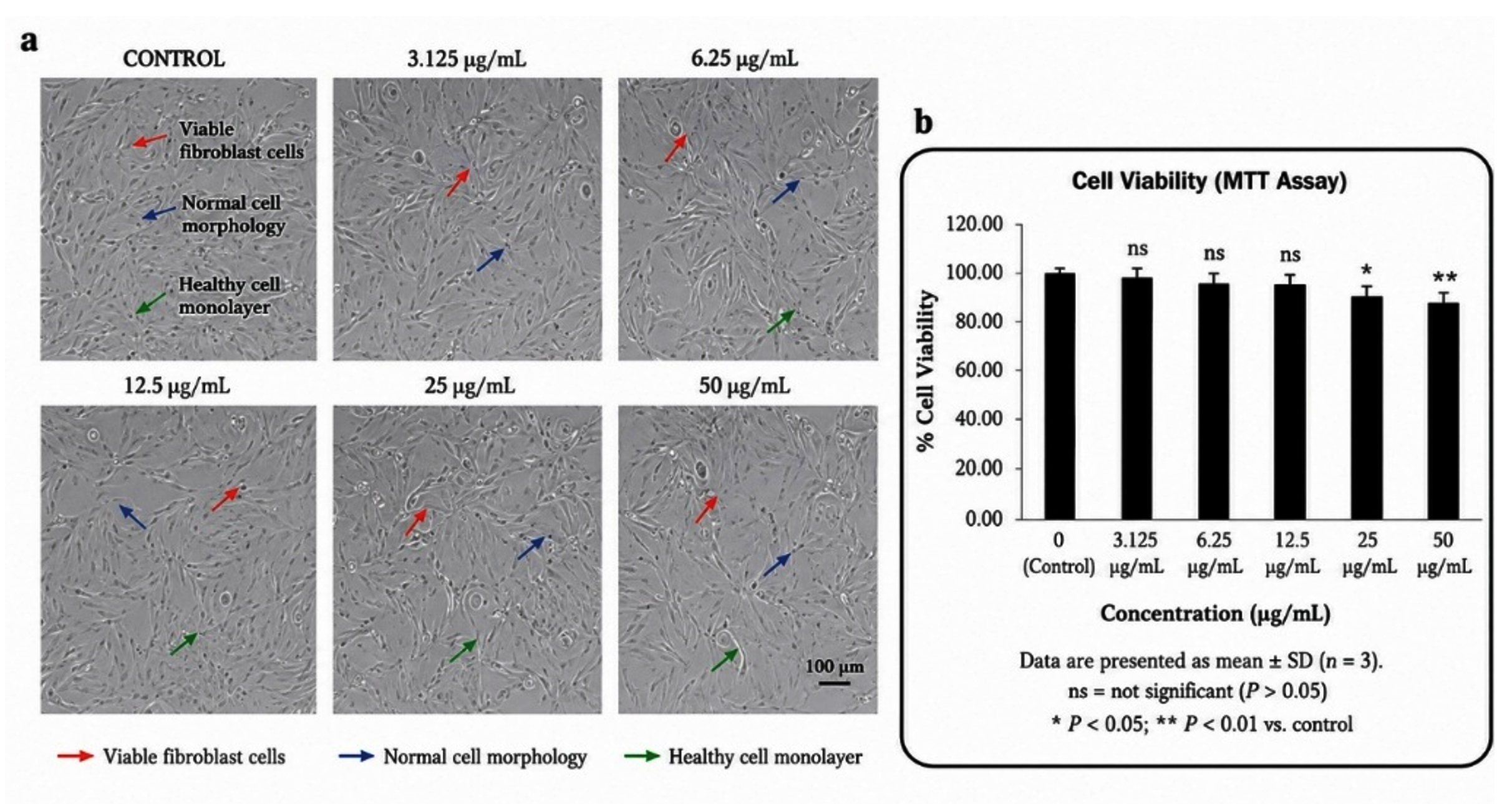
Figure 12. (a) and (b) Cytocompatibility of papain-derived collagen peptides on NIH-3T3 fibroblast cells after 48 hrs, showing preserved cell morphology, viable fibroblasts, and a healthy cell monolayer across all treatment concentrations. Cell viability is presented as mean ± SD (n = 3); P < 0.05 and *P < 0.01 compared with the untreated control
In vitro wound-healing activity (Scratch Assay)
The wound-healing potential of hydrolyzed collagen peptides was assessed using an in vitro scratch assay with NIH-3T3 fibroblast cells. At 0 hrs, all experimental groups exhibited an initial wound gap of 100%. Following 24 hrs of incubation, collagen peptide-treated cells demonstrated enhanced cell migration compared with the untreated control. After 48 hrs, the untreated control retained a wound gap of 24.6 ± 1.3%, whereas collagen peptide-treated cells exhibited a significantly reduced wound gap of 5.8 ± 0.6% (mean ± SD, n = 3; P < 0.05) (Figure 13). Microscopic observations clearly demonstrated accelerated fibroblast migration and nearly complete wound closure in the collagen peptide-treated group, confirming the wound-healing potential of the hydrolyzed collagen peptides.
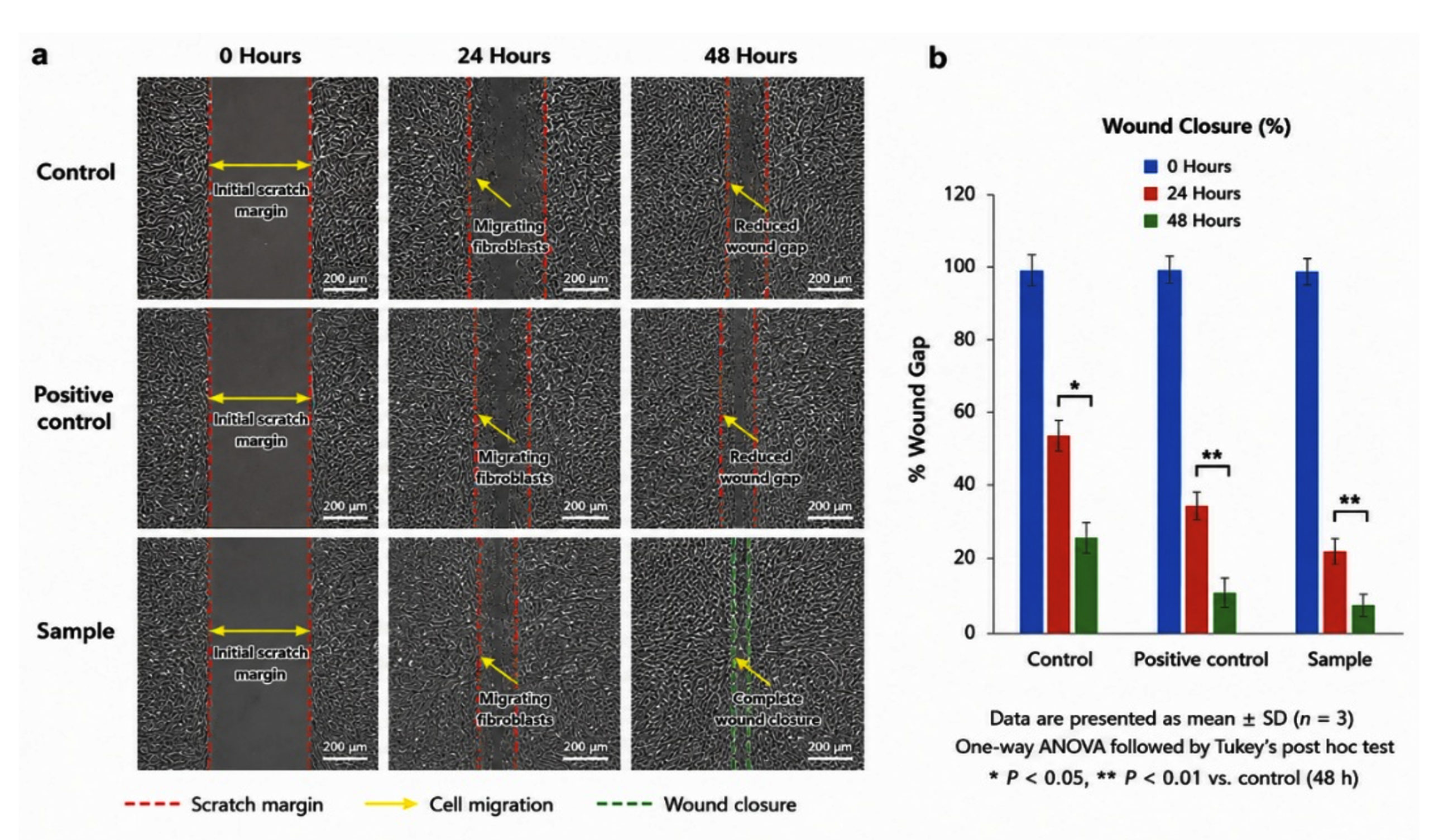
Figure 13. (a) Representative images of the in vitro scratch wound-healing assay showing fibroblast migration and progressive wound closure in the control, positive control, and collagen peptide-treated groups at 0, 24, and 48 hrs. (b) Quantitative analysis of wound closure (%) presented as mean ± SD (n = 3), showing significantly enhanced wound healing in the collagen peptide-treated group (P < 0.05)
Collagen has gained considerable attention as a naturally derived biomaterial because of its excellent biocompatibility, biodegradability, low immunogenicity, and essential role in extracellular matrix organization and tissue regeneration. In recent years, increasing emphasis has been placed on the sustainable utilization of fish-processing by-products as alternative collagen sources to reduce environmental waste while generating value-added biomaterials for pharmaceutical and biomedical applications. Among these by-products, fish swim bladders are particularly attractive because they contain highly organized collagen fibers with relatively low levels of non-collagenous proteins, facilitating efficient extraction of structurally intact collagen. Consequently, the valorization of fish-processing waste not only contributes to environmental sustainability but also supports the development of high-value biomaterials within the framework of the circular bioeconomy.31-33
In the present study, the swim bladder of Piaractus brachypomus served as an effective source of acid-soluble collagen with a comparatively high extraction yield. The efficiency of collagen recovery can be attributed to the organized connective tissue architecture of the swim bladder and the mild acetic acid extraction procedure, which selectively disrupts weak intermolecular interactions while preserving the native triple-helical configuration. Differences in collagen recovery among fish species have been associated with variations in tissue composition, age, habitat, collagen cross-linking, and extraction conditions. The relatively high collagen yield observed in the present investigation therefore indicates that P. brachypomus swim bladder represents a promising alternative source of collagen for biomedical applications when compared with many conventionally utilized marine resources.34,35
Protein quantification further demonstrated the successful recovery of collagen following extraction and purification. Maintaining a high protein concentration is particularly important because it directly influences enzymatic hydrolysis, scaffold fabrication, mechanical stability, and the biological performance of collagen-based biomaterials. Efficient removal of non-collagenous proteins during dialysis and lyophilization while minimizing protein loss indicates that the extraction protocol preserved collagen integrity without extensive denaturation. Similar observations have been reported for fish-derived collagens prepared under optimized extraction conditions, where preservation of protein quality significantly enhanced their functional and biomedical properties.36
Electrophoretic analysis by SDS-PAGE confirmed that the extracted material possessed the characteristic molecular profile of Type I collagen through the presence of distinct α₁, α₂, β, and γ chains. Preservation of these molecular components indicates that the extraction procedure maintained the native structural organization of collagen while preventing excessive degradation. The β- and γ-components further demonstrate the retention of intermolecular cross-linking, which contributes to collagen fibril stability and mechanical strength. Maintenance of the native molecular architecture is particularly important because intact Type I collagen provides appropriate binding sites for integrin-mediated cell adhesion, fibroblast proliferation, and extracellular matrix remodeling during tissue repair. Consequently, the electrophoretic profile obtained in the present study confirms that the extracted collagen retained the structural characteristics required for subsequent enzymatic hydrolysis and biomedical applications, including wound healing and tissue engineering.37,38
The physicochemical integrity of the extracted collagen was comprehensively evaluated using UV–visible spectroscopy, Fourier transform infrared spectroscopy (FTIR), scanning electron microscopy (SEM), and energy-dispersive X-ray spectroscopy (EDAX). These complementary analytical techniques provide valuable information regarding collagen purity, molecular conformation, fibrillar architecture, and elemental composition, all of which influence its biological performance and suitability for regenerative medicine. Preservation of the native collagen structure is particularly important because disruption of the triple-helical configuration can compromise mechanical stability, enzymatic resistance, and cellular interactions required for tissue repair.39,40
The UV–visible spectrum of the extracted collagen exhibited a characteristic absorption maximum at 218 nm, corresponding to the peptide bonds present within the collagen backbone. The absence of significant absorbance between 260 and 280 nm further indicated minimal contamination by aromatic amino acids and other non-collagenous proteins, demonstrating the effectiveness of the extraction and purification procedures. Previous investigations have shown that purified collagen typically exhibits an absorption peak within the range of 210-230 nm, whereas increased absorbance near 280 nm generally reflects residual protein impurities. Therefore, the spectral profile observed in the present study confirms that the isolated collagen possessed high purity and retained its native molecular characteristics following extraction.41
The existence of typical amide A, B, I, II, and III bands further confirmed the preservation of the collagen triple helix using FTIR spectroscopy. Collectively, these absorption bands constitute the molecular fingerprint of collagen; they correspond to the following vibrations: N-H stretching, asymmetric CHk stretching, C=O stretching, N-H bending, and C-N stretching. The amide III to 1451 cm-1 absorbance ratio of approximately 0.90 strongly suggests that the native triple-helical structure remained largely intact throughout extraction and purification. Maintenance of this ordered conformation is essential because hydrogen bonding within the triple helix contributes to collagen stability, resistance to enzymatic degradation, and interactions with extracellular matrix proteins. Preservation of the native structure is therefore expected to enhance the biological functionality of the collagen in tissue engineering and wound-healing applications.42-44
The SEM micrographs demonstrated that the lyophilized collagen possessed a porous, sponge-like fibrillar architecture with interconnected collagen fibers. Such morphology is characteristic of freeze-dried collagen and closely resembles the three-dimensional extracellular matrix environment. An interconnected porous network is particularly advantageous because it facilitates nutrient diffusion, oxygen transport, cell infiltration, and neovascularization, all of which are essential for effective tissue regeneration. Furthermore, the high surface area associated with porous collagen matrices improves water absorption and provides favorable conditions for the incorporation and controlled release of therapeutic agents in wound-dressing systems. Similar microstructural characteristics have been widely reported for fish-derived collagen scaffolds exhibiting excellent biocompatibility and regenerative performance.45
The preservation of an organized fibrillar architecture also suggests that the extraction and lyophilization processes caused minimal disruption of collagen fibrillogenesis. Maintenance of this native microstructure is biologically significant because collagen fibrils provide structural support for fibroblast attachment and guide cellular migration during extracellular matrix remodeling. These structural characteristics may therefore have contributed to the excellent cytocompatibility and wound-healing properties observed for the papain-derived collagen peptides in the present investigation.46
Elemental analysis by EDAX demonstrated that carbon and oxygen constituted the major elemental components of the extracted collagen, confirming its proteinaceous nature. Minor quantities of nitrogen, sulfur, phosphorus, sodium, calcium, chlorine, magnesium, and silicon were also detected, reflecting the amino acid composition and naturally occurring mineral constituents of connective tissues. The absence of substantial inorganic contaminants further supports the high purity of the isolated collagen and indicates that the extraction protocol effectively removed unwanted impurities without altering its chemical composition. Comparable elemental profiles have been reported for purified fish collagens intended for biomedical and pharmaceutical applications, supporting the reliability of the present extraction strategy.45 Overall, the spectroscopic, microscopic, and elemental analyses consistently demonstrate that the acid-soluble collagen isolated from the swim bladder of Piaractus brachypomus retained its native molecular organization, physicochemical stability, and fibrillar architecture. Preservation of these structural characteristics is fundamental for maintaining collagen bioactivity and provides a suitable platform for subsequent enzymatic hydrolysis and the production of multifunctional bioactive collagen peptides with potential applications in regenerative medicine.
The conversion of native collagen into bioactive peptides through enzymatic hydrolysis has attracted considerable interest because hydrolysis enhances the biological functionality and bioavailability of collagen without compromising its amino acid composition. In the present study, papain extracted from Carica papaya latex efficiently hydrolyzed acid-soluble collagen into low-molecular-weight peptide. Papain is a cysteine protease with broad substrate specificity that selectively cleaves peptide bonds under relatively mild conditions, thereby preserving biologically active peptide sequences that may otherwise be destroyed by chemical hydrolysis. Consequently, enzymatic hydrolysis has become the preferred approach for producing collagen peptides intended for pharmaceutical, nutraceutical, and tissue-engineering applications because it yields peptides with improved solubility, digestibility, and biological activity.47,48
The characteristic 23 kDa band observed for purified papain confirmed successful enzyme isolation, whereas the appearance of a predominant peptide band of approximately 6 kDa following hydrolysis demonstrated efficient cleavage of native collagen into low-molecular-weight peptides. Peptide size plays a crucial role in determining biological activity because smaller peptides possess greater aqueous solubility, enhanced cellular uptake, and improved diffusion through extracellular matrices. Several studies have reported that collagen peptides with molecular weights below 10 kDa exhibit superior antioxidant, antimicrobial, anti-inflammatory, and wound-healing activities owing to their increased accessibility to biological targets and greater interaction with cellular receptors.49,50
The positive ninhydrin reaction together with the TLC profile further confirmed successful collagen hydrolysis through the detection of hydroxyproline-rich peptides. Hydroxyproline is a unique amino acid that stabilizes the collagen triple helix by promoting hydrogen bonding between adjacent polypeptide chains.51 Release of hydroxyproline-containing peptides during enzymatic hydrolysis therefore reflects efficient degradation of collagen while retaining amino acid sequences responsible for biological activity. These peptides have been associated with stimulation of fibroblast proliferation, extracellular matrix synthesis, and antioxidant defense, all of which are essential for tissue regeneration.52
The hydrolyzed collagen peptides demonstrated selective antimicrobial activity against Salmonella spp., Staphylococcus spp., Aspergillus niger, and Penicillium spp. The antimicrobial activity of collagen-derived peptides depends largely on peptide sequence, molecular weight, hydrophobicity, and net positive charge. Cationic peptides interact electrostatically with negatively charged microbial membranes, resulting in membrane destabilization, increased permeability, leakage of intracellular contents, and ultimately microbial death. Variations in cell wall composition, membrane architecture, and outer membrane permeability may explain the absence of activity against Escherichia coli, Klebsiella spp., and Bacillus spp. Similar selective antimicrobial properties have previously been reported for fish-derived collagen peptides, supporting their potential as naturally derived antimicrobial agents for biomedical applications.53
The antioxidant activity observed in the DPPH assay further highlights the multifunctional nature of the collagen peptides. The concentration-dependent increase in radical scavenging activity is consistent with previous reports demonstrating that enzymatic hydrolysis exposes hydrophobic amino acid residues capable of donating electrons or hydrogen atoms to neutralize reactive oxygen species. By neutralizing free radicals, binding transition metals, and halting oxidative chain reactions, amino acids including glycine, proline, alanine, valine, and leucine greatly enhance the antioxidant capabilities of collagen peptides. Because oxidative stress plays a major role in inflammation, delayed wound healing, and tissue degeneration, the antioxidant activity demonstrated in the present study further supports the therapeutic potential of papain-derived collagen peptides for regenerative medicine and functional food applications.54
Successful translation of collagen-derived biomaterials into biomedical applications requires not only biological activity but also excellent compatibility with mammalian cells. In the present investigation, papain-derived collagen peptides maintained high viability of NIH-3T3 fibroblast cells, indicating that enzymatic hydrolysis generated biologically safe peptides without producing cytotoxic degradation products. Fish collagen possesses inherently low immunogenicity and excellent biocompatibility owing to its structural similarity to mammalian extracellular matrix proteins while avoiding concerns associated with mammalian pathogens and religious restrictions. Moreover, collagen peptides promote cell adhesion through interactions with integrin receptors and activate signaling pathways involved in cell survival, proliferation, and matrix synthesis, thereby supporting tissue regeneration.55
The preservation of normal fibroblast morphology and dense monolayer formation further confirmed that the collagen peptides did not induce membrane damage or morphological alterations. Maintenance of normal cellular architecture is a critical indicator of cytocompatibility because fibroblasts are responsible for collagen synthesis, extracellular matrix remodeling, and wound contraction during tissue repair. Previous studies have demonstrated that collagen peptides stimulate fibroblast proliferation while reducing oxidative stress-induced cellular injury, thereby accelerating regeneration of damaged tissues.56
The scratch assay demonstrated significantly enhanced fibroblast migration following treatment with collagen peptides, indicating accelerated wound closure. Fibroblast migration represents one of the earliest events during wound healing because these cells repopulate the wound bed, synthesize extracellular matrix proteins, and coordinate tissue remodeling. The enhanced wound closure observed in the present study is consistent with previous reports demonstrating that marine collagen peptides stimulate transforming growth factor-β (TGF-β), focal adhesion kinase (FAK), and mitogen-activated protein kinase (MAPK) signaling pathways that regulate cell migration, proliferation, and extracellular matrix deposition. The combined effects of antioxidant activity, antimicrobial activity, and collagen-derived amino acid supplementation likely contributed synergistically to the accelerated wound-healing response observed in this investigation.56
The present study demonstrates that acid-soluble collagen extracted from the swim bladder of Piaractus brachypomus can be converted into multifunctional bioactive peptides possessing antioxidant, antimicrobial, cytocompatible, and wound-healing properties. Utilization of freshwater fish-processing by-products provides an environmentally sustainable alternative to conventional mammalian collagen sources while simultaneously supporting circular bioeconomy strategies through value addition to biological waste. The integrated characterization of collagen extraction, enzymatic hydrolysis, structural analysis, biological evaluation, and wound-healing assessment represents a comprehensive approach that expands current knowledge regarding the biomedical potential of P. brachypomus collagen.
Despite these promising findings, the study has certain limitations. The biological activities were evaluated exclusively under in vitro conditions, and the molecular mechanisms underlying collagen peptide-mediated tissue regeneration were not investigated. Furthermore, peptide sequence identification and long-term biocompatibility studies were beyond the scope of the present investigation. Future studies should therefore focus on LC-MS/MS-based peptide characterization, elucidation of signaling pathways involved in collagen-mediated wound healing, fabrication of collagen-based composite scaffolds, and validation of therapeutic efficacy using appropriate preclinical animal models. Such investigations will facilitate the translation of fish-derived collagen peptides into clinically relevant biomaterials for regenerative medicine.
The present study successfully demonstrated that the swim bladder of Piaractus brachypomus is a promising and sustainable source of high-quality Type I acid-soluble collagen. The extracted collagen retained its native structural integrity and was efficiently converted into low-molecular-weight bioactive peptides through papain-mediated enzymatic hydrolysis. These collagen peptides exhibited significant antioxidant, selective antimicrobial, excellent cytocompatibility, and enhanced wound-healing activities, highlighting their multifunctional biological potential. Collectively, these findings establish P. brachypomus swim bladder as a valuable alternative to conventional mammalian collagen sources and support its potential application in wound dressings, tissue engineering, regenerative medicine, nutraceuticals, and biomedical formulations. A limitation of the present study is that the biological activities were evaluated only under in vitro conditions. Therefore, future investigations should focus on peptide sequence identification, elucidation of the underlying molecular mechanisms, development of collagen-based biomaterials, and validation of therapeutic efficacy through in vivo preclinical studies to facilitate clinical translation.
ACKNOWLEDGMENTS
The authors gratefully acknowledge the PG & Research Department of Microbiology, D.K.M. College for Women (Autonomous), Vellore, Tamil Nadu, India, for providing laboratory facilities and technical support for this research.
CONFLICT OF INTEREST
The authors declare that there is no conflict of interest.
AUTHORS’ CONTRIBUTION
All authors listed have made a substantial, direct and intellectual contribution to the work, and approved it for publication.
FUNDING
None.
DATA AVAILABILITY
The datasets generated or analyzed during this study are included in the manuscript and the rest can be obtained from the corresponding author upon reasonable request.
ETHICS STATEMENT
The present investigation involved only fish-processing by-products obtained from commercially available fish and established NIH-3T3 fibroblast cell lines. No live animal experimentation or human participants were involved in this study. All cell culture experiments were conducted in accordance with institutional laboratory biosafety guidelines.
- Holwerda AM, van Loon LJC. The impact of collagen protein ingestion on musculoskeletal connective tissue remodeling: a narrative review. Nutr Rev. 2022;80(6):1497-1514.
Crossref - Meyer M. Processing of collagen-based biomaterials and the resulting material properties. BioMed Eng OnLine. 2019;18(1):24.
Crossref - Nurilmala M, Hizbullah HH, Karnia E, Kusumaningtyas E, Ochiai Y. Characterization and antioxidant activity of collagen, gelatin, and derived peptides from yellowfin tuna (Thunnus albacares) skin. Mar Drugs. 2020;18(2):98.
Crossref - Zhu R, Huang Z, Zhang J, et al. The potential of collagen-based materials for wound management. Mater Today Chem. 2024;41:102295.
Crossref - Murugadoss P, Karna AK, Rajendran NK, et al. Collagen and its derivatives in the food industry: a review of sources, extraction, functional properties, and applications. Results Chem. 2026;27:103500.
Crossref - Lim YS, Ok YJ, Hwang SY, Kwak JY, Yoon S. Marine collagen as a promising biomaterial for biomedical applications. Mar Drugs. 2019;17(8):467.
Crossref - González-Serrano DJ, Hadidi M, Varcheh M, Jelyani AZ, Moreno A, Lorenzo JM. Bioactive peptide fractions from collagen hydrolysate of common carp fish by-product: antioxidant and functional properties. Antioxidants. 2022;11(3):509.
Crossref - Lan X, Luo M, Li M, et al. Swim bladder-derived biomaterials: structures, compositions, properties, modifications, and biomedical applications. J Nanobiotechnol. 2024;22:186.
Crossref - Li Y, Yang L, Wu S, Chen J, Lin H. Structural, functional, rheological and biological properties of swim bladder collagen extracted from grass carp (Ctenopharyngodon idella). LWT. 2022;153:112518.
Crossref - Zhao WH, Luo QB, Pan X, Chi CF, Sun KL, Wang B. Preparation, identification, and activity evaluation of ten antioxidant peptides from protein hydrolysate of swim bladders of miiuy croaker (Miichthys miiuy). J Funct Foods. 2018;47:503-511.
Crossref - Kumar GP, Nidheesh T, Govindaraju K, Jyoti, Suresh PV. Enzymatic extraction and characterization of thermostable collagen from swim bladder of rohu (Labeo rohita). J Sci Food Agric. 2017;97(5):1451-1458.
Crossref - Zhang X, Adachi S, Ura K, Takagi Y. Properties of collagen extracted from Amur sturgeon (Acipenser schrenckii) swim bladder and assessment of collagen fibrils in vitro. Int J Biol Macromol. 2019;137:809-820.
Crossref - Xiao L, Lv J, Liang Y, et al. Structural and physicochemical properties of swim bladder collagen promoting fibroblast viability and collagen synthesis. LWT. 2023;173:114294.
Crossref - Toniasso DPW, da Silva CG, Junior BDSB, et al. Collagen extracted from rabbit meat and by-products: Isolation and physicochemical assessment. Food Res Int. 2022;162:111967.
Crossref - Hashim P, Ridzwan MM, Bakar J. Isolation and characterization of collagen from chicken feet. Int J Bioeng Life Sci. 2014;8(3):250-254.
Crossref - Lowry OH, Rosebrough NJ, Farr AL, Randall RJ. Protein measurement with the Folin phenol reagent. J Biol Chem. 1951;193(1):265-275.
Crossref - Nurhayati T, Astiana I. Characteristics of papain-soluble collagen from redbelly yellowtail fusilier (Caesio cuning). IOP Conf Ser Earth Environ Sci. 2018;196:012034.
Crossref - Zhang N, Guo S, Zheng Y, Li W. Isolation and Characterisation of Acid Soluble Collagens and Pepsin Soluble Collagens from Eel (Anguilla japonica Temminck et Schlegel) Skin and Bone. Foods. 2025;14(3):502.
Crossref - Ge B, Wang H, Li J, et al. Comprehensive Assessment of Nile Tilapia Skin (Oreochromis niloticus) Collagen Hydrogels for Wound Dressings. Mar Drugs. 2020;18(4):178.
Crossref - Shaik MI, Rahman SHA, Yusri AS, et al. A review on the processing technique, physicochemical, and bioactive properties of marine collagen. J Food Sci. 2024;89(9):5205-5229.
Crossref - Wang JC, Kinsella JE. Functional properties of novel proteins: Alfalfa leaf protein. J Food Sci. 1976;41(2):286-292.
Crossref - Khatun MN, Saeid A, Mozumder NHMR, Ahmed M. Extraction, purification and characterization of papain enzyme from papaya. Food Res. 2023;7(2):241-247.
Crossref - Dhivya R, Rashma RS, Vinothini B, Pavithra R. Extraction and purification of papain enzyme from Carica papaya for wound debridement. Int J Pure Appl Math. 2018;119(15):1265-1274
- Cruz-López H, Rodríguez-Morales S, Enríquez-Paredes LM, et al. Swim bladder of farmed Totoaba macdonaldi: A source of value-added collagen. Mar Drugs. 2023;21(3):173.
Crossref - DeMeglio DC, Svanberg GK. A Thin-layer chromatographic procedure for separation of proline and hydroxyproline from biological samples. J Liq Chromatogr Relat Technol. 1996;19(6):969-975.
Crossref - Langrock T, Hoffmann R. Analysis of hydroxyproline in collagen hydrolysates. Methods Mol Biol. 2019;2030:47-56.
Crossref - Balouiri M, Sadiki M, Ibnsouda SK. Methods for in vitro evaluating antimicrobial activity: A review. J Pharm Anal. 2016;6(2):71-79.
Crossref - Song Y, Fu Y, Huang S, et al. Identification and antioxidant activity of bovine bone collagen-derived novel peptides prepared by recombinant collagenase from Bacillus cereus. Food Chem. 2021;349:129143.
Crossref - Ghasemi M, Turnbull T, Sebastian S, Kempson I. The MTT Assay: Utility, Limitations, Pitfalls, and Interpretation in Bulk and Single-Cell Analysis. Int J Mol Sci. 2021;22(23):12827.
Crossref - Yang F, Jin S, Tang Y. Marine collagen peptides promote fibroblast proliferation via NF-κB signaling pathway. Molecules. 2019;24(22):4201.
Crossref - Subhan F, Hussain Z, Tauseef I, Shehzad A, Wahid F. A review on recent advances and applications of fish collagen. Crit Rev Food Sci Nutr. 2021;61(6):1027-1037.
Crossref - Sharma S, Rai VK, Narang RK, Markandeywar TS. Collagen-based formulations for wound healing: A literature review. Life Sci. 2022;290:120096.
Crossref - de Souza A, Cruz MA, de Araújo TAT, et al. Fish collagen for skin wound healing: A systematic review in experimental animal studies. Cell Tissue Res. 2022;388(3):489-502.
Crossref - Jaziri AA, Shapawi R, Mokhtar RAM, Noordin WNMd, Sukoso, Huda N. Recent progress in sustainable fish byproduct utilization: unveiling fish collagen as a potential wound healing agent. Ann Anim Sci. 2025;26(1):75-106.
Crossref - Rajabimashhadi Z, Gallo N, Salvatore L, Lionetto F. Collagen derived from fish industry waste: progresses and challenges. Polymers. 2023;15(3):544.
Crossref - Ahmed R, Haq M, Chun BS. Characterization of marine-derived collagen extracted from the by-products of bigeye tuna (Thunnus obesus). Int J Biol Macromol. 2019;135:668-676.
Crossref - Hayashi T, Nagai Y. Separation of the alpha chains of type I and III collagens by SDS-polyacrylamide gel electrophoresis. J Biochem. 1979;86(2):453-459.
Crossref - Nurubhasha R, Kumar NSS, Thirumalasetti SK, Simhachalam G, Dirisala VR. Extraction and characterization of collagen from the skin of Pterygoplichthys pardalis and its potential application in food industries. Food Sci Biotechnol. 2019;28(6):1811-1817.
Crossref - Pesterau AM. Extraction, identification and characterization by SDS-PAGE method of collagen extracted from Rhizostoma pulmo, a jellyfish found in the Black Sea Basin. Eur J Med Nat Sci. 2025;8(1):53-61.
Crossref - Silva T, Moreira-Silva J, Marques A, Domingues A, Bayon Y, Reis R. Marine Origin Collagens and its potential applications. Mar Drugs. 2014;12(12):5881-5901.
Crossref - Zhong M, Chen T, Hu C, Ren C. Isolation and characterization of collagen from the body wall of sea cucumber Stichopus monotuberculatus. J Food Sci. 2015;80(4):C671-C679.
Crossref - Rasouli R, Yaghoobi H, Frampton J. A comparative study of the effects of different crosslinking methods on the physicochemical properties of collagen multifilament bundles. ChemPhysChem. 2024;25(14):e202400259.
Crossref - Olaru M, Simionescu N, Doroftei F, David G. Strategy based on Michael addition reaction for the development of bioinspired multilayered and multiphasic 3D constructs. Polymers. 2023;15(7):1635.
Crossref - Nurubhasha R, Sampath Kumar NS, Thirumalasetti SK, Simhachalam G, Dirisala VR. Extraction and characterization of collagen from the skin of Pterygoplichthys pardalis and its potential application in food industries. Food Sci Biotechnol. 2019;28(6):1811-1817.
Crossref - Kim SK, Wijesekara I. Development and biological activities of marine-derived bioactive peptides: A review. J Funct Foods. 2010;2(1):1-9.
Crossref - Sila A, Bougatef A. Antioxidant peptides from marine by-products: Isolation, identification and application in food systems. A review. Journal of Functional Foods. 2015;21:10-26.
Crossref - Tacias-Pascacio VG, Castañeda-Valbuena D, Morellon-Sterling R, et al. Bioactive peptides from fisheries residues: a review of use of papain in proteolysis reactions. Int J Biol Macromol. 2021;184:415-428.
Crossref - Gómez-Guillén MC, Giménez B, López-Caballero ME, Montero MP. Functional and bioactive properties of collagen and gelatin from alternative sources: a review. Food Hydrocoll. 2011;25(8):1813-1827.
Crossref - Zhang JB, Zhao YQ, Wang YM, Chi CF, Wang B. Eight collagen peptides from hydrolysate fraction of Spanish mackerel skins: isolation, identification, and in vitro antioxidant activity evaluation. Mar Drugs. 2019;17(4):224.
Crossref - Zamorano-Apodaca JC, García-Sifuentes CO, Carvajal-Millán E, Vallejo-Galland B, Scheuren-Acevedo SM, Lugo-Sánchez ME. Biological and functional properties of peptide fractions obtained from collagen hydrolysate derived from mixed by-products of different fish species. Food Chem. 2020;331:127350.
Crossref - Shoulders MD, Raines RT. Collagen structure and stability. Annu Rev Biochem. 2009;78:929-958.
Crossref - Ohara H, Ichikawa S, Matsumoto H, et al. Collagen-derived dipeptide, proline-hydroxyproline, stimulates cell proliferation and hyaluronic acid synthesis in cultured human dermal fibroblasts.
J Dermatol. 2010;37(4):330-338.
Crossref - Ohara H, Ichikawa S, Matsumoto H, et al. Collagen-derived dipeptide, proline-hydroxyproline, stimulates cell proliferation and hyaluronic acid synthesis in cultured human dermal fibroblasts. J Dermatol. 2010;37(4):330-338.
Crossref - Wang WY, Zhao YQ, Zhao GX, Chi CF, Wang B. Antioxidant peptides from collagen hydrolysate of redlip croaker (Pseudosciaena polyactis) scales: preparation, characterization, and cytoprotective effects on H2O2-damaged HepG2 cells. Mar Drugs. 2020;18(3):156.
Crossref - Yamamoto K, Igawa K, Sugimoto K, et al. Biological safety of fish (tilapia) collagen. Biomed Res Int. 2014;2014:630757.
Crossref - Mistry K, Van Der Steen B, Clifford T, et al. Potentiating cutaneous wound healing in young and aged skin with nutraceutical collagen peptides. Clin Exp Dermatol. 2021;46(1):109-117.
Crossref
© The Author(s) 2026. Open Access. This article is distributed under the terms of the Creative Commons Attribution 4.0 International License which permits unrestricted use, sharing, distribution, and reproduction in any medium, provided you give appropriate credit to the original author(s) and the source, provide a link to the Creative Commons license, and indicate if changes were made.