


ISSN: 0973-7510
E-ISSN: 2581-690X
 , Ganavalli S. Ajantha1, Padival Shruptha2, Oli Ajaykumar3 and K. Naveen4
Complex procedures are required for DNA extraction from filamentous fungi owing to the structural complexity of their cell walls. Disruption of fungal cells results in the release of large amounts of polysaccharides and proteins. The objective of the present study was to improve the yield and purity of DNA through the development of a refined protocol for extracting dermatophyte genomic DNA from the culture medium and to standardize each step of the extraction procedure. Different culture media compositions (dextrose/maltose) were compared to assess fungal growth. The initial biomass was standardized, and the mycelial biomass was subjected to liquid nitrogen grinding. A modified phenol-chloroform extraction method was used with a five-step washing procedure. Genomic DNA was subjected to quantitative and qualitative analyses by spectrophotometry and 1% agarose gel electrophoresis, respectively. The average amount of DNA obtained in the study from Sabouraud’s dextrose broth with Tween 80 was 1218.5 ng/µL. Amplification was performed using internal transcribed spacer primers. The A260/A280 ratios ranged from 1.7 and 2.0. The genomic DNA obtained had a sufficient molecular weight (>1,000 kb). Successful amplification with ITS primers designed for fungal DNA confirmed sufficient removal of PCR inhibitors during both fungal cell preparation and DNA extraction. The standardized, improved protocol for dermatophyte DNA provides a useful guideline for mycology researchers to work with fungal genomic DNA of sufficient molecular weight, which is best suited for downstream processes such as PCR, sequencing, and marker-based phylogenetic studies.
Trichophyton mentagrophytes, Phenol-Chloroform Method, Fungal DNA, Sabouraud Dextrose Broth, Tween 80
Extracting high-quality genomic DNA from filamentous fungi for PCR requires technical expertise, primarily because of the inherent complexity of fungal cell structure. The rigid, multilayered cell wall of fungal cells, which contains chitin, various polysaccharides, and glycoproteins, necessitates a targeted, specific, and aggressive method for disrupting the cell and isolating fungal genomic DNA. The next challenge is to separate DNA from a dense matrix of polysaccharides, proteins, endogenous RNA, and the residual chemicals used during extraction.1,2 Remnants of any of the above factors hinder downstream molecular processes such as PCR or lead to suboptimal results in targeted amplicon sequencing.2 A critical initial factor determining DNA quality and quantity is the mycelial mass utilized for extraction, which can be optimized through optimizing of the culture media and growth conditions. Researchers have shown that the relationship between mycelial mass and final DNA yield varies among filamentous fungi.3 Therefore, optimizing the initial mycelial quantity and the entire extraction procedure is essential to obtain the best yield of genomic DNA from the fungal species of interest. In molds, the cell wall is composed of a complex matrix primarily containing chitin, glucan, melanin, and glycoproteins. Therefore, cell disruption requires mechanical and chemical methods, but these methods need to be implemented without causing DNA shearing, as harsh treatment can sometimes fragment filamentous DNA.4-6 After the lysis process, the release of large amounts of fungal cell wall contents into the solution requires stringent purification steps. If the amount of mycelia is greater, it can deplete the available extraction reagents during the extraction process; on the other hand, too little mycelial mass can yield minute amounts of DNA after a series of processes involved in the extraction procedure. Therefore, optimizing the initial number of mycelia is critical.3 Multiple steps in the extraction process can lead to the risk of DNA loss during processing and carryover of phenol and other chemicals to the final product. Residual phenol in the supernatant slows DNA precipitation and act as PCR inhibitors.7,8 Although commercial spin column methods are available for extracting DNA from plant and human fungal pathogens, there is limited applicability of these methods across different fungal isolates.2 Therefore, the conventional phenol-chloroform method has been used by many researchers.9-12 DNA obtained using the phenol-chloroform method generally has a higher yield and remains stable under long-term storage at -20 °C. Considering the technical challenges described above involved in filamentous fungal DNA extraction, in the present study, we aimed to develop an improved, simple protocol for DNA extraction from dermatophytes with the following objectives: to compare and evaluate relevant culture media and conditions to maximize fungal biomass yield, and to optimize the protocol for dermatophyte fungal DNA extraction by refining every step to obtain both high DNA yield and purity. Therefore, this study provides a practical guide for researchers in this field.
This study aimed to standardize an experimental protocol for genomic DNA extraction from dermatophytes.
Dermatophyte Isolates
To optimize culture conditions, four Trichophyton mentagrophytes isolates were selected that were previously isolated from skin scraping samples obtained from clinically suspected cases of dermatophytosis. Samples were collected after obtaining approval from the institutional ethics committee, and informed consent was obtained from all patients. The fungi were accurately identified to the species level and stored (-80 °C) until further use. The specific laboratory numbers assigned to the isolates for experimental purposes were 1028, 1536, 286, and 976. Molecular biology-grade reagents were used for all experiments.
Fungal culture media optimization: Four different culture media were prepared as per standard guidelines.13
Culture media and conditions
- Plain Sabouraud’s dextrose broth (SDB): the ingredients were as follows: 4 g dextrose, 1 g peptone, and 100 mL distilled water.
- SDB supplemented with Tween 80 (0.1%): For this medium, 100 mL of SDB was added with 100 µL of Tween 80 during preparation only.
- Plain Sabouraud’s maltose broth (SMB): 4 g maltose, 1 g peptone, and 100 mL distilled water.
- SMB supplemented with Tween 80 (0.1%): For this medium, 100 mL of SMB was added with 100 µL of Tween 80 during preparation only.
Once the ingredients for each culture medium had completely dissolved, the pH was adjusted to ~5.6 for all four types of media, and the media were autoclaved for sterilization. Each of these sterile growth medium was inoculated with 1 mL of the fungal inoculum in sterile normal saline prepared from a 7 day-old dermatophyte colony grown on a Sabouraud dextrose agar plate. Inoculated flasks were then incubated at 28 °C at 100-120 rpm for 5 days. Four Trichophyton mentagrophytes isolates were used in this study.
Quantification of mycelial growth
The fungal biomass obtained after 5 days of incubation was quantified using previously described methods.14,15 Briefly, 10 mL of growth medium from the flasks was filtered through a preweighed Whatman filter paper No. 1. These filter papers were then placed aseptically onto sterile glass Petri dishes and dried in a hot-air oven at 40 °C until completely dry. The dried filter paper was carefully weighed. The fungal mass was calculated by subtracting the initial filter paper weight from the final weight after filtration. The fungal biomass harvested from the remaining 90 mL of broth from all 16 flasks was adjusted to a uniform turbidity and used for liquid nitrogen pulverization. A sterile mortar and pestle were used for liquid nitrogen grinding.
Modified phenol chloroform extraction method
Genomic DNA extraction from dermatophytes was standardized based on earlier studies, with the incorporation of necessary modifications.11,16 Briefly, 5 day-old cultures grown in different types of Sabouraud broth were harvested by centrifugation at 5,000 rpm (2,376 × g) for 10 min and washed twice with sterile PBS (pH 7.2). The washed mycelia were ground in liquid nitrogen for a maximum of 2 min. This preparation was either subjected to extraction immediately or stored at -80 °C until use. For the DNA extraction, first, approximately 700 µL of liquid nitrogen-ground mycelial preparation was transferred into microcentrifuge tubes and added an equal volume of lysis buffer (100 mM Tris-HCl, 50 mM EDTA, 150 mM NaCl, and 10% SDS). Tubes were incubated at 60 °C for 5 min and then cooled to room temperature. After cooling, 20 µL of proteinase K (20 mg/ml) and RNase (10 mg/ml) were added, and tubes were incubated at 56 °C for 1 hrs to a maximum of 2 hrs to ensure complete enzymatic digestion. After complete lysis, multiple washing steps were carried out using a refrigerated centrifuge at 4 °C and 13,000 rpm (15,626 × g) for 10 min for each step. The first two washes were performed with water-saturated phenol, followed by two washes with phenol:chloroform:isoamyl alcohol (25:24:1). A fifth wash was performed using chloroform only. After each wash step, the clear upper solution was carefully transferred to a fresh round microcentrifuge tube without disturbing the lower debris. The supernatant obtained from each step was mixed with an equal volume of the respective washing reagent. After the fifth and last wash, the supernatant was transferred to a conical-bottomed microcentrifuge tube (1.5 mL), and DNA in the supernatant was precipitated by adding 50 µL of 3 M sodium acetate (equivalent to 10% of the total volume of supernatant) and two volumes of ice-cold absolute alcohol (1 mL). DNA precipitation was facilitated by overnight incubation at -80 °C. The precipitated DNA was pelleted by centrifuging at 13,000 rpm for 15 min, and the supernatant was discarded. DNA was washed twice with chilled 70% ethanol at 13,000 rpm for 5 min. The residual ethanol was removed by pipetting with a 10 µL pipette, followed by air-drying at 37 °C. After drying, DNA was resuspended in 50 µL of Tris-EDTA (TE) buffer (pH 8.0) and eluted by incubating the tubes at 37 °C for 30 min. The purity and concentration of the DNA were assessed using spectrophotometry and 1% agarose gel electrophoresis, respectively (Table 1). The DNA was stored at -20 °C for further use. The entire procedure is presented in the flowchart (Figure 1). Following successful optimization of the growth medium and extraction procedure, 20 dermatophytes were subjected to modified phenol-chloroform extraction (Table 2).
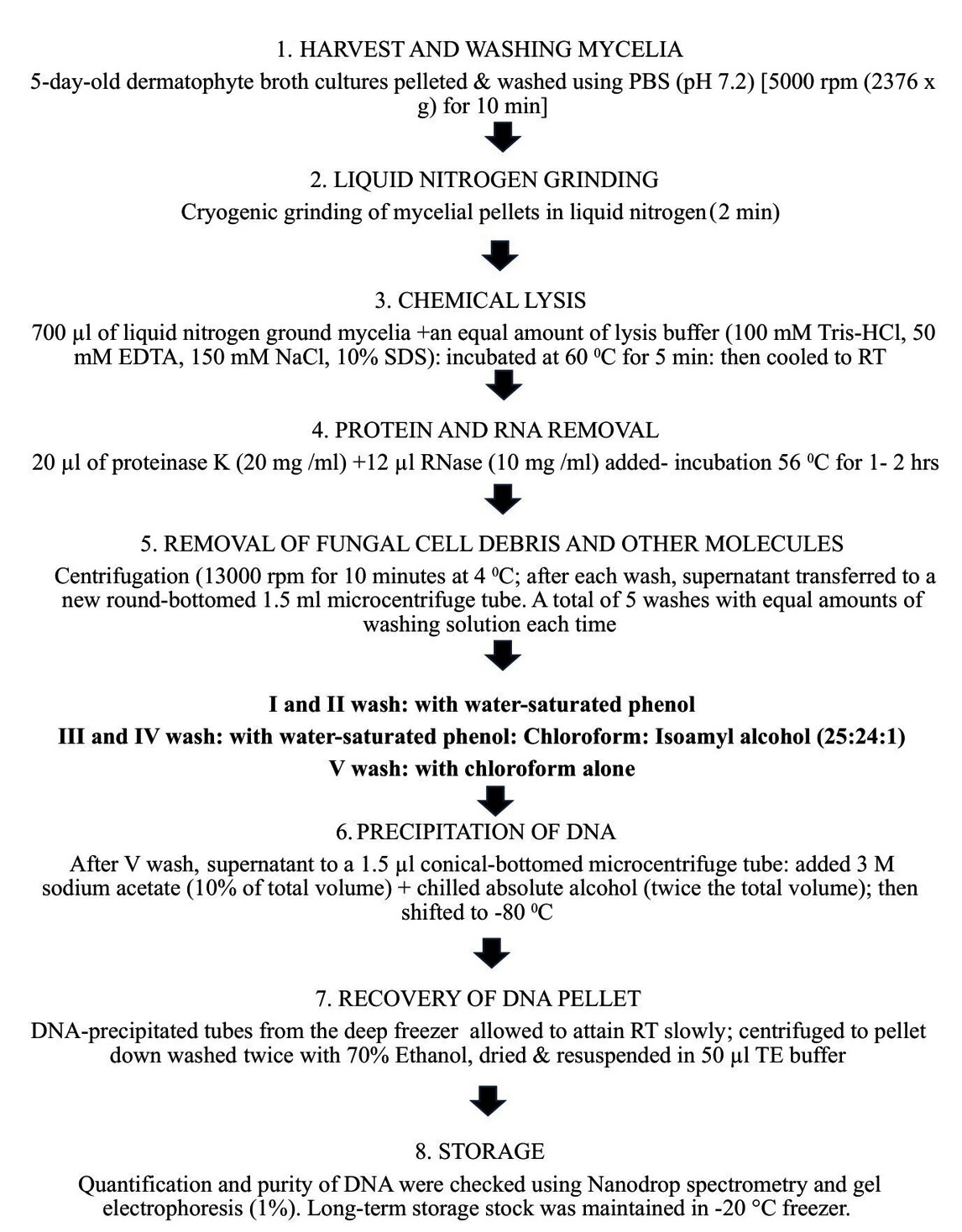
Figure 1. Sequential flow chart of dermatophyte DNA extraction and purification protocol
Table 1. Comparison of DNA yield and purity (A260/A280) across various culture media used (SDB = Sabouraud’s Dextrose Broth; SMB = Sabouraud’s Maltose Broth)
No. |
Isolate No. |
Sample details |
A260/A280 |
DNA Quantity (ng/µl) |
|---|---|---|---|---|
1 |
1028-1 |
SDB Plain |
1.7 |
659 |
2 |
1028-2 |
SMB Plain |
2.0 |
515 |
3 |
1028-3 |
SDB with Tween 80 |
1.8 |
1039 |
4 |
1028-4 |
SMB with Tween 80 |
2.01 |
803.6 |
5 |
1536-1 |
SDB Plain |
1.97 |
660 |
6 |
1536-2 |
SMB Plain |
2.0 |
576 |
7 |
1536-3 |
SDB with Tween 80 |
1.8 |
1130.2 |
8 |
1536-4 |
SMB with Tween 80 |
2.01 |
886.9 |
9 |
286-1 |
SDB Plain |
1.9 |
700 |
10 |
286-2 |
SMB Plain |
2.0 |
612 |
11 |
286-3 |
SDB with Tween 80 |
2.0 |
1231 |
12 |
286-4 |
SMB with Tween 80 |
1.7 |
964.8 |
13 |
976-1 |
SDB Plain |
2.01 |
780 |
14 |
976-2 |
SMB Plain |
2.0 |
663 |
15 |
976-3 |
SDB with Tween 80 |
1.92 |
1474 |
16 |
976-4 |
SMB with Tween 80 |
2.06 |
938.5 |
Table 2. Details of the dermatophyte isolates subjected to DNA extraction by the phenol-chloroform method
No. |
Species |
No. of isolates tested |
|---|---|---|
1 |
Trichophyton mentagrophytes |
10 |
2 |
Trichophyton rubrum |
4 |
3 |
Trichophyton indotineae |
4 |
4 |
Trichophyton tonsurans |
1 |
5 |
Microsporum canis |
1 |
Total |
20 |
Internal Transcribed Spacer (ITS) region-specific PCR amplification
Fungal genomic DNA was amplified from the internal transcribed spacer region of ribosomal DNA. The primers used were ITSF (GCATCGATGAAGAACGCAGC) and ITSR (TCCTCCGCTTATTGATATGC). The amplification conditions were as follows: initial denaturation at 94 °C for 3 min, followed by 30 cycles of denaturation at 94 °C for 1 min, annealing at 67 °C for 1 min, and extension at 72 °C for 3 min. The final extension step was performed at 72 °C for 10 min. The amplicons were subjected to 1% agarose gel electrophoresis and the resulting bands were visualized under UV illumination.17-19
Effect of culture media on fungal biomass production
The dry weight of mycelial growth showed that SDB with Tween 80 yielded the highest fungal biomass compared with all media types; the results were consistent with all four strains of Trichophyton mentagrophytes. The addition of Tween 80 increased fungal dispersion in the broth compared with that in plain media. The average fungal biomass obtained from SDB broth with Tween 80 was 412.5 mg/100 mL, which was higher than that obtained from plain SDB and SMB broths and SMB with Tween 80. However, SMB broth with Tween 80 yielded higher biomass (385.4 ± 21.1 mg/100 mL) than plain SMB (210.8 ± 15.4 mg/100 mL). Small clumps of mycelia were observed in both SDB and SMB plain broths, whereas uniform spreading of hyphal growth was observed in Tween 80 containing broths. The initial biomass from each broth was counted microscopically after diluting 100 µL of pellets from each tube; approximately 1-5 mycelia x 107 /mL were found in all pellet preparations, with slight differences. Fungal biomass grown from SDB with Tween 80 (0.1%) after extraction yielded a significantly higher quantity of DNA compared with the other culture media used (P < 0.01), as determined by two-way ANOVA (Table 1).
Quantitative and qualitative assessment of extracted genomic DNA
The modified phenol-chloroform method consistently yielded high concentrations of fungal DNA. The agarose gel runs were clean, indicating sufficient removal of complex fungal cell debris and RNA, along with chemicals used during the washing steps. Spectrophotometric readings of all 16 samples revealed an average yield of 852 ng/µL of genomic DNA. The A260/A280 ratios of all 16 samples ranged from 1.7- 2.0. The 1% agarose gel electrophoresis of 10 µL of sample yielded distinct, high-molecular-weight bands with no smearing, indicating that there was no contamination with chemicals or RNA, and the grinding in liquid nitrogen and vigorous chemical cleaning did not cause DNA shearing. The absence of a low-molecular-weight fluorescent smear at the bottom indicated the absence of RNA contamination. Thus, these results supported the purity of the DNA for downstream applications.
Results of PCR amplification
All samples were successfully amplified using ITS primers, yielding amplicons of approximately 432 bp. The bands obtained were sharp and distinct, without evidence of nonspecific amplification, supporting the absence of substantial PCR inhibition and the successful recovery of amplifiable DNA (Figure 2).
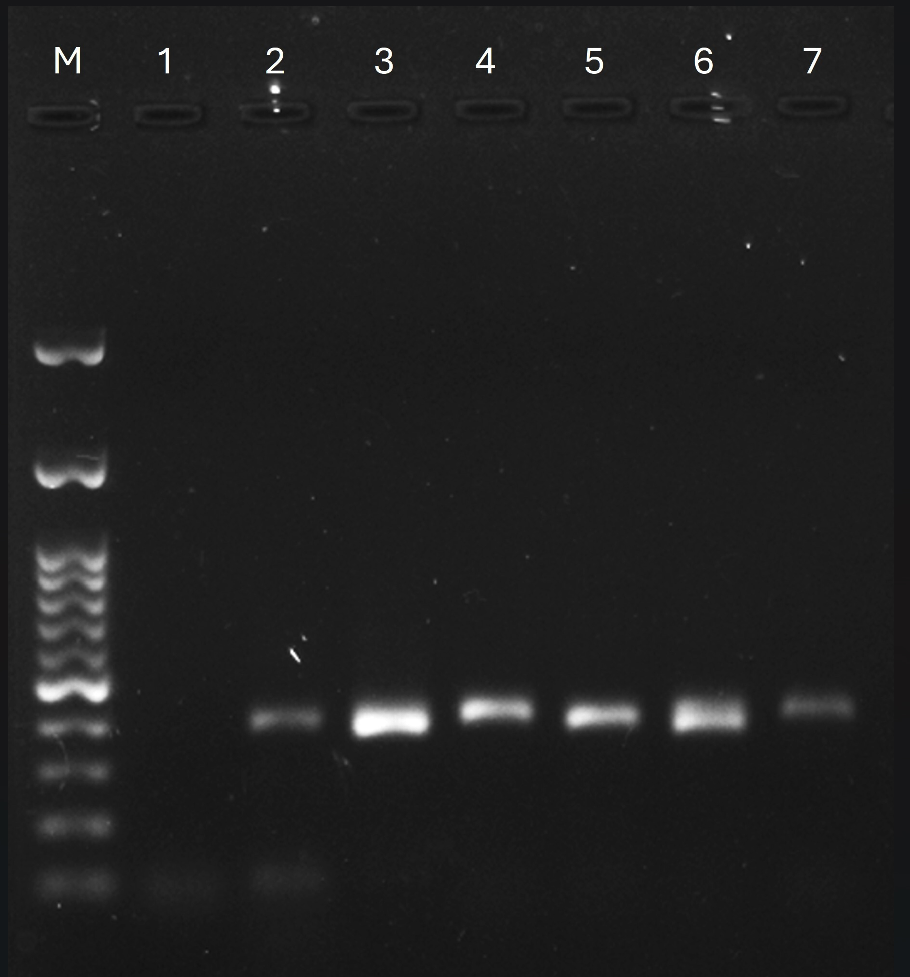
Figure 2. PCR products on 1% agarose gel; Lane M: 100 bp ladder, Lane 1: Negative control, Lane 2-7: Fungal PCR products obtained from the modified phenol-chloroform method
The structural complexity of the fungal cell wall and the coprecipitation of inhibitory secondary metabolites make the extraction of fungal DNA challenging. In this study, the growth conditions required for maximum fungal biomass production were analyzed by comparing two different carbohydrate-based media and the presence of a surfactant. During the extraction process, five washing steps were performed to maximize DNA purity and yield.
In the growth medium, the inclusion of 0.1% Tween 80 facilitated uniform dispersion of the fungal biomass. Therefore, the addition of Tween 80 resulted in a cleaner, more uniform, and higher biomass yield. These findings are consistent with those of previous studies.14,20 The mycelial ends, which are typically hydrophobic in Trichophyton species, receive more exposure to nutrients because of the addition of Tween 80. The addition of Tween 80 reduces the surface tension; therefore, surface pellicle formation is prevented, ensuring adequate exposure of the hyphae to nutrients and oxygen, which is also facilitated by shaking. Sabouraud dextrose broth with Tween 80 (0.1%) led to the highest yield, suggesting that fungi rely on glucose as a carbohydrate source, because it is a monosaccharide that can enter glycolysis directly. If the fungal biomass is dense and forms clumps in liquid culture, these pelleted clumps lead to anaerobic centers, which can cause the release of secondary metabolites that can coprecipitate with DNA. Tween 80, a nonionic surfactant, resulted in a finely dispersed mycelial suspension, thus maintaining homogeneous aerobic conditions within the broth and minimizing secondary metabolites.14 Therefore, the surface area available for liquid nitrogen grinding was also increased, and the grinding efficiency and lysis buffer penetration were also improved.
In the lysis buffer, the use of adequate proteinase K and RNase removed abundant fungal proteins and RNA. While standardizing the extraction procedure, we detected RNA smearing using agarose gel electrophoresis of genomic DNA. Optimizing the amount of RNA from 5 µL per tube in the initial trial to 12 µL led to clear RNA-free bands on agarose gel electrophoresis. However, the use of an excessive amount of RNase can cause shearing of genomic DNA.20,21
During the washing process, exactly five steps were used, which addressed the “polysaccharide paradox”. Two washes with water-saturated phenol facilitated denaturation of robust fungal proteins, followed by two washes with phenol:chloroform:isoamyl alcohol (25:24:1) facilitated the removal of complex polysaccharides from the interface. The final wash with pure chloroform facilitated phenol removal. Residual alcohol was removed from the final DNA preparation by pipetting with a 10 µL pipette and briefly drying at 37 °C. Traces of alcohol remaining in the final DNA preparation decreases the stability of polymerases during PCR.22 The A260/A280 ratio ranged from 1.7-2.0, consistent with relatively pure DNA. Thus, each step of the extraction process was systematically evaluated and optimized.
In the PCR, we used an annealing temperature of 67 °C. The stringent optimization of annealing temperature supported the absence of substantial PCR inhibition commonly present in fungal preparations, and the primers target a fungal-specific region. Achieving an optimal annealing temperature plays a key role in standardizing PCR.23
The DNA thus obtained using the modified method was suitable for downstream applications, including PCR amplification and sequencing. Based on the experimental data, SDB supplemented with 0.1% Tween 80 was identified as optimal for dermatophyte growth, with a combination of liquid nitrogen grinding and a five-step washing procedure incorporated in the modified phenol-chloroform extraction protocol, yielding high-quality genomic DNA.
The standardized “growth to amplification” framework provided a clean, cost-effective, and reproducible alternative to more costly commercial methods. The technical challenge in isolating high-quality genomic DNA from dermatophytes was addressed by integrating surfactant-enhanced biomass production and a five-step organic extraction procedure, forming a basis for a practical laboratory protocol for researchers who aim to obtain DNA of sufficient molecular weight that can meet the requirements of phylogenetic and advanced molecular biology applications.
ACKNOWLEDGMENTS
The authors acknowledge the Department of Microbiology, SDMCMSH, SDM Central Research Laboratory, SDM Research Institute for Biomedical Sciences and Centre for Cellular and Molecular Sciences; Shri Dharmasthala Manjunatheshwara University, Dharwad, for providing all the necessary facilities to carry out this research work.
CONFLICT OF INTEREST
The authors declare that there is no conflict of interest.
AUTHORS’ CONTRIBUTION
AJP and GSA conceived the idea and developed the work plan. AJP and PS performed the experimental work and contributed to data collection. AJP, GSA, PS, and OA verified the methods. OA further refined the work plan. KN provided the samples. AJP wrote the manuscript. All authors reviewed, revised, and approved the final manuscript for publication.
FUNDING
None.
DATA AVAILABILITY
All datasets generated or analyzed during this study are included in the manuscript.
ETHICS STATEMENT
This study was approved by the Institutional Ethics Committee, SDM College of Medical Sciences and Hospital, Karnataka, India, vide reference number SDMIEC:2023/542, dated 09/10/2023.
INFORMED CONSENT
Written informed consent was obtained from the participants before enrolling in the study.
- El Khatib A, Sabbagh D, Dimassi S, et al. Enhanced DNA extraction from dimorphic fungi: Exploring the advantages of blastospores versus mycelia. Biotechnol Appl Biochem. 2025;72(6):1788-1797.
Crossref - Kumar CM, Mugunthan M. Evaluation of three DNA extraction methods from fungal cultures. Med J Armed Forces India. 2018;74(4):333-336.
Crossref - Reyes Marquez FC, Auxier B, Briggeman B, Snelders E. High-Throughput culture and DNA isolation methods for Aspergillus fumigatus. Curr Protoc. 2025;5(4):e70112.
Crossref - Ova AO, Joffre E, Shafagh RZ, et al. Improved isolation of ultra-high-molecular-weight genomic DNA suitable for third-generation sequencing. Microorganisms. 2025;13(3):534.
Crossref - Zhang Y, Song Z, Schilling JS. Evaluation of DNA extraction methods for microbial community profiling in deadwood decomposition. Microbiologyopen. 2024;13(6):e70007.
Crossref - Langsiri N, Meyer W, Irinyi L, et al. Optimizing fungal DNA extraction and purification for Oxford Nanopore untargeted shotgun metagenomic sequencing from simulated hemoculture specimens. mSystems. 2025;10(6):e01166-24.
Crossref - Uakhit R, Smagulova A, Kukhar E, Bailina G. Development of DNA isolation methods to study dermatophytosis. Eurasian J Appl Biotechnol. 2025;(4):112-117.
Crossref - Rana AK. Challenging biological samples and strategies for DNA extraction. J Investig Med. 2025;73(6):443-459.
Crossref - Abid C, Zouari-Mechichi H, Benmarzoug R, Mechichi T, Kharrat N. Impact of DNA Extraction strategies on genomic and bioinformatic outcomes in eight selected fungal strains. J Fungi. 2026;12(5):299.
Crossref - Kenjar AR, Mohan Raj JR, Girisha BS, Karunasagar I. Diagnostic ability of Peptidase S8 gene in the Arthrodermataceae causing dermatophytoses: A metadata analysis. PLoS One. 2024;19(7):e0306829.
Crossref - Arantes BBA, Cabral AKLF, Dos Santos KS, et al. Characterization of biofilm formation by the dermatophyte Nannizzia gypsea. J Fungi. 2025;11(6):455.
Crossref - Alehashemi R, Arghavan B, Abastabar M, Niknejad F, Aghili SR. Molecular epidemiology of dermatophytosis in Golestan, Iran: A cross-sectional study. Microb Pathog. 2025;199:107223.
Crossref - Folorunso TR, Silva G, Girón ME, et al. Optimized protocol for culturing and extracting DNA from fungal isolates associated with brown spot needle blight in pine trees. PLoS One. 2025;20(11):e0337218.
Crossref - Li F, Fan H, Sun Q, Di Y, Xia H. Effects of medium additives on the mycelial growth and polysaccharide biosynthesis in submerged culture of Bjerkandera fumosa. Molecules. 2024;29(2):422.
Crossref - Zwinkels J, van Oorschot S, van Mastrigt O, Smid EJ. The potential of mycelium from mushroom-producing fungi in alternative protein production: a focus on fungal growth, metabolism, and nutrition. Curr Res Food Sci. 2025;12:101278.
Crossref - Gnat S, Nowakiewicz A, Ziolkowska G, Troscianczyk A, Majer Dziedzic B, Zieba P. Evaluation of growth conditions and DNA extraction techniques used in the molecular analysis of dermatophytes. J Appl Microbiol. 2017;122(5):1368-1379.
Crossref - Kenjar AR, Girisha BS, Jayaraman J, et al. Resistance & virulence traits in dermatophytes isolated from Mangaluru, India. Indian J Med Res. 2025;162(1):95-104.
Crossref - McTaggart LR, Cronin K, Ruscica S, Patel SN, Kus JV. Emergence of terbinafine-resistant Trichophyton indotineae in Ontario, Canada, 2014–2023. J Clin Microbiol. 2025;63(1):e01535-24.
Crossref - De Hoog GS, Dukik K, Monod M, et al. Toward a novel multilocus phylogenetic taxonomy for the dermatophytes. Mycopathologia. 2017;182(1):5-31.
Crossref - Liu YS, Wu JY. Effects of Tween 80 and pH on mycelial pellets and exopolysaccharide production in liquid culture of a medicinal fungus. J Ind Microbiol Biotechnol. 2012;39(4):623-628.
Crossref - El-Ashram S, Al Nasr I, Suo X. Nucleic acid protocols: Extraction and optimisation. Biotechnol Rep (Amst). 2016;12:33-39.
Crossref - Schrader C, Schielke A, Ellerbroek L, Johne R. PCR inhibitors – occurrence, properties and removal. J Appl Microbiol.2012;113(5):1014-1026.
Crossref - McDonald C, Taylor D, Linacre A. PCR in Forensic Science: A Critical Review. Genes. 2024;15(4):438.
Crossref
© The Author(s) 2026. Open Access. This article is distributed under the terms of the Creative Commons Attribution 4.0 International License which permits unrestricted use, sharing, distribution, and reproduction in any medium, provided you give appropriate credit to the original author(s) and the source, provide a link to the Creative Commons license, and indicate if changes were made.