


ISSN: 0973-7510
E-ISSN: 2581-690X
 , Wuttipong Phumrattanaprapin2,3
 and Akarapon Chantongsri4
Bacterial infections in companion animals involve multiple body systems and are commonly encountered in veterinary practice. This retrospective study aimed to describe bacterial species recovered from canine and feline clinical samples submitted to a private veterinary diagnostic laboratory in Thailand and to evaluate their antimicrobial susceptibility and multidrug-resistance profiles. A total of 212 clinical records from 2023-2025 were reviewed. After exclusion of incomplete records, duplicate entries, culture-negative cases, and records lacking antimicrobial susceptibility testing results, 131 bacterial isolates were included in the final analysis, comprising 44 isolates from dogs and 87 from cats. Sample types included wound and abscess swabs, nasal discharge, urine, pleural fluid, abdominal fluid, ear swabs, blood, feces, and prostate gland abscesses. Wound and abscess samples were the most common submissions (49.62%). The most frequently isolated bacteria were Escherichia coli (21/131, 16.03%), Pseudomonas aeruginosa (18/131, 13.74%), Klebsiella pneumoniae/Klebsiella spp. (17/131, 12.98%), Staphylococcus pseudintermedius (10/131, 7.63%), and Staphylococcus aureus (10/131, 7.63%). Antimicrobial susceptibility varies across bacterial species and drug classes. Among the isolates tested, aminoglycosides and meropenem showed comparatively favorable in vitro activity, whereas lower susceptibility was observed for cephalexin, doxycycline, and several β-lactam agents. Multidrug-resistance was most prominent among Gram-negative isolates, particularly Enterobacter cloacae, Pseudomonas aeruginosa, Acinetobacter spp., Klebsiella pneumoniae, and Escherichia coli. These findings provide laboratory-based data from a veterinary diagnostic setting in Thailand and support the value of culture-based antimicrobial selection and ongoing local antimicrobial resistance surveillance.
Antimicrobial Susceptibility Testing, Antimicrobial Resistance, Canine, Feline, Bacterial Isolates, Multidrug Resistance, Retrospective Analysis
Antimicrobial resistance (AMR) remains one of the most pressing global health challenges, threatening the effective treatment of infectious diseases across human and veterinary medicine.1 The acceleration of AMR is primarily driven by the widespread and often inappropriate use of antimicrobial agents, which promotes the selection and persistence of resistant bacterial populations.2 Within the One Health framework, companion animals are increasingly recognized as important reservoirs of antimicrobial-resistant bacteria, given their close and frequent interactions with humans and shared environments.3,4
In small animal clinical practice, dogs and cats frequently present with bacterial infections such as skin infections, urinary tract infections, otitis externa, and postoperative wound infections, which often require antimicrobial treatment.5 Repeated empirical therapy, particularly in the absence of culture and susceptibility testing, contributes significantly to the emergence of resistant strains. Of particular concern is multidrug-resistance (MDR), defined as resistance to at least one agent in three or more antimicrobial classes.6 Recent studies have reported increasing trends of MDR among common veterinary pathogens, including Escherichia coli, Staphylococcus pseudintermedius, and Klebsiella pneumoniae, with significant implications for treatment outcomes and infection control.7-11
Despite growing global awareness, surveillance data on AMR in companion animals remain limited and geographically imbalanced. Most large-scale studies originate from Europe and North America, whereas data from Southeast Asia are still relatively scarce and often lack longitudinal analysis.3,12 Furthermore, many existing studies focus on specific pathogens or infection types rather than providing a comprehensive evaluation of MDR patterns across multiple clinical sample types.
This highlights a critical knowledge gap, as the absence of region-specific, longitudinal data on MDR prevalence restricts the ability of veterinarians to make informed empirical treatment decisions and limits the development of effective antimicrobial stewardship strategies. Additionally, the lack of updated local resistance profiles hinders early detection of emerging MDR threats within companion animal populations.
Therefore, this study aimed to investigate the prevalence and distribution of multidrug-resistant bacterial isolates from canine and feline clinical samples over a three-year period. By evaluating antimicrobial susceptibility patterns and temporal trends, this study seeks to generate clinically relevant evidence to support rational antimicrobial use and strengthen antimicrobial stewardship in veterinary practice, contributing to the global One Health effort to mitigate AMR.
Study setting and design
This retrospective study reviewed medical and laboratory records of canine and feline patients submitted to a private veterinary diagnostic laboratory receiving samples from companion animal clinics in Thailand between 1 January 2023 and 31 December 2025. A total of 212 clinical records were initially retrieved from the laboratory database. After exclusion of duplicate entries, incomplete records, negative bacterial cultures, and cases without antimicrobial susceptibility testing results, 131 bacterial isolates met the inclusion criteria and were included in the final analysis. Of the 131 isolates included in the final analysis, 44 were obtained from dogs and 87 were obtained from cats.
Animals were eligible for inclusion if they met all of the following criteria:
(a) Species: dog or cat;
(b) Availability of clinical samples, including wound and abscess swabs, urine, nasal discharge, pleural or abdominal fluid, ear swabs, blood, feces, prostate gland abscesses, or ocular wound samples collected during the study period; and
(c) Availability of both bacterial culture identification and antimicrobial susceptibility testing results performed directly from the submitted clinical sample.
Data collection
Clinical information extracted from the laboratory database included animal species, sample type, bacterial species identified, and antimicrobial susceptibility testing results. Sample types included wound and abscess swabs, urine, nasal discharge, pleural and abdominal fluids, ear swabs, blood, feces, prostate gland abscesses, and ocular wound samples.
For each isolate, antimicrobial susceptibility testing results were recorded for all available antimicrobial agents tested. Only records with complete bacterial identification, at least one antimicrobial susceptibility result, and no duplicate or negative culture findings were included in the analysis.
Multidrug-resistance (MDR) was defined as acquired non-susceptibility to at least one antimicrobial agent in three or more antimicrobial classes. Possible extensively drug-resistant (XDR) isolates were interpreted according to the criteria proposed by Magiorakos et al.,6 in which non-susceptibility must be demonstrated in all but two or fewer antimicrobial classes. Because antimicrobial testing panels varied among isolates in this retrospective dataset, strict XDR classification could not be uniformly confirmed for all isolates. Therefore, isolates showing this pattern were reported as possible XDR and interpreted with caution. Antimicrobial class categorization was based on the World Organisation for Animal Health (WOAH) list of antimicrobial agents of veterinary importance, the World Small Animal Veterinary Association (WSAVA), Essential Medicines List, and the World Health Organization (WHO), Access, Watch, and Reserve (AWaRe) classification framework. To ensure data quality, 10% of the dataset was independently re-entered and cross-checked for accuracy and validity.
Data analysis
The data obtained from the laboratory database were manually entered into a Microsoft Excel spreadsheet (Microsoft Corporation, Redmond, WA, USA). The variables recorded included the microbiology report date, host species, sex, age, isolated pathogens, and antimicrobial susceptibility profiles, and minimum inhibitory concentrations (MICs-µg/mL) when available. Case identifiers were anonymized using a four-digit coding system, in which the first digit represented the clinic and the remaining three digits represented the individual patient identification number. All included variables were summarized as frequencies and percentages of total positive isolates. Descriptive statistics were used to summarize categorical variables as frequencies and percentages. The distribution of bacterial isolates according to animal species and specimen type was reported. Antimicrobial susceptibility profiles were summarized as the proportions of susceptible, intermediate, and resistant isolates for each bacterial species.
Antimicrobial susceptibility testing
Bacterial species identification was performed using colony morphology, Gram staining, and standard biochemical methods routinely used in the diagnostic laboratory. Antimicrobial susceptibility testing (AST) was conducted using the Kirby-Bauer disk diffusion method according to CLSI VET01-A5 and CLSI M100 guidelines where applicable. The antibiotics tested included amikacin (AK-30), amoxicillin (AML-10), amoxicillin-clavulanic acid (AMC-30), ampicillin (AMP-10), azithromycin (AZM-15), bacitracin (B-10), cefaclor (CEC-30), cefixime (CFM-5), cefovecin (CVN-30), cefoxitin (FOX-30), ceftazidime (CAZ-3), ceftiofur (EFT-30), ceftriaxone (CRO-30), cephalexin (CL-30), cephazolin (KZ-30), chloramphenicol (C-30), ciprofloxacin (CIP-5), clindamycin (DA-2), doxycycline (DO-30), enrofloxacin (ENR-5), erythromycin (E-15), gentamicin (CN-10), imipenem (IPM-10), kanamycin (K-30), marbofloxacin (MAR-5), meropenem (MEM-10), moxifloxacin (MXF-5), mupirocin (MUP-5), norfloxacin (NOR-10), oxacillin (OX-1), oxytetracycline (OT-30), penicillin G (P-10), pradofloxacin (PRA-5), sulbactam/ampicillin (SAM-20), spectinomycin (SH-10), sulbactam/cefoperazone (SCF-105), trimethoprim-sulfamethoxazole (SXT-25), and tetracycline (TE-30). Susceptibility results were interpreted as susceptible (S), intermediate (I), or resistant (R) according to CLSI criteria. Negative bacterial culture results were excluded from the analysis.
Characteristics of clinical samples
A total of 212 clinical records were retrieved from the laboratory database. After excluding incomplete records, duplicate entries, samples without bacterial identification, culture-negative cases, and records lacking antimicrobial susceptibility testing results, 131 bacterial isolates were eligible for inclusion in the final analysis. Of these, 44 isolates were obtained from dogs and 87 from cats.
The distribution of bacteriological samples by animal species and specimen type is presented in Table 1. Overall, wound and abscess samples were the most frequently submitted specimens, accounting for 65 of 131 samples (49.62%), followed by nasal discharge samples (22/131, 16.79%) and urine (voided/unspecified) samples (17/131, 12.98%). Urine collected by sterile methods accounted for 8 of 131 samples (6.11%).
Table 1. Distribution of bacteriological samples collected from canine and feline patients according to specimen type
Sample Type |
Dog, n (%) |
Cat, n (%) |
Total, n (%) |
|---|---|---|---|
Wound/Abscess |
11 (25.00) |
54 (62.07) |
65 (49.62) |
Nasal discharge |
16 (36.36) |
6 (6.90) |
22 (16.79) |
Urine (Voided/Unspecified)* |
6 (13.64) |
11 (12.64) |
17 (12.98) |
Urine (Sterile collection)** |
1 (2.27) |
7 (8.05) |
8 (6.11) |
Pleural Fluid |
6 (13.64) |
0 (0.00) |
6 (4.58) |
Ear swab |
1 (2.27) |
3 (3.45) |
4 (3.05) |
Feces |
1 (2.27) |
2 (2.30) |
3 (2.29) |
Abdominal Fluid |
0 (0.00) |
3 (3.45) |
3 (2.29) |
Blood |
1 (2.27) |
1 (1.15) |
2 (1.53) |
Prostate gland abscess |
1 (2.27) |
0 (0.00) |
1 (0.76) |
Total |
44 (100.00) |
87 (100.00) |
131 (100.00) |
*Urine (Voided/Unspecified) included voided urine and urine samples without a specified collection method
**Urine (Sterile collection) included catheterized urine and cystocentesis samples
When stratified by species, nasal discharge was the most common specimen type in dogs, representing 16 of 44 canine samples (36.36%), followed by wound and abscess samples (11/44, 25.00%). Urine (voided/unspecified) and pleural fluid each accounted for 6 of 44 canine samples (13.64%). In contrast, wound and abscess samples predominated in cats, accounting for 54 of 87 feline samples (62.07%), followed by urine (voided/unspecified) samples (11/87, 12.64%), urine collected by sterile methods (7/87, 8.05%), and nasal discharge (6/87, 6.90%).
Less frequently submitted specimens included ear swabs (4/131, 3.05%), feces (3/131, 2.29%), abdominal fluid (3/131, 2.29%), blood (2/131, 1.53%), and prostate gland abscesses (1/131, 0.76%). Pleural fluid was submitted only from dogs, whereas abdominal fluid was identified only among cats.
Distribution of bacterial isolates by animal species and specimen type
The distribution of bacterial isolates according to animal species is presented in Table 2. Overall, Gram-negative bacteria predominated in both dogs and cats. In canine samples, Klebsiella pneumoniae/Klebsiella spp. was the most frequently isolated organism (9/44, 20.45%), followed by Pseudomonas aeruginosa (6/44, 13.64%) and Escherichia coli (5/44, 11.36%). In feline samples, Escherichia coli was the most common isolate (16/87, 18.39%), followed by Pseudomonas aeruginosa (12/87, 13.79%), Klebsiella pneumoniae/Klebsiella spp. (8/87, 9.20%), and Staphylococcus pseudintermedius (7/87, 8.05%). Acinetobacter spp., Enterococcus faecalis, and Enterococcus spp. were recovered only from cats, whereas no isolation of Acinetobacter spp. was identified in dogs.
Table 2. Distribution of bacterial isolates recovered from canine and feline clinical samples
Bacterial Isolate |
Canine isolates, n (%) |
Feline isolates, n (%) |
Total isolates, n (%) |
|---|---|---|---|
Escherichia coli |
5 (11.36) |
16 (18.39) |
21 (16.03) |
Pseudomonas aeruginosa |
6 (13.64) |
12 (13.79) |
18 (13.74) |
Klebsiella pneumoniae/Klebsiella spp. |
9 (20.45) |
8 (9.20) |
17 (12.98) |
Staphylococcus pseudintermedius |
3 (6.82) |
7 (8.05) |
10 (7.63) |
Staphylococcus aureus |
4 (9.09) |
6 (6.90) |
10 (7.63) |
Staphylococcus intermedius group |
4 (9.09) |
3 (3.45) |
7 (5.34) |
Streptococcus spp.* |
1 (2.27) |
6 (6.90) |
7 (5.34) |
Pseudomonas spp. |
3 (6.82) |
3 (3.45) |
6 (4.58) |
Enterobacter cloacae |
1 (2.27) |
4 (4.60) |
5 (3.82) |
Acinetobacter spp. |
0 (0.00) |
5 (5.75) |
5 (3.82) |
Coagulase-negative Staphylococcus spp. |
1 (2.27) |
3 (3.45) |
4 (3.05) |
Pasteurella multocida |
2 (4.55) |
2 (2.30) |
4 (3.05) |
Proteus mirabilis |
1 (2.27) |
2 (2.30) |
3 (2.29) |
Kocuria spp. |
1 (2.27) |
1 (1.15) |
2 (1.53) |
Pasteurella spp. |
1 (2.27) |
1 (1.15) |
2 (1.53) |
Enterobacter spp. |
1 (2.27) |
1 (1.15) |
2 (1.53) |
Enterococcus faecalis |
0 (0.00) |
2 (2.30) |
2 (1.53) |
Enterococcus spp. |
0 (0.00) |
2 (2.30) |
2 (1.53) |
Others** |
1 (2.27) |
3 (3.45) |
4 (3.05) |
Total isolates (3 years) |
44 (100.00) |
87 (100.00) |
131 (100.00) |
*α-Hemolytic Streptococcus spp. were grouped under Streptococcus spp. for analysis
**Others included Stenotrophomonas spp. (dog, n = 1), Staphylococcus schleiferi (cat, n = 1), Staphylococcus felis (cat, n = 1), and Staphylococcus epidermidis (cat, n = 1)
When examined by specimen type, wound and abscess samples yielded the greatest number and diversity of bacterial isolates, as shown in Figure 1. The most frequently recovered organisms from wound/abscess samples were Staphylococcus pseudintermedius (n = 9), Pseudomonas aeruginosa (n = 8), Staphylococcus aureus (n = 7), Escherichia coli (n = 7), and Streptococcus spp. (n = 6). Klebsiella pneumoniae/Klebsiella spp. was most commonly identified in nasal discharge samples (n = 7), followed by wound/abscess samples (n = 5). Escherichia coli was recovered mainly from urine collected by sterile methods (n = 8) and wound/abscess samples (n = 7), whereas Pseudomonas aeruginosa was most often isolated from wound/abscess (n = 8), nasal discharge (n = 6), and urine (voided/unspecified) samples (n = 4).
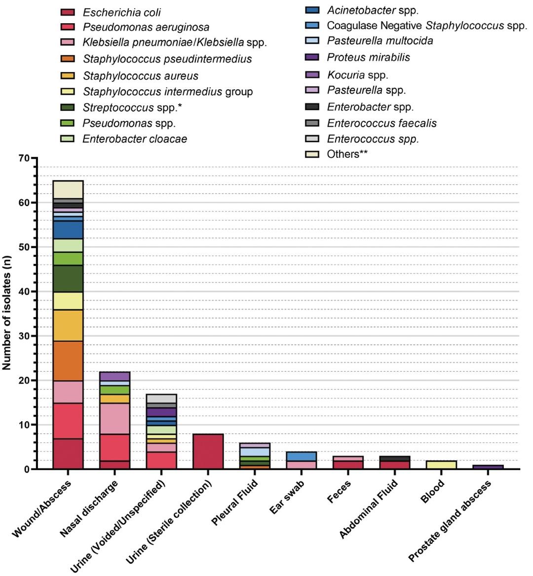
Figure 1. Distribution of the bacterial isolates according to specimen type
*α-Hemolytic Streptococcus spp. were grouped under Streptococcus spp. for analysis.
**Others included Stenotrophomonas spp. (dog, n = 1), Staphylococcus schleiferi (cat, n = 1), Staphylococcus felis (cat, n = 1), and Staphylococcus epidermidis (cat, n = 1)
Among other specimen types, pleural fluid was mainly associated with Pasteurella multocida (n = 2), whereas ear swab samples yielded Klebsiella pneumoniae/Klebsiella spp. and coagulase-negative staphylococci (n = 2 each). Blood samples were associated only with isolates belonging to the Staphylococcus intermedius group (n = 2). Fecal samples yielded Escherichia coli (n = 2) and Klebsiella pneumoniae/Klebsiella spp. (n = 1), while prostate gland abscess samples yielded Proteus mirabilis (n = 1). Overall, these findings indicate that wound and abscess specimens constituted the principal source of bacterial recovery and harbored the broadest range of bacterial species in this study.
Antimicrobial susceptibility patterns by bacterial species
Antimicrobial susceptibility profiles of the major bacterial isolates are illustrated in Figure 2. Detailed results are provided in Supplementary Table 1. Overall, susceptibility patterns varied across bacterial species and antimicrobial classes. Among Gram-negative isolates, Escherichia coli showed relatively high susceptibility to amikacin, gentamicin, ceftriaxone, and meropenem, whereas low susceptibility was observed for amoxicillin, ampicillin, and oxytetracycline. Pseudomonas aeruginosa remained highly susceptible to amikacin, gentamicin, meropenem, norfloxacin, and marbofloxacin, but showed low or absent susceptibility to amoxicillin, cefaclor, ceftiofur, ceftriaxone, cephalexin, imipenem, and kanamycin. Klebsiella pneumoniae/Klebsiella spp. demonstrated complete or high susceptibility to cefoxitin, meropenem, and sulbactam/cefoperazone, whereas susceptibility to amoxicillin, cefixime, and cefovecin was low.
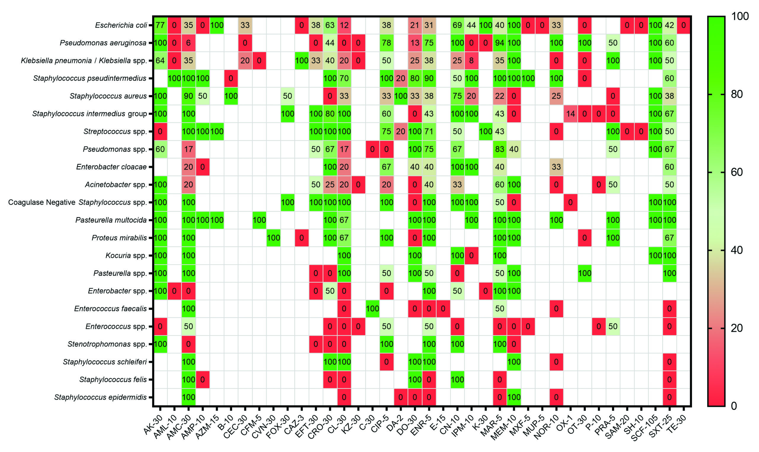
Figure 2. Heatmap showing antimicrobial susceptibility patterns among the major bacterial isolates. The heatmap illustrates antimicrobial susceptibility profiles of the predominant bacterial isolates recovered from canine and feline clinical samples. Green indicates susceptible isolates, whereas red indicates resistant isolates
Abbreviations: amikacin (AK-30), amoxicillin (AML-10), amoxicillin-clavulanic acid (AMC-30), ampicillin (AMP-10), azithromycin (AZM-15), bacitracin (B-10), cefaclor (CEC-30), cefixime (CFM-5), cefovecin (CVN-30), cefoxitin (FOX-30), ceftazidime (CAZ-30), ceftiofur (EFT-30), ceftriaxone (CRO-30), cephalexin (CL-30), cefazolin (KZ-30), chloramphenicol (C-30), ciprofloxacin (CIP-5), clindamycin (DA-2), doxycycline (DO-30), enrofloxacin (ENR-5), erythromycin (E-15), gentamicin (CN-10), imipenem (IPM-10), kanamycin (K-30), marbofloxacin (MAR-5), meropenem (MEM-10), moxifloxacin (MXF-5), mupirocin (MUP-5), norfloxacin (NOR-10), oxacillin (OX-1), oxytetracycline (OT-30), penicillin G (P-10), pradofloxacin (PRA-5), sulbactam/ampicillin (SAM-20), spectinomycin (SH-10), sulbactam/cefoperazone (SCF-105), trimethoprim-sulfamethoxazole (SXT-25), and tetracycline (TE-30)
Among Gram-positive isolates, Staphylococcus pseudintermedius showed high susceptibility to amoxicillin, amoxicillin-clavulanate, ampicillin, imipenem, marbofloxacin, meropenem, and mupirocin, whereas no susceptibility was observed for bacitracin and oxytetracycline. Staphylococcus aureus showed high susceptibility to amikacin, bacitracin, clindamycin, and sulbactam/cefoperazone, but low susceptibility to ceftriaxone, meropenem, and pradofloxacin. The Staphylococcus intermedius group retained high susceptibility to amikacin, amoxicillin-clavulanate, cefoxitin, ceftiofur, ceftriaxone, cephalexin, gentamicin, and imipenem, while showing poor susceptibility to meropenem, oxytetracycline, penicillin G and pradofloxacin. Streptococcus spp. showed high susceptibility to amoxicillin-clavulanate, ampicillin, azithromycin, ceftiofur, ceftriaxone, cephalexin, doxycycline, kanamycin, and sulbactam/cefoperazone, whereas susceptibility to amikacin, norfloxacin, spectinomycin and sulbactam/ampicillin was not observed.
Taken together, these findings indicate that susceptibility profiles differed substantially by bacterial species, with several Gram-negative isolates showing reduced susceptibility to commonly used first-line agents, whereas selected aminoglycosides, carbapenems, and sulbactam/cefoperazone retained favorable in vitro activity against many of the major pathogens.
Overall Susceptibility Profiles of Commonly Tested Antimicrobial Agents
The overall susceptibility profiles of the commonly tested antimicrobial agents are shown in Figure 3. Among the evaluated agents, amikacin showed one of the most favorable overall profiles, with 66 susceptible and 13 resistant isolates. Gentamicin also demonstrated substantial activity, with 51 susceptible and 23 resistant isolates. Enrofloxacin and marbofloxacin showed high numbers of susceptible isolates (61 and 70, respectively), although both agents were also associated with considerable resistant counts (38 and 48, respectively), indicating variable activity across the tested bacterial population.
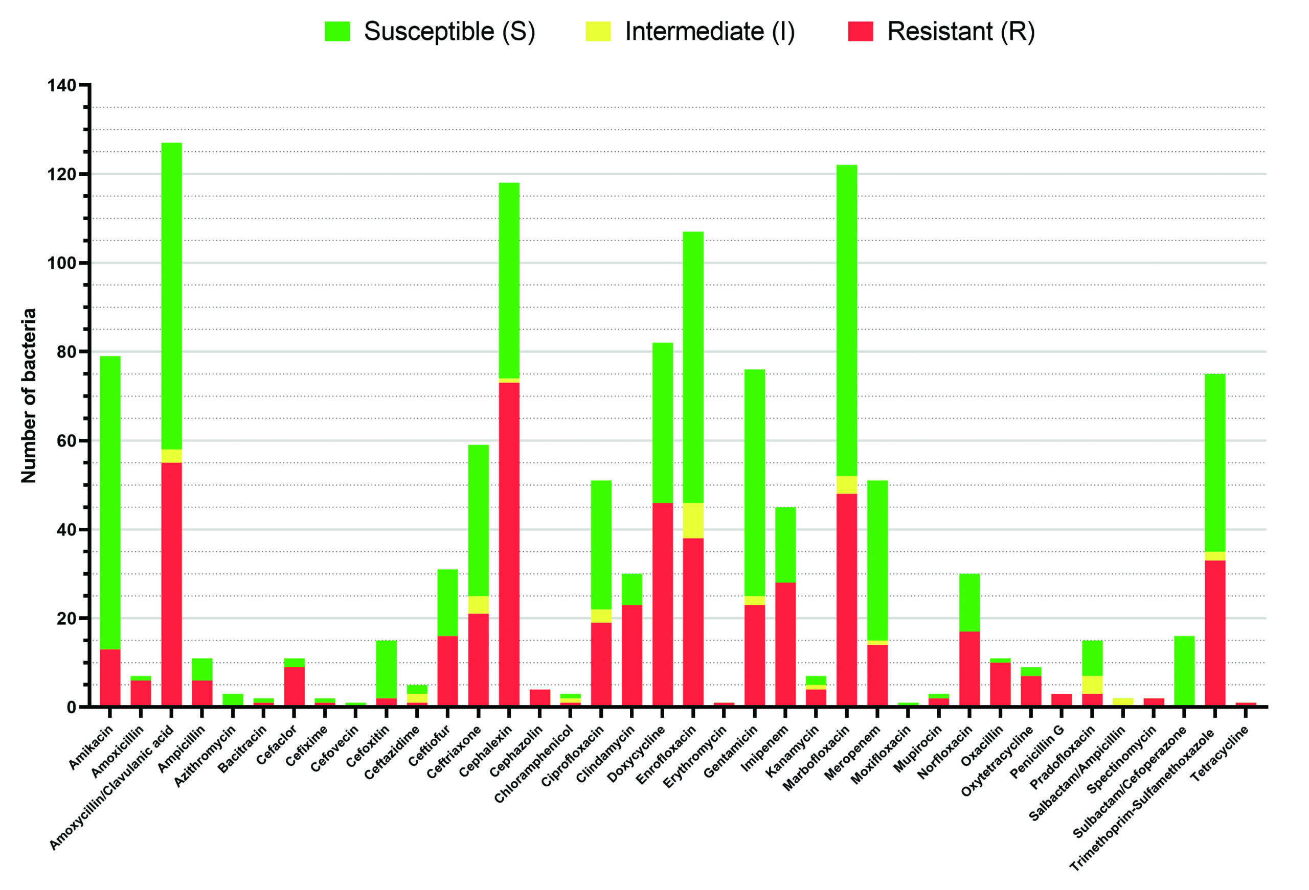
Figure 3. Stacked bar chart of susceptibility profiles of bacterial isolates from canine and feline samples according to CLSI interpretation categories. Each column represents a single antimicrobial agent, with stacked bars showing the proportion of isolates classified as susceptible (S, green), intermediate (I, yellow), or resistant (R, red). The graph provides a visual summary of resistance patterns across all tested antibiotics. The susceptibility testing was interpreted using the Kirby–Bauer disk diffusion method according to CLSI guidelines specific to companion animals and EUCAST guidelines for reference data not available in veterinary medicine
Meropenem showed a relatively favorable overall profile, with 36 susceptible and 14 resistant isolates, whereas trimethoprim-sulfamethoxazole showed 40 susceptible and 33 resistant isolates. Amoxicillin-clavulanate also yielded a high number of susceptible isolates (69), but this was accompanied by a substantial number of resistant isolates (55), suggesting heterogeneous susceptibility across bacterial species. Doxycycline similarly showed mixed results, with 36 susceptible and 46 resistant isolates.
By contrast, cephalexin showed the poorest overall profiles, with resistant counts of 73, exceeding the corresponding susceptible counts. Intermediate interpretations were generally uncommon across most antimicrobial agents, although enrofloxacin (I = 8), ceftriaxone (I = 4), pradofloxacin (I = 4), marbofloxacin (I = 4), amoxicillin-clavulanate (I = 3), and ciprofloxacin (I = 3) showed small but notable intermediate proportions.
Overall, the results of all susceptibility profiles indicate that although several antimicrobial agents retained considerable in vitro activity, resistance to a number of commonly used agents remained frequent, particularly for cephalexin, doxycycline, and amoxicillin-clavulanate.
Multidrug-resistant and Possible Extensively Drug-Resistant Profiles
Multidrug-resistant (MDR) and possible extensively drug-resistant (XDR) profiles of the major bacterial isolates are summarized in Table 3. Overall, the burden of resistance was concentrated predominantly among Gram-negative organisms. The highest proportion of MDR isolates was observed in Enterobacter cloacae (4/5, 80.0%), followed by Pseudomonas aeruginosa (14/18, 77.8%), Acinetobacter spp. (3/5, 60.0%), Klebsiella pneumoniae (11/17, 64.7%), and Escherichia coli (12/21, 57.1%). These findings indicate that multidrug-resistance was especially prominent among the major Gram-negative pathogens recovered in this study.
Table 3. Multidrug-resistant (MDR) and possible extensively drug-resistant (XDR) profiles among major bacterial isolates recovered from canine and feline clinical samples
Bacterial species |
No. of isolates (n) |
MDR, n (%) |
XDR, n (%) |
Predominant resistant antimicrobial classes* |
|---|---|---|---|---|
Escherichia coli |
21 |
12 (57.1) |
2 (9.5) |
Penicillin, tetracyclines, folate pathway inhibitors, fluoroquinolones |
Pseudomonas aeruginosa |
18 |
14 (77.8) |
4 (22.2) |
Penicillin, cephalosporins, tetracyclines, folate pathway inhibitors, fluoroquinolones |
Klebsiella pneumoniae |
17 |
11 (64.7) |
3 (17.6) |
Penicillin, cephalosporins, tetracyclines, folate pathway inhibitors |
Staphylococcus pseudintermedius |
10 |
5 (50.0) |
0 (0.0) |
Penicillin, tetracyclines, fluoroquinolones |
Staphylococcus aureus |
10 |
5 (50.0) |
1 (10.0) |
Penicillin, tetracyclines, fluoroquinolones |
Staphylococcus intermedius group |
6 |
3 (50.0) |
0 (0.0) |
Penicillin, tetracyclines, fluoroquinolones |
Enterobacter cloacae |
5 |
4 (80.0) |
2 (40.0) |
Penicillin, cephalosporins, tetracyclines, folate pathway inhibitors |
Acinetobacter spp. |
5 |
3 (60.0) |
1 (20.0) |
Penicillin, cephalosporins, tetracyclines, folate pathway inhibitors |
Proteus mirabilis |
3 |
1 (33.3) |
0 (0.0) |
Penicillin, tetracyclines, folate pathway inhibitors |
Enterococcus spp. |
3 |
2 (66.7) |
0 (0.0) |
Penicillin, tetracyclines, fluoroquinolones |
*Predominant resistant antimicrobial classes were assigned according to the main drug classes represented in the antimicrobial susceptibility testing panel. MDR was defined as acquired non-susceptibility to at least one antimicrobial agent in three or more antimicrobial classes. XDR was defined as non-susceptibility to at least one antimicrobial agent in all but two or fewer antimicrobial classes
Possible XDR phenotypes were also identified mainly among Gram-negative isolates. Specifically, possible XDR profiles were observed in Enterobacter cloacae (2/5, 40.0%), Acinetobacter spp. (1/5, 20.0%), Pseudomonas aeruginosa (4/18, 22.2%), and Klebsiella pneumoniae (3/17, 17.6%). Because antimicrobial testing panels varied among isolates, these possible XDR profiles should be interpreted with caution and should not be regarded as definitive XDR classifications.
Among Gram-positive organisms, MDR was also evident but at a lower overall burden than that observed among Gram-negative bacteria. MDR was identified in 5 of 10 Staphylococcus pseudintermedius isolates (50.0%), 5 of 10 Staphylococcus aureus isolates (50.0%), and 3 of 6 isolates belonging to the Staphylococcus intermedius group (50.0%). In addition, Enterococcus spp. showed notable resistance, with 2 of 3 isolates (66.7%) classified as MDR.
Taken together, these findings indicate that MDR and possible XDR patterns were concentrated mainly among Gram-negative bacteria, particularly Enterobacter cloacae, Pseudomonas aeruginosa, Acinetobacter spp., and Klebsiella pneumoniae, highlighting the importance of culture-based antimicrobial selection and continued surveillance of antimicrobial resistance in canine and feline clinical infections.
The present study provides a laboratory-based overview of bacterial distribution, antimicrobial susceptibility, and multidrug-resistance (MDR) patterns among canine and feline clinical isolates submitted to a private veterinary diagnostic laboratory in Thailand. The predominance of Gram-negative bacteria, particularly Escherichia coli, Pseudomonas aeruginosa, and Klebsiella pneumoniae, observed in this study is broadly consistent with previous reports highlighting the increasing clinical importance of these pathogens in companion animal infections.3,9-11 The high proportion of isolates recovered from wounds and abscess samples further reflects the common occurrence of skin and soft tissue infections in small animal practice, where opportunistic and environmental bacteria frequently play a pathogenic role.
A key finding of this study is the substantial burden of MDR among Gram-negative organisms, with particularly high rates observed in Escherichia coli, Enterobacter cloacae, P. aeruginosa, Acinetobacter spp., and K. pneumoniae. These findings are in line with previous reports indicating that Gram-negative bacteria can contribute importantly to the burden of antimicrobial resistance due to their intrinsic resistance mechanisms, including efflux pumps, biofilm formation, and horizontal gene transfer.3,13 The detection of possible extensively drug-resistant (XDR) phenotypes was limited to a subset of isolates; however, these findings should be interpreted with caution because antimicrobial testing panels varied among isolates in this retrospective dataset.
The antimicrobial susceptibility profiles observed in this study reveal important implications for empirical therapy. Aminoglycosides, particularly amikacin and gentamicin, demonstrated relatively favorable activity across multiple bacterial species, which is consistent with previous studies reporting their retained efficacy against resistant Gram-negative pathogens.14 Similarly, carbapenems such as meropenem showed high in vitro effectiveness; however, their use in veterinary medicine should be approached with caution due to their classification as critically important antimicrobials in human medicine.15 Overuse of such agents may accelerate the emergence of carbapenem-resistant organisms, posing significant One Health concerns.
In contrast, commonly used first-line antimicrobials, including cephalexin, amoxicillin, and doxycycline, exhibited relatively low susceptibility rates across several bacterial species. This pattern may reflect long-standing selective pressure from widespread empirical use in veterinary practice and is consistent with reports of increasing resistance to β-lactams and tetracyclines in companion animals.16-18 The high resistance observed for these agents suggests that reliance on empirical therapy without culture and susceptibility testing may lead to suboptimal treatment outcomes.
The study also highlights notable differences in resistance patterns between Gram-negative and Gram-positive organisms. While MDR was present in both groups, the overall burden was more pronounced among Gram-negative isolates. Nevertheless, the detection of MDR in Staphylococcus pseudintermedius and Staphylococcus aureus is clinically significant, as these pathogens are commonly associated with dermatological infections and may contribute to recurrent or chronic disease.19,20 The presence of MDR among these organisms reinforces the importance of targeted antimicrobial therapy and infection control measures in veterinary clinics.
From a clinical perspective, the high prevalence of MDR (exceeding 50% in several pathogens) strongly supports the routine use of antimicrobial susceptibility testing (AST) to guide treatment decisions. Although empirical therapy remains necessary in acute or emergency situations, culture-based approaches enable optimization of antimicrobial selection and may reduce unnecessary exposure to broad-spectrum agents. This aligns with current antimicrobial stewardship recommendations, which emphasize evidence-based prescribing to mitigate the development of resistance.5
The prevalence of multidrug-resistance observed in the present study appeared numerically higher than that reported in some studies from Europe and North America, where MDR rates in companion animal isolates are generally reported at moderate levels depending on pathogen and clinical context.10,21 However, such comparisons should be interpreted cautiously, as differences in laboratory submission pattern, infection types, sample sources, antimicrobial panels, and case selection may substantially influence reported resistance rates. This variation may also reflect differences in antimicrobial usage practices, accessibility of diagnostic testing, and the implementation of antimicrobial stewardship programs across regions. In many low- and middle-income settings, including parts of Southeast Asia, empirical antimicrobial use and limited routine culture-based diagnostics may contribute to increased selective pressure and the emergence of resistant strains.22-24
This study addresses an important regional knowledge gap by providing three-year laboratory-based data on MDR patterns in companion animals in Thailand. Previous studies from Southeast Asia have been limited in scope, often focusing on specific pathogens or lacking temporal analysis.25 By incorporating multiple specimen types and a three-year dataset, the present study offers a more comprehensive understanding of resistance dynamics in a clinical veterinary setting.
However, several limitations should be acknowledged. First, the retrospective design may have introduced selection bias, as only submitted samples with available culture and susceptibility results were included. Second, the relatively small sample size and single-laboratory setting may limit the generalizability of the findings. Third, molecular characterization of resistance mechanisms was not performed. Finally, interpretation of extensively drug-resistant profiles should be made with caution because antimicrobial testing panels varied among isolates in this retrospective dataset. According to Magiorakos et al.,6 XDR classification requires testing against all or nearly all relevant antimicrobial classes; therefore, the term “possible XDR” was used in the present study. Future studies incorporating larger multicenter datasets and genomic analyses are warranted to better understand the epidemiology and transmission of antimicrobial resistance in companion animals.
Taken together, these findings have important clinical implications within the context of submitted diagnostic laboratory samples in Thailand. The high prevalence of MDR and the reduced susceptibility to commonly used first-line antimicrobials highlight the urgent need to transition from empirical to evidence-based antimicrobial prescribing in small animal practice. Routine implementation of antimicrobial susceptibility testing should be strongly encouraged, particularly in recurrent, non-responsive, or severe infections. In addition, the preserved efficacy of certain critically important antimicrobials, such as carbapenems, must be interpreted with caution and reserved for cases with clear clinical justification, in order to prevent further escalation of resistance. Strengthening antimicrobial stewardship programs, improving diagnostic accessibility, and promoting rational drug use are essential steps to mitigate the ongoing threat of AMR in companion animals and to safeguard both animal and public health within the One Health framework.
This study describes bacterial isolates recovered from selected canine and feline clinical submissions to a veterinary diagnostic laboratory in Thailand. Gram-negative pathogens, particularly Escherichia coli, Pseudomonas aeruginosa, and Klebsiella pneumoniae, were among the most frequently isolated organisms. Antimicrobial susceptibility varied markedly across bacterial species, with aminoglycosides and meropenem retaining comparatively favorable in vitro activity, while reduced susceptibility was observed for several commonly used first-line agents, including cephalexin, doxycycline, and β-lactams. MDR was identified in both Gram-negative and Gram-positive organisms, although the burden was greater among Gram-negative isolates. These findings underscore the importance of culture-based antimicrobial selection and the need for continuous local surveillance to support evidence-based prescribing in veterinary practice. Within the One Health context, the emergence and persistence of multidrug-resistant pathogens in companion animals warrant increased attention, as they may contribute to the broader dissemination of antimicrobial resistance. Continued surveillance of antimicrobial resistance trends in companion animals is crucial for preserving therapeutic efficacy in veterinary medicine and mitigating their potential contribution to the broader global AMR crisis. This study highlights the importance of antimicrobial susceptibility testing in guiding the effective management of bacterial infections in dogs and cats. Integrating routine susceptibility testing with antimicrobial stewardship practices may help limit the emergence and spread of resistance while improving clinical outcomes.
Additional file: Table S1.
ACKNOWLEDGMENTS
The authors would like to express their sincere gratitude to the participating veterinary clinics and the veterinary diagnostic laboratory in Thailand for their support in providing access to laboratory data used in this study. The authors are also grateful to the technical staff for their valuable assistance in bacterial culture, identification, antimicrobial susceptibility testing, and data management throughout the study period.
CONFLICT OF INTEREST
The authors declare that there is no conflict of interest.
AUTHORS’ CONTRIBUTION
All authors listed have made a substantial, direct and intellectual contribution to the work, and approved it for publication.
FUNDING
None.
DATA AVAILABILITY
All datasets generated or analyzed during this study are included in the manuscript and/or in the supplementary files.
ETHICS STATEMENT
Not applicable.
- World Health Organization. Global Antimicrobial Resistance and Use Surveillance System (GLASS). World Health Organization. https://www.who.int/initiatives/glass (accessed 2026-05-13).
- Centers for Disease Control and Prevention. Antimicrobial Resistance Threats in the United States, 2021-2022. Antimicrobial Resistance. https://www.cdc.gov/antimicrobial-resistance/data-research/threats/update-2022.html (accessed 2026-05-13).
- Antimicrobial Resistance C. Global burden of bacterial antimicrobial resistance in 2019: a systematic analysis. Lancet. 2022;399(10325):629-655.
Crossref - Pomba C, Rantala M, Greko C, et al. Public health risk of antimicrobial resistance transfer from companion animals. J Antimicrob Chemother. 2017;72(4):957-968.
Crossref - Weese JS, Blondeau JM, Boothe D, et al. Antimicrobial use guidelines for treatment of urinary tract disease in dogs and cats: antimicrobial guidelines working group of the international society for companion animal infectious diseases. Vet Med Int. 2011;2011:263768.
Crossref - Magiorakos AP, Srinivasan A, Carey RB, et al. Multidrug-resistant, extensively drug-resistant and pandrug-resistant bacteria: an international expert proposal for interim standard definitions for acquired resistance. Clin Microbiol Infect. 2012;18(3):268-81.
Crossref - Lapcharoen K, Bumrungpun C, Chumpol W, et al. Investigation of Bacterial Species and Their Antimicrobial Drug Resistance Profile in Feline Urinary Tract Infection in Thailand. Animals. 2025;15(15):2235.
Crossref - Lee DE, Hyeon JY, Kang SW, Lee DY, Kim JH. Antibiotic efficacy and resistance patterns of urinary tract infection-causing bacteria in dogs and resistome of multidrug-resistant Klebsiella pneumoniae via whole genome sequencing in South Korea. Front Vet Sci. 2024;11:1455021.
Crossref - Marco-Fuertes A, Marin C, Lorenzo-Rebenaque L, Vega S, Montoro-Dasi L. Antimicrobial Resistance in Companion Animals: A New Challenge for the One Health Approach in the European Union. Vet Sci. 2022;9(5):208
Crossref - Monteiro HIG, Silva V, de Sousa T, et al. Antimicrobial Resistance in European Companion Animals Practice: A One Health Approach. Animals. 2025;15(12):1708.
Crossref - Saputra S, Jordan D, Mitchell T, et al. Antimicrobial resistance in clinical Escherichia coli isolated from companion animals in Australia. Vet Microbiol. 2017;211:43-50.
Crossref - Afshar MF, Sarwary NA, Ishak S, et al. Prevalence and antibiotic resistance profile of Staphylococcus pseudintermedius in pyodermic dogs in Asia: a systematic review and meta-analysis. Vet Res Commun. 2026;50(4)
Crossref - Bassetti M, Peghin M, Vena A, Giacobbe DR. Treatment of Infections Due to MDR Gram-Negative Bacteria. Front Med. 2019;6:74.
Crossref - Thy M, Timsit JF, de Montmollin E. Aminoglycosides for the Treatment of Severe Infection Due to Resistant Gram-Negative Pathogens. Antibiotics. 2023;12(5):860.
Crossref - Smith A, Wayne AS, Fellman CL, Rosenbaum MH. Usage patterns of carbapenem antimicrobials in dogs and cats at a veterinary tertiary care hospital. J Vet Intern Med. Jul 2019;33(4):1677-1685.
Crossref - Zhou Y, Ji X, Liang B, et al. Antimicrobial Resistance and Prevalence of Extended Spectrum β-Lactamase-Producing Escherichia coli from Dogs and Cats in Northeastern China from 2012 to 2021. Antibiotics. 2022;11(11):1506.
Crossref - Stefanska I, Kwiecien E, Kizerwetter-Swida M, Chrobak-Chmiel D, Rzewuska M. Tetracycline, Macrolide and Lincosamide Resistance in Streptococcus canis Strains from Companion Animals and Its Genetic Determinants. Antibiotics. 2022;11(8):1034.
Crossref - Kizerwetter-Swida M, Chrobak-Chmiel D, Stefanska I, Kwiecien E, Nejfeld R, Rzewuska M. Tetracycline Resistance Among Canine Methicillin-Resistant Staphylococcus pseudintermedius (MRSP) Clinical Isolates: Is Minocycline a Viable Treatment Option? Antibiotics. 2025;15(1):9.
Crossref - An NV, Hai LHL, Luong VH, et al. Antimicrobial Resistance Patterns of Staphylococcus Aureus Isolated at a General Hospital in Vietnam Between 2014 and 2021. Infect Drug Resist. 2024;17:259-273.
Crossref - Fungwithaya P, Sontigun N, Boonhoh W, Boonchuay K, Wongtawan T. Antimicrobial resistance in Staphylococcus pseudintermedius on the environmental surfaces of a recently constructed veterinary hospital in Southern Thailand. Vet World. 2022;15(4):1087-1096.
Crossref - Sobkowich KE, Weese JS, Poljak Z, Plum A, Szlosek D, Bernardo TM. Epidemiology of companion animal AMR in the United States of America: filling a gap in the one health approach. Front Public Health. 2023;11:1161950.
Crossref - Foxlee ND, Townell N, Heney C, McIver L, Lau CL. Strategies Used for Implementing and Promoting Adherence to Antibiotic Guidelines in Low- and Lower-Middle-Income Countries: A Systematic Review. Trop Med Infect Dis. 2021;6(3):166.
Crossref - Sartelli M, Hardcastle TC, Catena F, et al. Antibiotic Use in Low and Middle-Income Countries and the Challenges of Antimicrobial Resistance in Surgery. Antibiotics. 2020;9(8):497.
Crossref - Seale AC, Gordon NC, Islam J, Peacock SJ, Scott JAG. AMR Surveillance in low and middle-income settings – A roadmap for participation in the Global Antimicrobial Surveillance System (GLASS). Wellcome Open Res. 2017;2:92.
Crossref - Sihombing B, Bhatia R, Srivastava R, Aditama TY, Laxminarayan R, Rijal S. Response to antimicrobial resistance in South-East Asia Region. Lancet Reg Health Southeast Asia. 2023;18:100306.
Crossref
© The Author(s) 2026. Open Access. This article is distributed under the terms of the Creative Commons Attribution 4.0 International License which permits unrestricted use, sharing, distribution, and reproduction in any medium, provided you give appropriate credit to the original author(s) and the source, provide a link to the Creative Commons license, and indicate if changes were made.