


ISSN: 0973-7510
E-ISSN: 2581-690X
 , Tamanna Aktar1, Aditi Bhattacharya2, Debjani Bhowmik3, Dipankar Das4, Shukdeb Acharjee5, Abhijit Bhattacharya6, Debasish Maiti1 and Manash C. Das7
 To characterize the phytoconstituents and explore the antibacterial efficacy, cytotoxicity of Moringa oleifera leaf extract, assessing its potential as natural therapeutic alternative for antimicrobial and anticancer applications. Moringa oleifera leaves were collected, air-dried, and extracted using 50% aqueous methanol. Fractionation was carried out using solvents of increasing polarity and concentrated for analysis. The method of disc diffusion was utilized to evaluate the antimicrobial effects against Escherichia coli, Shigella boydii, and Staphylococcus aureus, with standard antibiotics as controls. Cytotoxicity was assessed via the MTT assay on A-549 (human lung adenocarcinoma cell lines) and BEAS-2B (non-tumorigenic human bronchial epithelium cell lines). Spectroscopic characterization (UV-Vis and IR) was conducted to determine the bioactive compounds responsible for the observed biological activities. The Moringa oleifera methanolic extract exhibited a strong antibacterial effect against bacteria such as E. coli and S. aureus, increasing the diameter of the zone based on their dose. However, Shigella boydii showed resistance. Cytotoxicity analysis revealed selective toxicity, with up to 70% cell death in A-549 cancer cells at 50 µg/ml, while normal BEAS-2B cells exhibited 38% cytotoxicity at the same concentration. Spectroscopic analysis confirmed the presence of flavonoids, phenolic acids, carotenoids, and chlorophyll derivatives, known for their antimicrobial, anticancer, and antioxidant properties. The calculated band gap (3.34 eV) indicated strong UV absorption, which may contribute to reactive oxygen species mediated antimicrobial and anticancer mechanisms. Moringa oleifera leaf extract demonstrated potent antibacterial activity and selective cytotoxicity against cancer cells while exhibited minimal toxicity to normal cells. Its rich phytochemical profile suggests promising role as natural antimicrobial and anticancer agent.
Moringa oleifera, Phytochemicals, Antibacterial Activity, Cytotoxicity, Anticancer Activity
Moringa oleifera Lam. is a softwood tree, a tropical slender plant belonging to the family Moringaceae, and is a well-known tree as an ethnomedicinal plant softwood of Tripura. This tree is rich in protein, also a good source of β-carotene, saponin. The major phytochemicals which are reported- Saponins, Myricyl alcohol, Cholesterol, Sitosterol, Glycosides, a few diterpenoid acids etc. The leaf and fresh tender shoot are used as vegetables and also available in the local market all over Tripura. The plant cooked with a little water for 20-30 minutes is eaten to cure stomach disorder, dysentery & calculus. Leaf extract is given as antidote to skin diseases, jaundice, insomnia and nervous ailments.1,2 It is also used as laxative.
Natural products have been widely recognized for their therapeutic potential, owing to their diverse bioactive compounds. Among them, plant-derived compounds have demonstrated significant anticancer properties, with multiple studies supporting their efficacy.3 Globally, cancer continues as a leading cause of mortality, requiring the active and innovative strategies of treatment. Moringa oleifera, a medicinal plant rich in bioactive phytochemicals, has exhibited promising antibacterial and anticancer properties with minimal cytotoxic effects.4,5 Further research on such natural compounds could pave the way for safer, more sustainable therapeutic alternatives.
Antibiotics have played a crucial role in managing bacterial infections; however, the rise of antibiotic resistance poses a serious global health challenge. The misuse of antibiotics can control the development of multidrug-resistant (MDR) pathogens, which gradually hard to treat the infections, while escalating morbidity, mortality, and healthcare costs. This growing resistance crisis necessitates the urgent discovery of novel antimicrobial agents with distinct mechanisms of action.5-8 Natural products, including plant extracts, have emerged as promising candidates for new antibiotic discovery.
Given the increasing concerns surrounding antibiotic resistance, this study emphasizes the necessity of exploring Moringa oleifera for its antibacterial potential while ensuring its non-cytotoxic nature.8-12 With its rich phytochemical profile and reported medicinal properties, investigating Moringa oleifera leaf extract could contribute to the identification of novel bioactive compounds.11-14 This research highlights the importance of developing plant-based antimicrobial agents as sustainable and effective alternatives to conventional antibiotics.
All bacterial culture chemicals and cell culture chemicals were collected from Himedia, India. From SRL, India, all the chemicals for extraction were obtained. All glass and plastic wares used for the work were collected from Borosil, India.
Collection of plant material
From the BBM College, Agartala, Tripura, leaves of the Moringa oleifera Lam. were collected in their flowering stage, and Prof. B. Datta from the Department of Botany, Tripura University (A Central University) was taxonomically verified the herbarium. In the herbarium, Moringa oleifera Lam. voucher specimens were stored for future reference.
Preparation of plant extract
For seven days, the Moringa oleifera Lam. leaves were dried with air and then it blended to form a 600 g total weight of powder. This was exhaustively removed with 50% aqueous-methanol at room temperature for 72 hours. Then, Whatman filter paper No. 2 was used to filter the leaf extract, and then focused under lowered pressure. The crude extract, which was dissolved in the purified water and subsequently in chloroform, ethyl acetate, n-butanol, and n-hexane distributed sequentially in order of maximum polarity.1,8,9 The solvent extracts found in the fraction were utilized a rotary evaporator to evaporate to dryness, the obtained solid extract weighed 12 gm, and then screened for their anti-bacterial and anti-cancer activity.
Antibacterial activity study
Against human pathogenic bacteria such as Shigella Boydii (Q), Gram-negative E. coli (I), and Gram-positive Staphylococcus aureus (J), we examined the antibacterial properties of Moringa oleifera by measuring ZOI (zone of inhibition) under laboratory conditions as previously described by Debnath et al.15 In the present work, we have used antibiotics such as Gentamicin and Polymyxin-B which are standard positive control for the above bacterial strains. E. coli and Shigella Boydii, which are Gram-negative bacteria sensitive to gentamicin whereas Gram-positive Staphylococcus aureus is sensitive to polymyxin-B. Bacterial strains were taken from overnight culture and made a lawn on the surface of the nutrient agar plates. On the agar plate, we placed four paper discs where different concentrations of 4 µl (1 µg/disc, 10 µg/disc and 100 µg/disc) of test drug and antibiotics were added and one disc contains only 4 µl of distilled water as blank. Then petri dishes were placed at 37 °C in an incubator for 24 hrs. A clear zone of inhibition was found in active compound after overnight incubation and by ruler, diameter of zone of inhibition was measured (in mm).
MTT assay
Utilizing the MTT assay, the cytotoxicity assessment was carried out on the methanol extract from Moringa oleifera previously outlined.16 In a complete DMEM media, A-549 cells (human lung adenocarcinoma cell line) and BEAS-2B (non-tumorigenic human bronchial epithelium Cell line) were seeded during their exponential growth phase in a 96-well polystyrene coated plate (FALCON) with a flat-bottomed at a concentration of 1 × 10⁴ cells/mL and were incubated for 24 hrs at 37 °C in a 5% CO2 incubator. After overnight culture, the media was replaced with serum-free media and incubated for 4 hrs at 37 °C incubator. Subsequently, at different dose of concentrations (10 ng/ml, 500 ng/ml, 1 µg/ml, and 50 µg/ml) in the form of triplicate, cells were treated with Moringa oleifera methanol extract. In this experiment, cisplatin served as a positive control. After 24 hrs of incubation, 10 µl of 12 mM MTT was added to each well at 37 °C incubated for 4 hrs (in 5% CO2). After the treatment, from each well were removed 75 µl of media (without disrupting the purple-hued formazan crystal) and then in each well, the removed media was mixed in 50 µl DMSO (Biotek).
Spectroscopic characterization of extract
The optical absorption spectra of moringa leaf extract were examined to discover the states of energy or the defect level of energy, analyzed by the UV-vis-NIR spectrophotometer. The material depicted in Figure 1 was studied by optical absorption spectroscopy. At 325 nm, 404 nm, and 663 nm, distinct absorption peaks were displayed by the absorption spectra. The peak at 325 nm is commonly associated with flavonoids and phenolic acids,1 which are known for their potent antioxidant properties. These compounds can hunt free radicals and decrease oxidative stress, a key factor in cancer development and progression.2 By justifying oxidative damage to cellular DNA, proteins, and lipids, these antioxidants can inhibit carcinogenesis and tumour growth. The absorption peak at 404 nm suggests the presence of carotenoids and other pigmented compounds.3 Carotenoids have been shown to induce apoptosis in cancer cells as well as inhibit their proliferation.4 They also possess anti-inflammatory activities to decrease the chronic inflammation often related to cancer and microbial infections. The peak at 663 nm is characteristic of chlorophyll and its derivatives.5 Chlorophyll has demonstrated antimicrobial and anti-biofilm activities by disrupting microbial cell membranes and inhibiting biofilm formation.6 This disrupts the protective environment of microbial communities and making them more susceptible to antimicrobial agents. As well chlorophyll’s antioxidant properties further contribute to its anti-cancer effects by protecting cells from oxidative damage.7
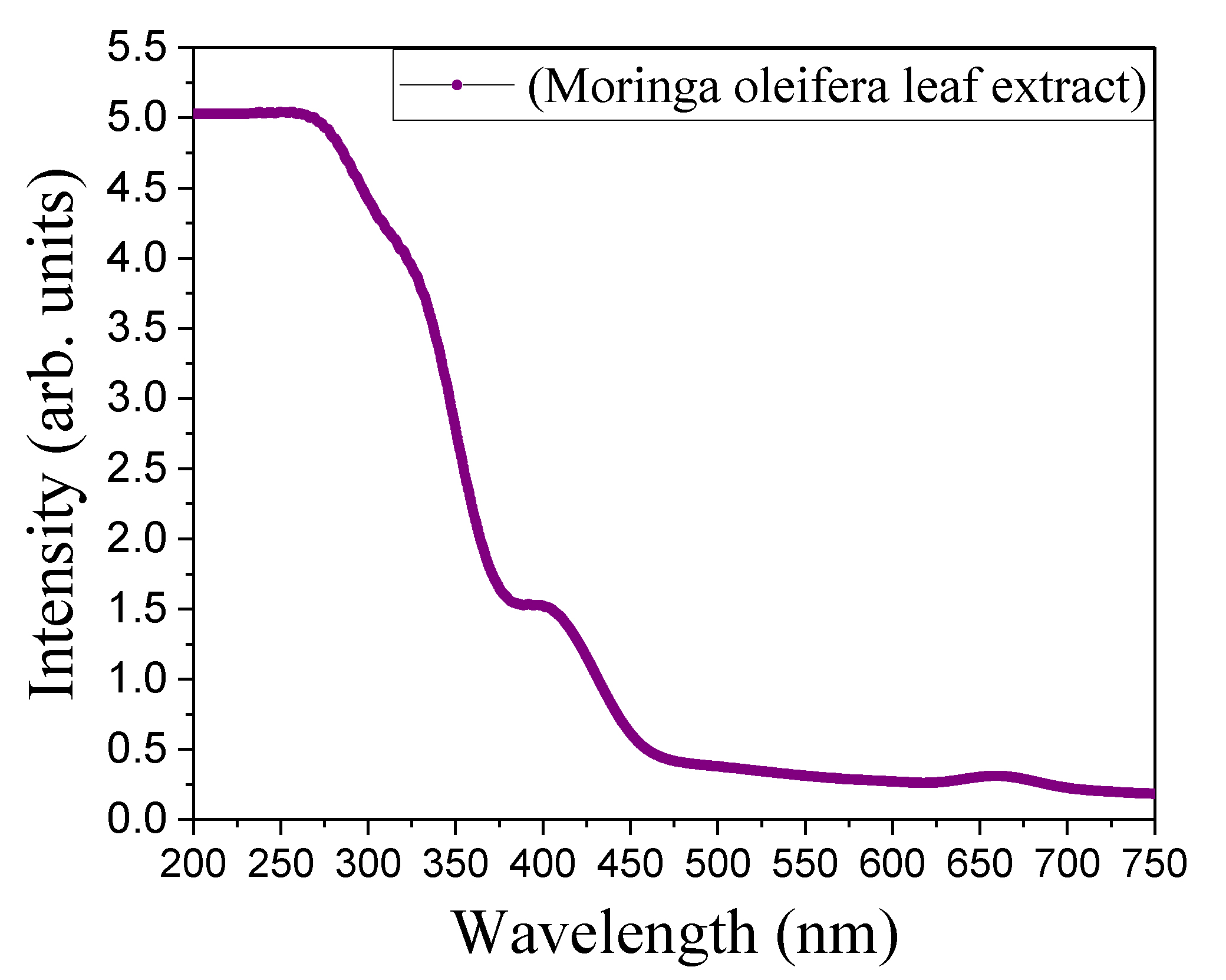
Figure 1. Optical absorption spectra of Moringa oleifera leaf extract by UV-Visible spectrophotometer from 250-750 nm for determination of absorption peaks
The band gap of the extract have been measured using the fundamental relationship –
(αhν)1/n = A(hν-Eg) … (1)
From the previous outline of their relation has been found the energy band gap using the graph of (αhν)1/n vs hν for the extract material. The energy band gap value determined utilizing the intercept of the linear portion of the graph when the hν axis obtained by extrapolation, as shown in Figure 2. The energy band gap was found as 3.34 eV (Figure 3).
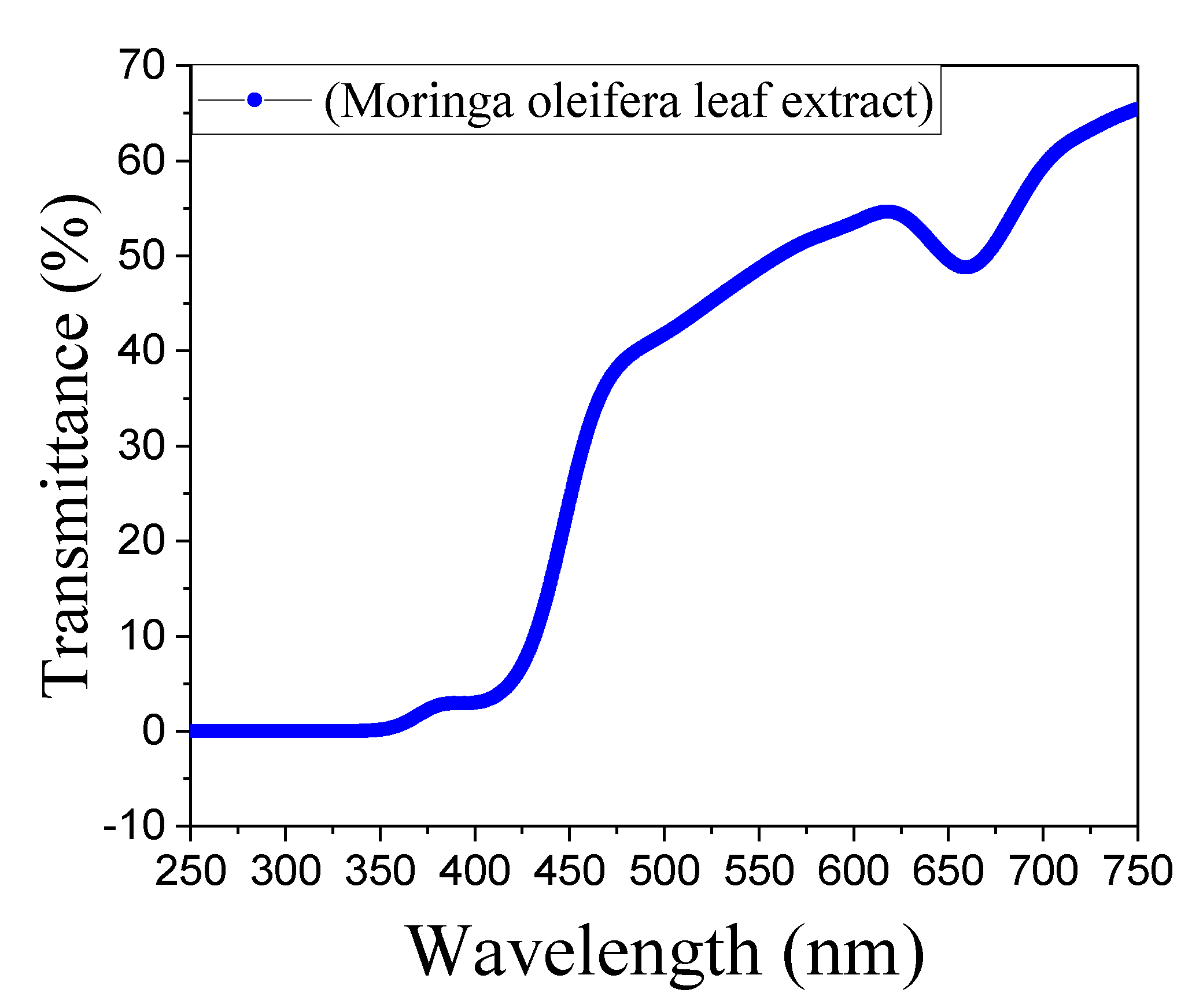
Figure 2. Optical transmission spectra of Moringa oleifera leaf extract by UV-Visible spectrophotometer from 250-750 nm for determination of transmission peaks
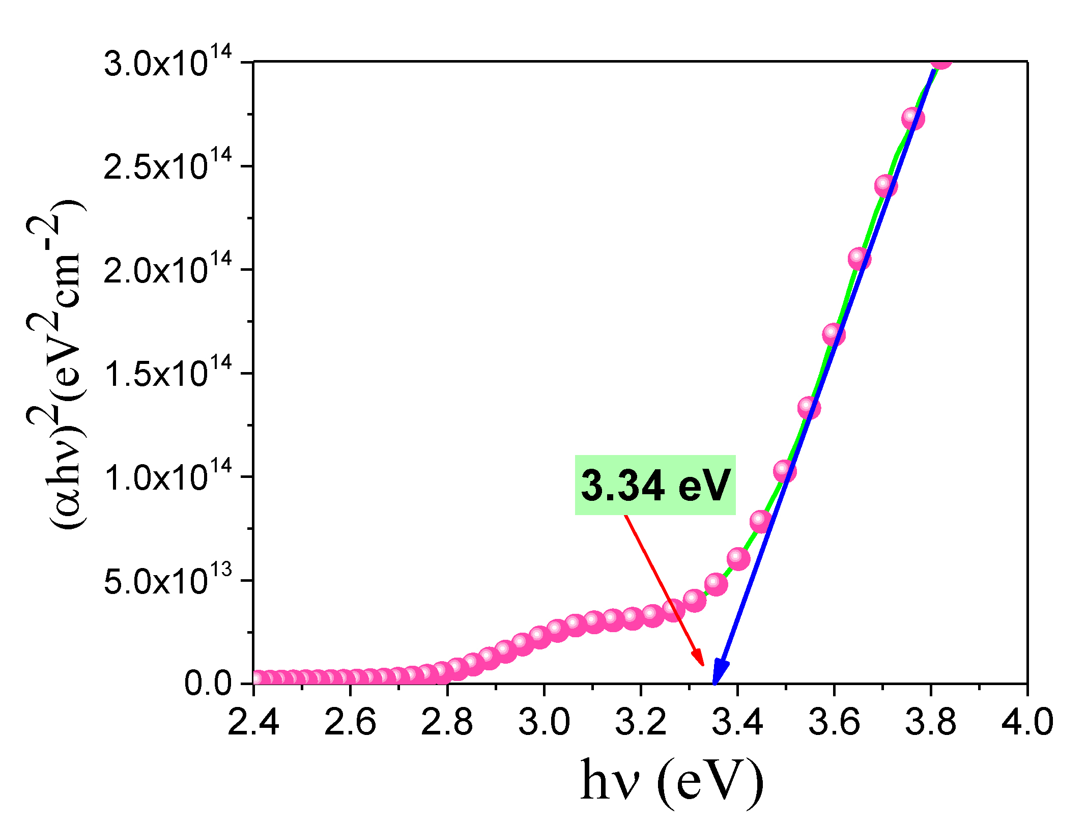
Figure 3. Optical band gap of Moringa oleifera leaf extract has been measured using the fundamental relationship-(αhν)1/n = A(hν-Eg). Further, the band gap of energy has been found using the graph of -(αhν)1/n vs hν of the Moringa oleifera leaf extract
The band gap 3.34 eV of Moringa oleifera indicates strong UV absorption, that’s leading to the generation of Reactive Oxygen Species (ROS).8 Methanolic extract of the air dried plant showed UV absorption maxima in MeOH at 278 nm and 328 nm, are the characteristic of flavones; (1) The IR spectrum of the plant extract in KBr (Figure 4) showed the bands for: hydroxyl (3348 cm-1), α, β-unsaturated ketone (1623 cm-1) and aromatic rings (15170 cm-1). (2) Both UV and IR spectral analysis (Figure 4) indicate the probability of presence of flavone type of organic compounds as major component. These species exhibit anti-cancer and antibacterial activities by damaging cellular structures and inhibiting bacterial growth (Figure 5 and Figure 6).
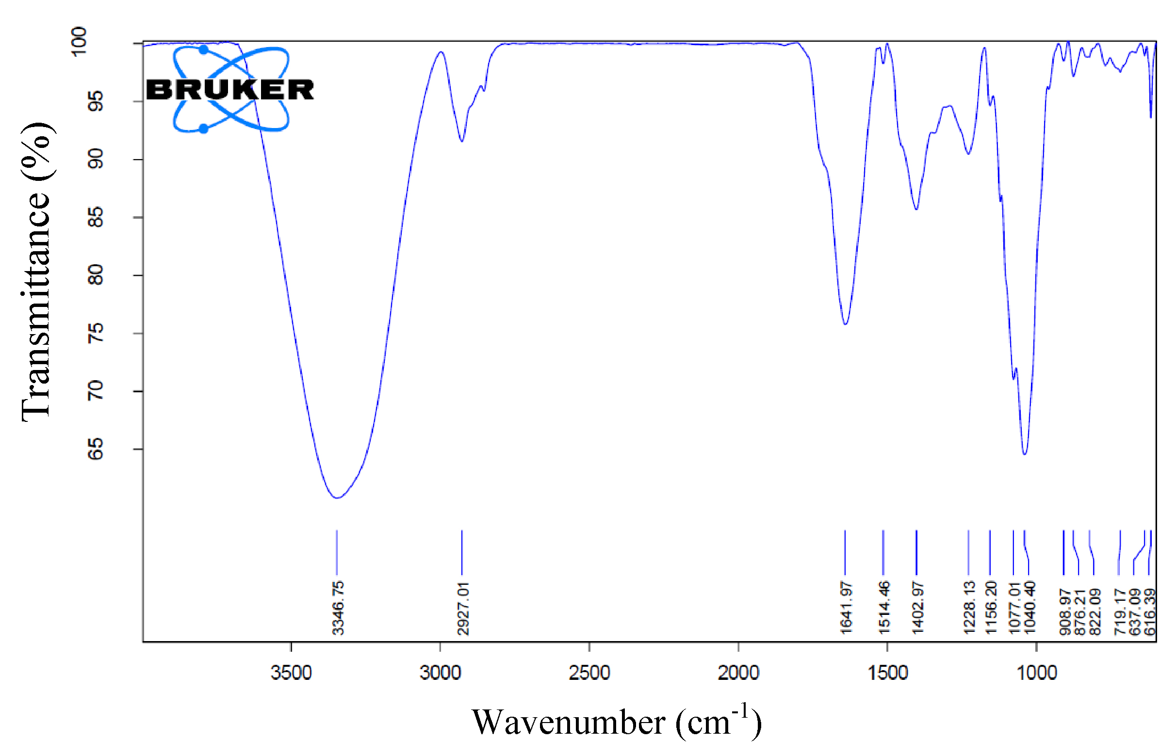
Figure 4. The FTIR spectrum of the Moringa oleifera leaf extract in KBr was determined between wave numbers 4000 cm-1 upto 500 cm-1
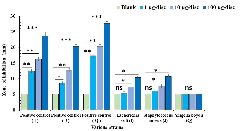
Figure 5. Diameter of the inhibited zone at different concentrations of Moringa oleifera compound against strain- E. coli, Staphylococcus aureus & Shigella boydii. Results are represented as mean ± SD of the three different doses of concentration (*P < 0.05, **P < 0.01, *** P < 0.001, ns – no significance, P > 0.05). Gentamicin & Polymyxin-B were used as standard antibiotics (positive control) against these bacterial strains at different dose concentrations
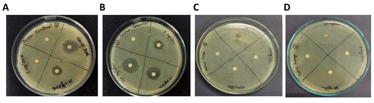
Figure 6. Representative images showing antibacterial activity of Moringa oleifera leaf extract against strain- E. coli, Staphylococcus aureus & Shigella boydii at different concentrations. A & B represents activity of standard antibiotic such as Gentamicin & Polymyxin-B against strain E. coli & Staphylococcus aureus respectively. C & D represents activity of Moringa oleifera leaf extract against strain E. coli & Staphylococcus aureus at various concentrations
Cytotoxicity and anti-bacterial activity study
The methanolic extract of Moringa oleifera induced cell death up to approximately 70 percent showing the highest cytotoxicity against A-549 cells (human lung adenocarcinoma cell line) at a concentration of 50 µg/ml. At concentration of 500 ng/ml and 1 µg/ml it showed up to approximately 10 and 40 percent respectively (Figure 7). When the same drug at same concentrations were subjected to assess the cytotoxicity in normal cells (BEAS-2B), it showed, that at highest concentration i.e., 50 µg/ml, there is 38% cell death was found (Figure 8). The methanolic extract of Moringa oleifera has the ability to be considered as a potential anti-cancer agent against A-549 cell line which further supported by the fact that it has been found less toxic against BEAS-2B normal human broncho-epithelial cell line even at higher concentrations. But the specific compound which is active against cancer cells are not known. The mechanism how the cancer cells are killed is also not known to us. But in future the active compound can be isolated and the same can be treated in in-vivo cancer model which may give us a promising effect of Moringa oleifera in future.
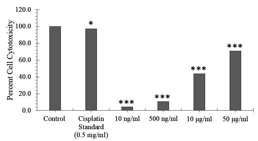
Figure 7. The impact of Moringa oleifera leaf extract on A-549 cell line at various dose concentrations. Cisplatin was used as standard anticancer drug. The results are presented as mean ± SD of the three various doses of concentration (*P < 0.05, **P < 0.01, ***P < 0.001, ns – no significance, P > 0.05)
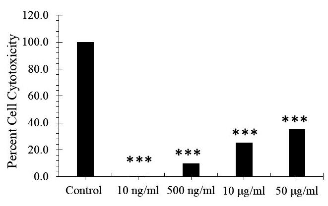
Figure 8. The impact of Moringa oleifera leaf extract on BEAS-2B cell line at various dose concentrations. Results are presented as mean ± SD of three different concentration of doses (*P < 0.05, **P < 0.01, ***P < 0.001, ns – no significance, P > 0.05)
Certain plants and plant products are recognised by the international standard medical system for their potent therapeutic properties and suggesting that, in the specific health condition, the botanical-based products, plant materials, and their plant extracts could be helpful for treatment.17 Therefore, we evaluated whether the extract from Moringa oleifera leaves could stop the growth of the bacterial species or not. At the different concentrations of the drug Moringa oleifera, which has antibacterial properties was studied against the specific bacterial strains, including Shigella boydii (Q), E. coli (I), and Staphylococcus aureus (J). Our results showed Moringa oleifera has very mild antibacterial activity by comparing its activity with that of standard antibiotics, which were used as a positive control. A moderate activity can be seen between blank and 10 µg/disc dose against both the strains E. coli and Staphylococcus aureus with a zone diameter of 7.33 mm ± 0.58 and 7.67 mm ± 0.58 respectively. No such antimicrobial activity has been found against strain Shigella boydii. The highest activity of Moringa oleifera can be seen against both the strains E. coli and Staphylococcus aureus in a dose concentration of 100 µg/disc with 10.33 mm ± 0.58 and 10.67 mm ± 0.58 zone diameters, respectively (Figure 5). Representative images of zone of bacterial growth inhibition are produced in Figure 6. Earlier El-Sherbiny et al. also described that the methanol leaf extract from the Moringa oleifera has potent the antibacterial effects against bacterial strains Klebsiella pneumoniae and Salmonella typhimurium.17 Additionally, the Moringa oleifera fruit extract had a wide range of antibacterial effects against a range of microorganisms, was shown by Sayeed et al.18 Flavonoids, terpenoids, tannins, and alkaloids are the Numerous chemical components found in the plant extract, which are well-known for their effects including anticancer, antioxidants, and antibacterial properties.17 These phenolic and chemical components may have a direct impact on the antibacterial and antioxidative effect.19
In the present work, Moringa oleifera leaf extract has been extensively characterised through spectroscopic technique and observed presence of various carotenoids, phenolic compounds like flavonoids etc. Further the extract also executed potential cytotoxic effect against lung adenocarcinoma cell line A-549. In addition, the extract also showed potential antibacterial effect against various bacterial species. Thus, in a nutshell, from the present work it can be concluded that Moringa oleifera extract has significant effect against adenocarcinoma and bacterial growth as well. Thus, Moringa oleifera plant may be highlighted for its potential therapeutic effect.
ACKNOWLEDGMENTS
The authors would like to thank State Biotech Hub, Tripura University for extending their all support to complete the work. The authors are also thankful to the Dept. of Chemistry, BBM College and the Dept. of Medical Lab Technology, Women’s Polytechnic for extending their support to complete this work.
CONFLICT OF INTEREST
The authors declare that there is no conflict of interest.
AUTHORS’ CONTRIBUTION
SM, TA, AdB, AbB, DM and MCD have contributed to performing all in vitro experiments. DB, DD, SA, AbB and MCD have contributed in data analysis. DB, SA, AbB, DM and MCD wrote the manuscript. All authors reviewed, revised and approved the final manuscript for publication.
FUNDING
None.
DATA AVAILABILITY
All datasets generated or analyzed during this study are included in the manuscript.
ETHICS STATEMENT
Not applicable.
- Kamble MT, Gallardo W, Salin KR, et al. Effect of Moringa oleifera leaf extract on the growth performance, hematology, innate immunity, and disease resistance of Nile tilapia (Oreochromis niloticus) against Streptococcus agalactiae Biotype 2. Animals. 2024;14(6):953.
Crossref - Chaudhary P, Janmeda P, Docea AO, et al. Oxidative stress, free radicals and antioxidants: Potential crosstalk in the pathophysiology of human diseases. Front Chem. 2023;11:1158198.
Crossref - Jamkhande PG, Ajgunde BR, Jadge DR. Annona cherimola Mill. (Custard apple): A review on its plant profile, nutritional values, traditional claims and ethnomedicinal properties. Oriental Pharmacy and Experimental Medicine. 1993;17(3):189-201.
Crossref - Shin J, Song MH, Oh JW, Keum YS, Saini RK. Pro-oxidant actions of carotenoids in triggering apoptosis of cancer cells: A review of emerging evidence. Antioxidants. 2020;9(6):532.
Crossref - Ngcobo S, Bada SO, Ukpong AM, Risenga I. Optimal chlorophyll extraction conditions and postharvest stability in Moringa (M. Oleifera) leaves. Food Measure. 2024;18(3):1611-1626.
Crossref - Wang G, Yang C, Shan M, et al. Synergistic poly (lactic acid) antibacterial surface combining superhydrophobicity for antiadhesion and chlorophyll for photodynamic therapy. Langmuir. 2022;38(29):8987-8998.
Crossref - Martins T, Barros AN, Rosa E, Antunes L. Enhancing health benefits through chlorophylls and chlorophyll-rich agro-food: A comprehensive review. Molecules. 2023;28(14):5344. https://doi.org/10.3390/molecules28145344
- Elsherbiny G, Alluqmani A, Elsehemy, Islam & Kalaba, Mohamed. Antibacterial, antioxidant, cytotoxicity, and phytochemical screening of Moringa oleifera leaves. Scientific Reports. 2024;14
Crossref - Malhotra SPK, Mandal TK. Phytochemical screening and in vitro antibacterial activity of Moringa oleifera (Lam.) leaf extract. Arch Agric Environ Sci. 2018;3(4):367-372
- Ghebremichael KA, Gunaratna KR, Henriksson H, Brumer H, Dalhammar G. A simple purification and activity assay of the coagulant protein from Moringa oleifera seed. Water Res. 2005;39(11):2338-2344.
Crossref - Sutherland JP, Folkard GK, Mtawali MA, Grant WD. Moringa oleifera as a natural coagulant. In: Pickford J, ed. Affordable Water Supply and Sanitation: Proceedings of the 20th WEDC Conference; Colombo, Sri Lanka. Loughborough University; 1994:297-299
- Fuglie LJ. The Miracle Tree: Moringa oleifera: Natural Nutrition for the Tropics. Dakar: Church World Service. 1999.
- Goyal BR, Agrawal BB, Goyal RK, Mehta AA. Phytopharmacology of Moringa oleifera Lam.: an overview. Nat Prod Radiance. 2007;6(4):347-353.
- Ju J, Gothai S, Hasanpourghadi M, et al. Anticancer potential of Moringa oleifera flower extract in human prostate cancer PC-3 cells via induction of apoptosis and downregulation of AKT pathway. Pharmacogn Mag. 2018;14(58):477-481.
Crossref - Debnath P, Debnath P, Roy M, et al. Novel Organotin(IV) Complexes of 2-[4-Hydroxy-3-((2-hydroxyethylimino)methyl)phenylazo]benzoic Acid: Synthesis, Structure, Noncovalent Interactions and In Vitro Antibacterial Activity. Crystals. 2022;12(11):1582.
Crossref - Boruah SR, Bhattacharjee M, Purkayastha RND, et al. Insights into Synthesis, Crystal Structure, Bioactivity and Computational Studies of Cu (II) and Zn (II) Carboxylates Containing Aminopyridine. Chemistry Select. 2023;8(19):e202204937.
Crossref - El-Sherbiny GM, Alluqmani AJ, Elsehemy IA, Kalaba MH. Antibacterial, antioxidant, cytotoxicity, and phytochemical screening of Moringa oleifera leaves. Sci Rep. 2024;14(1):30485.
Crossref - Sayeed MA, Hossain MS, Chowdhury MEH, Haque M. In vitro antimicrobial activity of methanolic extract of Moringa oleifera Lam. fruits. J Pharmacogn Phytochem. 2012;1(4):94-98
- Barchan A, Bakkali M, Arakrak A, Pagan R, Laglaoui A. The effects of solvents polarity on the phenolic contents and antioxidant activity of three Mentha species extracts. Int J Curr Microbiol Appl Sci. 2014;3(11):399-412.
© The Author(s) 2026. Open Access. This article is distributed under the terms of the Creative Commons Attribution 4.0 International License which permits unrestricted use, sharing, distribution, and reproduction in any medium, provided you give appropriate credit to the original author(s) and the source, provide a link to the Creative Commons license, and indicate if changes were made.