


ISSN: 0973-7510
E-ISSN: 2581-690X
 ,
, Nagalakshmi Rajan4
, Komala Sivakumar5 and Manjunath Jagadeesan6Luffa acutangula aqueous peel extract was used as a reducing and stabilizing agent in the effective green and sustainable synthesis of nickel oxide nanoparticles (NiO NPs). By using ridge gourd peel for the environmentally friendly synthesis of metal oxide nanoparticles with possible medicinal uses, the current study highlights the valorization of agricultural waste. The current work explicitly uses Luffa acutangula peel waste as a low-cost bioresource for nanoparticle synthesis and antibacterial assessment, in contrast to previously reported plant-mediated syntheses of NiO nanoparticles. Field Emission Scanning Electron Microscopy (FESEM), X-ray Diffraction (XRD), Fourier Transform Infrared Spectroscopy (FTIR), and UV-visible spectroscopy were used to analyze the biosynthesized NiO nanoparticles. The development of nanoparticles was confirmed by UV-visible analysis, which revealed a distinctive absorption peak at 320 nm. Phenols, flavonoids, alcohols, and carboxylic groups were among the phytochemicals that were involved in the reduction and stability of NiO nanoparticles, according to FTIR spectra. The synthesised compound was confirmed by XRD investigation. Particle diameters ranging from 13-18 nm were found to be almost spherical and somewhat agglomerated by FESEM examination. Nickel NPS’s antibacterial properties can engage with the bacterial cell membrane’s ROS. Assess the agent’s possible antibacterial capabilities against harmful bacteria. This study sheds insight on the environmentally friendly method of producing nickel oxide nanoparticles with potential medical uses.
Luffa acutangula, Nickel Oxide, Characterization, Antibacterial Activity
Green synthesis has become a viable and sustainable method for creating metal and metal oxide nanoparticles. Traditional synthesis techniques often include toxic reducing agents, dangerous chemicals, and energy-intensive processes that could be harmful to the environment and human health. By using naturally available phytochemicals as reducing and stabilizing agents, plant-mediated synthesis, on the other hand, provides an easy, affordable, and environmentally beneficial alternative.1-4 Nickel oxide (NiO) nanoparticles have garnered significant interest among other metal oxide nanomaterials because of their superior physicochemical stability, catalytic effectiveness, optical characteristics, and biological applications. NiO nanoparticles have been extensively studied for sensing, photocatalytic, antioxidant, and antibacterial uses.5 Their high surface area, nanoscale size, and capacity to produce reactive oxygen species (ROS), which can harm intracellular components and microbial cell membranes, are primarily responsible for their increased biological activity.6
The synthesis of NiO nanoparticles utilizing plant extracts from Aegle marmelos, Syzygium cumini, Allium cepa, and Sesbania grandiflora has been documented in a number of research.7-10 However, the use of agricultural peel waste as a renewable feedstock for the creation of nanoparticles has received little attention. Utilizing plant waste materials promotes sustainable nanotechnology and waste-to-wealth conversion while also reducing environmental impact.
Ridge gourd, or Luffa acutangula, is a popular vegetable in India and other Asian nations. It is a member of the Cucurbitaceae family. Despite being rich in phytochemicals like flavonoids, triterpenoids, saponins, glycosides, and phenolic components, the peel of Luffa acutangula is typically thrown away as agricultural waste. During the creation of nanoparticles, these bioactive components can function as organic reducing and capping agents.11,12 The use of L. acutangula extracts for the manufacture of metal oxide nanoparticles, such as CuO and TiO2 nanoparticles, has been investigated in earlier papers; however, there are still few investigations on the synthesis of NiO nanoparticles using L. acutangula peel waste.13,14
Thus, the current study concentrates on the environmentally friendly production of NiO nanoparticles utilizing Luffa acutangula aqueous peel extract. The optical, structural, and morphological characteristics of the produced nanoparticles were assessed by FTIR, XRD, FESEM, and UV-visible spectroscopy. Additionally, the biosynthesized NiO nanoparticles’ antibacterial efficacy against particular harmful bacterial strains was examined. The study also emphasizes the potential mechanism of antibacterial activity mediated by ROS production and interactions between nanoparticles and cell membranes (Figure 1).
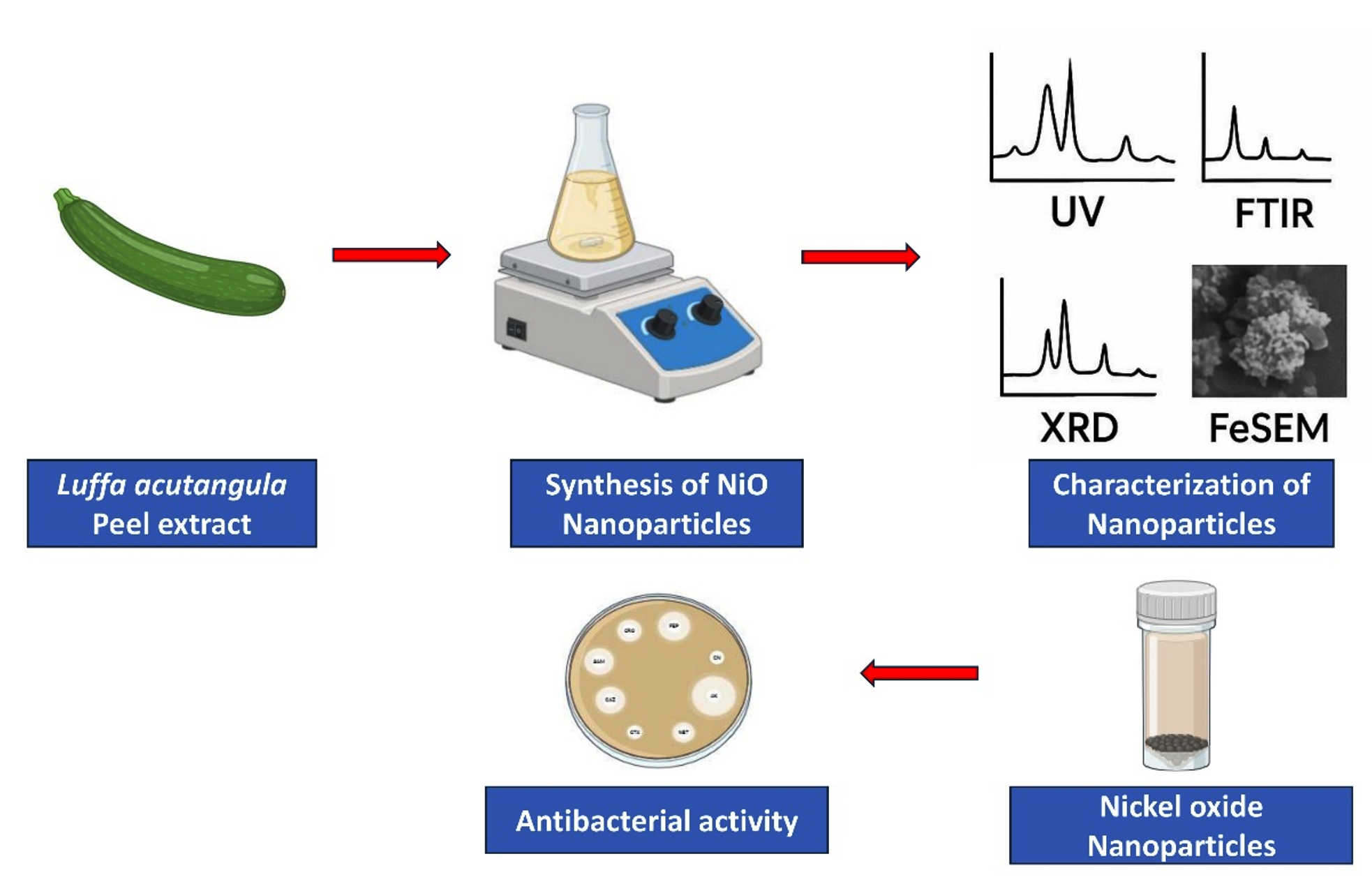
Figure 1. Schematic Representation of Nickel oxide nanoparticle synthesis from Luffa acutangula peel and antibacterial activity
Sample collection
The Luffa acutangula was gathered from neighbourhood markets in Chennai, and after being cleaned with water, the skin was removed. After three days of sun-drying, the gathered peeled material was powdered to a fine consistency.
Extraction using aqueous solution
20 g of powdered dried Luffa acutangula peel were combined with 400 mL of sterile distilled water, and the mixture was heated to 80 degrees Celsius for thirty minutes while being constantly stirred at 500 revolutions per minute. Whatman No. 1 filter paper was used to filter the extract once it had cooled to room temperature. For future research, the filtrate was kept at 4 °C.
Synthesis of nickel oxide nanoparticles
The necessary amount of nickel nitrate hexahydrate [Ni(NO3)2·6H2O] was dissolved in 50 mL of distilled water to create a 1 mM aqueous solution. After that, 20 mL of Luffa acutangula peel extract was added dropwise while being continuously stirred by a magnetic stirrer at 80 °C for three hours at 600 rpm. Dropwise additions of 1 M NaOH solution were used to bring the reaction mixture’s pH down to 12. The synthesis of nickel hydroxide precursor and subsequent NiO nanoparticles was indicated by a noticeable color shift from greenish to mustard yellow.
The resulting precipitate was filtered, repeatedly cleaned with ethanol and distilled water to get rid of contaminants, and then dried in a hot air oven at 60 °C for 12 hours. NiO nanoparticles were produced by calcining the dry material at 400 °C for three hours in ambient air. Gravimetric analysis was used to determine the ultimate yield percentage of the produced nanoparticles.
Characterization of NiO nanoparticles
The synthesized nickel oxide nanoparticles UV-Vis spectra (Shimadzu UV-1900i Plus) were captured for the analytical research utilizing in the 200-800 nm wavelength range.12 The functional groups were investigated using the FTIR spectrum (Agilent-Cary 630), which was obtained in the 400-4000 cm-1 mid-IR range.13 X-ray diffractometer (Rigaku ULTIMA IV) was used to record the XRD patterns of NiO NPs.14 The FEI Apreo LoVac field emission scanning electron microscope (FeSEM) was used to analyse the nanorods’ surface appearance and structure.15
Anti-bacterial activity
Bacillus cereus, E. coli, Staphylococcus aureus, and Pseudomonas aeruginosa strains were used to test the antibacterial activity of the synthesized nickel oxide NPs. The well diffusion technique was used to track the produced NiO NPs antibacterial efficacy. Nickel oxide nanoparticles in different concentrations (20, 30, and 40 µg/ml) were placed onto the well, while a sterile, positive control streptomycin served as a reference. Room temperature was used to incubate the plates for the entire night. The development of the clear zone of inhibition was calculated, and the experiment was run in triplicate.16,17
Synthesis of NiO Nanoparticle from L. acutangula peel
A useful method to determine if the precursors are completely reduced was UV-visible spectroscopy analysis. As the nickel oxide solution was incubated, a colour shift was discernible in the extract of L. acutangula peel. There was no confirmation of the formation of nickel oxide nanoparticles, and the pure NiNO3 concentrate of L. acutangula peel without watery leaves did not exhibit any color variation. After the NiNo3 and L. acutangula peel extract reacted, the colour turned mustard yellow for three hours during a stirred conditional incubation phase. After that, no significant changes happened.18 The stimulation of plasmonic resonance at the surface during the production of Nickle oxide NPs was the origin of this distinctive colour variation. Now, the optical range of 200-800 nm was employed to document the absorption NiO NPs from UV to visible light. The absorption of UV-visible spectra of NiO NPs is shown in Figure 2. The creation of NiO nanoparticles was indicated by the greatest absorption peak, which was detected at 320 nm. The absorption peak for green produced nickel nanoparticles was observed around 342 nm in earlier studies.19 The produced titanium dioxide nanoparticles exhibit a comparable absorption peak in comparison to the prior research.20
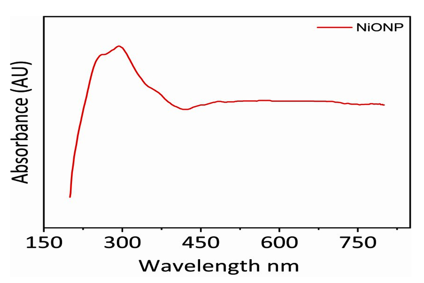
Figure 2. UV spectroscopy absorption of NiO Nanoparticles
FTIR Analysis
The involvement of phytochemicals found in Luffa acutangula peel extract in the formation of nanoparticles was verified by FTIR analysis. Phenols and alcohols’ O-H stretching vibrations are shown by the wide absorption band at 3456 cm-1. Carbonyl groups’ C=O stretching vibrations are responsible for the peak at 1680 cm-1, whereas alkanes’ C-H stretching is indicated by peaks close to 3034 cm-1. C=C or C-N stretching vibrations may be represented by the peaks seen at 1447 cm-1.
The C-O stretching vibrations of alcohols and carboxylic acids are represented by the peaks at 1183, 1061, and 1006 cm-1, indicating the role of phytochemicals in the reduction and stabilization process (Figure 3). The effective production of NiO nanoparticles is confirmed by the distinctive Ni-O stretching vibration seen below 700 cm-1.21-23
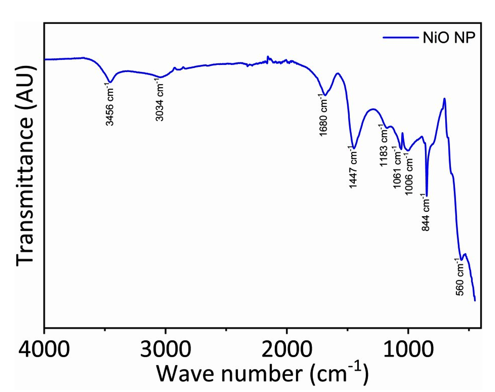
Figure 3. Characterization of NiO nanoparticles in FT-IR
XRD analysis
The crystalline nature of the synthesised NiO nanoparticles was verified by the XRD pattern. The (111), (200), (220), (311), and (222) crystal planes, which are typical of the face-centered cubic (fcc) rock-salt structure of NiO nanoparticles, are represented by the diffraction peaks seen at 2θ values of 37.31°, 43.35°, 62.91°, 75.53°, and 79.45° (Figure 4). The creation of phase-pure cubic NiO nanoparticles with no discernible impurities was confirmed by the diffraction data’s good agreement with standard JCPDS card No. 47-1049.
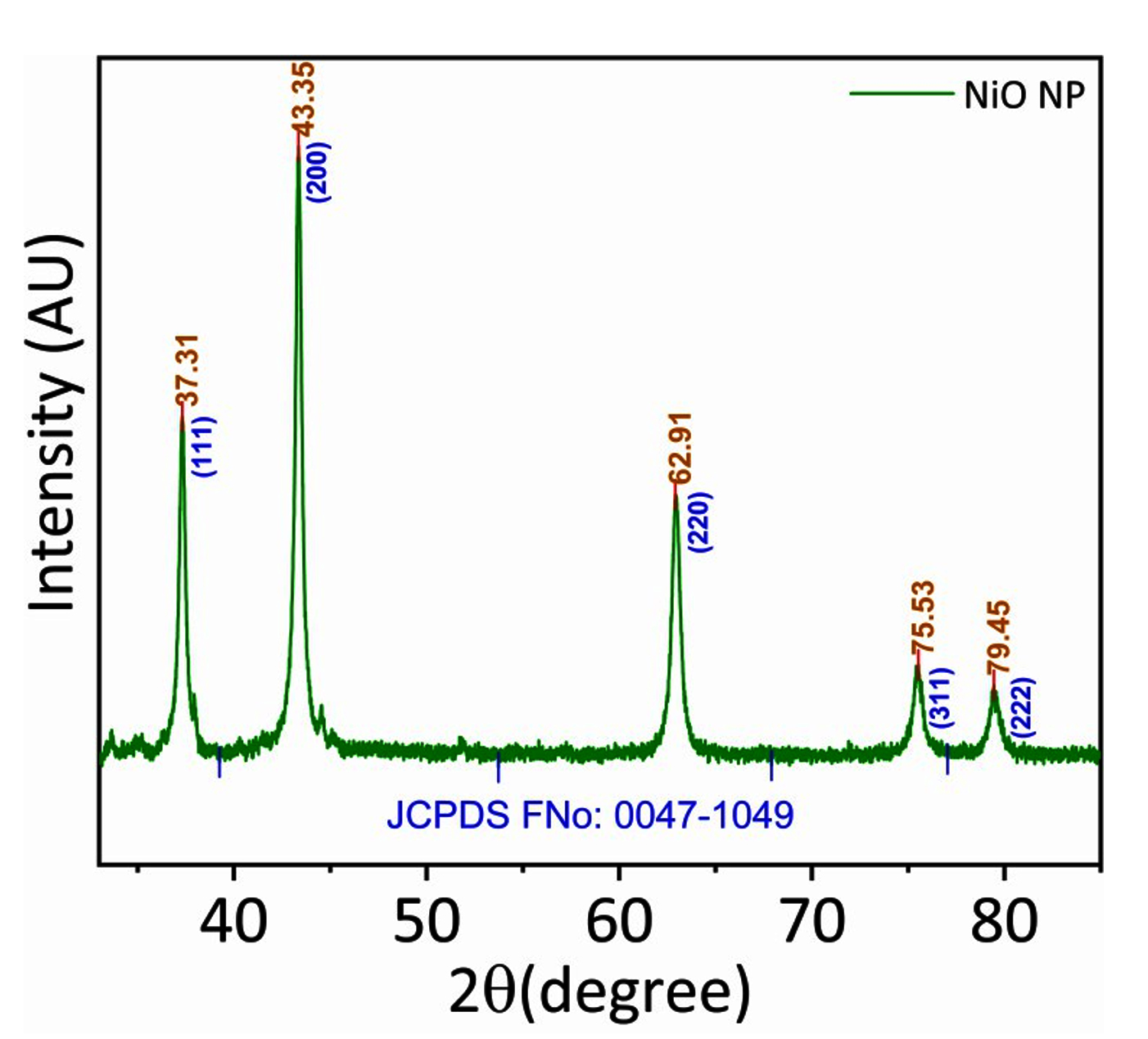
Figure 4. XRD pattern of NiO nanoparticle synthesized from L. acutangula peel
The average crystallite size was estimated using the Debye-Scherrer equation:
D = 0.94λ / βcosθ
where D is the crystallite size, λ is the wavelength of CuKα radiation (0.15406 nm), β is the full width at half maximum (FWHM), and θ is the Bragg diffraction angle. The average crystallite size was found to be approximately 15-18 nm, which agrees well with FESEM observations.24,25
FESEM analysis
The surface morphological characteristics of the produced NPs were examined using FESEM. FESEM pictures of the NiO nanoparticle are shown in Figure 5 at a 65k magnification. Nearly spherical and clumped nanoparticles with an average size of 13-18 nm are visible in the image. The tight size distribution shown by the measured sizes (13.85 nm, 15.84 nm, and 17.68 nm) validates the successful production of NiO nanoparticles in the nanometer range.
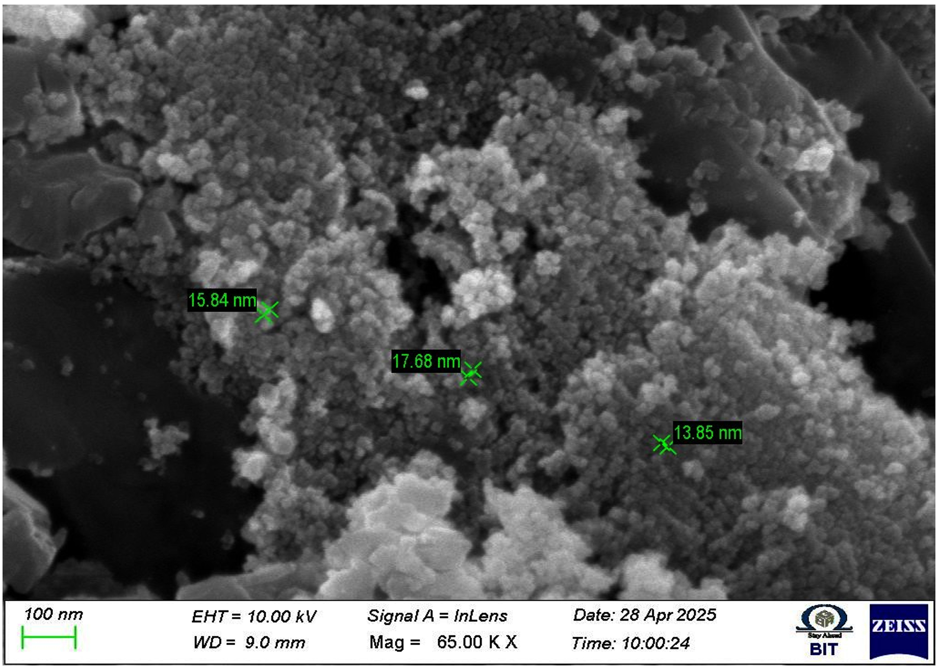
Figure 5. FESEM image of Nio nanoparticle synthesized from L. acutangula peel
Surface hydroxyl groups and remaining phytochemicals from the L. acutangula peel extract form hydrogen bonds, which causes the mild aggregation typical of nickel oxide nanoparticles made from the peel. The plant metabolites may have served as stabilizing and capping agents, preventing overproduction during calcination, as indicated by the tiny, porous structure.26
Anti-bacterial activity
The synthesised NiO nanoparticles shown concentration-dependent antibacterial activity against Pseudomonas aeruginosa, Bacillus cereus, and Staphylococcus aureus. At a concentration of 100 µL, Bacillus cereus showed the largest zone of inhibition (32 ± SD mm). On the other hand, there was very little inhibition against Escherichia coli. (Figure 6; Table). The dosage of nanoparticles ranges from 50-100 µL, depending on how much the zone produces as the concentration increases.
Table. The zone of inhibition (mm, mean ± SD) of NiO nanoparticles synthesised from Luffa acutangula peel
| Name of The Pathogens | Zone of Inhibition | ||
|---|---|---|---|
| NiO NPs | |||
| 100 µl | 75 µl | 50 µl | |
| Staphylococcus aureus | 28 | 27 | 26 |
| Pseudomonas aeruginosa | 30 | 29 | 28 |
| Bacillus cereus | 32 | 30 | 29 |
| E. coil | NZ | NZ | NZ |
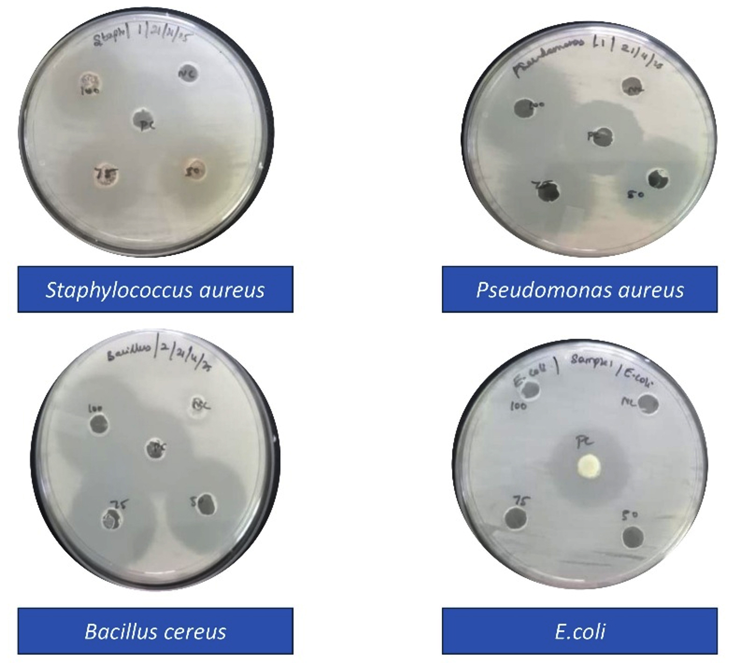
Figure 6. Antibacterial activity using NiO nanoparticle synthesised from Luffa acutangula peel
The complex outer lipopolysaccharide membrane seen in Gram-negative bacteria, which limits nanoparticle penetration and lessens ROS-mediated damage, may be the cause of E. coli’s decreased susceptibility. Gram-positive bacteria, on the other hand, have a somewhat porous peptidoglycan layer that makes it easier for nanoparticles to connect with the bacterial membrane. NiO nanoparticles’ antibacterial action is primarily linked to the production of reactive oxygen species (ROS), oxidative stress, membrane rupture, denaturation of proteins, and intracellular component leakage. Additionally, the phytochemicals adsorbed on the surface of the nanoparticles may enhance the antibacterial efficiency in a synergistic way.27-34
In this study, a straightforward and environmentally friendly green synthesis method was used to successfully create nickel oxide nanoparticles utilizing an aqueous peel extract of Luffa acutangula. The creation of nanoparticles was confirmed by UV-visible spectroscopy, which showed an absorption peak at 320 nm. Crystalline cubic-phase NiO nanoparticles with an average crystallite size of roughly 15-18 nm were formed, according to XRD examination. Nearly spherical and somewhat agglomerated nanoparticles were shown by FESEM investigation. Phytochemicals’ role in the stabilization and reduction of nanoparticles was verified by FTIR analysis. The biosynthesized NiO nanoparticles showed weak antibacterial action against Escherichia coli, but strong antibacterial activity against Bacillus cereus, Staphylococcus aureus, and Pseudomonas aeruginosa. The production of ROS and membrane damage caused by nanoparticles may be linked to the antibacterial action. The study emphasizes the potential use of NiO nanoparticles made from agricultural waste in the sectors of biomedicine and antimicrobials.
ACKNOWLEDGMENTS
The authors would like to thank Vels Institute of Science, Technology and Advanced Studies, Chennai, India, for providing the facilities to carry out the work.
CONFLICT OF INTEREST
The authors declare that there is no conflict of interest.
AUTHORS’ CONTRIBUTION
All authors listed have made a substantial, direct and intellectual contribution to the work, and approved it for publication.
FUNDING
None.
DATA AVAILABILITY
All datasets generated or analyzed during this study are included in the manuscript.
ETHICS STATEMENT
Not Applicable.
- Aigbe UO, Osibote OA. Green synthesis of metal oxide nanoparticles, and their various applications. J Hazard Mater Adv.2024;13:100401.
Crossref - Nair GM, Sajini T, Mathew B. Advanced green approaches for metal and metal oxide nanoparticles synthesis and their environmental applications. Talanta Open.2022;5(6):100080.
Crossref - Tailor G, Chaudhary J, Jandu S, Mehta C, Yadav M, Verma D. A review on green route synthesized nickel nanoparticles: biological and photo-catalytic applications. Results in Chemistry. 2023;6:101195.
Crossref - Moradnia F, Fardood ST, Zarei A, Heidarzadeh S, Ramazani A, Sillanpaa M. Green synthesis of nickel oxide nanoparticles using plant extracts: an overview of their antibacterial, catalytic, and photocatalytic efficiency in the degradation of organic pollutants. Iranian Journal of Catalysis. 2024;14(1).
Crossref - Abdallah Y, Ogunyemi SO, Bi J, Wang F, et al. Nickel oxide nanoparticles: A new generation nanoparticles to combat bacteria Xanthomonas oryzae pv. oryzae and enhance rice plant growth. Pestic Biochem Physiol.2024;200:105807.
Crossref - Raji P, Balachandra KK. Green synthesis and characterization of copper oxide nanoparticles using Luffa acutangula peel extract and its antibacterial activity. Results Surf Interfaces.2024;16:100261.
Crossref - Anbumani D, vizhi Dhandapani K, Manoharan J, et al. Green synthesis and antimicrobial efficacy of titanium dioxide nanoparticles using Luffa acutangula leaf extract. J King Saud Univ Sci.2022;34(3):101896.
Crossref - Singh S, Salodiya R, Tailor G, Sharma TC, Mehta C. Comprehensive studies: biogenic constructed nickel oxide nanoparticles and their broad spectrum biomedical applications. Chemistry Select. 2024;9(38):e202402099.
Crossref - Abegunde SM, Owoeye SS, Lamidi YD. Green synthesis of Nickel oxide nanoparticles using Parkia biglobosa flower extracts for the removal of malachite green and acid red 88 dyes from aqueous solution. Sustainable Chemistry One World. 2025:100119.
Crossref - Chowdhury MAS, Islam MM, Jamal M. Green synthesis of nickel oxide nanoparticles using Allium cepa stalks and investigation of their antibacterial activity. Results Chem.2025;16:102328.
Crossref - Haider A, Ijaz M, Ali S, et al. Green synthesized phytochemically (Zingiber officinale and Allium sativum) reduced nickel oxide nanoparticles confirmed bactericidal and catalytic potential. Nanoscale Res Lett. 2020;15(1):50.
Crossref - Berhe A, Tilahun F, Wendu A, Lakew W. Plant-mediated biosynthesis of Nickel (II) oxide nanoparticles from Calpurnia Aurea Leaf extract: A promising photocatalyst for malachite green degradation. Chemical Physics Impact.2025;11:100906.
Crossref - Suresh L, Snega R, Sravanthy PG, Saravanan M. Phytosynthesis of Nickel Oxide nanoparticles and their antioxidant and Antibacterial Efficacy studies. Cureus.2024;16(4):e58064.
Crossref - Istrate D, Oproescu M, Modan EM, Moga SG, Negrea DA, Schiopu AG. Nanoscale Nickel Oxide: Synthesis, Characterization, and Impact on Antibacterial Activity Against Representative Microorganisms. ChemEngineering. 2025;9(4):77.
Crossref - Kesmez O, Kuruca T, Odabas E, Cihanoglu N, Akarsu E, Demir F. Synthesis and Characterization of Nickel Oxide Nanoparticles via Polyol Mediated Hydrothermal Process with Antibacterial Properties. ChemistrySelect. 2025;10(9):e202405811.
Crossref - Riaz T, Munnwar A, Shahzadi T, et al. Phyto-mediated synthesis of nickel oxide (NiO) nanoparticles using leaves’ extract of Syzygium cumini for antioxidant and dyes removal studies from wastewater. Inorg Chem Commun.2022;142:109656.
Crossref - Khan KR, Dastageer NS, Munde AS, Momin NP. Characterization and Microbial Activity of Nickel Oxide Nanoparticles Synthesized from Aquatic Hydrilla Plant Leaves. Oriental Journal of Chemistry. 2025;41(3):1021-1027.
Crossref - Roshid MH, Alam MS, Amin KAI, Ferdousi FK, Rahman MH, Islam S. Green synthesis of nickel oxide nanoparticles using Lagerstroemia speciosa, Bombax ceiba, and Piper chaba extracts and evaluating their potential antioxidant, antidiabetic and antibacterial properties. Heliyon.2025;11(6):e42953.
Crossref - Gobinath E, Dhatchinamoorthy M, Saran P, Vishnu D, Indumathy R, Kalaiarasi G. Synthesis and characterization of NiO nanoparticles using Sesbania grandiflora flower to evaluate cytotoxicity. Results Chem. 2023;6:101043.
Crossref - Prabhu S, Thangadurai TD, Bharathy PV, Kalugasalam P. Synthesis and characterization of nickel oxide nanoparticles using Clitoria ternatea flower extract: Photocatalytic dye degradation under sunlight and antibacterial activity applications. Results Chem.2022;4:100285.
Crossref - Al-Fakeh MS, O. Alsaedi RO, Aldoghaim M, Ibrahim AB, Mostafa AM. Nickel Oxide Nanoparticles Derived from Coordination Polymer of PVA and Aminobenzoic Acid Derivative: Synthesis, Characterization and Antimicrobial Activity. Polymers. 2025;17(3):301.
Crossref - Barzinjy AA, Azeez HH. Green synthesis and characterization of zinc oxide nanoparticles using Eucalyptus globulus Labill. leaf extract and zinc nitrate hexahydrate salt. SN Appl Sci.2020;2(5):991.
Crossref - Jawahar S, Priya M, Venkatesan R, Sathiasivan K, Khan MR, Kim SC. Green Synthesis of Nickel Oxide Nanoparticles Using Leaf Extract of Aegle marmelos and Their Antibacterial, Anti Oxidant, and In Vitro Cytotoxicity Activity. Microscopy Research and Technique.2025;88(10):2830-2842.
Crossref - Asaldoust F, Mabhouti K, Jafari A, Taleb-Abbasi M. Structural, magnetic, and optical characteristics of undoped and chromium, iron, cobalt, copper, and zinc doped nickel oxide nanopowders. Sci Rep. 2025;15(1):1088.
Crossref - Kumar N, Singh A, Devra V. Experimental investigation on plant extract-induced biosynthesis of Nickel nanoparticles. Next Nanotechnol.2025;7:10010.
Crossref - Hong SJ, Mun HJ, Kim BJ, Kim YS. Characterization of nickel oxide nanoparticles synthesized under low temperature. Micromachines. 2021;12(10):1168.
Crossref - Ingalagondi PK, Sannaikar MS, Mruthunjaya K, Horti NC. Optical and Antibacterial Properties of Nickel Oxide (NiO) Nanoparticles: Effect of Annealing Temperature. Results Surf Interfaces.2025:100571.
Crossref - Faizan M, Fatima M, Shams F, et al. Plant extract mediated biogenic synthesis and characterization of nickel oxide nanoparticles and its environmental and antibacterial applications. Journal of Chinese Entrepreneurship. 2023;2(2):109-121.
Crossref - Kumar V, Kaushal S, Singh Y. Biogenic synthesis of zinc oxide nanoparticles using cell-free extract of Spirogyra crassa(Kutz.) Kutz for sustainable biomedical and environmental application. New J Chem. 2025;49:16145-16159.
Crossref - Kumar V, Kaushal S, Singh Y, Kumar R. Bioinspired synthesis of copper oxide nanoparticles using aqueous extracts of Cladophora glomerata (L.) Kuetz and their potential biomedical applications. Bioprocess Biosyst Eng. 2025;48(4):633-646.
Crossref - Sodhi R, Singh P, Lal B, et al. Biogenic synthesis of ZnO nanoparticles using Polystichum squarrosum extract and its applications as anti-oxidant, anti-diabetic agent and industrial waste water treatment. Emergent Materials. 2023;7(1):285-298.
Crossref - Singh Y, Sodhi RS, Singh PP, Kaushal S. Biosynthesis of NiO nanoparticles using Spirogyra sp. Cell- free extract and their potential biological applications. Mater Adv. 2022;3(12):4991-5000.
Crossref - Kumar V, Kumar S, Kumar S, et al. 1 monolinoleoylglycerol and 3,4 dihydroxymandelic acids-mediated ZnO for antibiotic degradation and anticancer activity. J Environ Chem Eng. 2026;14(3);122605.
Crossref - Bano K, Singh PP, Kumar S, Saeed SM, Aggarwal S, Kumar R, Kaushal S. Construction of honey bee hive-like CuO/PbO heterojunction photocatalysts with enhanced antibiotic and dye degradation activity under visible light. Environmental Science: Water and Technology. 2024;10(7):1714-1725.
Crossref
© The Author(s) 2026. Open Access. This article is distributed under the terms of the Creative Commons Attribution 4.0 International License which permits unrestricted use, sharing, distribution, and reproduction in any medium, provided you give appropriate credit to the original author(s) and the source, provide a link to the Creative Commons license, and indicate if changes were made.