


ISSN: 0973-7510
E-ISSN: 2581-690X
Nirmala Deo1  , Indu Singh2,3, Krishnamurthy Venkatesan1 and
Divakar Sharma3
Fluoroquinolones (FQs) manifest bactericidal activity in a concentration-dependent manner as they inhibit DNA gyrase and topoisomerase activity that are crucial in the replication process. They are the key component of a multidrug-resistant tuberculosis treatment regimen. FQs are effective against bacterial infections that cause skin, urinary tract, and prostate-related infections. Ofloxacin is also being tried as part of modified Multidrug Treatment (MDT) regimens against leprosy. This study examined the accumulation of ofloxacin in M. smegmatis, M. kansasii, and M. tuberculosis. These mycobacteria were grown in Sauton’s liquid medium up to the exponential phase. Finally, cells were harvested, cleaned by washing, and resuspended. Suspensions were incubated with ofloxacin at 37 °C, and ofloxacin concentration in supernatants was observed using spectrofluorimetry. Ofloxacin concentrations were also observed in the absence or presence of Carbonyl cyanide m-chlorophenylhydrazone (CCCP), which is a proton motive force inhibitor. Steady state concentration (SSC) of ofloxacin was achieved in M. smegmatis (03 minutes), M. kansasii (03 minutes), and M. tuberculosis (05 minutes), and the ofloxacin accumulation level was 102 ng/mg, 100 ng/mg, and 107 ng/mg (dry weight of the bacilli), respectively. Ofloxacin accumulation was found at 10 µg/ml concentrations; however, CCCP addition was unable to influence the ofloxacin accumulation. This study concludes that ofloxacin accumulation is a simple diffusion in M. smegmatis, M. kansasii, and M. tuberculosis. Through simple diffusion, ofloxacin is transported across the mycobacterial cell envelope, inhibiting type II DNA isomerase (gyrase), which is required for DNA replication. Thus, it is effective in the treatment of disease.
Mycobacteria, Ofloxacin, Fluoroquinolones (FQ), Carbonyl Cyanide m-chlorophenyl-hydrazone
The reduced permeation of antimicrobials through the cell wall of mycobacteria is crucial in antibiotic resistance in different mycobacterial species.1-3 Rare antimicrobial agents are efficiently active against Mycobacterium tuberculosis as well as atypical mycobacteria (Mycobacterium avium complex). Tuberculosis (TB) is the foremost contributor to the high mortality rate worldwide. Only a few antimycobacterial agents are clinically active against Mycobacterium tuberculosis,4 which is frequently prescribed in the present era, specifically in combinations due to the emergence of multidrug-resistance (MDR). MDR is often viewed as a consequence of interrupted treatment of active TB during the longer course regimens, weak monitoring, poor patient compliance, healthcare facilities, and socioeconomic factors.5,6 For the bacterial dormancy or antibiotic-resistant phase, antibiotic pressure is also required. Fluoroquinolones (FQs) in combination are used clinically both as first and second-line treatment, and thus are considered valuable weapons against tuberculosis. Due to the higher potential and broad-spectrum activity of FQs, they are widely used as antibacterial drugs.7 FQs bind to DNA gyrase, topoisomerase II, and IV, leading to the inhibition of replication and transcription processes.8 M. tuberculosis, M. bovis, M. leprae, M. marinum, M. kansasii, and M. xenopi growth was repressed in vitro by ofloxacin/ciprofloxacin 0.5-2.0 mg/L concentrations.9-10 Broeck et al. reported the DNA gyrase structure of E. coli in the form of a nucleoprotein complex trapped by the gepotidacin antibiotic.11 Disentanglement of the DNA-binding and cleavage domain leads to an enhanced insight into the enzyme complex’s allosteric movements, which opens the avenue for targeting the conformational complexes or intermediates. As DNA gyrase is intracellular, FQs should reach inside the cell to exhibit the antibacterial effect. Therefore, permeability plays a crucial role in the FQs accumulation among the mycobacterial species.12 Ofloxacin is extensively used as part of modified multidrug therapy regimens in the treatment of leprosy. A pilot study was done to check the accumulation of ofloxacin drug in M. smegmatis, M. tuberculosis, and M. kansasii by fluorometry. M. smegmatis was considered a model organism because of its fast-growing nature and ability to produce homogeneous suspensions as compared to other species.13 The “TB Lipid Man”, Minnikin, proposed a chemical model depicting structural elucidation of the cell envelope in mycobacteria, the presence of mycolic acid, a virulence factor,14 and applied it to studies of M. smegmatis and other mycobacteria.15 Furthermore, for exploring the mycobacterial genetics, M. smegmatis has been considered as a model strain.16 M. kansasii is a non-tuberculosis mycobacterium (NTM), a slow grower, and shows the clinical pattern of infection in six different ways, as in other mycobacterial species. M. kansasii infection usually causes chronic pulmonary cavitary disease, and due to this, initially, the patients are misdiagnosed with pulmonary tuberculosis.17 Apart from this, it also causes musculoskeletal infections, skin and soft tissue disease, lymphadenitis, sacroiliitis, skin lesions, and catheter-associated disease.18 TB is the biggest killer among infectious diseases despite the availability of the BCG vaccine and chemotherapy. Emergence of the drug-resistant M. tuberculosis, co-infection with HIV, and SARS-CoV-2, is still considered the biggest public health issue in the 21st century.19 An earlier study evaluated the different quinolone accumulation in other mycobacteria.20 The present study aimed to speculate on the OFX accumulation in M. smegmatis, M. kansasii, and M. tuberculosis using fluorometric analysis.
Antibiotics and chemicals
FQs (Ofloxacin, Sigma-Aldrich), carbonyl cyanide m-chlorophenyl-hydrazone (CCCP) were acquired from Merck (USA), 50 mM Sodium Phosphate Buffer (pH 7), and 0.1 M Glycine-HCl were procured from Merck (USA), and double-distilled water was purchased from Central Drug House (Delhi). Other chemicals were procured from local vendors.
Ethical consideration
The study deals with a mycobacterial strain, which has been taken from the repository of our Institute. This study does not deal with human subjects; it is exempt from the human ethical committee.
Type of sampling and reasons for selection
Mycobacteria were used in this study. The reason for the selection of Mycobacteria for the study was that ofloxacin transports across the cell envelope through simple diffusion.
Inclusion criteria
To compare the accumulation of ofloxacin in non-tuberculous mycobacteria and tuberculous mycobacteria.
Exclusion criteria
Mycobacteria other than M. tuberculosis, which causes infection in humans, were excluded.
Bacterial strain
Standard strains of Mycobacterium smegmatis (ATCC-607), Mycobacterium kansasii, and Mycobacterium tuberculosis, which are sensitive to ofloxacin, were obtained from the repository of ICMR- National JALMA Institute for Leprosy and Other Mycobacterial Diseases, India.
Ofloxacin accumulation in M. kansasii, M tuberculosis, and M. smegmatis
M. kansasii, M tuberculosis, and M. smegmatis were cultured separately in Sauton’s liquid medium until the exponential phase. Cell pellets, centrifuged and suspended in buffer (0.1 M phosphate buffer, pH 7.2, O.D. of 0.4-0.5) and incubated in the presence of ofloxacin (10 µg/ml) at 37 °C. One ml of each aliquot was taken at various time intervals (minutes) for twenty minutes, and centrifuged, the ofloxacin concentration was tested in the supernatants using spectrofluorimetry. A similar set of experiments was also done with Carbonyl cyanide m-chlorophenylhydrazone (CCCP), at 100 µM, 10 minutes before and after ofloxacin addition.21
Dry cell weight
To proceed with every set of experiments, 1 mL aliquots of each M. smegmatis, M. kansasii, and M. tuberculosis suspension were taken in 1.5 mL tubes separately. Then, cells were centrifuged at 3000 x g for 15 minutes at 4 °C. The cell pellet was again washed with HPLC-grade water (10 mL) and then centrifuged at 3003 x g for 15 minutes at 4 °C. Finally, all pellets were air-dried at 60 °C for approximately twelve hours, and weights were calculated. All the tests were performed in triplicate.
Ofloxacin accumulation
The method employed to determine ofloxacin accumulation was initially developed by Williams et al. for norfloxacin accumulation in M. aurum and M. smegmatis.21 To achieve a better fluorescence, the optimum concentration was set at 10 µg/ml for accumulation studies. The ofloxacin accumulation kinetics for M. smegmatis and M. kansasii have been shown (Figure 1) and (Table 1). The accumulation kinetics in M. tuberculosis are tabulated in Table 2 and shown in Figure 2.
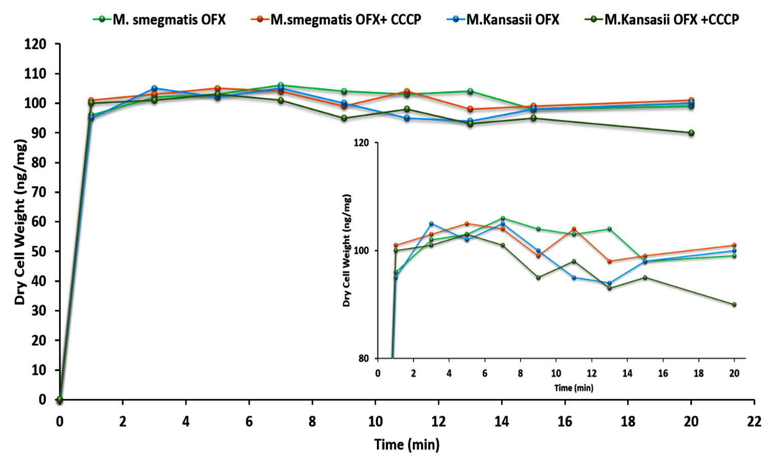
Figure 1. Accumulation of ofloxacin (OFX) in M. smegmatis and M. kansasii without and with CCCP (ng/mg dry cell weight; *Mean of 3 values)
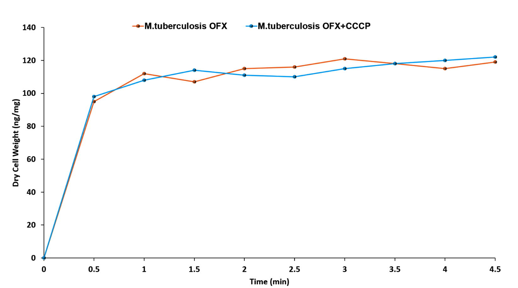
Figure 2. Accumulation of ofloxacin (OFX) in M. tuberculosis without and with CCCP (ng/mg dry cell weight; *Mean of 3 values)
Steady State Concentration (SSC) of ofloxacin in M. smegmatis was achieved within 3 minutes, and the ofloxacin accumulation level was 102 ng/mg of the bacilli dry weight. For M. kansasii and M. tuberculosis, the time span for SSC was 5 minutes, and the levels of ofloxacin accumulation were 100 ng/mg and 107 ng/mg, respectively. The accumulated ofloxacin concentration was approximately 10 µg/ml of exposed concentrations, which indicated that CCCP was unable to influence the ofloxacin accumulation. The addition of 100 µM CCCP for 10 minutes before the drug exposure did not influence the accumulation kinetics.
Table 1. Accumulation of ofloxacin (ofx) in M. smegmatis and M. kansasii without and with CCCP (ng/mg dry cell weight; *Mean of 3 values)
Time (Minutes) |
M. smegmatis OFX |
M. smegmatis OFX + CCCP |
M. kansasii OFX |
M. kansasii OFX + CCCP |
|---|---|---|---|---|
0 |
0 |
0 |
0 |
0 |
1 |
96 |
101 |
95 |
100 |
3 |
102 |
103 |
105 |
101 |
5 |
103 |
105 |
102 |
103 |
7 |
106 |
104 |
105 |
101 |
9 |
104 |
99 |
100 |
95 |
11 |
103 |
104 |
95 |
98 |
13 |
104 |
98 |
94 |
93 |
15 |
98 |
99 |
98 |
95 |
20 |
99 |
101 |
100 |
90 |
Table 2. Accumulation of ofloxacin (ofx) in M. tuberculosis without and with CCCP (ng/mg dry cell weight; *Mean of 3 values)
Time (Minutes) |
M. tuberculosis OFX |
M. tuberculosis OFX + CCCP |
|---|---|---|
0 |
0 |
0 |
0.5 |
95 |
98 |
1 |
112 |
108 |
1.5 |
107 |
114 |
2 |
115 |
111 |
2.5 |
116 |
110 |
3 |
121 |
115 |
3.5 |
118 |
118 |
4 |
115 |
120 |
4.5 |
119 |
122 |
FQs belong to the family of broad-spectrum antibiotics, are greatly effective towards Gram-negative bacteria, and tremendously impart bactericidal action.22-25 Bove et al.26 mentioned that due to their broad-spectrum activity of FQs and their physicochemical properties are mostly used as antimicrobials in human and animal medicine, which has led to great attraction in clinical situations.24,26 Over the last two decades, FQs have become popular due to their promising group of antimicrobial agents to treat the infection caused by MDR bacterial strains, such as aminoglycosides, β-lactam, and macrolide-resistant microbes.26,27 The addition of a fluorine atom to nalidixic acid leads to the derivation of FQs. Due to this, the activity against bacteria increases and leads to improved absorption, metabolism, and excretion, including high tissue and intracellular distribution.28-30 Compared to other groups of antibacterial agents, FQs are more effective at lower concentrations.
Against most of the susceptible microbes, FQs’ minimum inhibitory concentrations (MICs) range from 0.001-1.0 µg/ml,25 which was due to the prolonged post-antibiotic effect. Molecularly, Intracellular enzymes such as DNA gyrase and topoisomerase IV are the major targets of FQs. While in the microbe, FQs enter via pores and quickly accumulate with specific steady state concentrations.31,32 The enzyme-DNA complex was stabilized by FQs’ covalent binding, where the DNA is usually cleaved into two strands.33 The binding is in a cooperative manner by DNA gyrase with the help of electrostatic binding. This proposes the binding kinetics of FQs, which can fit into the deep pocket and lead to gyrase inhibition.34 Type II isomerase unwinds, cuts, and consecutively reseals the DNA; however, FQs inhibit the resealing and lead to fragment generation, which are later destroyed by exonucleases.35,36 FQs’ resistance does not emerge quickly because it is usually due to a mutation in the intrinsic genes of microbes, and it is rarely reported in plasmids.23 Plasmid-mediated FQs resistance has not been documented.37
In vitro and in vivo studies showed FQs as potent anti-mycobacterial agents.38,39 There is a tremendous effort to inculcate the FQs into frontline antibiotics like ofloxacin, moxifloxacin, and ciprofloxacin.40-43 Industrial efforts have also been seen in the discovery and development of newer FQs, which might have potential in TB treatment. However, FQs’ selection for TB treatment is based on their efficacy in murine models; therefore, few FQs are tested in these murine models. The cell wall of mycobacteria comprises 60% of lipids by dry weight, giving it extreme hydrophobicity, and has been used in chemotaxonomy and pathogenesis.44-46 For maintaining cellular integrity and virulence, M. tuberculosis has a special structure of cell envelope (peptidoglycan layer) and composition.47 Jacobo-Delgado explored the convolution of the M. tuberculosis cell wall and analyzed the antimicrobial peptides’ efficacy to clear the bacilli.48 In M. chelonae, permeability for β-lactams is three times less than E. coli and 10-times higher than P. aeruginosa.1 FQs are usually hydrophobic, and passive diffusion via lipid bilayer or porin is the existing mechanism in Gram-negative bacteria49; however, in bacteria (Gram-positive), it is simply diffusion across the membrane.50 Due to the hydrophilic nature of norfloxacin, its accumulation was non-saturable in M. aurum up to the 0-100 mg/ml concentration range, and its transportation was not affected in CCCP presence.51 Therefore, they suggested that noroxacin is transported through a non-saturable porin pathway. Porin efficiency in M. smegmatis and M. chelonae was lower than that of other bacterial porins.52,53 Ofloxacin is a broader-spectrum analog of norfloxacin, a second-generation fluoroquinolone, and its uptake could be due to the amino acid transporter.54 Newer generation FQs have an extended spectrum and improved activity against mycobacteria and other bacteria compared to older generation FQs. For ofloxacin accumulation, the cell wall does not serve as a barrier.
Earlier, Deo et al. reported the level of OFX, NFX, LFX, and CFX accumulation in M. smegmatis by simple diffusion.55 The SSC observed in the present study is similar to NFX-SSCs in M. smegmatis mc2155, as shown in Liu et al.56 Another previous report showed that the SSC of NFX was 35 ng/mg for M. smegmatis (strain CNCM 7326). Kocagoz et al.57 also found that NFX-SSC was 110 pmol/mg of cells (35 ng/mg of cells) for avirulent strain M. tuberculosis, which suggests that NFX is less permeable compared to other mycobacteria.58,59 The present study shows the OFX accumulation in M. smegmatis, M. kansasii, and M. tuberculosis. The SSC of OFX in M. smegmatis was 3 minutes, and the 102 ng/mg dry weight was the level of accumulation level. In M. kansasii, SSC time was five minutes, and 100 ng/mg was the accumulation level; however, in M. tuberculosis, SSC was 107 ng/mg in five minutes. The permeability of mycobacteria to ofloxacin varies between mycobacterial species.
In this study, the transportation of ofloxacin across the cell envelopes of M. smegmatis, M. tuberculosis, and M. kansasii was studied by diffusion. The intracellular level of drug in the presence of CCCP inhibitor was not affected, which suggests that FQs are transported by other means, not by efflux pumps, in these mycobacterial drug-susceptible clinical isolates. For validation, more studies are needed with FQ-resistant clinical isolates.
ACKNOWLEDGMENTS
The authors are grateful to the Director, ICMR-NJIL, and OMD for the support. This research was supported by ICMR, New Delhi. The authors are also thankful to the staff of the Biochemistry Division. The authors wish to acknowledge the invaluable contributions of Dr. Nirmala Deo to this study. Dr. Deo, who served as the first and co-corresponding author of this manuscript, sadly passed away in March 2026. We dedicate this work to her memory and her enduring contributions to science.
CONFLICT OF INTEREST
The authors declare that there is no conflict of interest.
AUTHORS’ CONTRIBUTION
All authors listed have made a substantial, direct and intellectual contribution to the work, and approved it for publication.
FUNDING
None.
DATA AVAILABILITY
All datasets generated or analyzed during this study are included in the manuscript.
ETHICS STATEMENT
Not applicable.
- Brennan PJ, Nikaido H. The envelope of mycobacteria. Ann Rev Biochemi. 1995;64(1):29-63.
Crossref - Jarlier V, Nikaido H. Permeability barrier to hydrophilic solutes in Mycobacterium chelonei. J Bacteriol. 1990;172(3):1418-23.
Crossref - Jarlier V, Nikaido H. Mycobacterial cell wall: structure and role in natural resistance to antibiotics. FEMS Microbiol Lett. 1994;123(1-2):11-18.
Crossref - Shandil RK, Jayaram R, Kaur P, et al. Moxifloxacin, ofloxacin, sparfloxacin, and ciprofloxacin against Mycobacterium tuberculosis: evaluation of in vitro and pharmacodynamic indices that best predict in vivo efficacy. Antimicrob Agents Chemother. 2007;51(2):576-582.
Crossref - Wanger A, Chavez V, Huang RS, Wahed A, Actor JK, Dasgupta A. Antibiotics, antimicrobial resistance, antibiotic susceptibility testing, and therapeutic drug monitoring for selected drugs. Microbiol Mol Diagn Pathol. 2017:119-153.
Crossref - Goshiye SM, Dube L, Mogas SB, Marine BT. Predictive Factors Associated With Multidrug Resistant Tuberculosis Among Tuberculosis Patients in Southwest Ethiopia: An Unmatched Case Control Study. Health Sci Rep. 2025;8(11):e71408.
Crossref - Pais JP, Policarpo M, Pires D, et al. Fluoroquinolone derivatives in the treatment of Mycobacterium tuberculosis infection. Pharmaceuticals. 2022;15(10):1213.
Crossref - Hooper DC, Jacoby GA. Topoisomerase inhibitors: fluoroquinolone mechanisms of action and resistance. Cold Spring Harb Perspect Med. 2016;6(9):a025320.
Crossref - Leysen DC, Haemers A, Pattyn SR. Mycobacteria and the new quinolones. Antimicrob Agents Chemother. 1989;33(1):1-5.
Crossref - Garcia-Rodriguez JA, Garcia AG. In-vitro activities of quinolones against mycobacteria. J Antimicrob Chemother. 1993;32(6):797-808.
Crossref - Vanden Broeck A, Lotz C, Ortiz J, Lamour V. Cryo-EM structure of the complete E. coli DNA gyrase nucleoprotein complex. Nat Commun. 2019;10(1):4935.
Crossref - Piddock LJV, Ricci V. Accumulation of five fluoroquinolones by Mycobacterium tuberculosis H37Rv. J Antimicrob Chemother. 2001;48(6):787-91.
Crossref - Chantefort A, Lellouche M. Bactericidal efficacy of a disinfectant by AFNOR standards NF T 72-150 and NF T 72-151. A collaborative study. Pathol Bio (Paris). 1984;32(5 Pt 2):623-627. PMID: 6431382.
- Lee OY, Wu HH, Besra GS. Professor David Minnikin Memorial Lecture: An era of the mycobacterial cell wall lipid biomarkers. Tuberculosis. 2023;1(143):102415.
Crossref - Minnikin DE. Chemical principles in the organization of lipid components in the mycobacterial cell envelope. Res Microbiol. 1991;142(4):423-427.
Crossref - Revel V, Cambau E, Jarlier V, Sougakoff W. Characterization of mutations in Mycobacterium smegmatis involved in resistance to fluoroquinolones. Antimicrob Agents Chemother. 1994;38(9):1991-6.
Crossref - Machineni NVK, Gullapalli D, Tangutoori S, et al. Dust to disease: cavitary lung lesion with Mycobacterium kansasii in a black lung patient. J Investig Med High Impact Case Rep. 2025;13:23247096251348906.
Crossref - Andalibi F, Bostanghadiri N, Amirmozafari N, Irajian G, Mirkalantari S. Efficacy and treatment outcome of infected patients with pulmonary Mycobacterium kansasii: A systematic review. J Clin Tuberc Other Mycobact Dis. 2024;1(36):100463.
Crossref - Yang J, Zhang L, Qiao W, Luo Y. Mycobacterium tuberculosis: Pathogenesis and therapeutic targets. MedComm. 2023;4(5):e353.
Crossref - Corti S, Chevalier J, Cremieux A. Intracellular accumulation of norfloxacin in Mycobacterium smegmatis. Antimicrob Agents and Chemother. 1995;39(11):2466-2471.
Crossref - Williams KJ, Chung GAC, Piddock LJV. Accumulation of norfloxacin by Mycobacterium aurum and Mycobacterium smegmatis. Antimicrob Agents Chemother. 1998;42(4):795-800.
Crossref - European Medicines Agency. Fluoroquinolone Antibiotics: Reminder of Measures to Reduce the Risk of Long-Lasting, Disabling and Potentially Irreversible Side Effects. 2023. https://www.ema.europa.eu/en/news/fluoroquinolone-antibiotics-reminder-measures-reduce-risk-long-lasting-disabling-potentially-irreversible-side-effects. (accessed 28 July 2023).
- Brown SA, Cooper J, Gauze JJ, Greco DS, Weise DW, Buck JM. Pharmacokinetics of norfloxacin in dogs after single intravenous and single and multiple oral administrations of the drug. Am J Vet Res. 1990;51(7):1065-1070.
Crossref - Petri WA. Sulfonamides, trimethoprim, sulfamethoxazole, quinolones, and agents for urinary tract infections. Goodman & Gilman’s the Pharmacological Basis of Therapeutics. New York (NY): McGraw-Hill. 2006:1111-25.
- McKellar Q, Gibson I, Monteiro A, Bregante M. Pharmacokinetics of enrofloxacin and danofloxacin in plasma, inflammatory exudate, and bronchial secretions of calves following subcutaneous administration. Antimicrob agents chemother, 1999; 43(8): 1988-1992.
Crossref - Bove C, Baldock RA, Champigneulle O, Martin L, Bennett CL. Fluoroquinolones: old drugs, putative new toxicities. Expert Opin Drug Safety. 2022;21(11):1365-1378.
Crossref - Rodrigues CF, Silva F. The rise, fall, and rethink of (Fluoro) quinolones: A quick rundown. Pathogens. 2025;14(6):525.
Crossref - Pascual A, Garcia I, Ballesta S, Perea EJ. Uptake and intracellular activity of trovafloxacin in human phagocytes and tissue-cultured epithelial cells. Antimicrob Agents Chemother. 1997;41(2):274-277.
Crossref - Ito T, Yano I, Masuda S, Hashimoto Y, Inui KI. Distribution characteristics of levofloxacin and grepafloxacin in rat kidney. Pharm Res. 1999;16(4):534-539.
Crossref - Ezelarab HAA, Abbas SH, Hassan HA, Abuo Rahma GE. Recent updates of fluoroquinolones as antibacterial agents. Archiv der Pharmazie. 2018;351(9):1800141.
Crossref - Chapman JS, Georgopapadakou NH. Routes of quinolone permeation in Escherichia coli. Antimicrob Agents Chemother. 1988;32(4):438-442.
Crossref - Piddock LJ. New quinolones and gram-positive bacteria. Antimicrob Agents Chemother. 1994;38(2):163-9.
Crossref - Bush NG, Diez-Santos I, Abbott LR, Maxwell A. Quinolones: mechanism, lethality and their contributions to antibiotic resistance. Molecules. 2020;25(23):5662.
Crossref - Wolfson JS, Hooper DC. The fluoroquinolones: structures, mechanisms of action and resistance, and spectra of activity in vitro. Antimicrob Agents Chemother. 1985;28(4):581-586.
Crossref - Bourguignon GJ, Levitt M, Sternglanz R. Studies on the mechanism of action of nalidixic acid. Antimicrob Agents Chemother. 1973;4(4):479-486.
Crossref - Shen LL, Pernet AG. Mechanism of inhibition of DNA gyrase by analogues of nalidixic acid: the target of the drugs is DNA. Proc Natl Acad Sci USA. 1985;82(2):307-311.
Crossref - Dowling PM, Baptiste KE. Miscellaneous antimicrobials: ionophores, nitrofurans, nitroimidazoles, rifamycins, and others. In: Dowling PM, Prescott JF, Baptiste KE, eds. Antimicrobial Therapy in Veterinary Medicine. 6th ed. Wiley Blackwell; 2025. Chapter 18.
Crossref - Lowther J, Bryskier A. Fluoroquinolones and tuberculosis. Expert Opin Investig Drugs. 2002;11(2):233-258.
Crossref - Ginsburg AS, Grosset JH, Bishai WR. Fluoroquinolones, tuberculosis, and resistance. Lancet Infect Dis. 2003;3(7):432-442.
Crossref - Nuermberger EL, Yoshimatsu T, Tyagi S, et al. Moxifloxacin-containing regimen greatly reduces time to culture conversion in murine tuberculosis. Am J Respirat Crit Care Med. 2004;169(3):421-426.
Crossref - Narayanan P. Tuberculosis Research Centre. Shortening short course chemotherapy a randomised clinical trial for treatment of smear positive pulmonary tuberculosis with regimens using ofloxacin in the intensive phase. Ind J Tub. 2002;49:27-38.
- Yoshimatsu T, Nuermberger E, Tyagi S, Chaisson R, Bishai W, Grosset J. Bactericidal activity of increasing daily and weekly doses of moxifloxacin in murine tuberculosis. Antimicrob Agents Chemother. 2002;46(6):1875-1879.
Crossref - Falzon D, Schunemann HJ, Harausz E, et al. World Health Organization treatment guidelines for drug-resistant tuberculosis, 2016 update. Eur Respirat J. 2017;49(3):1602308.
Crossref - Jackson M. The mycobacterial cell envelope—lipids. Cold Spring Harb Perspect Med. 2014;4(10):a021105.
Crossref - Singh P, Rameshwaram NR, Ghosh S, Mukhopadhyay S. Cell envelope lipids in the pathophysiology of Mycobacterium tuberculosis. Future Microbiol. 2018;13(6):689-710.
Crossref - Ghazaei C. Mycobacterium tuberculosis and lipids: Insights into molecular mechanisms from persistence to virulence. J Res Med Sci. 2018;23(1):63.
Crossref - Maitra A, Munshi T, Healy J, et al. Cell wall peptidoglycan in Mycobacterium tuberculosis: An Achilles’ heel for the TB-causing pathogen. FEMS Microbiol Rev. 2019;43(5):548-75.
Crossref - Jacobo-Delgado YM, Rodriguez-Carlos A, Serrano CJ, Rivas-Santiago B. Mycobacterium tuberculosis cell-wall and antimicrobial peptides: a mission impossible? Front Immunol. 2023;14:1194923.
Crossref - Piddock LJV. Mechanism of quinolone uptake into bacterial cells. J Antimicrob Chemother. 1991;27(4):399-403.
Crossref - Furet YX, Deshusses J, Pechere JC. Transport of pefloxacin across the bacterial cytoplasmic membrane in quinolone-susceptible Staphylococcus aureus. Antimicrob Agents Chemother. 1992;36(11):2506-2511.
Crossref - Asuquo AE, Piddock LJV. Accumulation and killing kinetics of fifteen quinolones for Escherichia coil, Staphylococcus aureus and Pseudonwnas aeruginosa. J Antimicrob Chemother. 1993;31(6):865-880.
Crossref - Trias J, Jarlier V, Benz R. Porins in the cell wall of mycobacteria. Science. 1992;258(5087):1479-81.
Crossref - Trias J, Benz R. Characterization of the channel formed by the mycobacterial porin in lipid bilayer membranes. Demonstration of voltage gating and of negative point charges at the channel mouth. J Biol Chem. 1993;268(9):6234-6240.
Crossref - Pascual A, Garcia I, Perea EJ. Fluorometric measurement of ofloxacin uptake by human polymorphonuclear leukocytes. Antimicrob Agents Chemother. 1989;33(5):653-656.
Crossref - Deo N, Sharma D, Bisht D, Venkatesan K. Evaluation of the intracellular accumulation of fluoroquinolones in mycobacteria by fluorometric assays. Int J Mycobacteriol. 2020;9(1):34-38.
Crossref - Liu J, Takiff HE, Nikaido H. Active efflux of fluoroquinolones in Mycobacterium smegmatis mediated by LfrA, a multidrug efflux pump. J Bacteriol. 1996;178(13):3791-5.
Crossref - Kocagoz T, Hackbarth CJ, Unsal I, Rosenberg EY, Nikaido H, Chambers HF. Gyrase mutations in laboratory-selected, fluoroquinolone-resistant mutants of Mycobacterium tuberculosis H37Ra. Antimicrob Agents Chemother. 1996;40(8):1768-74.
Crossref - Woo SR, Sotos J, Hart AP, Barletta RG, Czuprynski CJ. Bovine monocytes and a macrophage cell line differ in their ability to phagocytose and support the intracellular survival of Mycobacterium avium subsp. paratuberculosis. Vet Immunol Immunopathol. 2006;110(1-2):109-120.
Crossref - Vacher S, Pellegrin JL, Leblanc F, Fourche J, Maugein J. Comparative antimycobacterial activities of ofloxacin, ciprofloxacin, and grepafloxacin. J Antimicrob Chemother. 1999;44(5):647-652.
Crossref
© The Author(s) 2026. Open Access. This article is distributed under the terms of the Creative Commons Attribution 4.0 International License which permits unrestricted use, sharing, distribution, and reproduction in any medium, provided you give appropriate credit to the original author(s) and the source, provide a link to the Creative Commons license, and indicate if changes were made.