


ISSN: 0973-7510
E-ISSN: 2581-690X
 , Fatih Buyuk2, Yasemin Bayram3 and Yaren Ersoy2
This study aimed to determine the distribution of bacterial and fungal pathogens in neonatal sepsis cases in a neonatal unit during the COVID-19 pandemic and to investigate antimicrobial susceptibility/resistance profiles using phenotypic and molecular methods. A total of 940 samples were analysed in this study. Pathogens were identified using conventional culture methods and the Siemens MicroScan Walkaway 96 Plus system. Antimicrobial susceptibility of the isolates was determined phenotypically using the Siemens MicroScan Walkaway 96 Plus system and by PCR. Selected resistance genes were detected using multiplex PCR. A total of 113 isolates were recovered, predominantly Gram-negative bacilli (72%). The most frequent pathogens were coagulase-negative staphylococci (n = 28), Acinetobacter baumannii/haemolyticus (n = 22), Klebsiella spp. (n = 16), Escherichia coli (n = 10), Enterococcus spp. (n = 10), Candida albicans (n = 8), Stenotrophomonas maltophilia (n = 6), Serratia marcescens (n = 5), Staphylococcus aureus (n = 5) and Pseudomonas aeruginosa (n = 3). High resistance rates to beta-lactam antibiotics were observed, particularly among Gram-negative isolates. ESBL production ranged from 33%-50% in Enterobacterales, while MDR rates varied between 20% and 95.45% across species. All C. albicans isolates were susceptible to amphotericin B and to caspofungin. These findings highlight the urgent need for strengthened infection control measures and tailored empirical therapy.
Neonatal Sepsis, Antimicrobial Resistance, Pathogens, ESBL, MDR
Neonatal sepsis, characterized as a systemic infection within the neonatal period (0-28 days), continues to be a major contributor to infant mortality globally, posing a persistent clinical and public health concern worldwide, particularly in low- and middle-income countries.1-3
The emergence of resistance among bacterial pathogens has become a significant public health concern worldwide.4 The COVID-19 pandemic has further intensified these issues in NICUs.3,4 Increased patient loads, staffing shortages, disrupted infection control practices, and excessive antibiotic use have been associated with surges in MDR infections, including carbapenem-resistant Enterobacterales and non-fermenters.3-5
This study, which was conducted with a particular emphasis on neonatal infections, aimed to determine the distribution of bacterial and fungal agents in cases of neonatal sepsis in the neonatal unit, offering contemporary local data on pathogen resistance patterns, and implications for management during the COVID-19 pandemic.
Study design and setting
This study was conducted at Van Yuzuncu Yil University, Turkey, between 2020 and 2022. The study included microbiological samples obtained for routine diagnostic purposes from infants aged 0-28 days who presented with a preliminary diagnosis of sepsis.
Ethical approval for this study was obtained from the Non-Interventional Clinical Research Ethics Committee of Van Yüzüncü Yıl University (Türkiye) (decision no. 2020/01-19, dated 17 January 2020). The study was conducted in accordance with the Declaration of Helsinki. Written informed consent was obtained from all participants prior to their enrollment in the study.
Study material
A total of 940 clinical samples were analyzed, including 647 blood, 133 urine, 70 cerebrospinal fluid (CSF), 56 catheter, 27 total parenteral nutrition (TPN) fluid, 3 abscesses, 2 paracentesis, 1 ear swab, and 1 nasal swab. Samples yielding the growth of potential pathogens were included, and probable contaminants were excluded.
Isolation and identification of microorganisms
The samples were inoculated onto 5% sheep blood agar and Eosin Methylene Blue (EMB) agar and incubated aerobically at 37 °C for 24-48 hours. The Gram characteristics of the pure colonies were revealed using the Gram staining method. Bacterial identification was performed using the identification (ID) panel of the MicroScan WalkAway 96 Plus automated system (Siemens Healthcare Diagnostics, West Sacramento, CA, USA).
For the fungal agents, samples were inoculated onto Sabouraud Dextrose Agar (SDA) medium and incubated aerobically at 37 °C for up to 7 days. The preliminary identification was performed based on their macroscopic and microscopic morphologies.
Antimicrobial susceptibility tests
Antimicrobial susceptibility testing (AST) of bacterial isolates was performed using the appropriate AST panels on a MicroScan WalkAway 96 Plus system. The results were interpreted according to the Clinical and Laboratory Standards Institute (CLSI M100, 30th ed. 2020) guidelines.6 Different panels were employed depending on the sample origin. Screening and confirmation of extended-spectrum β-lactamase (ESBL) production, vancomycin-resistant enterococci (VRE), and methicillin-resistant Staphylococcus aureus (MRSA) were performed according to the algorithm of the automated system and CLSI recommendations.
Antimicrobial susceptibility analyses of the fungal isolates were performed using the gradient test (E-test) method.7 E-test strips (BM Bioanalyse, Turkey) of fluconazole (FLU, 0.016-256 µg/ml), voriconazole (VO, 0.002-32 µg/ml), amphotericin B (AMB, 0.002-32 µg/ml), and caspofungin (CAS, 0.002-32 µg/ml) were used.
Multidrug-resistance (MDR) was defined as non-susceptibility to at least one agent in three or more antimicrobial categories.5
Molecular analysis
DNA extraction
DNA extraction from bacteria and yeast isolates was performed according to the phenol-chloroform-isoamyl alcohol method reported by Saran et al.8
Candida albicans specific PCR
Candida albicans-specific PCR was performed using forward (CA,5′ -TCAACTTGTCACACCAGATTATT-3′) and reverse (ITS4, 5′-TCCTCCGCTTATTGATATGC-3′) primers designed by Li et al.9 and White et al.10 respectively. The products of 402 bp in size were identified as C. albicans based on 1.5% agarose gel electrophoresis.
PCR analysis of antimicrobial resistance genes
Antimicrobial resistance gene analysis was performed in parallel with MicroScan WalkAway 96 Plus AST and gradient assay results. Common genes of clinical importance that are responsible for resistance were selected. PCRs was performed using the primer pairs listed in Table 1.11-20
Table 1. Primer pairs used for the PCR process
| Gene | Target | Primers | Annealing (°C) | Product (bp) | Ref. |
|---|---|---|---|---|---|
| TEM | ESBL | F-5’-ATGAGTATTCAACATTTCCGTG-3’ | 55 | 861 | [11] |
| R-5’-TTACCAATGCTTAATCAGTGAG-3’ | |||||
| CTX-M | ESBL | F-5’-TTTGCGATGTGCAGTACCAGTAA-3’ | 57 | 544 | [12] |
| R-5’-CGATATCGTTGGTGGTGCCATA-3’ | |||||
| CMY-2 | AmpC beta-lactamase | F-5’-GACAGCCTCTTTCTCCACA-3’ | 50 | 1000 | [13] |
| R-5’-TGGAACGAAGGCTACGTA-3’ | |||||
| VIM | Carbapenemase | F-5’-GATGGTGTTTGGTCGCATA-3’ | 54 | 390 | [14] |
| R-5’-CGAATGCGCAGCACCAG-3’ | |||||
| NDM | Carbapenemase | F-5’-GGTTTGGCGATCTGGTTTTC-3’ | 54 | 621 | |
| R-5’-GAATGGCTCATCACGATC-3’ | |||||
| KPC | Carbapenemase | F-5’-ATGTCACTGTATCGCCGTCT-3’ | 54 | 893 | |
| R-5’-TTTTCAGAGCCTTACTGCCC-3’ | |||||
| SUL-2 | Sulfonamide | F-5’-CCAATACCGCCAGCCCGTCG-3’ | 56 | 489 | [15] |
| R-5’-TGCCTTGTCGCGTGGTGTGG-3’ | |||||
| MecA | Methicillin | F-5’-CCTAGTAAAGCTCCGGAA-3’ | 60 | 314 | [16] |
| R-5’-CTAGTCCATTCGGTCCA-3’ | |||||
| vanA | Glycopeptide | F-5’-GGGAAAACGACAATTGC-3’ | 54 | 732 | [17] |
| R-5’-GTACAATGCGGCCGTTA-3’ | |||||
| vanB | Glycopeptide | F-5’-ATGGGAAGCCGATAGTC-3’ | 54 | 647 | |
| R-5’-GATTTCGTTCCTCGACC-3’ | |||||
| vanC | Glycopeptide | F-5’-GAAAGACAACAGGAAGACCGC-3’ | 54 | 796 | |
| R-5’-ATCGCATCACAAGCACCAATC-3’ | |||||
| AaDB | Aminoglycoside | F-5’-TTACGCAGCAGGGCAGTCGC-3’ | 56 | 551 | [15] |
| R-5’-GCGGCACGCAAGACCTCAAC-3’ | |||||
| AaDA25 | Aminoglycoside | F-5’-GCAGTGGATGGCGGCCTGAA-3’ | 56 | 503 | |
| R-5’-TCGGCGCGATTTTGCCGGTT-3’ | |||||
| ERG11 | Fluconazole | F-5’-CATAACTCAAATATGGCTATT-3’ | 50 | 245 | [18] |
| R-5’-CTTTTGACGACATGATTCGA-3’ | |||||
| F-5’-TTGAAACTGTCATTGATGGC-3’ | 193 | ||||
| R-5’-GGTTGTTGACCATATGAAGC-3’ | |||||
| FUR1 | Flucytosine | F-5’-CGCAACCTGATTTTGTCCATA-3’ | 50 | 340 | [19] |
| R-5’-ATCGGAAGAATATCATGAAAATCC-3’ | |||||
| FKS1 | Caspofungin | F-5’-GAAATCGGCATATGCTGTGTC-3’ | 50 | 450 | [20] |
| R-5’-AATGAACGACCAATGGAGAAG-3’ |
ESBL, Extended-Spectrum β-Lactamases
The PCR products were evaluated for the relevant resistance genes by electrophoresis on a 1.5% agarose gel (Figure 1). Positive controls (S. aureus, K. pneumoniae, E. coli, E. faecalis, C. albicans) belonging to the culture collection of the Department of Microbiology, Faculty of Veterinary Medicine, were used in the study.
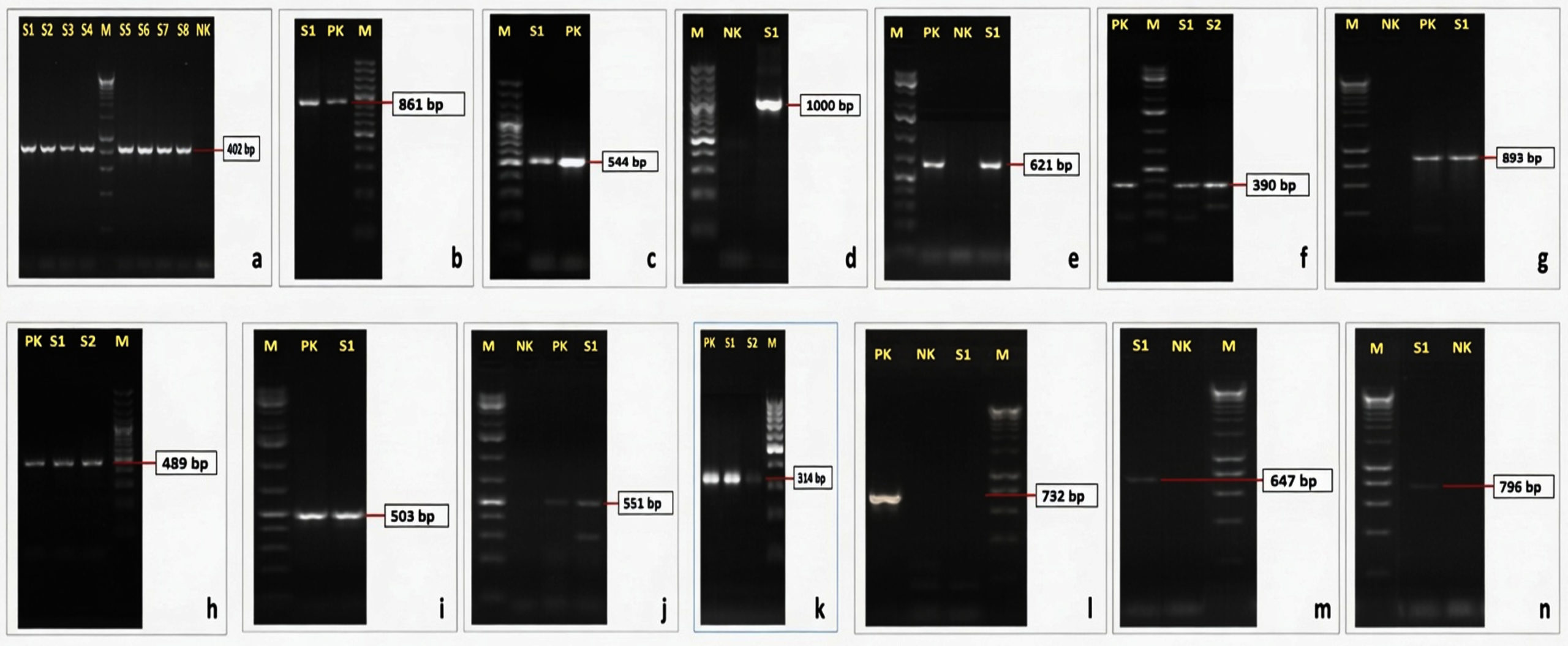
Figure 1. Electrophoresis of PCR products. C. albicans species-specific PCR (a); TEM (b), CTX-M (c), CMY-2 (d), NDM (e), VIM (f), KPC (g), SUL-2 (h), AaDA25 (i), AaDB (j), mecA (k), vanA (l), vanB (m), and vanC (n) resistance gene-specific PCR. M: DNA ladder (GeneRuler 1 kb Plus DNA ladder, SM1331, Thermo Fisher Sci. (a, e, f, i, j), GeneRuler 100 bp plus DNA ladder, SM0321, Thermo Fisher Sci. (b, c, d, h), HyperLadder 1 kb DNA ladder, BIO-33053, Bioline (g, l, m, n), GeneRuler 100 bp DNA ladder, SM0241, Thermo Fisher Sci.) (k). S1, S2, …, Sn: Field strains, PK: Positive control, NK: Negative control
Statistical analysis
The data were analyzed using the IBM SPSS Version 25.0 software. Chi-square and Fisher’s exact tests were used for the data analysis of variables to determine whether there was a relationship between categorical variables. P < 0.05 was taken statistically significant. Due to the limited number of isolates for some species and antibiotics, confidence intervals and advanced statistical tests were not routinely performed.
Identification findings
Analysis results of the 940 clinical samples were found to be positive for bacterial agents in 105 (11.17%) and fungal agents in 8 (0.85%) (Table 2). The culture positivity was calculated at rates that varied significantly by sample type (Pearson’s chi-square test). The culture positivity was calculated as 100%, 100%, 50%, 33.33%, 30.36%, 13.53%, 10.66%, 5.71%, and 3.7% for ear, nose, paracentesis, abscess, catheter, urine, blood, CSF, and TPN samples, respectively. The most common sources of isolated microorganisms were blood 69 (7.34%), urine 18 (1.91%), and catheter 17 (1.80%).
The distribution of the isolates is shown in Table 2. The most frequent pathogens were coagulase-negative staphylococci (28 isolates, 24.8%), A. baumannii/haemolyticus (22, 19.5%), Klebsiella spp. (16, 14.2%), E. coli and Enterococcus spp. (10 each, 8.8%), C. albicans (8, 7.1%), S. maltophilia (6, 5.3%), S. marcescens (5, 4.4%), S. aureus (5, 4.4%), and P. aeruginosa (3, 2.7%). All Candida isolates were confirmed to be C. albicans using species-specific PCR.
Table 2. Distribution of the isolates according to species and sample origin
| Microorganism | No. of isolates | Distribution of isolates by sample origin | ||||||||
|---|---|---|---|---|---|---|---|---|---|---|
| Blood n = 647 |
Urine n = 133 |
Catheter n = 56 |
CSF n = 70 |
Abscess n = 3 |
Paracentesis n = 2 |
Ear svap n = 1 |
Nasal svap n = 1 |
TPN n = 27 |
||
| CoNS | 28 (24.78%) | 24 (21.24%) | 0 (0%) | 3 (2.65%) | 1 (0.88%) | 0 (0%) | 0 (0%) | 0 (0%) | 0 (0%) | 0 (0%) |
| A. baumanii/ haemolyticus | 22 (19.47%) | 15 (13.27%) | 2 (1.77%) | 1 (0.88%) | 2 (1.77%) | 0 (0%) | 1 (0.88%) | 0 (0%) | 0 (0%) | 1 (0.88%) |
| Klebsiella spp. | 16 (14.16%) | 9 (7.96%) | 4 (3.54%) | 3 (2.65%) | 0 (0%) | 0 (0%) | 0 (0%) | 0 (0%) | 0 (0%) | 0 (0%) |
| E. coli | 10 (8.85%) | 3 (2.65%) | 6 (5.31%) | 1 (0.88%) | 0 (0%) | 0 (0%) | 0 (0%) | 0 (0%) | 0 (0%) | 0 (0%) |
| Enterococcus spp. | 10 (8.85%) | 6 (5.31%) | 3 (2.65%) | 1 (0.88%) | 0 (0%) | 0 (0%) | 0 (0%) | 0 (0%) | 0 (0%) | 0 (0%) |
| S. maltophilia | 6 (5.31%) | 1 (0.88%) | 0 (0%) | 4 (3.54%) | 0 (0%) | 1 (0.88%) | 0 (0%) | 0 (0%) | 0 (0%) | 0 (0%) |
| S. marcescens | 5 (4.42%) | 4 (3.54%) | 1 (0.88%) | 0 (0%) | 0 (0%) | 0 (0%) | 0 (0%) | 0 (0%) | 0 (0%) | 0 (0%) |
| S. aureus | 5 (4.42%) | 2 (1.77%) | 0 (0%) | 2 (1.77%) | 0 (0%) | 0 (0%) | 0 (0%) | 0 (0%) | 1 (0.88%) | 0 (0%) |
| P. aeruginosa | 3 (2.65%) | 1 (0.88%) | 0 (0%) | 1 (0.88%) | 0 (0%) | 0 (0%) | 0 (0%) | 1 (0.88%) | 0 (0%) | 0 (0%) |
| Subtotal | 105 (11.17%) | 65 (10.05%) | 16 (12.03%) | 16 (28.57%) | 3 (4.29%) | 1 (33.33%) | 1 (50%) | 1 (100%) | 1 (100%) | 1 (3.7%) |
| C. albicans | 8 (0.85%) | 4 (0.62%) | 2 (1.5%) | 1 (1.79%) | 1 (1.43%) | 0 (0%) | 0 (0%) | 0 (0%) | 0 (0%) | 0 (0%) |
| Total | 113 (12.02%) | 69 (10.66%) | 18 (13.53%) | 17 (30.36%) | 4 (5.71%) | 1 (33.33%) | 1 (50%) | 1 (100%) | 1 (100%) | 1 (3.7%) |
CoNS, Coagulase-negative staphylococci; CSF, Cerebrospinal Fluid; TPN, Total Parenteral Nutrition
Phenotypic antimicrobial susceptibility findings
The antimicrobial susceptibility results are summarized in Table 3. Gram-positive isolates exhibited high resistance to penicillin, oxacillin, and aminoglycosides but universal susceptibility to glycopeptides. Vancomycin-resistance was not observed in enterococci. Methicillin-resistance was prevalent in CoNS and S. aureus. Gram-negative isolates showed extensive resistance to beta-lactam antibiotics, including high rates of resistance to carbapenems in A. baumannii/haemolyticus and Klebsiella spp. Multidrug-resistance (MDR) was frequent, reaching 95.45% in A. baumannii/haemolyticus and 81.25% in Klebsiella spp. The ESBL phenotype was detected in 33%-50% of Enterobacterales. All C. albicans isolates were susceptible to amphotericin B and caspofungin, with 50% resistance to fluconazole and voriconazole.
Table 3. Resistance percentages of microorganisms obtained from cultural analysis to antimicrobial agents
| Bacterial isolates | Antimicrobial Resistance Rates of Isolates (%) | |||||||||||||||||||||
|---|---|---|---|---|---|---|---|---|---|---|---|---|---|---|---|---|---|---|---|---|---|---|
| Beta-lactams | Carbapenems | Sulfonamides | Glycopeptides | Aminoglycosides | Fluoroquinolones | |||||||||||||||||
| AMC | AMP | AZT | CAZ | CTX | FEP | OX | P | TZP | ETP | IMP | MEM | SXT | TEC | VA | AK | CN | TOB | CIP | ||||
| CoNS | 92.86 | 100 | – | – | – | – | 96.43 | 100 | – | – | – | – | 29.63 | 0 | 0 | – | 100 | 100 | 88.89 | |||
| S. aureus | 75 | 80 | – | – | – | – | 100 | 80 | – | – | – | – | 0 | 0 | 0 | – | 100 | 100 | 0 | |||
| Enterococcus spp. | – | 50 | – | – | – | – | – | 100 | – | – | – | – | 100 | 28.57 | 28.57 | – | 100 | 100 | 71.43 | |||
| A. baumannii/ haemolyticus | – | – | – | 81.82 | 81.82 | 81.82 | – | – | – | – | 100 | 95.45 | 95.45 | – | – | 95.45 | 100 | 95.45 | – | |||
| Klebsiella spp. | 80 | 100 | 100 | 100 | 100 | 100 | – | – | 57.14 | 88.89 | 30.77 | 54.55 | 68.75 | 0 | – | 58.33 | 80 | 81.25 | 70 | |||
| E. coli | 55.56 | 100 | 100 | – | 80 | 100 | – | – | 14.29 | 40 | 0 | 0 | 75 | – | 100 | 33.33 | 30 | 33.33 | 75 | |||
| S. marcescens | 100 | 100 | 100 | 100 | 100 | 80 | – | – | 66.67 | 100 | 50 | 66.67 | 25 | – | – | 40 | 75 | 50 | 100 | |||
| P. aeruginosa | – | – | 0 | 0 | – | 50 | – | – | 50 | – | 0 | 0 | – | – | – | 50 | 50 | 50 | 50 | |||
| S. maltophilia | – | – | – | 66.67 | – | – | – | – | – | – | – | – | 0 | – | – | – | – | – | – | |||
| Fungal isolates | Polyene macrolides | Echinocandins | Azoles | |||||||||||||||||||
| AMB | CAS | FLU | VO | |||||||||||||||||||
| C. albicans | 0 | 0 | 50 | 50 | ||||||||||||||||||
AMC, Amoxicillin-Clavulanate; AMP, Ampicillin; AZT, Aztreonam; CAZ, Ceftazidime; CTX; Cefotaxime; FEP, Cefepime; OX, Oxacillin; P, Penicillin; TZP, Piperacillin/Tazobactam; ETP= ertapenem; IMP, Imipenem; MEM, Meropenem; SXT, Trimethoprim/Sulfamethoxazole; TEC, Teicoplanin; VA, Vancomycin; AK, Amikacin; CN, Gentamicin; TOB, Tobramycin; CIP, Ciprofloxacin; AMB, Amphotericin B; CAS, Caspofungin; FLU, Fluconazole; VO, Voriconazole; CoNS, Coagulase-negative staphylococci
Antimicrobial resistance gene analysis findings
At least one antimicrobial resistance gene was detected in 91 (86.67%) of the 105 bacterial isolates. The detailed gene distribution is shown in Figure 2. Among Gram-negative isolates, blaTEM was the most prevalent ESBL gene, ranging from 33.33% (P. aeruginosa) to 93.33% (Klebsiella spp.). blaCTX-M was detected in 66.67% of E. coli and Klebsiella spp., but was rare (6.25%) in A. baumannii/haemolyticus and absent in S. marcescens, S. maltophilia, and P. aeruginosa. blaCMY-2 (AmpC-type) was found in 46.67% of Klebsiella spp., 20% of S. marcescens, 17.65% of A. baumannii/haemolyticus, and 16.67% of E. coli. Carbapenemase genes showed distinct patterns: blaNDM was common in S. marcescens (80%) and Klebsiella spp. (33.33%), but low in A. baumannii/haemolyticus (11.76%) and absent in other species. blaVIM predominated in A. baumannii/haemolyticus (88.24%) and S. maltophilia (83.33%), with a low prevalence in Klebsiella (7.14%). blaKPC was detected only in Klebsiella spp. (23.08%) and E. coli (16.67%). The sulfonamide resistance gene, sul2, was highly prevalent in A. baumannii/haemolyticus (94.44%) and E. coli (60%). Aminoglycoside-modifying enzyme genes were frequently found in Klebsiella spp. (aadA25 60%, aadB 60%) and E. coli (aadA25 50%), with lower rates in other species. Among Gram-positive isolates, mecA was detected in 26 CoNS and two S. aureus isolates. Glycopeptide resistance genes were infrequent: vanA in 40% of S. aureus, 30% of Enterococcus spp., and one CoNS; vanB in 20% of S. aureus; and vanC in one CoNS.
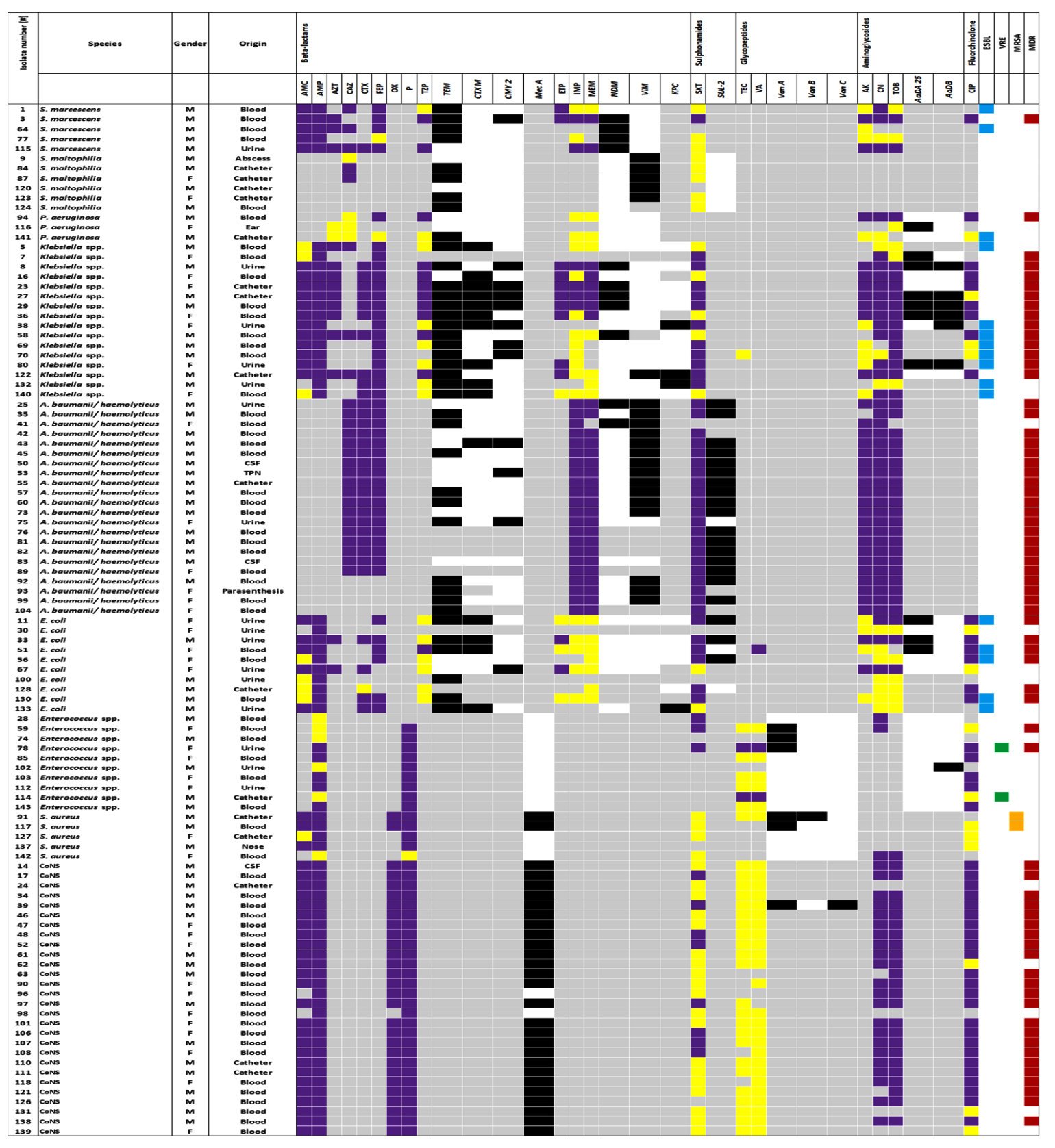
Figure 2. Antibiotic resistance and related gene positivity of bacterial isolates. ■ resistant; ■ sensitive; ■ not determined; ■ positive; ■ ESBL= extended-spectrum β-lactamases; ■ VRE= vancomycin-resistant enterococci; ■ MRSA= methicillin-resistant Staphylococcus aureus; ■ MDR= multidrug-resistance; CoNS= coagulase-negative staphylococci; CSF= cerebrospinal Fuid; TPN= total parenteral nutrition, AMC= amoxicillin-clavulanate; AMP= ampicillin; AZT= aztreonam, CAZ= ceftazidime; CTX= cefotaxime; FEP= cefepime, OX= oxacillin; P= penicillin; TZP= piperacillin/tazobactam, ETP= ertapenem; IMP= imipenem; MEM= meropenem; SXT= trimethoprim/sulfamethoxazole; TEC= teicoplanin; VA= vancomycin; AK= amikacin; CN= gentamicin; TOB= tobramycin; CIP= ciprofloxacin
No significant difference was observed in overall resistance rates between Gram-negative and Gram-positive isolates (chi-square test, P = 0.527), though Gram-negatives showed broader resistance patterns. Carbapenemase gene distribution showed species-specific patterns: blaVIM was significantly more prevalent in non-fermenters (A. baumannii/ haemolyticus 88.24%, S. maltophilia 83.33%) than in Enterobacterales (Fisher’s exact test, P = 0.048). The mecA gene was detected in 92.9% of CoNS and 40% of S. aureus isolates, with a significantly higher prevalence in CoNS than in other Gram-positive species (Fisher’s exact test, P < 0.0001). Glycopeptide resistance genes (vanA, vanB, and vanC) were infrequent, predominantly in S. aureus and Enterococcus spp.
At least one antifungal resistance gene was detected in seven (87.5%) C. albicans isolates. Among the isolates, FUR1 positivity was 75%, ERG11 positivity was 62.5%, and FKS1 positivity was 50%, respectively (Figure 3).
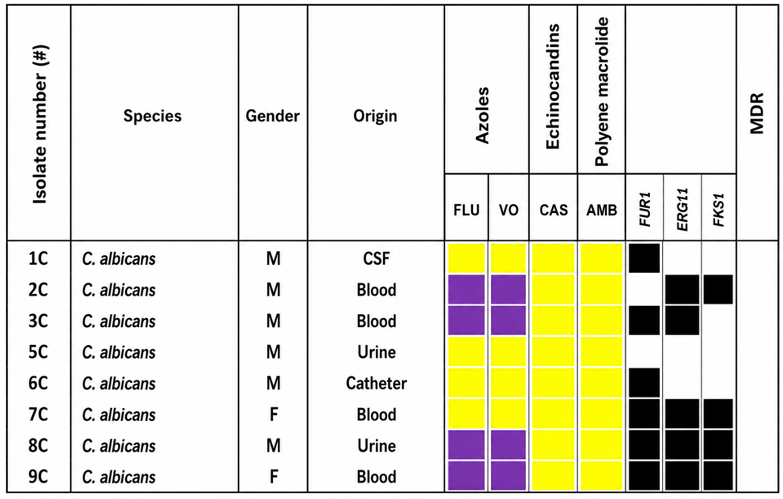
Figure 3. Antibiotic resistance and related gene positivity of bacterial isolates. ■ = resistant, ■ = sensitive, ■ = positive; CSF = cerebrospinal fluid, FLU = fluconazole, VO = voriconazole, CAS = caspofungin, AMB = amphotericin B, MDR = multidrug-resistance
Correlation between phenotypic resistance, resistance genes, and MDR patterns
Strong correlations were observed between phenotypic resistance and gene detection for beta-lactams and aminoglycosides in Klebsiella spp., Acinetobacter baumannii/ haemolyticus, E. coli, and other species, contributing to high rates of MDR. Notable findings included multiple gene positivity in MDR isolates of S. marcescens, S. maltophilia, and P. aeruginosa. However, discrepancies were evident, such as the presence of resistance genes in phenotypically susceptible isolates or the absence of tested genes in resistant isolates.
In C. albicans, azole resistance was associated with higher gene diversity, although genes were also present in the susceptible isolates.
Neonatal sepsis remains a major cause of morbidity and mortality in newborns, particularly in neonatal intensive care units (NICUs), where immature immune systems and invasive procedures increase susceptibility to infection.1,3,5 Advances in neonatal care have improved the survival rates of preterm and low-birth-weight infants; however, prolonged hospitalization and device use have also increased the risk of healthcare-associated infections. The widespread use of broad-spectrum antibiotics has driven the emergence of multidrug-resistant (MDR) pathogens, complicating empirical therapy and worsening patient outcomes.1,5
The bacterial profile of neonatal infections varies depending on the neonatal period.1,2 Streptococcus agalactiae, E. coli, S. aureus, Enterococcus spp., and Streptococcus pneumoniae are the most common bacteria reported in the early-period of neonatal infection cases, whereas, Gram-negative bacteria, CoNS, K. pneumoniae and A. baumannii are mostly reported in the late-period cases. Candida spp. are the most common fungal species detected in these cases.1,2 In this study, conducted during the COVID-19 pandemic, Gram-negative bacilli accounted for 77.1% of bacterial isolates, with CoNS (24.8%), A. baumannii/haemolyticus (19.5%), and Klebsiella spp. (14.2%) was the most common. In addition, C. albicans (n = 8, 7.1%) was also isolated in this study. This Gram-negative predominance is potentially exacerbated by pandemic-related factors, such as increased antibiotic pressure and infection control challenges.3,4 The data in this study indicate that critical changes occurred in pathogen profiles and resistance mechanisms, particularly during the COVID-19 pandemic, and these changes are consistent with studies reported in the current literature.3,4 This situation aligns with findings reported during the pandemic, where researchers determined that empirical antibiotic pressure and disruptions in infection control measures during the pandemic altered the epidemiology of neonatal sepsis in favor of Gram-negative pathogens.3 Gram-negative dominance is a major threat to neonatal survival, especially in low- and middle-income regions.4,21
The presence of ESBL, MRSA, VRE, and MDR in hospitals has become a significant problem in treatment and infection control.4,22,23 In the current study, phenotypic susceptibility testing showed high glycopeptide susceptibility among Gram-positive isolates, whereas marked resistance to penicillins, oxacillin, and aminoglycosides was observed. In contrast, Gram-negative isolates exhibited extensive resistance to beta-lactams, with particularly high carbapenem resistance in A. baumannii/haemolyticus and Klebsiella spp. MDR rates reached 95.5% in A. baumannii/haemolyticus and 81.3% in Klebsiella. The ESBL phenotype was detected in 33%-50% of Enterobacterales isolates. This study showed that ESBL positivity and MDR rates can vary significantly among different bacterial species. It is striking that special resistances, such as MRSA and VRE, were determined at similar rates in certain bacterial species.
Molecular analysis revealed species-specific patterns of resistance genes. blaTEM was the most prevalent ESBL gene among Gram-negatives (33%-93%), while blaVIM predominated in non-fermenters (A. baumannii/haemolyticus 88.2%; S. maltophilia 83.3%) compared with Enterobacterales (P = 0.048, Fisher’s exact test). blaNDM was frequently detected in S. marcescens (80%) and Klebsiella spp. (33.3%). The mecA gene was commonly found in CoNS isolates (92.9%) and showed a significant association with the species
(P < 0.0001). In this study, the detection of resistance genes in phenotypically susceptible isolates was a significant discrepancy; this is likely due to unexpressed genes or silent genes. Furthermore, genetically unproven but phenotypically expressed resistance is due to the presence of alternative operational genes. PCR-based data highlight the importance of genomic studies and the value of combining phenotypic and genotypic surveillance methods.23,24
The microorganisms that cause neonatal infections and their responses to antimicrobials vary depending on the time and geography.25 This could be due to factors such as different laboratory methods, sample origins, and infection control measures in different geographical regions. One of the most striking findings of our study was the 95.5% rate of multidrug-resistance (MDR) detected in A. baumannii/haemolyticus isolates. MDR has been reported to increase the risk of septic shock in Gram-negative sepsis.5 The predominance of blaNDM-producing Klebsiella spp. and blaVIM-producing A. baumannii/haemolyticus suggests possible nosocomial transmission of resistant clones within the unit, warranting enhanced infection control measures and molecular epidemiological typing in future investigations. Furthermore, the high prevalence of blaVIM and blaNDM at the molecular level was consistent with the global increase in carbapenem-resistant strains in the post-pandemic era.4
In this study, we found 100% susceptibility to amphotericin B and 50% resistance to azoles. These results contradict those of a study reporting 39.1% resistance to amphotericin B and complete susceptibility to azoles.26 This discrepancy is molecularly supported by the presence of genes in our unit. This is critical for demonstrating the decisive influence of antifungal surveillance on local empirical treatment decisions and the geographic/unit-based evolution of resistance. Among the C. albicans isolates, at least one antifungal resistance gene was identified in 87.5% of the isolates. FUR1 (75%) and ERG11 (62.5%) were the most common mutations associated with azole resistance. The presence of resistance genes in phenotypically susceptible isolates may indicate an emerging resistance potential under antifungal pressure.27,28
Molecular screening did not include common OXA-48 genes, potentially underestimating the rate of carbapenemase-producing Enterobacterales. Epidemiological typing was not performed to confirm the clonal relatedness of MDR isolates. The limited number of isolates for some species reduced the statistical power. The clinical outcomes were not correlated with the resistance profiles. Despite these limitations, the data clearly demonstrate that empirical treatment in the management of neonatal sepsis after the pandemic needs to be restructured based on local resistance genetics.
Antimicrobial resistance has become an urgent and significant problem among the identified microorganisms. This underscores the necessity of genomic surveillance for the early detection and management of outbreaks in neonatal intensive care units. The high rates of multidrug-resistance (MDR) and the presence of silent resistance genes have once again highlighted the importance of infection control and rational antibiotic use in neonatal units. PCR-based studies for detecting neonatal pathogens are insufficient. In particular, the detection of genetic resistance burden despite phenotypic susceptibility underscores the need to strengthen surveillance protocols using molecular methods.
ACKNOWLEDGMENTS
None.
CONFLICT OF INTEREST
The authors declare that there is no conflict of interest.
AUTHORS’ CONTRIBUTION
EK, FB and YB conceptualized the study and designed the experiments. EK, FB and YE screened the samples. EK, FB and YE performed the experiments and designed the figures. EK, FB, YB and YE wrote the manuscript. FB and YB edited the manuscript. FB critically analyzed and approved the final manuscript for publication.
FUNDING
None.
DATA AVAILABILITY
All datasets generated or analyzed during this study are included in the manuscript.
ETHICS STATEMENT
This study was approved by the Non-Interventional Clinical Research Ethics Committee of Van Yüzüncü Yıl University (Türkiye), approval number 2020/01-19, dated 17/01/2020.
INFORMED CONSENT
Written informed consent was obtained from the participants before enrolling in the study.
- Cortese F, Scicchitano P, Gesualdo M, et al. Early and Late Infections in Newborns: Where Do We Stand? A Review. Pediatr Neonatol. 2016;57(4):265-273.
Crossref - Attia Hussein Mahmoud H, Parekh R, Dhandibhotla S, et al. Insight Into Neonatal Sepsis: An Overview. Cureus. 2023;15(9):e45530.
Crossref - Sahebi L, Dalili H, Shariat M, Kosari NS, Omidi Z. Impact of the COVID-19 pandemic on neonatal sepsis: epidemiological shifts, antibiotic resistance patterns, and pathogen profiles in a tertiary referral hospital. Ann Clin Microbiol Antimicrob. 2025;24(1):49.
Crossref - Sakagianni A, Koufopoulou C, Koufopoulos P, Feretzakis G, Koumaki V. The Impact of COVID-19 on the Epidemiology of Carbapenem Resistance. Antibiotics. 2025; 14(9):916.
Crossref - Thatrimontrichai A, Premprat N, Janjindamai W, Dissaneevate S, Maneenil G. Multidrug-resistant Gram-negative bacilli sepsis from a neonatal intensive care unit: a case-case-control study. J Infect Dev Ctries. 2019;13(7):603-611.
Crossref - CLSI. Performance Standards for Antimicrobial Susceptibility Testing. 30th ed. CLSI supplement M100. Wayne, PA: Clinical and Laboratory Standards Institute. 2020.
- CLSI. Method for Antifungal Disk Diffusion Susceptibility Testing of Yeasts. 3rd ed. CLSI document M44-A2. Wayne, PA, USA: Clinical and Laboratory Standards Institute. 2018.
- Saran B, Karahan ZC, Agirbasli H, Tekeli F, Aksoy A. Comparison of different primers used for the genotyping of Candida albicans clinical isolates by Randomly Amplified Polymorphic DNA method. Mikrobiyol Bul. 2008;42(4):645-654.
- Li YL, Leaw SN, Chen JH, Chang HC, Chang TC. Rapid identification of yeasts commonly found in positive blood cultures by amplification of the internal transcribed spacer regions 1 and 2. Eur J Clin Microbiol Infect Dis. 2003;22(11):693-696.
Crossref - White TJ, Bruns T, Lee SB, Taylor J. Amplification and direct sequencing of fungal ribosomal RNA genes for phylogenetics. In: Innis MA, Gefland DH, Sninsky JJ, White TJ, eds. PCR protocols: A guide to methods and applications. Academic Press, San Diego. 1990;315- 322.
Crossref - Monstein HJ, Ostholm-Balkhed A, Nilsson MV, Nilsson M, Dornbusch K, Nilsson LE. Multiplex PCR amplification assay for the detection of blaSHV, blaTEM and blaCTX-M genes in Enterobacteriaceae. APMIS. 2007;115(12):1400-1408.
Crossref - Boyd DA, Tyler S, Christianson S, et al. Complete nucleotide sequence of a 92-kilobase plasmid harboring the CTX-M-15 extended-spectrum beta-lactamase involved in an outbreak in long-term-care facilities in Toronto, Canada. Antimicrob Agents Chemother. 2004;48(10):3758-3764.
Crossref - Zhao S, White DG, McDermott PF, et al. Identification and expression of cephamycinase blaCMY genes in Escherichia coli and Salmonella isolates from food animals and ground meat. Antimicrob Agents Chemother. 2001;45(12):3647-3650.
Crossref - Poirel L, Walsh TR, Cuvillier V, Nordmann P. Multiplex PCR for detection of acquired carbapenemase genes. Diagn Microbiol Infect Dis. 2011;70(1):119-123.
Crossref - Klima CL, Zaheer R, Cook SR, et al. Pathogens of bovine respiratory disease in North American feedlots conferring multidrug resistance via integrative conjugative elements. J Clin Microbiol. 2014;52(2):438-448.
Crossref - Choi SM, Kim SH, Kim HJ, et al. Multiplex PCR for the detection of genes encoding aminoglycoside modifying enzymes and methicillin resistance among Staphylococcus species. J Korean Med Sci. 2003;18(5):631-636.
Crossref - Kankalil George S, Suseela MR, El Safi S, et al. Molecular determination of vangenes among clinical isolates of enterococci at a hospital setting. Saudi J Biol Sci. 2021;28(5):2895-2899.
Crossref - Makene VA, Hosea KMM, Kivaisi AK. Rapid screening of Candida albicans yeasts associated with vulvovaginal candidiasis in Tanzania using Nested PCR. Int J Res Biol Sci. 2013;3(3):136-140.
- Gopinathan S, Janagond AB, Agatha D, et al. Detection of FUR1 gene in 5-flucytosine resistant Candida isolates in vaginal candidiasis patients. J Clin Diagn Res. 2013; 7(11):2452-2455.
Crossref - Baixench MT, Aoun N, Desnos-Ollivier M, et al. Acquired resistance to echinocandins in Candida albicans: case report and review. J Antimicrob Chemother. 2007;59(6):1076-1083.
Crossref - Dickson BFR, Harrison M, Villanueva-Uy MET, et al. Pathogen distribution and antimicrobial resistance among neonatal bloodstream infections in Southeast Asia: results from NeoSEAP, a multicentre retrospective study. Lancet Reg Health West Pac. 2025;62:101617.
Crossref - Nikita Payal N, Bedi N, Sharma M, Khandait M, Shailja. Antimicrobial Resistance Patterns and Multidrug-resistance among Neonatal Sepsis Cases in a Tertiary Care Hospital. J Pure Appl Microbiol. 2025;19(4):3225-3232.
Crossref - Dong Q, Liu Y, Li W, et al. Phenotypic and Molecular Characteristics of Community-Associated Staphylococcus aureusInfection in Neonates. Infect Drug Resist. 2020;13:4589-4600.
Crossref - Silva-Junior WP, Martins AS, Xavier PCN, Appel KLA, Oliveira Junior SA, Palhares DB. Etiological profile of early neonatal bacterial sepsis by multiplex qPCR. J Infect Dev Ctries. 2016;10(12):1318-1324.
Crossref - Topcu KF, Hasbek M, Çabuk A. Investigation of the agents isolated from the blood cultures of a university hospital neonatal intensive care unit and their antimicrobial susceptibility. J Curr Pediatr. 2023;21(2):171-181.
Crossref - Kooshki P, Rezaei-Matehkolaei A, Mahmoudabadi AZ. The patterns of colonization and antifungal susceptibility of Candida, isolated from preterm neonates in Khorramabad, South West of Iran. J Mycol Med. 2018;28(2):340-344.
Crossref - Ferreras-Antolin L, Chowdhary A, Warris A. Neonatal Invasive Candidiasis: Current Concepts. Indian J Pediatr. 2025;92(7):765-773.
Crossref - Czajka KM, Venkataraman K, Brabant-Kirwan D, et al. Molecular Mechanisms Associated with Antifungal Resistance in Pathogenic CandidaSpecies. Cells. 2023;12(22):2655.
Crossref
© The Author(s) 2026. Open Access. This article is distributed under the terms of the Creative Commons Attribution 4.0 International License which permits unrestricted use, sharing, distribution, and reproduction in any medium, provided you give appropriate credit to the original author(s) and the source, provide a link to the Creative Commons license, and indicate if changes were made.