


ISSN: 0973-7510
E-ISSN: 2581-690X
 , Maysaa El Sayed Zaki2, Eman Hosney Mohammad Salem3, Lobna Abdelaziz Kassem4, Dina Mohammed Abdel-Hady1 and Omnia A. Salem1
Staphylococcus aureus (S. aureus) is a leading cause of infections in children. The emergence of multidrug-resistant (MDR) strains, especially methicillin-resistant S. aureus (MRSA), presents significant therapeutic challenges. Data on molecular resistance and virulence determinants in pediatric isolates from Egypt remain limited. This retrospective study analysed 120 non-duplicate S. aureus isolates recovered from pediatric patients. Antimicrobial susceptibility testing (AST) was performed using the CLSI disk diffusion method. Conventional PCR detected resistance genes (mecA, blaZ, ermA/B/C, msrA, mphC, tetK, tetM, aminoglycoside-modifying enzyme (AME) genes, and qacA/B) and the Panton–Valentine leukocidin (PVL) gene. Logistic regression assessed associations with MDR. Of the 120 isolates examined, 55% were from hospital-acquired infections (HAI). The blaZ gene was present in 83.3% of isolates, and mecA in 36.7%. Genes tetK, tetM, ermC, and mphC were significantly associated with MDR. The PVL gene was detected in 18.3% of isolates. Approximately one-third of isolates met the definition of MDR, showing resistance to three or more antimicrobial categories. MDR in pediatric S. aureus in Egypt is primarily driven by genetic determinants, underscoring the significance of molecular surveillance and antimicrobial stewardship (AMS).
S. aureus, MRSA, Tetracycline Resistance, Macrolide Resistance, PVL, Molecular Surveillance
Staphylococcus aureus (S. aureus) infection remains a major cause of pediatric infections, ranging from skin and soft tissue infections to severe invasive diseases such as bloodstream infections, pneumonia, and sepsis. The increasing emergence of multidrug-resistant (MDR) strains has further amplified the clinical and public health burden of these infections in children.1,2
The pathogenic success of Staphylococcus aureus is largely attributed to its remarkable genetic adaptability, which enables the acquisition of antimicrobial resistance (AMR) and virulence determinants through mobile genetic elements, including plasmids, transposons, and the staphylococcal cassette chromosome mec (SCCmec).3,4 The mecA gene, carried by SCCmec, confers methicillin resistance by producing the altered penicillin-binding protein PBP2a.5 In addition, several resistance genes confer resistance to multiple antimicrobial classes, including β-lactams, macrolides, tetracyclines, aminoglycosides, and antiseptics/disinfectants.6-8 Virulence determinants, particularly the Panton–Valentine leukocidin (PVL) genes (lukS/F-PV), are associated with severe skin infections and necrotising pneumonia in pediatric patients.9,10
Although previous studies have investigated selected resistance markers in S. aureus, most reports from pediatric populations have focused on limited gene targets or phenotypic resistance patterns alone. Comprehensive molecular characterisation integrating a broad spectrum of resistance determinants and virulence-associated genes in pediatric isolates remains scarce, particularly in low- and middle-income countries such as Egypt.11-13 Furthermore, regional epidemiological variations in AMR and virulence profiles necessitate continuous local surveillance to guide empirical therapy and infection control strategies.
The novelty of the present study lies in the simultaneous molecular investigation of an extensive panel of antimicrobial resistance genes together with PVL virulence genes among pediatric S. aureus isolates collected from a tertiary care children’s hospital in Egypt. By combining resistance and virulence profiling, this study provides updated local epidemiological data and deeper insight into the molecular characteristics of circulating pediatric isolates, which may contribute to improved antimicrobial stewardship and infection prevention policies.
Therefore, this study aimed to characterize the molecular profiles of antimicrobial resistance and virulence genes among pediatric S. aureus isolates using PCR-based detection of mecA, blaZ, femA, ermA, ermB, ermC, msrA, mphC, tetK, tetM, aac(6′)-Ie-aph(2″)-Ia, aph(3″)-IIIa, ant(4″)-Ia, qacA/B, and lukS/F-PV genes.
Study design
This was a prospective, laboratory-based study of children conducted at Mansoura University Children’s Hospital (MUCH), Egypt, from January 2024 to July 2025. The study enrolled 120 non-duplicate clinical isolates of S. aureus. This study was approved by the Research Ethics Committee, Faculty of Medicine, Mansoura University, Egypt (Approval No.: R.25.11.3455). Written informed consent was obtained from the legal guardians of the contributing children before sample collection.
Clinico-demographic characteristics were retrieved from laboratory and hospital records. These included patient age, sex, hospital ward, specimen source, and outcome. Isolates were classified as community-acquired (CAI) if infection occurred within 48 hours of admission without prior hospitalisation. Hospital-acquired infections (HAI) developed 2 or more days after admission or within 30 days of discharge, as defined by Magill et al.14 for healthcare-associated infections.
Inclusion and exclusion criteria
Pediatric patients aged <18 years who had clinically significant Staphylococcus aureus isolates recovered during the study period were eligible for inclusion. Only cases with available clinical and demographic data and preserved bacterial isolates suitable for laboratory confirmation and molecular analysis were included.
Duplicate isolates obtained from the same patient were excluded to avoid bias from repeated sampling. When multiple isolates were recovered from the same patient, only the first isolate was included in the analysis. Isolates with incomplete clinical information or unavailable/non-viable stored samples were also excluded.
Sample size
The sample size was calculated to estimate the prevalence of antimicrobial resistance gene carriage among pediatric Staphylococcus aureus isolates using the single-proportion formula:
n = (Z² × p × (1 – p)) / d²
where Z = 1.96 for a 95% confidence interval, d = 0.09 (absolute precision), and P = 0.5 (expected prevalence).
A prevalence estimate of 50% was selected because no prior local data were available regarding the molecular prevalence of resistance genes among pediatric S. aureus isolates in the study setting. Using P = 0.5 is considered the most conservative approach in sample size estimation, as it yields the maximum required sample size and ensures adequate study power when prevalence is uncertain.
Accordingly
n = (1.96² × 0.5 × (1 – 0.5)) / 0.09²
n = (3.8416 × 0.25) / 0.0081
n = 0.9604 / 0.0081
n ≈ 118.57
The calculated sample size was rounded to 120 isolates. This sample size was considered sufficient to achieve the study objective, assuming simple random sampling (design effect = 1). The calculation was based on World Health Organisation (WHO) recommendations15 and standard biostatistical methods.16
Isolation and identification of S. aureus
Clinical specimens—including blood, wound swabs, urine, cerebrospinal fluid (CSF), and other sterile body fluids—were inoculated onto mannitol salt agar and blood agar plates, followed by aerobic incubation at 37 °C for one to two days.
Colonies displaying characteristic morphology were subjected to Gram staining and microscopic examination and assessed for catalase and tube coagulase activity. Molecular identification was confirmed by PCR amplification of the femA and 16S rDNA genes.17
Antimicrobial Susceptibility Testing (AST)
Antimicrobial susceptibility testing was performed utilising the Kirby–Bauer disk diffusion method, in accordance with the Clinical and Laboratory Standards Institute (CLSI, 2024) guidelines.18
The antimicrobial agents tested in this study included oxacillin, penicillin, erythromycin, clindamycin, tetracycline, gentamicin, trimethoprim-sulfamethoxazole, fusidic acid, and linezolid.
Cefoxitin (30 µg) disk diffusion testing was additionally conducted as a surrogate marker for methicillin resistance, following Clinical and Laboratory Standards Institute recommendations. Cefoxitin is recognised as a more reliable inducer of mecA-mediated resistance than oxacillin. Isolates exhibiting resistance to cefoxitin were classified as methicillin-resistant Staphylococcus aureus (MRSA) per CLSI interpretive criteria.
Isolates exhibiting linezolid resistance by disk diffusion were further confirmed by broth microdilution minimum inhibitory concentration (MIC) testing, performed in accordance with Clinical and Laboratory Standards Institute (CLSI) guidelines. Resistance was defined as an MIC ≥8 µg/mL, based on CLSI breakpoints.
MDR isolates are those that exhibit resistance to at least 3 antimicrobial classes.
DNA extraction
Genomic DNA was extracted using the boiling lysis method. Two to three colonies were suspended in 200 µL sterilized distilled water, heated at 100 degrees Celsius for ten minutes (min), and centrifuged at 12,000 rpm for five min. The PCR template was made from the DNA-containing supernatant.
Detection of resistance and virulence genes by conventional PCR
Conventional PCR was performed to detect antimicrobial resistance and virulence genes in Staphylococcus aureus isolates using previously validated primers (Table 1).
Each PCR reaction mixture (25 µL) contained 1 × PCR buffer, 1.5 mM MgCl2, 200 µM dNTPs, 0.5 µM of each primer, 1 U Taq DNA polymerase, and 2 µL of DNA template.
PCR amplification was performed under optimised cycling conditions based on the melting temperatures of the respective primers. The cycling protocol generally included an initial denaturation at 95 °C for 5 min, followed by 35 cycles of denaturation at 94 °C for 30 sec, annealing at gene-specific temperatures for 30 sec, and extension at 72 °C for 45 sec, with a final extension step at 72 °C for 5 min. The annealing temperatures used were optimised for each target gene and ranged from 52-60 °C (55 °C for mecA and blaZ, 56 °C femA, 52 °C for ermA, ermB, and tetK, 54 °C for ermC, msrA, and tetK, 58 °C for mphC), according to previously published protocols and primer characteristics (Table 1). PCR products were visualised by electrophoresis on 1.5% agarose gels stained with ethidium bromide and examined under ultraviolet illumination.20,21
Table 1.
Conventional PCR Primers for Staphylococcus aureus
Gene (target) |
Forward primer (5’→3′) |
Reverse primer (5’→3′) |
Amplicon (bp) |
Ref. |
|---|---|---|---|---|
mecA |
AAAATCGATGGTAAAGGTTGGC |
AGTTCTGCAGTACCGGATTTGC |
533 |
(19) |
blaZ |
ACTTCAACACCTGCTGCTTTC |
TGACCACTTTTATCAGCAACC |
173 |
(20) |
femA |
AAAAAAGCACATAACAAGCG |
GATAAAGAAGAAACCAGCAG |
132 |
(20) |
16S rDNA |
CAGCTCGTGTCGTGAGATGT |
AATCATTTGTCCCCACCTTCG |
420 |
(20) |
ermA |
AAGCGGTAAACCCCTCTGA |
TTCGCAAATCCCTTCTCAAC |
190 |
(20) |
ermB |
CTATCTGATTGTTGAAGAAGGATT |
GTTTACTCTTGGTTTAGGATGAAA |
142 |
(20) |
ermC |
AATCGTCAATTCCTGCATGT |
TAATCGTGGAATACGGGTTTG |
299 |
(20) |
msrA |
TCCAATCATTGCACAAAATC |
AATTCCCTCTATTTGGTGGT |
163 |
(20) |
mphC |
GAGACTACCAAGAAGACCTGACG |
CATACGCCGATTCTCCTGAT |
722 |
(1) |
tetK |
GTAGCGACAATAGGTAATAGT |
GTAGTGACATAAACCTCCTA |
360 |
(20) |
tetM |
AGTGGAGCGATTACAGAA |
CATATGTCCTGGCGTGTCTA |
158 |
(20) |
aac(6′)-Ie- aph(2″)-Ia |
GAAGTACGCAGAAGAGA |
ACATGGCAAGCTCTAGGA |
491 |
(20) |
aph(3′)-IIIa |
AAATACCGCTGCGTA |
CATACTCTTCCGAGCAA |
242 |
(20) |
ant(4′)-Ia |
AATCGGTAGAAGCCCAA |
GCACCTGCCATTGCTA |
135 |
(20) |
Quality control
Reference strains S. aureus ATCC 25923 (methicillin-susceptible) and S. aureus ATCC 43300 (MRSA) were utilised as positive controls in all susceptibility and PCR assays to ensure accuracy and reproducibility.
All collected data were anonymised, and no patient identifiers were used.
Statistical analysis
Data entry was conducted using Microsoft Excel 365. Statistical analyses were performed using IBM SPSS Statistics version 29 (IBM Corp., Armonk, NY, USA). Categorical variables, including distribution of antibiotic resistance genes, multidrug-resistance (MDR) status, infection source, and isolate type, were summarised as frequencies and percentages. Continuous variables, such as the number of resistance genes per isolate, were expressed as mean ± standard deviation (SD).
Associations between categorical variables were evaluated using the Chi-square (χ²) test, while Fisher’s exact test was applied when expected cell counts were less than five. The relationship between the presence of individual resistance genes and MDR status was also assessed using the Chi-square test.
To identify factors independently associated with MDR, multivariable logistic regression analysis was performed. Variables with potential clinical or statistical relevance identified in univariate analysis were included in the regression model, including infection source (hospital- versus community-acquired) and isolation source (e.g., CSF, urine, and other clinical specimens). The strength of associations was expressed as odds ratios (ORs) with corresponding 95% confidence intervals (CIs). Model adequacy was assessed using the Hosmer–Lemeshow goodness-of-fit test, and predictive performance was evaluated using classification accuracy. A two-tailed P < 0.05 was considered statistically significant.
Table 2. The analysis revealed that Staphylococcus aureus isolates were more frequently associated with HAI (55%) than with CAI (45%). Hospital-acquired isolates were predominantly recovered from neonatal intensive care unit (NICU) and pediatric intensive care unit (PICU) wards, whereas community-acquired isolates were mainly obtained from outpatient samples.
Table 2. Clinico-demographic Characteristics of Staphylococcus aureus Isolates (N = 120)
| Variable | Category | Hospital- acquired n (%) | Community- acquired n (%) | P-value |
|---|---|---|---|---|
| Age (years) | Mean ± SD | 8.82 ± 3.20 | 9.00 ± 3.10 | |
| Sex | Male | 38 (57.6%) | 31 (57.4%) | 1.000 |
| Female | 28 (42.4%) | 23 (42.6%) | ||
| Source of sample | Blood | 33 (27.5%) | 18 (15.0%) | 0.0003 |
| CSF | 11 (9.2%) | 0 (0.0%) | ||
| Other | 7 (5.8%) | 4 (3.3%) | ||
| Urine | 5 (4.2%) | 9 (7.5%) | ||
| Wound | 10 (8.3%) | 23 (19.2%) |
Chi-square tests were utilised to compare HAI and CAI. Age is presented as mean ± SD. P < 0.05 was deemed significant
Blood and wound specimens were the most common clinical sources of infection, together accounting for more than two-thirds of all isolates. There was a significant association between the source of the sample and hospital acquisition (P < 0.05), suggesting that invasive samples, such as blood and CSF, were more likely to be collected in hospital settings.
The sex distribution didn’t differ significantly between hospital and community infections (P > 0.05). The mean age of pediatric patients spanned infancy and childhood, consistent with prior epidemiological reports of pediatric S. aureus infection.
Table 3 summarises the distributions of significant AMR and virulence genes among the S. aureus isolates. The β-lactamase gene (blaZ) was the most predominant resistance determinant, identified in 83.3% of isolates, indicating widespread penicillin resistance among CAI and HAI. The methicillin-resistance gene (mecA) was identified in 36.7% of isolates, confirming a substantial proportion of MRSA within the cohort.
Table 3. Frequency of resistance genes
Gene |
Positive Isolates |
Percentage (%) |
|---|---|---|
mecA |
44 |
36.7 |
blaZ |
100 |
83.3 |
ermA |
17 |
14.2 |
ermB |
13 |
10.8 |
ermC |
30 |
25.0 |
msrA |
22 |
18.3 |
mphC |
22 |
18.3 |
tetK |
36 |
30.0 |
tetM |
33 |
27.5 |
aac(6′)-Ie-aph(2″)-Ia |
35 |
29.2 |
aph(3′)-IIIa |
23 |
19.2 |
ant(4′)-Ia |
16 |
13.3 |
qacA |
17 |
14.2 |
qacB |
8 |
6.7 |
PVL |
22 |
18.3 |
Among the macrolide-lincosamide-streptogramin (MLS) resistance genes, ermC was the most frequent (25.0%), followed by ermA (14.2%) and ermB (10.8%). In comparison, efflux-mediated resistance genes (msrA and mphC) were each present in 18.3% of isolates. This pattern suggests heterogeneous mechanisms of macrolide resistance, combining ribosomal methylation and active efflux pathways.
Tetracycline resistance was also common, with tetK (30.0%) and tetM (27.5%) found at comparable rates, indicating the coexistence of plasmid-mediated efflux and ribosomal protection genes. In addition, AME genes, including aac(6′)-Ie-aph(2″)-Ia (29.2%), aph(3′)-IIIa (19.2%), and ant(4′)-Ia (13.3%), were determined at moderate frequencies, consistent with multidrug-resistant phenotypes observed in hospital isolates.
Notably, qacA (14.2%) and qacB (6.7%) were identified, representing biocide resistance genes associated with reduced susceptibility to antiseptics, such as chlorhexidine—an emerging concern for infection control in healthcare environments. The PVL gene, a main virulence factor associated with SSTIs, was identified in 18.3% of isolates, denoting that a subset of strains retains high virulence potential.
Table 4. The resistance profile of the Staphylococcus aureus isolates displayed variable susceptibility across antimicrobial categories. Increased resistance ratios to beta-lactams, particularly penicillin and oxacillin, were observed, consistent with the widespread presence of β-lactamases and mecA-mediated resistance. Moderate resistance frequencies were recorded for macrolides (erythromycin) and tetracyclines, while lower resistance levels were observed for gentamicin and trimethoprim-sulfamethoxazole. Fusidic acid and linezolid maintained the highest activity and the lowest rates of resistance among the tested agents. Overall, these data reflect a trend toward multidrug-resistance among clinical S. aureus isolates, emphasising the need for ongoing antimicrobial stewardship (AMS) in pediatric settings.
Table 4. Frequency of Antibiotic Resistance among S. aureus Isolates (N = 120)
Antibiotic |
Resistant (n) |
Resistant (%) |
|---|---|---|
Oxacillin |
27 |
22.5 |
Penicillin |
25 |
20.8 |
Erythromycin |
23 |
19.2 |
Clindamycin |
22 |
18.3 |
Tetracycline |
26 |
21.7 |
Gentamicin |
19 |
15.8 |
Trimethoprim- sulfamethoxazole |
26 |
21.7 |
Fusidic acid |
23 |
19.2 |
Linezolid |
26 |
21.7 |
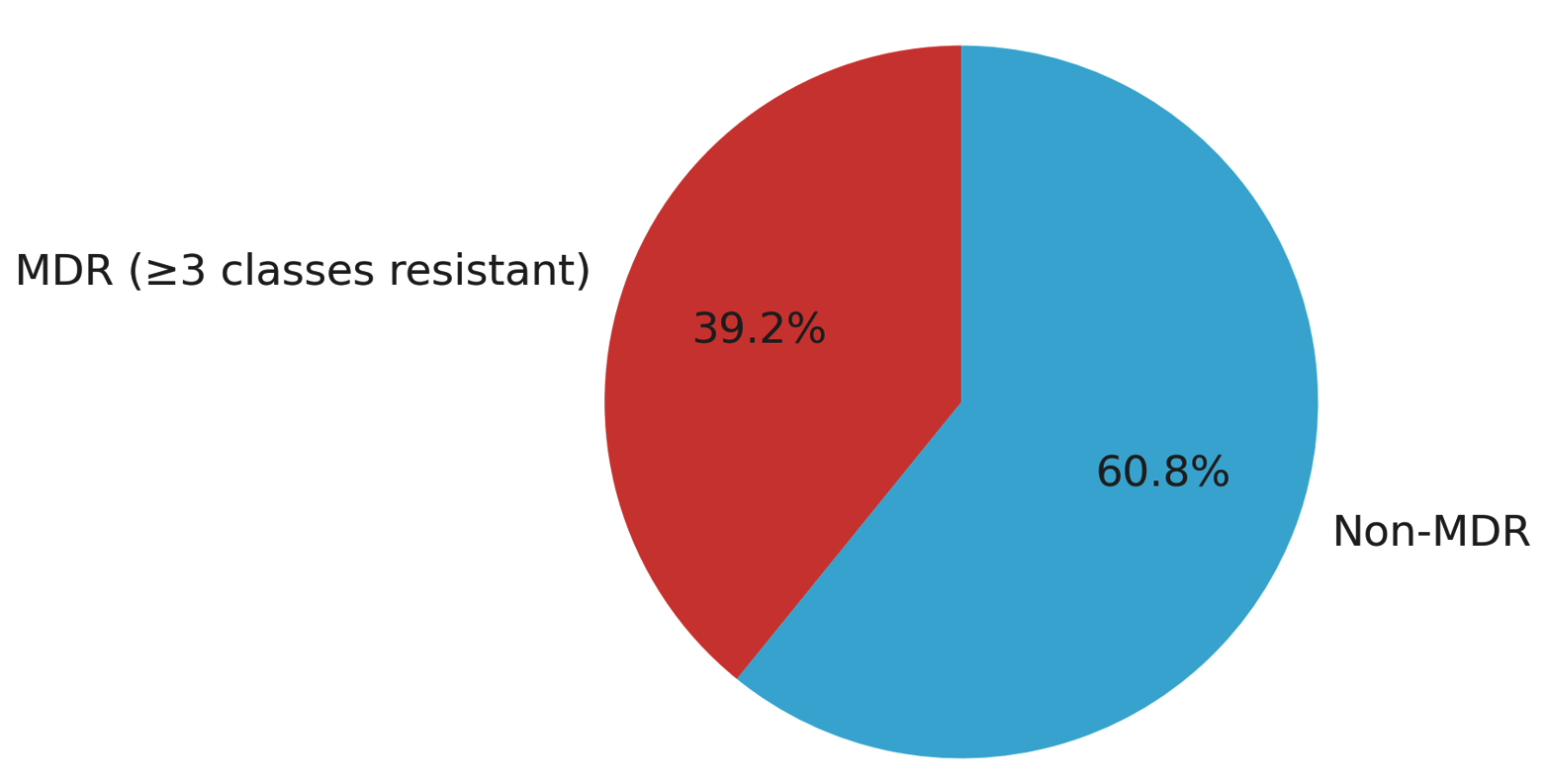
Figure. Proportion of Multidrug-resistant (MDR) S. aureus Isolates
Figure illustrates the distributions of MDR and non-MDR S. aureus isolates obtained from pediatric patients. “Multidrug-resistance (MDR), defined as resistance to three or more antimicrobial classes, was identified in 47 of 120 S. aureus isolates, corresponding to a prevalence of 39.2%, while 73 isolates (60.8%) were classified as non-MDR”.
Table 5. Among the genes examined, tetK, tetM, PVL, ermC, and mphC showed significant associations with MDR (P < 0.05). The genes tetK and tetM showed the strongest associations (OR ≈ 7.9 and 8.1, respectively), indicating that isolates carrying these genes were approximately eight times more likely to exhibit MDR. Similarly, PVL, ermC, and mphC exhibited moderate but significant associations (ORs ranging from 4.1-6.5), suggesting their co-occurrence with MDR phenotypes.
Table 5. Association between antimicrobial resistance genes and multidrug-resistance (MDR) among S. aureus isolates using logistic regression analysis
Resistance Gene |
Odds Ratio (OR) |
95% Confidence Interval (CI) |
P-value |
Interpretation |
|---|---|---|---|---|
tetM |
7.39 |
2.45-22.31 |
Significant association with MDR |
|
tetK |
6.82 |
2.21-21.04 |
0.001 |
Significant association with MDR |
PVL |
5.12 |
1.78-14.74 |
0.002 |
Significant association with MDR |
mphC |
3.94 |
1.42-10.93 |
0.008 |
Significant association with MDR |
ermA |
3.61 |
1.26-10.31 |
0.017 |
Significant association with MDR |
ermC |
3.42 |
1.20-9.74 |
0.021 |
Significant association with MDR |
aac(6′)-Ie-aph(2″)-Ia |
2.81 |
1.04-7.57 |
0.041 |
Significant association with MDR |
aph(3′)-IIIa |
2.63 |
1.01-6.89 |
0.048 |
Significant association with MDR |
mecA |
2.41 |
0.94-6.17 |
0.067 |
Not statistically significant |
blaZ |
1.95 |
0.76-5.02 |
0.165 |
Not statistically significant |
ant(4′)-Ia |
1.42 |
0.51-3.95 |
0.503 |
Not statistically significant |
ermB |
1.36 |
0.49-3.80 |
0.556 |
Not statistically significant |
msrA |
1.28 |
0.46-3.54 |
0.639 |
Not statistically significant |
qacA |
0.72 |
0.19-2.65 |
0.621 |
Not statistically significant |
qacB |
0.51 |
0.08-3.12 |
0.471 |
Not statistically significant |
In contrast, other tested genes demonstrated odds ratios near zero, with no statistically significant association with MDR, indicating that their presence was not predictive of multidrug-resistance. Overall, the data suggest that tetracycline resistance genes (tetK, tetM) and macrolide resistance genes (ermC, mphC) are major contributors to the MDR phenotype. At the same time, PVL-primarily a virulence determinant may be linked to MDR through co-selection or genetic linkage within mobile genetic elements.
Table 6 displays the findings of a multivariable logistic regression assessing the relationship between multidrug-resistance (MDR) and two predictor variables: infection source (hospital- or community-acquired) and isolate source (e.g., CSF, urine, or other clinical specimens). The regression model used the Chi-square test to evaluate statistical significance.
Table 6. Multivariable logistic regression analysis between MDR and infection source and isolate type
Variable |
Coefficient (β) |
Std. Error |
z |
P-value |
95% CI |
|---|---|---|---|---|---|
Intercept |
-0.162 |
0.386 |
-0.42 |
0.674 |
(-0.918, 0.594) |
(Hospital_Acquisition) [Hospital] |
0.434 |
0.409 |
1.06 |
0.288 |
(-0.367, 1.235) |
(Source)[CSF] |
-1.253 |
0.748 |
-1.68 |
0.094 |
(-2.718, 0.213) |
(Source)[Other] |
0.070 |
0.671 |
0.10 |
0.917 |
(-1.245, 1.385) |
(Source)[Urine] |
0.298 |
0.624 |
0.48 |
0.633 |
(-0.925, 1.522) |
(Source)[Wound] |
-0.277 |
0.472 |
-0.59 |
0.557 |
(-1.203, 0.649) |
Chi-square test was utilized to assess the relationship between MDR status and predictor variables. No significant relationships were detected (P > 0.05)
The findings indicate no statistically significant associations between MDR status and either infection source or isolate type (P > 0.05). Although the coefficient for hospital-acquired infections (β = 0.43, P = 0.29) suggested slightly higher odds of MDR, this trend was not significant. Similarly, CSF isolates showed a non-significant tendency toward reduced MDR odds (β = –1.25, P = 0.09). In contrast, isolates from urine and other sources showed no meaningful difference compared to the reference group.
Overall, the regression analysis suggests that neither infection source nor isolate type independently predicts MDR status in this dataset. This implies that MDR occurrence may be primarily driven by genetic determinants of resistance rather than by the clinical source or infection setting.
S. aureus remains an important pathogen causing a broad spectrum of pediatric infections, ranging from skin and soft tissue infections to severe invasive diseases such as bacteremia, pneumonia, and meningitis.22,23 The emergence of MDR and MRSA strains further increases the clinical and epidemiological burden of these infections.24,25
In the present study, hospital-acquired infections were more frequent than community-acquired infections, consistent with previous reports identifying healthcare settings, particularly intensive care units, as important reservoirs for MDR S. aureus because of selective antibiotic pressure and cross-transmission.26-28 Blood and wound specimens represented the most common sources of isolates, in agreement with previous studies reporting S. aureus as a leading cause of bacteremia and wound infections.29,30
Invasive specimens, particularly blood and CSF, were significantly associated with hospital-acquired infections, supporting earlier findings linking invasive S. aureus infections to healthcare exposure and invasive procedures.31,32 These findings underscore the importance of continuous surveillance, infection-control measures, and antimicrobial stewardship programs in pediatric healthcare settings.33,34
The high prevalence of the blaZ gene (83.3%) indicates widespread β-lactamase-mediated resistance among pediatric isolates, while the detection of mecA in 36.7% confirms a substantial burden of MRSA.35-37 Although blaZ was highly prevalent, phenotypic penicillin resistance was observed less frequently, which may reflect differences between gene carriage and actual gene expression under laboratory conditions. Similar genotype–phenotype discrepancies have previously been described in S. aureus isolates. These findings suggest that empirical penicillin therapy may be unreliable and emphasize the importance of local resistance surveillance and appropriate antimicrobial selection.38-40
Among macrolide resistance determinants, ermC was the most prevalent, followed by ermA and ermB, consistent with previous studies identifying ermC as the dominant MLS resistance determinant in clinical S. aureus isolates.6,41 The presence of msrA and mphC suggests coexistence of multiple macrolide resistance mechanisms, including active efflux and ribosomal methylation.42 Tetracycline resistance genes (tetK and tetM) and aminoglycoside-modifying enzyme genes were also frequently detected, similar to findings reported in Egyptian and international studies.43,44 In addition, detection of qacA/qacB may indicate reduced susceptibility to antiseptics such as chlorhexidine, while the presence of PVL-positive isolates reflects persistence of virulent pediatric strains.45,46
Notably, tetK and tetM demonstrated the strongest association with MDR, followed by ermC, mphC, and PVL. Similar findings have been reported in pediatric and clinical S. aureus studies, where tetracycline- and macrolide-resistance genes were frequently associated with MDR phenotypes and mobile genetic elements facilitating resistance dissemination. Previous clinical studies have identified tetK, tetM, and ermC as common resistance determinants among MRSA isolates and important contributors to multidrug resistance.47-49 These findings highlight the importance of molecular surveillance and targeted antimicrobial stewardship programs for controlling MDR S. aureus in pediatric settings. In contrast, infection source and isolate type were not independent predictors of MDR, suggesting that resistance is driven primarily by specific genetic determinants rather than by clinical origin alone.48-50 These findings highlight the importance of molecular surveillance and targeted antimicrobial stewardship to control the spread of MDR S. aureus.
The observed linezolid resistance rate was higher than that reported in many previous pediatric studies and should therefore be interpreted cautiously. Although susceptibility testing and broth microdilution confirmation were performed according to CLSI recommendations, further molecular investigations targeting resistance determinants such as cfr and optrA are warranted.
Limitations
The present study has several limitations that should be acknowledged. First, the D-test for inducible clindamycin resistance was not performed despite the analysis of MLS resistance genes. Consequently, phenotypic confirmation of inducible MLS_B resistance could not be assessed. Future studies should incorporate D-zone testing according to CLSI recommendations to provide a more comprehensive evaluation of clindamycin resistance and improve genotype–phenotype correlation.
Second, the study was conducted at a single tertiary-care center with a relatively limited sample size, which may restrict the generalizability of the findings to other pediatric populations and healthcare settings. In addition, molecular characterization was limited to selected resistance and virulence genes, while other clinically relevant resistance determinants, including linezolid resistance genes such as cfr and optrA, were not investigated. Finally, the cross-sectional design prevented assessment of temporal trends in antimicrobial resistance and transmission dynamics of S. aureus isolates over time.
In summary, S. aureus remains an important pediatric pathogen, with hospital-acquired infections, particularly in NICU and PICU settings, representing a considerable proportion of cases in the present study. A high prevalence of β-lactamase-associated (blaZ) and methicillin-resistance (mecA) genes was identified, together with diverse resistance determinants including tetracycline (tetK, tetM), macrolide (ermC, mphC), and aminoglycoside-modifying enzyme genes. PVL-positive isolates were also detected, indicating the coexistence of resistance and virulence characteristics among some isolates.
Tetracycline and macrolide resistance genes showed significant associations with multidrug-resistance (MDR), whereas infection source and isolate type were not independently associated with MDR in the regression analysis. These findings support the value of continued antimicrobial resistance surveillance and infection-control measures in pediatric healthcare settings.
ACKNOWLEDGMENTS
None.
CONFLICT OF INTEREST
The authors declare that there is no conflict of interest.
AUTHORS’ CONTRIBUTION
RHH conceptualized and designed the study. OAS and DMAH performed clinical data collection. LAK, EHMS and MESZ performed laboratory work. EHMS, MESZ, DMAH and RHH performed data analysis. OAS, RHH, MESZ, EHMS, LAK and DMAH wrote the manuscript. MESZ, DMAH and RHH reviewed and revised the manuscript. All authors read and approved the final manuscript for publication.
FUNDING
None.
DATA AVAILABILITY
The datasets generated and/or analysed during the current study are available from the corresponding author on reasonable request.
ETHICS STATEMENT
This study was approved by the Research Ethics Committee, Faculty of Medicine, Mansoura University, Egypt (Approval No.: R.25.11.3455).
INFORMED CONSENT
Written informed consent was obtained from the participants’ legal guardians before enrollment in the study.
- Tong SYC, Davis JS, Eichenberger E, Holland TL, Fowler Jr VG. Staphylococcus aureus infections: epidemiology, pathophysiology, clinical manifestations, and management. Clin Microbiol Rev. 2015;28(3):603-661.
Crossref - Cassat JE, Thomsen I. Staphylococcus aureus infections in children. Curr Opin Infect Dis. 2021;34(5):510-518.
Crossref - Otto M. Staphylococcus aureus toxins. Curr Opin Microbiol. 2014;17:32-7.
Crossref - Argemi X, Hansmann Y, Prola K, Prevost G. Coagulase-negative staphylococci pathogenomics. Int J Mol Sci. 2019;20(5):1215.
Crossref - Peacock SJ, Paterson GK. Mechanisms of Methicillin Resistance in Staphylococcus aureus. Annual Review of Biochemistry. 2015;84(1):577-601.
Crossref - Sedaghat H, Esfahani BN, Mobasherizadeh S, et al. Phenotypic and genotypic characterization of macrolide resistance among Staphylococcus aureus isolates in Isfahan, Iran. Iran J Microbiol. 2017;9(5):264-270.
- Duran N, Ozer B, Duran GG, Onlen Y, Demir C. Antibiotic resistance genes and susceptibility patterns in staphylococci. Indian J Med Res. 2012;135(3):389-96.
- Selvaraj V, Puthukkudi N, Chandran A, et al. Biocide susceptibility and resistance genes in udder surface Staphylococcus from Indian peri-urban dairy farms. Microb Pathog. 2025;205:107601.
Crossref
Crossref - Edelstein M, Kearns A, Cordery R. Panton-Valentine leukocidin-associated Staphylococcus aureus infections in London, England: clinical and sociodemographic characterization, management, burden of disease, and associated costs. J Infect Public Health. 2011;4(3):145-153.
Crossref - Di Bella S, Marini B, Stroffolini G, et al. The virulence toolkit of Staphylococcus aureus: a comprehensive review of toxin diversity, molecular mechanisms, and clinical implications. Eur J Clin Microbiol Infect Dis. 2025;44(8):1797-1816.
Crossref - Turner NA, Sharma-Kuinkel BK, Maskarinec SA, et al. Methicillin-resistant Staphylococcus aureus: an overview of basic and clinical research. Nat Rev Microbiol. 2019;17(4):203-218.
Crossref - Hassoun A, Linden PK, Friedman B. Incidence, prevalence, and management of MRSA bacteremia across patient populations: a review of recent developments in MRSA management and treatment. Crit Care. 2017;21(1):211.
Crossref - Rasmi AH, Ahmed EF, Darwish AMA, Gad GFM. Virulence genes distributed among Staphylococcus aureus causing wound infections and their correlation to antibiotic resistance. BMC Infect Dis. 2022;22:652.
Crossref - Magill SS, O’Leary E, Janelle SJ, et al. Changes in prevalence of healthcare-associated infections in U.S. hospitals, 2011-2015. N Engl J Med. 2018;379(18):1732-1744.
Crossref - Lwanga SK, Lemeshow S. Sample Size Determination in Health Studies: A Practical Manual. World Health Organization; 1991. https://iris.who.int/items/9c2e5da4-3785-4fec-9dbc-841e4ae0d98c
- Daniel WW. Biostatistics: a foundation for analysis in the health sciences. 7th ed. New York: John Wiley & Sons, 1999.
- Ignak S, Nakipoglu Y, Gurler B. Frequency of antiseptic resistance genes in clinical staphylococci and enterococci isolates in Turkey. Antimicrob Resist Infect Control. 2017;6(1):88.
Crossref - Clinical and Laboratory Standards Institute. Performance standards for antimicrobial susceptibility testing. 34th ed. CLSI supplement M100. Wayne (PA): CLSI; 2024.
- Murakami K, Minamide W, Wada K, Nakamura E, Teraoka H, Watanabe S. Identification of methicillin-resistant strains of staphylococci by polymerase chain reaction. J Clin Microbiol. 1991;29(10):2240-2244.
Crossref - Khan S, Shetty P, Lakshmi Y, et al. Detection of mecA genes of methicillin- resistant Staphylococcus aureus by polymerase chain reaction. Int J Health Rehabil Sci. 2012;1(1):64-68.
Crossref - Unal S, Hoskins J, Flokowitsch JE, Wu CYE, Preston DA, Skatrud PL. Detection of methicillin-resistant Staphylococci by using the polymerase chain reaction. J Clin Microbiol. 1992;30(7):1685-1691.
Crossref - Lowy FD. Staphylococcus aureus infections. N Engl J Med. 1998;339(8):520-532.
Crossref - Chambers HF, Deleo FR. Waves of resistance: Staphylococcus aureus in the antibiotic era. Nat Rev Microbiol. 2009;7(9):629-641.
Crossref - Grundmann H, Aires-de-Sousa M, Boyce J, Tiemersma E. Emergence and resurgence of meticillin-resistant Staphylococcus aureus as a public-health threat. Lancet. 2006;368(9538):874-885.
Crossref - Kock R, Becker K, Cookson B, et al. Methicillin-resistant Staphylococcus aureus (MRSA): burden of disease and control challenges in Europe. Euro Surveill. 2010;15(41):19688.
Crossref - Tacconelli E, Cataldo MA, Dancer SJ, et al. ESCMID guidelines for the management of infection control measures to reduce transmission of multidrug-resistant Gram-negative bacteria in hospitalized patients. Clin Microbiol Infect. 2014;20(Suppl 1):1-55.
Crossref - Saiman L, Cronquist A, Wu F, et al. An outbreak of methicillin-resistant Staphylococcus aureus in a neonatal intensive care unit. Infect Control Hosp Epidemiol. 2003;24(5):317-321.
Crossref - Hidron AI, Edwards JR, Patel J, et al. Antimicrobial-Resistant Pathogens Associated with Healthcare-Associated Infections: Annual Summary of data reported to the National Healthcare Safety Network at the Centers for Disease Control and Prevention, 2006–2007. Infect Control Hosp Epidemiol. 2008;29(11):996-1011.
Crossref - David MZ, Daum RS.Community-associated methicillin-resistant Staphylococcus aureus: epidemiology and clinical consequences of an emerging epidemic. Clin Microbiol Rev. 2010;23(3):616-687.
Crossref - Mertz D, Frei R, Jaussi B, et al. Throat swabs are necessary to reliably detect carriers of Staphylococcus aureus. Clin Infect Dis. 2007;45(4):475-477.
Crossref - Dulon M, Peters C, Schablon A, Nienhaus A. MRSA carriage among healthcare workers in non-outbreak settings in Europe and the United States: a systematic review. BMC Infect Dis. 2014;14:363.
Crossref - Tacconelli E, Pezzani MD. Public health burden of antimicrobial resistance in Europe. Lancet Infect Dis. 2019;19(1):4-6.
Crossref - World Health Organization (WHO). Prioritization of pathogens to guide discovery, research and development of new antibiotics for drug-resistant bacterial infections, including tuberculosis Geneva: WHO. 2017. https://www.who.int/publications/i/item/WHO-EMP-IAU-2017.12
- Zahid PS, Mahdy ZA, Solim EA. Molecular identification of methicillin resistant Staphylococcus aureus and vancomycin resistant Staphylococcus aureus strains isolated from milk and milk products. Benha Vet Med J. 2023;45(1):213-217.
Crossref - Lowy FD. Antimicrobial resistance: the example of Staphylococcus aureus. J Clin Invest. 2022;132(5):e18535.
Crossref - Arede P, Ministro J, Oliveira DC. Redefining the Role of the β-Lactamase Locus in Methicillin-Resistant Staphylococcus aureus: β-Lactamase Regulators Disrupt the MecI-Mediated Strong Repression on mecA and Optimize the Phenotypic Expression of Resistance in Strains with Constitutive mecA Expression. Antimicrob Agents Chemother. 2013;57(7):3037-45.
Crossref - Pantosti A, Sanchini A, Monaco M. Mechanisms of antibiotic resistance in Staphylococcus aureus. Future Microbiol. 2007;2(3):323-334.
Crossref - Gerber JS, Coffin SE, Smathers SA, Zaoutis TE. Trends in the incidence of methicillin-resistant Staphylococcus aureus infection in children’s hospitals in the United States. Clin Infect Dis. 2009;49(1):65-71.
Crossref - Liu C, Bayer A, Cosgrove SE, et al. Clinical practice guidelines for the treatment of methicillin-resistant Staphylococcus aureus infections in adults and children. Clin Infect Dis. 2011;52(3):e18-55.
Crossref - Zomorodi AR, Motamedifar M, Rahmanian K, et al. Investigation of integron classes 1, 2, and 3 among multi-drug resistant Staphylococcus aureus isolates in Iran: a multi-center study. BMC Infect Dis. 2024;24(1):1430.
Crossref - Hu Y, Ouyang L, Li D, et al. The antimicrobial activity of cethromycin against Staphylococcus aureus compared with erythromycin and telithromycin. BMC Microbiol. 2023;23:109
Crossref - Firouzjaei MD, Halaji M, Yaghoubi S, et al. Inducible clindamycin-resistant and biofilm formation in Staphylococcus aureus isolated from healthcare workers’ anterior nasal carriage. BMC Res Notes. 2024;17(1):252.
Crossref - Gad WA, Osman SA, Abd El-Razik KAE, Soror AH, Soliman YA, Fouad EA. Prevalence and molecular characterization of resistant Staphylococcus aureus strains in bulk milk tanks of dairy cattle in Northern Egypt. Vet Res Forum. 2025;16(6):317-323.
- Yadegar A, Sattari M, Mozafari NA, Goudarzi GR. Prevalence of genes encoding aminoglycoside-modifying enzymes and methicillin resistance among clinical isolates of Staphylococcus aureus in Tehran, Iran. Microb Drug Resist. 2009;15(2):109-113.
Crossref - Ghanim SO, Eldegla HE, Abdel-Fattah GM. Detection of chlorhexidine-resistant genes in methicillin-resistant Staphylococcus aureus in Mansoura University Hospitals. Mansoura J Biol. 2023;64(3):12-18.
Crossref - Mandour S, Fadl G, Abdalla S, Mohamed F, Mohamed H. The prevalence of Panton-Valentine leukocidin gene among Staphylococcus aureus strains isolated from medical staff and community people in Minia Governorate, Egypt. Egypt J Med Microbiol. 2023;32(1):125-132.
Crossref - Dormanesh B, Siroosbakhat S, Darian E, et al. Methicillin-Resistant Staphylococcus aureus Isolated From Various Types of Hospital Infections in Pediatrics: Panton-Valentine Leukocidin, Staphylococcal Chromosomal Cassette mec SCCmec Phenotypes and Antibiotic Resistance Properties. Jundishapur J Microbiol. 2015;8(11):e11341.
Crossref - Abdulmanea AA, Alharbi NS, Somily AM, Khaled JM, Algahtani FH. The Prevalence of the Virulence Genes of Staphylococcus aureus in Sickle Cell Disease Patients at KSUMC, Riyadh, Saudi Arabia. Antibiotics. 2023;12(7):1221.
Crossref - Shrief R, El-Kholy R. M, Rizk M. A, Zaki M. E. Prevalence of Tetracycline Resistant Genes in Staphylococcus aureus Isolates from Surgical Site Infections Egypt. Biosci Biotech Res Asia. 2019;16(2): 1-7.
Crossref - Lim KT, Hanifah YA, Yusof Mym, Thong KL. ermA, ermC, tetM and tetK are essential for erythromycin and tetracycline resistance among methicillin-resistant Staphylococcus aureus strains isolated from a tertiary hospital in Malaysia. Indian J Med Microbiol. 2012;30(2):203-7.
Crossref
© The Author(s) 2026. Open Access. This article is distributed under the terms of the Creative Commons Attribution 4.0 International License which permits unrestricted use, sharing, distribution, and reproduction in any medium, provided you give appropriate credit to the original author(s) and the source, provide a link to the Creative Commons license, and indicate if changes were made.