


ISSN: 0973-7510
E-ISSN: 2581-690X
 , Nino Gachechiladze1, Nino Chikadze2, Irina Datikashvili-David3, Peter M Lydyard2,4, Nina Porakishvili2,4and Marina Tediashvili2,5
Chronic lymphocytic leukemia (CLL) is characterized by immune dysfunction and increased susceptibility to bacterial infections. Growing antimicrobial resistance has renewed interest in bacteriophage therapy (PT) as a complementary antimicrobial approach. However, naturally occurring anti-phage antibodies may affect its efficacy. This study investigated the upper respiratory tract microbiota of newly diagnosed, untreated CLL patients with no prior exposure to therapeutic phages. A total of 51 bacterial isolates were recovered from 22 CLL patients and 14 isolates from 12 healthy volunteers. Staphylococcus aureus was the predominant species, followed by coagulase-negative staphylococci (CoNS). Antimicrobial susceptibility testing revealed the highest susceptibility (82.35%) to linezolid and moxifloxacin, whereas several S. aureus isolates showed reduced susceptibility to glycopeptides. Commercial phage preparations showed weak or no lytic activity against most isolates, particularly CoNS. In contrast, all 12 S. aureus isolates from CLL patients and healthy volunteers were susceptible to phage PSA-1 from the Pyophage preparation, with an efficiency of plating (EOP) of 0.7-1.0. Naturally occurring anti-phage antibodies were detected in both groups. The mean phage neutralization constant (K) was 0.64 ± 0.12 in CLL patients and 0.28 ± 0.06 in healthy controls, indicating variable pre-existing humoral immunity that may influence phage activity. These findings indicate that upper respiratory isolates from untreated CLL patients remain largely susceptible to conventional antibiotics. Phage therapy may represent a promising adjunctive strategy for managing S. aureus infections in CLL patients. Assessment of phage susceptibility and anti-phage antibody levels may optimize future phage-based therapies.
Chronic Lymphocytic Leukemia, Bacterial Infection, Antibiotic Susceptibility, Bacteriophage, Phage Therapy, Anti-Phage Antibodies
Chronic lymphocytic leukemia (CLL) is the most frequently diagnosed leukemia in adults worldwide and is characterized by the progressive accumulation of mature, immunophenotypically defined CD19 + CD5 + CD23 + B lymphocytes. Malignant cells expand within proliferative niches in the bone marrow and lymphoid tissues and persist due to impaired apoptotic mechanisms.1 This pathological accumulation is accompanied by broad immune dysfunction, resulting in weakened cellular and humoral immune responses and a markedly increased vulnerability to infections.
Multiple components of the immune system are affected in CLL, including hypogammaglobulinemia, dysregulation of T-cell subsets, impaired complement activation, neutropenia, and expansion of immunosuppressive myeloid populations. In addition, interactions between leukemic cells and the surrounding microenvironment promote tumor survival through direct contact and soluble mediators, further contributing to immune impairment.2 Importantly, immunodeficiency associated with CLL is frequently intensified by antineoplastic therapy. Conventional chemotherapy and some targeted agents disrupt bone marrow function, damage mucosal barriers, and suppress both B- and T-cell activity, thereby increasing susceptibility to opportunistic infections. As a result, infectious complications remain the leading cause of morbidity and mortality in CLL patients. On other hand, it is believed that there is a bidirectional relationship between CLL and infection since common community-acquired infections are considered as a risk factor for the development of CLL.3 The healthcare-associated infections (HAIs) further increase treatment complexity and healthcare burden4,5 with the expanded spectrum of causative agents. Thus, regular monitoring of patients with CLL is very important to ensure immediate action upon any evidence of infection and early intervention to decrease the infection-related risk of hospitalization.
Infections in CLL may be viral, fungal, or bacterial in origin and can involve either single or multiple microbial species. Among bacterial pathogens, Staphylococcus aureus, Streptococcus pneumoniae, Pseudomonas aeruginosa, Escherichia coli, Klebsiella pneumoniae, and Haemophilus influenzae are most commonly reported. Sporadic cases of invasive fungal infections, including Candida and Aspergillus species, have also been described, particularly in patients with advanced disease and prolonged neutropenia.6 Despite extensive research conducted on infectious complications in CLL, the data on colonizing microbiota, especially from particular geographic regions, remain scarce. To our knowledge no previous studies from Georgia have systematically examined the composition of colonizing bacterial communities in patients with CLL.
Given the strong association between infections and adverse clinical outcomes in CLL, increasing attention has been directed toward advanced molecular diagnostic approaches that enable rapid pathogen detection and identification, and more accurate clinical decision-making improving preventive and therapeutic strategies.7,8 While broad-spectrum antibiotics remain the cornerstone of treatment for bacterial infections, the rapid emergence of antimicrobial resistance complicates empirical therapy and limits available options. Although numerous studies report antibiotic susceptibility profiles of clinical isolates from CLL patients,9 there is comparatively limited information regarding the effectiveness of non-antibiotic antimicrobial approaches, such as bacterial vaccines (bacterial lysates) and bacteriophages, against bacteria most frequently colonizing or infecting this population.
Bacteriophage therapy (PT) has re-emerged as a promising complementary antimicrobial strategy due to its high specificity toward bacterial hosts and the capacity of certain phages to disrupt biofilms, known to be key contributors to chronic and recurrent infections and antimicrobial resistance.10 Bacteriophages are ubiquitous in natural environments, an integral part of the human microbiome, being detectable in body fluids such as saliva, urine, feces, and blood. By contributing substantially to bacterial turnover, phages play an important role in maintaining microbial ecosystem balance.11 From a therapeutic perspective, phages offer several advantages, including minimal toxicity to mammalian cells, preservation of commensal microbiota, and potential synergistic activity with antibiotics.12-14 In some settings, phages have been shown to restore bacterial susceptibility to antimicrobial agents.13 Moreover, individualized phage preparations can be generated from phage libraries to match patient-specific bacterial isolates, supporting the concept of personalized antimicrobial therapy.15
At the same time, interactions between bacteriophages and the human immune system are complex and not yet fully understood. Exposure to phages can induce humoral immune responses, predominantly IgM, but also IgG and IgA antibodies, which may neutralize phage activity and potentially reduce therapeutic efficacy.16 Importantly, little is known about baseline anti-phage immunity in immunocompromised individuals, including patients with CLL. It is plausible that naturally occurring antibodies against commonly circulating phages may already be present prior to therapeutic administration, which could influence both infection dynamics and responses to phage-based interventions. In addition to antimicrobial treatment, prevention of infections in CLL relies on multiple strategies, including vaccination against respiratory pathogens, immunoglobulin replacement therapy in patients with severe antibody deficiency, early detection of colonizing or emerging pathogens and antimicrobial prophylaxis in selected high risk groups. Improved understanding of colonization patterns may therefore inform both preventive and therapeutic decision-making and help identify patients at increased risk of invasive disease.
Based on these considerations, the present study aimed to characterize the upper respiratory tract microbiota of CLL patients who had not previously been evaluated for bacterial colonization and had not received phage therapy. We further assessed the susceptibility of isolated bacteria to commonly used antibiotics and to selected bacteriophages, and evaluated the presence and phage-neutralizing capacity of naturally occurring anti-phage antibodies in patient sera. This integrated approach was designed to provide preliminary insight into the feasibility of phage-based antimicrobial strategies in CLL and to contribute baseline data relevant to infection prevention in this vulnerable population.
Sample collection
Peripheral blood samples, as well as nasopharyngeal and oropharyngeal swabs, were collected from 22 patients diagnosed with CLL (age range: 50-75 years; 10 males and 12 females) at the M. Zodelava Center of Hematology, Tbilisi, Georgia. All patients were newly diagnosed and had not received any CLL-specific therapy at the time of sampling (Table 1).
Table 1. Clinical and hematological characteristics of patients with Chronic Lymphocytic Leukemia (CLL)*
Parameters |
CLL Patients |
|---|---|
Number of participants (n) |
22 |
Age range (years) |
50-75 |
Gender (M/F) |
10/12 |
CLL stage (Rai) |
Rai 0-I (early-stage) |
Leukocytes (WBC) (×10⁹/L) |
41.3 ± 9.8 |
Lymphocytes (%) |
77.3 ± 3.3 |
Monocytes (%) |
3.7 ± 0.6 |
NLR |
0.20 ± 0.04 |
Infection signs (fever, cough, muscle pain, sore throat) at the time of admission |
1 patient |
Antibiotic treatment conducted prior to admission |
2 patients (antibiotics Moxifloxacin, Ciprofloxacin, and Ampicillin + Sulbactam) |
Comorbidities and associated conditions |
12 patients in total, with Type II diabetes mellitus (n = 1); chronic ischemic heart disease with coronary stents (n = 1); iron-deficiency anemia (n = 1); hyperlipidemia (n = 1); hyperuricemia (n = 1); severe/critical anemia (n = 1); thrombocytopenia (n = 1); hepatomegaly/hepatosplenomegaly (n = 2); allergic reactions/angioedema (n = 1); intestinal protozoal infection (n = 1); acute viral infection (n = 1) |
*Data are presented as mean ± standard deviation (SD) or number of patients (n), as appropriate. All patients were in the early stage of disease according to the Rai classification (Rai stages 0-I). WBC = white blood cell count; NLR = neutrophil-to-lymphocyte ratio. Comorbidities and associated clinical conditions observed in the study cohort are also summarized
Samples were collected after obtaining written informed consent from all participants and approval from the local ethics committee.
Fourteen age and sex matched healthy volunteers were also enrolled as a control group following written informed consent.
Microbiological analysis
Nasopharyngeal and oropharyngeal swabs obtained from patients and healthy volunteers were processed using standard culture-based microbiological methods.17 Briefly, swabs were streaked onto sheep blood agar (SBA) plates using the four-quadrant streaking technique and simultaneously inoculated into tubes containing tryptic soy broth (TSB) for enrichment. Plates and broths were incubated at 37 °C for 24-36 hours.
Following incubation, subcultures were performed on routinely used selective and differential media with emphasis on isolation of clinically relevant bacterial species, including Staphylococcus aureus, Streptococcus pneumoniae, Pseudomonas aeruginosa, Escherichia coli, and Klebsiella pneumoniae.17 Preliminary identification was based on colony morphology, Gram staining and microscopic examination, also growth characteristics on selective and differential media, followed by basic biochemical testing. The final phenotypic species-level identification was performed using Analytical Profile Index (API) systems, including API Staph, API 20 NE, API 20E, and API 20 STREP (bioMerieux, France).
Assessment of antibiotic and bacteriophage susceptibility
The antibiotic susceptibility of isolated bacteria was tested on Mueller-Hinton agar by Kirby-Bauer disc-diffusion method, according to European Committee on Antimicrobial Susceptibility Testing (EUCAST) standards (http://www.eucast.org). The following antibiotics were used: Tetracyclines, Tobramycin, Moxifloxacin and Levofloxacin, all effective against both Gram-positive and Gram-negative bacteria; Vancomycin and Cefuroxime, highly effective against Gram-positive microorganisms. For vancomycin and linezolid, determination of minimal inhibitory concentration (MIC) on solid and liquid media was also performed.
The susceptibility to bacteriophages was studied by the standard phage spot test, followed with titration by the double layer agar technique.18,19 The commercial preparations “Pyo Bacteriophage”, “Staph Bacteriophage”, “Ses Bacteriophage”, “Intesti Bacteriophage”, “Fersisi Bacteriophage”, “Enko bacteriophage” (all products of “Eliava Biopreparations”, Georgia) were used in the study. In addition, S. aureus specific phage PSA-1 cloned from the Pyophage preparation on the host strain S. aureus GMH17, was tested for lytic activity against Staphylococcus spp. isolates.
Phage virion morphology
Phage nucleocapsid morphology was studied by transmission electron microscopy (TEM). Concentrated phage suspensions (≥1 × 10¹⁰ PFU/mL) were applied to Formvar/carbon film-coated 300 mesh copper grids – CF300-CU (Electron Microscopy Sciences, PA, USA). Samples were negatively stained with 2% Phosphotungstate and examined using the microscope JEM 100 SX (Jeol, Japan). Up to 6 phage particles were measured, and the average head and tail dimensions were determined.
Detection of naturally-occurring anti-phage antibodies
The binding ability of sera from 10 randomly selected CLL patients and 14 age- and sex-matched healthy volunteers to an S. aureus PSA-1 phage was assessed using an indirect enzyme-linked immunosorbent assay (ELISA). Briefly, Nunc MaxisorpC 96-well flat-bottom microtiter plates were coated with 50 µL of semi-purified preparation of phage PSA-1 at a concentration of 108 PFU/mL in 0.05 M carbonate-bicarbonate buffer (CBB, pH 9.6; Sigma, USA). The plates were incubated overnight at 4 °C. Blocking was performed using Pierce™ Protein-Free Blocking Buffer (Thermo Fisher Scientific, USA). Sera were serially diluted 1:25, 1:50, and 1:100 in 5% bovine serum albumin (BSA; Thermo Fisher Scientific, USA) in phosphate-buffered saline (PBS) (Thermo Fisher Scientific, USA). Fifty microliters of each dilution were added to the corresponding wells in duplicates. For detection, a mixture of goat anti-human IgG, IgM, and IgA antibodies conjugated to horseradish peroxidase (HRP; Invitrogen, USA) was used along with the substrate 3,3′,5,5′-Tetramethylbenzidine (TMB; Thermo Fisher Scientific, USA). The plates were read for an optical density (OD) at 450 nm using a spectrophotometer (Selecta, Spain).
Phage neutralization assay
The standard methodology of phage neutralization reaction18,20,21 was used to assess the neutralizing activity of naturally occurring antibodies against S. aureus phages in sera obtained from CLL patients. The rate of inactivation of the S. aureus-specific phage PSA-1 was determined. Sera from 16 patients (dilutions 1:50 and 1:100) were incubated with phage suspension containing 1 × 10⁶ plaque-forming units per mL (PFU/mL) for 10 and 30 minutes at 37 °C. Following 100-fold dilution of the reaction mixtures in cold saline, surviving phages were quantified using the double-agar overlay method on the host strain, and percent neutralization was calculated. The neutralization constant (K) was calculated using the formula: K = 2.3 × (D/T) × log(P0/Pt), where D is the serum dilution, T is incubation time in minutes, P0 is the initial phage titer, and Pt is the phage titer after time T.
Statistical analysis
Statistical analysis was performed using Microsoft Excel and relevant statistical software. Proportions of bacterial susceptibility profiles were compared between cohorts using Fisher’s exact test. For quantitative data, an unpaired, two-tailed Student’s t-test was applied to determine the statistical significance of differences in anti-phage antibody levels, with data presented as mean ± standard error of the mean (SEM). Since Levene’s test indicated equal variances for the 1:50 dilution, the standard Student’s t-test was used, whereas an independent-samples t-test (Welch’s correction) was applied for the 1:100 dilutions due to unequal variances. A value of P < 0.05 was considered statistically significant.
A total of 51 bacterial strains were isolated from 44 oropharyngeal swabs collected from the nasal cavity and throat of 22 CLL patients and identified at the species level. Among these, 42 isolates belonged to opportunistic pathogenic species (17 species total), including Pseudomonas luteola (1), Staphylococcus epidermidis (8), S. salivarius (2), S. lentus (5), S. capitis (2), S. warneri (2), S. hominis (2), S. xylosus (1), S. haemolyticus (1), S. saprophyticus (1), S. cohnii (1), Kocuria varians/rosea (1), Aerococcus viridans (3), and Bacillus subtilis (3). Nine isolates were identified as Staphylococcus aureus. Most of these organisms are recognized as possible causes of infection in immunocompromised patients, including individuals with hematological malignancies.
From 12 nasopharyngeal swabs of healthy volunteers 14 bacterial strains were isolated, of which 11 were opportunistic bacteria – 3 species of CoN staphylococci, including S. epidermidis (6), S. salivarius (1), S. lentus (2), and S. xylosus (2). Three isolates were identified as S. aureus, frequently found as nasal carriage in practically healthy population.
Antibiotic susceptibility testing of bacterial isolates included representatives of several antimicrobial classes: tetracycline, vancomycin, linezolid, cefuroxime, levofloxacin, moxifloxacin, and tobramycin. Overall, isolates demonstrated moderate to high susceptibility to most antibiotics tested (≥58% of strains), with linezolid and moxifloxacin showing the highest activity (82.35%).
Interestingly, several S. aureus isolates from CLL patients exhibited intermediate resistance to vancomycin (MIC range 4-8 µg/mL), while susceptibility to other antibiotic classes remained high (Table 2 and Figure 1). This is typically characteristic for vancomycin-intermediate Staphylococcus aureus (VISA) strains, that can occur as a result of accumulations of mutations leading to a number of phenotypic changes, primarily the increased cell wall thickness, limiting the access of vancomycin to its target. VISA strains are mostly isolated in hospital settings from patients with underlying diseases (diabetes, kidney disorders etc.), manifested as skin or wound infections but can escalate to more serious systemic infections if left untreated. Thus, timely detection and rational management of VISA in CLL patients is of high medical importance.22,23
Table 2. Antibiotic susceptibility/resistance of bacterial isolates obtained from the URT of CLL patients (51 strains) and healthy volunteers (14 strains)
| Isolation | Antibiotics | Sensitive strains (%) | Resistant strains (%) |
|---|---|---|---|
| CLL Patients | Moxifloxacin 5 µg | 82.4 | 17.7 |
| Linezolid 10 µg | 82.4 | 17.7 | |
| Tobramycin 10 µg | 74.5 | 25.5 | |
| Tetracyclin 30 µg | 74.5 | 25.5 | |
| Cefuroxime 30 µg | 74.5 | 25.5 | |
| Levofloxacin 5 µg | 72.6 | 27.5 | |
| Vancomycin 30 µg | 68.6 | 31.4 | |
| Healthy Volunteers | Linezolid 10 µg | 92.9 | 7.1 |
| Tetracyclin 30 µg | 78.6 | 21.4 | |
| Moxifloxacin 5 µg | 78.6 | 21.4 | |
| Levofloxacin 5 µg | 71.4 | 28.6 | |
| Cefuroxime 30 µg | 64.3 | 35.7 | |
| Tobramycin 10 µg | 42.9 | 57.1 | |
| Vancomycin 30 µg | 85.7 | 14.3 |
In the antibiotic susceptibility testing of the isolates from healthy controls the same antimicrobial panel was used. Moxifloxacin and linezolid again demonstrated the highest activity, with susceptibility rates ranging from 82.4%- 92.9%, linezolid showing the greatest overall efficacy. Comparing to CLL patients, the percentage of vancomycin intermediate resistant strains was lower in healthy individuals (Table 2, Figure 1).
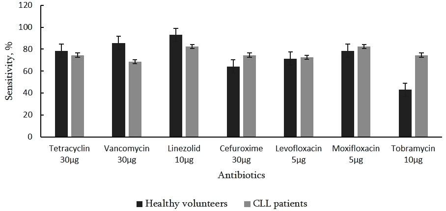
Figure 1. Antibiotic susceptibility profiles of nasopharyngeal bacterial strains isolated from CLL patients (n = 51) and healthy volunteers (n = 14). Statistical analysis was performed using Fisher’s exact test to compare the proportions of antibiotic susceptibility profiles (sensitive versus resistant strains) between CLL patients and healthy volunteers
To date, no randomized clinical trials have evaluated antibiotic strategies specifically in CLL patients, and no national evidence-based antimicrobial guidelines exist in Georgia for this population. Our findings indicating high in vitro activity of oxazolidinones against both pathogenic and opportunistic Gram-positive bacteria suggest that these agents may be effective options for treating secondary bacterial infections in CLL patients.
Commercial phage preparations (Pyo, Ses, Enko, Intesti, Pyophage, and Staph-phage) showed no lytic activity against the majority of isolated strains. This was not surprising and likely reflects the species composition of the isolates, as most belonged to coagulase-negative staphylococci usually not covered by available commercial phage cocktails. On the contrary, the lack of activity against S. aureus isolates was rather unexpected, as strains from diverse origins are typically highly susceptible to Sa- specific phages.24 These findings may indicate potential strain-specific differences in phage receptors or host defense mechanisms among S. aureus isolates from CLL patients.
Interestingly, certain lytic activity was demonstrated by the individual phage which has been cloned from the Pyophage preparation and propagated on the host strain S. aureus GMH17 (Table 3). This phage – Sa PSA-1 with Myoviridae morphotype (Figure 2), closely resembling staphylococcal virulent phage Sb-1,24 showed strong lytic activity against all S. aureus isolates while several coagulase-negative staphylococci exhibited weak sensitivity to it. These results highlight the importance of screening phage libraries to identify effective phages for therapeutic or prophylactic use, which will increase the feasibility of personalized phage preparations.
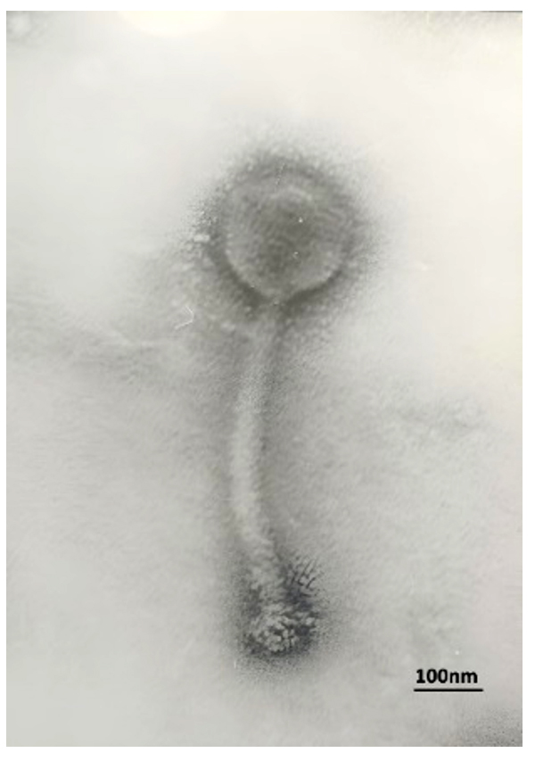
Figure 2. Transmission electron micrograph (TEM) of staphylococcal bacteriophage PSA-1
JEM 100 SX (Jeol, Japan), 80KV, instrumental magnification X 40,000
Table 3. Susceptibility of Staphylococcus spp. isolates to phage Sa PSA-1*
| Staphylococcus species | Source | No. of isolates tested | Susceptible (n) | Mean EOP (range) |
|---|---|---|---|---|
| S. aureus | CLL patients | 9 | 9 | 0.80-1.00 |
| Healthy volunteers | 3 | 3 | 0.70-1.00 | |
| S. epidermidis | CLL patients | 8 | 2 | <0.01-0.06 |
| Healthy volunteers | 6 | 0 | <0.01 | |
| S. lentus | CLL patients | 5 | 0 | <0.01 |
| Healthy volunteers | 2 | 0 | <0.01 | |
| S. hominis | CLL patients | 2 | 0 | <0.01 |
| S. capitis | CLL patients | 2 | 0 | <0.01 |
| S. warneri | CLL patients | 2 | 0 | <0.01 |
| S. haemolyticus | CLL patients | 1 | 0 | <0.01 |
| S. saprophyticus | CLL patients | 1 | 0 | <0.01 |
| S. cohnii | CLL patients | 1 | 0 | <0.01 |
| S. xylosus | Healthy volunteers | 2 | 0 | <0.01 |
| S. salivarius | Healthy volunteers | 1 | 0 | <0.01 |
*EOP (efficiency of plating) was calculated as the ratio of the phage titer obtained on the test strain to the phage titer obtained on the reference host strain Staphylococcus aureus GMH 17
Previous studies have demonstrated that phage administration may induce phage-specific humoral immune responses, though mechanisms regulating this response and its impact on treatment efficacy remain not sufficiently clear.16,25,26 Case reports of phage therapy in humans have provided conflicting evidence regarding whether anti-phage antibodies impair therapeutic outcomes.27,28
Therefore, we next evaluated the presence of naturally occurring antibodies against Sa- specific phage from the Pyophage cocktail propagated on S. aureus in the sera of 10 selected CLL patients and 14 healthy volunteers. As shown in Figure 3, moderate levels of phage-binding antibodies were detected in both groups (OD range 1.3-1.5), with no statistical difference between these two groups. This is not surprising, since staphylococcal infections frequently occur in the population both – practically healthy and with different diseases, including CLL. So, the human bodies naturally may contain also anti- staphylococcal phages, capable to induce human humoral response to them, especially if we measure antibodies against the most widespread type of staphylococcal phages, to which we also may attribute phage PSA-1.
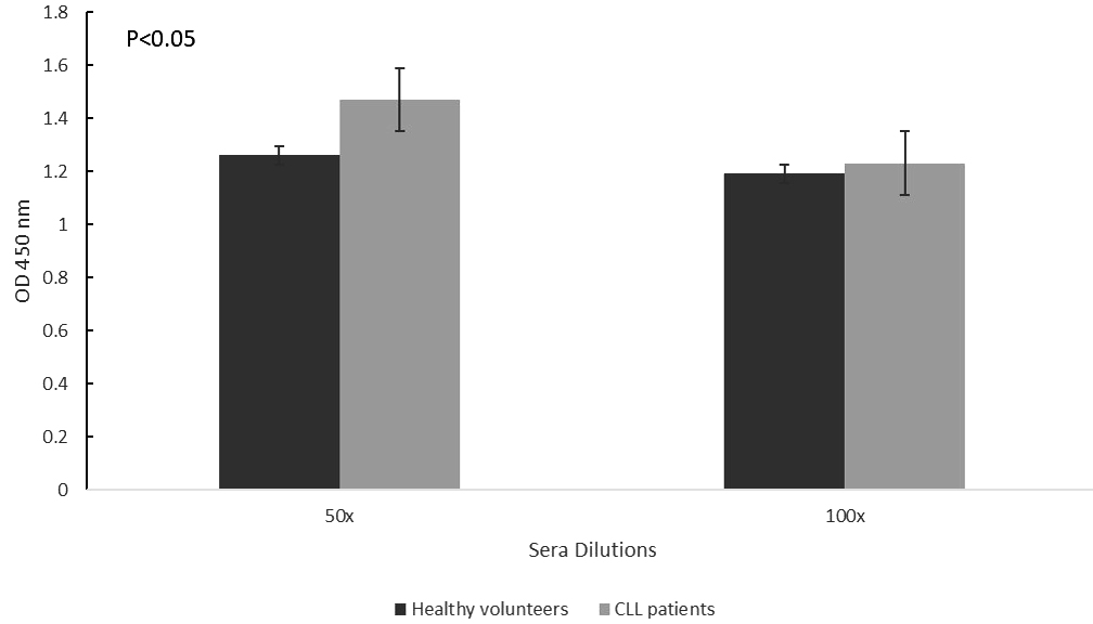
Figure 3. Naturally occurring total antibodies (IgG, IgM, and IgA) to S. aureus phage PSA-1, determined by indirect ELISA in the blood sera of CLL patients (n = 10) and age-matched healthy volunteers (n = 14)
Statistical analysis was performed using an unpaired two-tailed Student’s t-test. Anti-phage antibody levels were significantly higher in CLL patients than in healthy volunteers at the 1:50 serum dilution (P < 0.05), whereas no significant difference was observed between the groups at the 1:100 serum dilution. Data are presented as mean ± SE.
Considering that not all binding antibodies have phage neutralizing capability, and neutralization depends on epitope specificity, antibody isotype, and immune interactions,25,29,30 a series of classical phage neutralization tests was performed.18,20 Sera from CLL patients demonstrated partial inhibition of phage activity (Figure 4). At a serum dilution of 1:50, the mean neutralization rate was 31.4% ± 0.05 (P < 0.05) with K = 0.65 ± 0.22, whereas at 1:100 dilution, as expected, the neutralization rate decreased to 14.59% ± 0.05 (P < 0.05) with K = 0.63 ± 0.13. The average phage neutralization capacity of CLL patient’s sera comprised 0.64 ± 0.12 The sera from healthy volunteers exhibited a distinctly lower neutralization profile: at 1:50 dilution, the neutralization rate was 17.71% ± 0.05 (P < 0.05) with K = 0.33 ± 0.09, while at 1:100 dilution, it decreased to 6.18% ± 0.05 (P < 0.05) with K = 0.23 ± 0.08. The average K for the sera of healthy volunteers was determined as 0.28 ± 0.06. An independent-samples t-test (Welch’s correction) was conducted to compare the two groups, as Levene’s test indicated unequal variances (P = 0.026). The results showed a statistically significant difference between the groups.
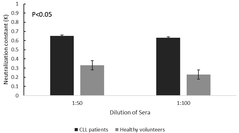
Figure 4. Sera from CLL patients (n = 16) and healthy volunteers (n = 14) were tested at 1:50 and 1:100 dilutions using the Phage Neutralization Reaction (PNR). Phage neutralization is represented as the neutralization constant (K)
Overall, obtained results indicate that naturally occurring antibodies in untreated CLL patients (as well as in healthy controls) partially inhibit the lytic activity of phage PSA-1 in vitro. Based on these preliminary findings and earlier studies on the virulent phage behavior (data not shown here), PSA-1 may represent a suitable therapeutic candidate for treatment of staphylococcal URT infections in CLL patients, either alone or in combination with antibiotics.
Comparison of phage neutralization rates (K constant) between CLL patients and healthy volunteers showed a statistically significant difference between the two groups at both serum dilutions (1:50 and 1:100) (P < 0.05), indicating a higher phage neutralization capacity in CLL patients. According Levene’s test indicated equal variances, the standard Student’s t-test was used for the serum dilutions (1:50) and an independent-samples t-test (Welch’s correction) for the serum dilutions (1:100). The results showed a statistically significant difference between the groups. Overall, both study groups – CLL patients and healthy volunteers exhibited the presence of APA (although at a very low level) in their serum samples, indicating the possible previous exposure to endogenous or exogenous naturally circulating phages.
The relatively small sample size was the main limitation of this study, which restricted the ability to draw robust and generalizable conclusions regarding the prevalence and role of bacterial pathogens in the upper respiratory tract of patients with CLL. Therefore, the findings cannot be interpreted as definitive evidence. The limited number of participants also constrained the assessment of phage susceptibility and the potential effectiveness of phage therapy for the control of bacterial infections in CLL patients. In addition, bacterial isolates obtained from both CLL patients and healthy controls were not screened against a broader panel of individual bacteriophages, particularly those available in the Eliava phage collection. Such screening might have identified additional phages with lytic activity against the target bacterial isolates, especially CoN staphylococci, thereby substantially increasing the proportion of phage-susceptible isolates detected in this study.
In conclusion, our data suggest that bacteria colonizing the URT in the newly diagnosed, untreated CLL patients remain largely susceptible to conventional antibiotics. At the same time, considering the possible development of antimicrobial resistance in the course of antibiotic treatment the phage therapy may be suggested as a complementary strategy within a broader, multidisciplinary approach to infection management in CLL patients. The effective implementation of phage therapy along with the use of commercially available phage mixtures will likely require the development of customized phage preparations based on careful selection from collections of therapeutic phages. The monitoring of neutralizing antibodies during preclinical evaluation and clinical application of therapeutic phages may be an important additional parameter for predicting of their therapeutic efficacy in vivo.
ACKNOWLEDGMENTS
The authors are thankful to Dr. Elene Didebulidze and Ms. Sopiko Sturua (Eliava Institute of Bacteriophages, Tbilisi, Georgia) for providing S. aureus phage PSA-1 and assistance in the phage neutralization assays, and to Mr. George Tsertsvadze (Eliava Institute of Bacteriophages, Tbilisi, Georgia) for performing the TEM investigation.
CONFLICT OF INTEREST
The authors declare that there is no conflict of interest.
AUTHORS’ CONTRIBUTION
NP, PML, and MT conceptualized the study. NG, NP, PML, and MT contributed to the study design. NC and KM conducted the experimental study. KM collected the data and performed the statistical analyses. NG, NC, and MT participated in data analysis. IDD contributed to the investigation, data curation, formal analysis, project administration, resource provision, supervision, and funding acquisition. KM drafted the original manuscript, while MT contributed to the manuscript writing. PML, MT, and NP reviewed the manuscript. IDD, KM, NG, NC, PML, MT, and NP revised the manuscript. All authors read and approved the final manuscript for publication.
FUNDING
This study was supported by a research grant from the Shota Rustaveli National Science Foundation of Georgia, Grant # PHDF-22-560.
DATA AVAILABILITY
All datasets generated or analyzed during this study are included in the manuscript.
ETHICS STATEMENT
This study was approved by the Ethics Committee of National Center for Disease Control and Public Health of Georgia (NCDC) (approval number #2024-071).
INFORMED CONSENT
Written informed consent was obtained from the participants before enrolling in the study.
- Edwards K, Manoussaka M, Sayed U, et al. MD-1 downregulation is associated with reduced cell surface CD180 expression in CLL. Leukemia Research. 2024;143:107540.
Crossref - Guven ZT. CLL-256 monocytosis developing in the course of chronic lymphocytic leukemia: Case report. Clinical Lymphoma, Myeloma & Leukemia. 2024;24(Suppl.):S349-S350.
Crossref - Lai R. Understanding the complex relationship between CLL and respiratory infection. Rare Disease Advisor. 2025. https://www.rarediseaseadvisor.com/insights/complex-relationship-cll-respiratory-infection/
- Guarana M, Nucci M. Infections in patients with chronic lymphocytic leukemia. Hematol Transfus Cell Ther. 2023;45(3):387-393.
Crossref - Asokan S, Banerjee N, Saleem M, et al. Healthcare-associated infections: Insights into epidemiology, microbiology, and diagnostics. Diagn Microbiol Infect Dis. 2026;115(3):117376.
Crossref - Korona-Glowniak I, Grywalska E, Grzegorczyk A, Roliński J, Glowniak A, Malm A. Bacterial colonization in patients with chronic lymphocytic leukemia and factors associated with infections and colonization. J Clin Med. 2019;8(6):861.
Crossref - Nosari A. Infectious complications in chronic lymphocytic leukemia. Mediterr J Hematol Infect Dis. 2012;4(1):e2012070.
Crossref - Asokan, S, Choudekar, A, Jagadeesan, A, et al. Molecular diagnostics in clinical microbiology: Advances, applications, and future directions. Diagn Microbiol Infect Dis. 2025;114(3):117223. 10.1016/j.diagmicrobio.2025.117223
- Skånland SS, Mato AR. Overcoming resistance to targeted therapies in chronic lymphocytic leukemia. Blood Advances. 2021;5(1):334-343.
Crossref - Asokan S, Pandey RK, Jalil MA, et al. Biofilm-associated infections on medical devices: Pathogenesis, diagnostic challenges, and control strategies. Microbe. 2026;11:100712.
Crossref - Naureen Z, Dautaj A, Anpilogov K, et al. Bacteriophages present in nature and their role in the natural selection of bacterial populations. Acta Bio Medica. 2020;91(Suppl. 13):e2020024.
Crossref - Ferriol-Gonzalez C, Domingo-Calap P. Phages for biofilm removal. Antibiotics. 2020;9(5):268.
Crossref - Altamirano FG, Forsyth JH, Patwa R, et al. Bacteriophage-resistant Acinetobacter baumannii are desensitized to antimicrobials. Nat Microbiol. 2021;6(2):157-161.
Crossref - Mu A, McDonald D, Jarmusch AK, et al. Assessment of the microbiome during bacteriophage therapy in combination with systemic antibiotics to treat a case of staphylococcal device infection. Microbiome. 2021;9(1):92.
Crossref - Islam MS, Fan J, Pan F. The power of phages: Revolutionizing cancer treatment. Front Oncol. 2023;13:1290296.
Crossref - Majewska J, Kazmierczak Z, Lahutta K, et al. Induction of phage-specific antibodies by two therapeutic staphylococcal bacteriophages administered per os. Front Immunol. 2019;10:2607.
Crossref - Tille PM. Bailey & Scott’s diagnostic microbiology (14th ed.). Elsevier. 2016
- Clokie MRJ, Kropinski AM, eds. Bacteriophages: Methods and Protocols. Volume 1: Isolation, Characterization, and Interactions. Methods in Molecular Biology. Vol 501. Humana Press; 2009.
Crossref - Suladze T, Jaiani E, Darsavelidze, et al. New bacteriophages with podoviridal morphotypes active against Yersinia pestis: Characterization and application potential. Viruses. 2023;15(7):1484.
Crossref - Adams MH. Methods of study of bacterial viruses. In Bacteriophages. 1959:443-522. Interscience Publishers.
- Archana A, Patel PS, Kumar R, Nath G. Neutralizing antibody response against subcutaneously injected bacteriophages in rabbit model. Virus Disease. 2021;32(1):38-45.
Crossref - Gardner SG, Marshall DD, Daum RS, Powers R, Somerville GA. Metabolic mitigation of Staphylococcus aureus vancomycin intermediate-level susceptibility. Antimicrob Agents Chemother. 2018;62(1):e01608-17.
Crossref - Hiramatsu K, Kayayama Y, Matsuo M, et al. Vancomycin-intermediate resistance in Staphylococcus aureus. J Glob Antimicrob Resist. 2014;2(4):213–224.
Crossref - Kvachadze L, Balarjishvili N, Meskhi T, et al. Evaluation of lytic activity of staphylococcal bacteriophage Sb-1 against freshly isolated clinical pathogens. Microb Biotechnol. 2011;4(5):643-650.
Crossref - Berkson JD, Wate CE, Allen GB, et al. Phage-specific immunity impairs efficacy of bacteriophage targeting vancomycin-resistant Enterococcus in a murine model. Nat Commun. 2024;15(1):2993.
Crossref - Zaczek M, Lusiak-Szelachowska M, Jonczyk-Matysiak E, et al. Antibody production in response to staphylococcal MS-1 phage cocktail in patients undergoing phage therapy. Front Microbiol. 2016;7:168.
Crossref - Dan JM, Lehman SM, Al-Kolla R, et al. Development of host immune response to bacteriophage in a lung transplant recipient on adjunctive phage therapy for multidrug-resistant pneumonia. J Infect Dis. 2022;227(2):311-316.
Crossref - Lusiak-Szelachowska M, Zaczek M, Weber-Dabrowska B, et al. Antiphage activity of sera during phage therapy in relation to its outcome. Future Microbiol. 2017;12:109-117.
Crossref - Hodyra-Stefaniak K, Kazmierczak Z, Majewska J, et al. Natural and induced antibodies against phages in humans: Induction kinetics and immunogenicity for structural proteins of PB1-related phages. Phage. 2020;1(2):91-99.
Crossref - Pescovitz MD, Torgerson TR, Ochs HD, et al. Effect of rituximab on human in vivo antibody immune responses. J Allergy Clin Immunol. 2011;128(6):1295-1302.
Crossref
© The Author(s) 2026. Open Access. This article is distributed under the terms of the Creative Commons Attribution 4.0 International License which permits unrestricted use, sharing, distribution, and reproduction in any medium, provided you give appropriate credit to the original author(s) and the source, provide a link to the Creative Commons license, and indicate if changes were made.