


ISSN: 0973-7510
E-ISSN: 2581-690X
 , Chanittha Phimon1, Chanthanat Thonglamol1 and Nattaya Srisawad2
The present study investigated the phytochemical composition and antibacterial activity of the hexane flower extract of Artabotrys hexapetalus (L.f.) Bhandari. The crude extract obtained from dried flowers was dark yellow, viscous, and possessed a characteristic floral odor, with an extraction yield of 20.78% (w/w). Gas chromatography-mass spectrometry (GC-MS) analysis detected 100 chromatographic peaks corresponding to 94 identified compounds, representing approximately 99.1% of the total peak area. The extract was predominantly composed of fatty acid derivatives (43.67%), followed by sesquiterpenes and oxygenated sesquiterpenes (31.09%), other identified constituents (18.22%), sterols and triterpenoids (5.57%), and monoterpenes (0.55%). The major constituents were 1H-cyclopropa[a]naphthalene derivative (10.08%), cyclohexane (9.50%), 9,12-octadecadienoic acid methyl ester (7.18%), hexadecanoic acid methyl ester (4.91%), and β-sitosterol (4.15%). The antibacterial activity of the extract was evaluated against representative Gram-positive and Gram-negative bacteria using the disc diffusion method. The extract inhibited the growth of all tested bacterial strains, with inhibition zones ranging from 6.33 ± 0.58 to 16.00 ± 1.73 mm. The strongest activity was observed against Salmonella enterica subsp. enterica serovar Typhi, followed by Escherichia coli ATCC 25922 and Bacillus cereus. However, the antibacterial activity was significantly lower than that of streptomycin (P < 0.05). The minimum inhibitory concentration (MIC) values ranged from 0.488 to 500 mg/mL, whereas the minimum bactericidal concentration (MBC) values were either 500 mg/mL or >500 mg/mL, depending on the bacterial strain. These findings demonstrate that the hexane flower extract of A. hexapetalus contains diverse bioactive phytochemicals, particularly fatty acid derivatives and sesquiterpenes, and exhibits broad-spectrum antibacterial activity against both Gram-positive and Gram-negative bacteria. The results provide a scientific basis for further studies on the isolation and characterization of antibacterial compounds from A. hexapetalus.
Artabotrys hexapetalus, GC-MS Analysis, Hexane Extract, Sesquiterpenes, Fatty Acid Derivatives, Phytosterols, Antibacterial Activity
Plant-derived metabolites have attracted considerable attention as valuable sources of bioactive compounds for pharmaceutical, agricultural, and food applications. Various classes of secondary metabolites, including terpenoids, phenolic compounds, alkaloids, and fatty acid derivatives, have been reported to possess a wide range of biological activities, such as antimicrobial, antioxidant, anti-inflammatory, and anticancer effects.1,2 Owing to their structural diversity and biological potential, medicinal plants continue to be extensively investigated for the discovery of novel natural products.3,4
Artabotrys hexapetalus (L.f.) Bhandari is an aromatic flowering plant widely cultivated in tropical and subtropical regions.5,6 The species is valued for its fragrant flowers and has long been used in traditional medicine and perfumery.6 Previous phytochemical investigations have revealed that A. hexapetalus contains diverse bioactive constituents, including essential oil components, sesquiterpenoids, and other secondary metabolites.5,6 Several of these constituents have been associated with antioxidant, antifungal, and other biological activities.6 Despite its medicinal importance, information regarding the phytochemical composition and antibacterial activity of A. hexapetalus flower hexane extracts remains limited. Therefore, characterization of its phytochemical constituents and evaluation of its antibacterial potential are necessary to bridge this knowledge gap and provide scientific evidence supporting its future application as a natural source of antimicrobial compounds.
Meanwhile, antimicrobial resistance has emerged as a major global public health concern, reducing the effectiveness of conventional antibiotics and increasing the need for alternative antimicrobial agents. Foodborne and opportunistic pathogenic bacteria, including Staphylococcus aureus, Bacillus cereus, Escherichia coli, and Salmonella enterica, remain important causes of human infections.7,8 The increasing prevalence of antibiotic-resistant microorganisms has intensified efforts to identify natural products with antibacterial properties. Plant-derived metabolites, particularly terpenoids and fatty acid derivatives, have attracted considerable attention because of their antimicrobial potential against a wide range of pathogenic microorganisms.9
Therefore, the present study aimed to characterize the phytochemical constituents of the hexane flower extract of A. hexapetalus using GC-MS analysis and subsequently evaluate its antibacterial activity against selected Gram-positive and Gram-negative bacteria. In addition, the minimum inhibitory concentration (MIC) and minimum bactericidal concentration (MBC) of the extract were determined to assess its antibacterial efficacy. The findings provide baseline information for future studies on the isolation, characterization, and application of bioactive compounds from A. hexapetalus.
Preparation of A. hexapetalus flower extract
Fresh flowers of A. hexapetalus were collected and sun-dried under ambient conditions for 1-2 days until suitable for grinding. The dried material was then ground into a fine powder. The powdered material (100 g) was extracted with 900 ml of hexane (1:9, w/v) by maceration in a sealed container at room temperature for 7 days. The extract was initially filtered through muslin cloth to remove plant residues. To increase extraction efficiency, the plant residue was re-extracted twice following the same extraction conditions.9 After filtration through Whatman No. 1 paper, solvent removal was performed under reduced pressure at 45 °C using a rotary evaporator. The extraction yield was calculated as follows:
% yield = [weight of dried extract / initial dry weight of plant material] ×100
The obtained crude extract was transferred into sterile amber containers and kept at -20 °C prior to further analysis.
Gas chromatography–mass spectrometry (GC-MS) analysis
Chemical constituents of the hexane extract were characterized using a gas chromatography-mass spectrometry (GC-MS) system (Agilent Technologies, 8890 GC coupled with 5977C MSD). Prior to analysis, 50 mg of the extract was dissolved in 1 ml hexane and passed through a 0.45 µm membrane filter. The GC-MS instrument was operated under electron impact (EI) ionization at 70 eV with an ion source temperature maintained at 230 °C. Mass spectra were recorded over a scan range of 35-550 amu. The oven program was initiated at 40 °C and maintained for 5 min, followed by heating to 200 °C at 8 °C/min, and subsequently increased to 280 °C at a rate of 5 °C/min with a final holding time of 20 min. The total analytical run time was 61 min. Compound identification was achieved by comparison of retention times and mass spectral data with entries in the Wiley and NIST libraries (version 2023).
Microorganisms
The bacterial strains used in this study included three Gram-positive bacteria (Bacillus cereus, Staphylococcus aureus, and Staphylococcus aureus DMST 20654) and four Gram-negative bacteria (Escherichia coli, Escherichia coli ATCC 25922, Pseudomonas aeruginosa, and Salmonella enterica subsp. enterica serovar Typhi). All strains were obtained from the Microbiology Laboratory, Department of Biology, Faculty of Science, Mahasarakham University, Thailand. Stock cultures were preserved in sterile 20% glycerol at -20 °C until further use.
Preparation of bacterial inoculum
Fresh bacterial cultures were prepared on nutrient agar (NA) and incubated at 37 °C for 18-24 hrs. Selected colonies were suspended in sterile normal saline to obtain a bacterial density equivalent to 0.5 McFarland standard (»1.5 × 10⁸ CFU/ml).
Antibacterial activity assay (disc diffusion method)
The antibacterial activity of the extract was initially evaluated using the paper disc diffusion method as a preliminary screening assay. Therefore, a single extract concentration (500 mg/ml) was selected to assess antibacterial activity prior to quantitative determination of the minimum inhibitory concentration (MIC) and minimum bactericidal concentration (MBC).10 Briefly, 100 µl of bacterial suspension was evenly spread onto Mueller-Hinton agar (MHA) plates using a sterile cotton swab, and the plates were allowed to dry for 3-5 min. Sterile paper discs were then placed on the agar surface, and 20 µl of the hexane extract (500 mg/ml) was applied to each disc. Streptomycin (3 mg/ml) and 10% dimethyl sulfoxide (DMSO) were used as positive and negative controls, respectively. Following incubation at 37 °C for 24 hrs, antibacterial activity was assessed by measuring the diameter of the inhibition zones, including the paper disc. All experiments were performed in triplicate.
Determination of Minimum Inhibitory Concentration (MIC)
Minimum inhibitory concentration (MIC) was evaluated using a broth microdilution assay in sterile 96-well microplates. The extract was prepared by 2-fold serial dilution with sterile distilled water to obtain concentrations ranging from 500-0.488 mg/ml. Each well received 100 µl of diluted extract together with 100 µl of bacterial suspension. Mueller-Hinton broth (MHB) without extract served as the negative control, whereas streptomycin (3 mg/ml) was included as the positive control. Following incubation at 37 °C for 24 hrs, bacterial growth was monitored by measuring optical density at 600 nm using a microplate reader (ASYS UVM 340). The MIC value was recorded as the lowest concentration showing no visible bacterial growth.
Determination of Minimum Bactericidal Concentration (MBC)
Bactericidal activity was assessed by subculturing samples from MIC wells lacking visible growth onto nutrient agar (NA) plates. After incubation at 37 °C for 24 hrs, the MBC was defined as the lowest extract concentration at which no viable bacterial colonies were observed.
Statistical analysis
All experiments were carried out in triplicate, and the results are presented as mean ± standard deviation (SD). Statistical analyses were conducted using IBM SPSS Statistics software licensed by Mahasarakham University. Differences between treatment groups were evaluated by one-way analysis of variance (ANOVA), followed by the least significant difference (LSD) test. Statistical significance was considered at P < 0.05.
Extraction yield, physicochemical characteristics and GC-MS chemical profiling
The extraction yield of A. hexapetalus flower hexane extract was 20.78% (w/w), which may be attributed to the efficiency of hexane in extracting lipophilic constituents such as fatty acids, sterols, and terpenoids. The crude extract exhibited a dark yellow color, viscous texture, and characteristic pungent odor, which are typically associated with non-polar phytochemical constituents. The dark coloration may be related to lipid-soluble pigments and oxidized terpenoid derivatives, whereas the viscous nature suggests the presence of fatty acids, fatty acid esters, and other hydrophobic compounds. The characteristic odor is likely due to volatile terpenoid and sesquiterpene constituents. GC-MS analysis of the hexane extract of A. hexapetalus flowers revealed a complex phytochemical profile, as presented in Table 1 and Figure. A total of 100 chromatographic peaks were detected, corresponding to 94 identified compounds. The chromatogram showed several prominent peaks distributed across different retention time regions. As illustrated in Figure, a cluster of peaks between 19 and 24 min represented sesquiterpenes and oxygenated sesquiterpenes, including copaene, longifolene, cadinene derivatives, spathulenol, and caryophyllene oxide. The most abundant sesquiterpene-related compound was 1H-cyclopropa[a]naphthalene derivative (RT 20.53 min), accounting for 10.08% of the total peak area. Peaks observed between 27 and 36 min were mainly associated with fatty acid derivatives, including hexadecanoic acid methyl ester (4.91%), 9,12-octadecadienoic acid methyl ester (7.18%), and 9,12,15-octadecatrienoic acid methyl ester (3.92%). In the later retention time region (48-56 min), sterols and triterpenoids such as g-sitosterol, campesterol, and stigmasterol were detected. Overall, the chromatographic pattern was consistent with the predominance of fatty acid derivatives (43.67%) and sesquiterpenes (31.09%) in the hexane extract.
Table 1. Chemical composition of A. hexapetalus flower extract as determined by GC-MS
| No. | RT | Name | Formula | % Relative peak areas |
|---|---|---|---|---|
| 1 | 3.164 | dimethyl-2-Hydroxyglutarate | C7H12O5 | 0.1 |
| 2 | 3.228 | Hexane, 3,4-dimethyl | C8H18 | 2.17 |
| 3 | 3.317 | Cyclohexane | C6H12 | 9.5 |
| 4 | 3.358 | Butane, 2,2,3-trimethyl | C7H16 | 0.28 |
| 5 | 3.564 | Pentane, 3,3-dimethyl | C7H16 | 0.12 |
| 6 | 3.623 | Cyclohexane | C6H12 | 0.84 |
| 7 | 3.681 | Cyclohexane | C6H12 | 4.37 |
| 8 | 3.728 | Pentane, 2,3-dimethyl | C7H16 | 0.11 |
| 9 | 3.811 | Cyclopentane, 1,3-dimethyl | C7H14 | 0.27 |
| 10 | 10.434 | (1R)-2,6,6-Trimethylbicyclo[3.1.1]hept-2-ene | C10H16 | 0.3 |
| 11 | 12.746 | D-Limonene | C10H16 | 0.25 |
| 12 | 18.992 | (1S,4S,4aS)-1-Isopropyl-4,7-dimethyl-1,2,3,4,4a,5- hexahydronaphthalene | C15H24 | 0.11 |
| 13 | 19.416 | 1,2,4-Metheno-1H-indene, octahydro-1,7a-dimethyl-5-(1-methylethyl)-, [1S-(1.alpha.,2.alpha.,3a.beta.,4. alpha.,5.alpha.,7a.beta.,8S*)] | C15H24 | 0.17 |
| 14 | 19.516 | Copaene | C15H24 | 1.72 |
| 15 | 19.716 | Bicyclo[5.3.0]decane, 2-methylene-5-(1-methylvinyl)-8-methyl | C15H24 | 0.5 |
| 16 | 20.057 | 1H-Cycloprop[e]azulene,1a,2,3,4,4a,5,6,7b-octahydro-1,1,4, 7-tetramethyl-, [1aR-(1a.alpha.,4.alpha.,4a.beta.,7b.alpha.)] | C15H24 | 0.24 |
| 17 | 20.139 | beta.-Cadinene | C15H24 | 1.06 |
| 18 | 20.286 | Longifolene-(V4) | C15H24 | 3.9 |
| 19 | 20.528 | 1H-Cyclopropa[a]naphthalene, 1a,2,3,5,6,7,7a,7b-octahydro-1, 1,7,7a-tetramethyl-, [1aR-(1a.alpha.,7.alpha.,7a.alpha.,7b.alpha.)] | C15H24 | 10.08 |
| 20 | 20.733 | 1H-Cycloprop[e]azulene,1a,2,3,4,4a,5,6,7b-octahydro-1,1,4,7- tetramethyl-, [1aR-(1a.alpha.,4.alpha.,4a.beta.,7b.alpha.)] | C15H24 | 0.12 |
| 21 | 20.857 | 1,4,7,-Cycloundecatriene,1,5,9,9-tetramethyl-,Z,Z,Z | C15H24 | 1.04 |
| 22 | 20.927 | Naphthalene,1,2,3,5,6,7,8,8a-octahydro-1,8a-dimethyl-7-(1-methylethenyl)-,[1R-(1.alpha.,7.beta.,8a.alpha.)] | C15H24 | 0.12 |
| 23 | 21.116 | gamma.-Muurolene | C15H24 | 0.63 |
| 24 | 21.251 | (1R,2S,6S,7S,8S)-8-Isopropyl-1-methyl-3-methylenetricyclo[4.4.0.0 2,7]decane-rel | C15H24 | 0.3 |
| 25 | 21.392 | Naphthalene, decahydro-4a-methyl-1-methylene-7-(1-methy- lethenyl)-,[4aR-(4a.alpha.,7.alpha.,8a.beta.)] | C15H24 | 0.22 |
| 26 | 21.48 | Naphthalene,1,2,4a,5,6,8a-hexahydro-4,7-dimethyl-1-(1-methy- lethyl)-,(1.alpha.,4a.alpha.,8a.alpha.) | C15H24 | 0.76 |
| 27 | 21.586 | beta.-Bisabolene | C15H24 | 0.11 |
| 28 | 21.727 | Naphthalene,1,2,3,4,4a,5,6,8a-octahydro-7-methyl-4-methylene-1-(1-methylethyl)-,(1.alpha.,4a.beta.,8a.alpha.) | C15H24 | 0.44 |
| 29 | 21.786 | delta.-Cadinene | C15H24 | 1.58 |
| 30 | 21.851 | cis-Calamenene | C15H22 | 0.16 |
| 31 | 22.08 | Naphthalene,1,2,4a,5,6,8a-hexahydro-4,7-dimethyl-1-(1-methy lethyl)-, [1S-(1.alpha.,4a.beta.,8a.alpha.)] | C15H24 | 0.12 |
| 32 | 22.457 | Junipercamphor | C15H26O | 0.22 |
| 33 | 22.522 | 1,7,7,7b-Tetramethyldecahydrocyclopropa[5,6]naphtho[1,8a-b] oxirene | C15H24O | 0.28 |
| 34 | 22.645 | (1aR,3aS,7S,7aS,7bR)-1,1,3a,7-Tetramethyldecahydro-1H- cyclopropa[a]naphthalen-7-ol | C15H26O | 0.47 |
| 35 | 22.733 | Spathulenol | C15H24O | 0.66 |
| 36 | 22.845 | Caryophyllene oxide | C15H24O | 1.88 |
| 37 | 22.886 | 1H-Cycloprop[e]azulen-4-ol, decahydro-1,1,4,7-tetramethyl-, [1aR-(1a.alpha.,4.beta.,4a.beta.,7.alpha.,7a.beta.,7b.alpha.)] | C15H26O | 1.04 |
| 38 | 23.169 | 2-Naphthalenemethanol,decahydro-.alpha.,.alpha.,4atrimethyl-8-methylene-,[2R-(2.alpha.,4a.alpha.,8a.beta.)] | C15H26O | 0.21 |
| 39 | 23.245 | Junicedranol | C15H26O | 0.67 |
| 40 | 23.521 | alpha.-Isonootkatol | C15H24O | 0.16 |
| 41 | 23.657 | alpha.-cadinol | C15H26O | 0.51 |
| 42 | 23.721 | 1-Naphthalenol,1,2,3,4,4a,7,8,8a-octahydro-1,6-dimethyl-4-(1-methylethyl)-, [1R(1.alpha.,4.beta.,4a.beta.,8a.beta.)] | C15H26O | 0.16 |
| 43 | 23.857 | (4aS,9aR)-3,5,5,9-Tetramethyl-2,4a,5,6,7,9a-hexahydro-1H-benzo[7]annulene | C15H24 | 0.42 |
| 44 | 24.074 | 14-Hydroxycaryophyllene | C15H24O | 0.55 |
| 45 | 24.157 | exo-Nootkatol | C15H24O | 0.12 |
| 46 | 24.698 | Methyl tetradecanoate | C15H30O2 | 0.12 |
| 47 | 25.363 | (E)-3-((4S,7R,7aR)-3,7-Dimethyl-2,4,5,6,7,7ahexahydro-1H-inden-4-yl)-2-methylacrylaldehyde | C15H22O2 | 0.2 |
| 48 | 26.098 | Cedran-diol, 8S,13 | C15H26O2 | 0.16 |
| 49 | 27.275 | Methyl hexadec-9-enoate | C17H32O2 | 0.37 |
| 50 | 27.586 | Hexadecanoic acid methyl ester | C17H34O2 | 4.91 |
| 51 | 28.145 | n-Hexadecanoic acid | C16H32O2 | 2.98 |
| 52 | 28.251 | Palmitoleic acid ethyl ester | C18H34O2 | 0.15 |
| 53 | 28.58 | Hexadecanoic acid, ethyl ester | C18H36O2 | 1.68 |
| 54 | 29.062 | Heptadecanoic acid, methyl ester | C18H36O2 | 0.16 |
| 55 | 30.144 | 9,12-Octadecadienoic acid (Z,Z),methyl ester | C19H36O2 | 7.18 |
| 56 | 30.233 | 9,12,15-Octadecatrienoic acid, methyl ester,(Z,Z,Z) | C19H34O2 | 3.92 |
| 57 | 30.303 | 9-Octadecenoic acid, methyl ester, (E)- | C19H36O2 | 1.25 |
| 58 | 30.615 | Methyl stearate | C19H38O2 | 1.09 |
| 59 | 30.765 | 9,12-Octadecadienoic acid (Z,Z)- | C18H32O2 | 2.61 |
| 60 | 30.815 | Oleic Acid | C18H34O2 | 1.25 |
| 61 | 30.898 | Hexadecanoic acid, 2-methylpropyl ester | C20H40O2 | 1.09 |
| 62 | 31.15 | Linoleic acid ethyl ester | C20H36O2 | 2.65 |
| 63 | 31.239 | Ethyl Oleate | C20H38O2 | 1.39 |
| 64 | 31.333 | Ethyl Oleate | C20H38O2 | 0.38 |
| 65 | 31.58 | Isopropyl linoleate | C21H38O2 | 0.19 |
| 66 | 31.65 | Octadecanoic acid, ethyl ester | C20H40O2 | 0.46 |
| 67 | 33.05 | 1-Methylbutyl hexadecanoate | C21H42O2 | 0.22 |
| 68 | 33.18 | 1-Methylbutyl hexadecanoate | C21H42O | 0.17 |
| 69 | 33.321 | Palmitic anhydride | C32H62O3 | 0.71 |
| 70 | 33.486 | Butyl 9,12-octadecadienoate | C22H40O2 | 0.62 |
| 71 | 33.586 | i-Propyl 9-octadecenoate (Z) | C21H40O2 | 0.24 |
| 72 | 33.727 | Eicosanoic acid, methyl ester | C21H42O2 | 0.4 |
| 73 | 35.044 | Pentyl linoleate | C23H42O2 | 0.45 |
| 74 | 35.621 | Isopropyl linoleate | C21H38O2 | 0.63 |
| 75 | 35.75 | Isopropyl linoleate | C21H38O2 | 1.12 |
| 76 | 35.865 | 9,12,15-Octadecatrienoic acid, 1-methylethylester, (Z,Z,Z)- | C21H36O2 | 0.44 |
| 77 | 35.938 | 9,12-Octadecadienoic acid (Z,Z)-, 2,3-dihydroxypropyl ester | C21H38O4 | 0.75 |
| 78 | 36.033 | E,E,Z-1,3,12-Nonadecatriene-5,14-diol | C19H48O2 | 0.34 |
| 79 | 36.356 | Octan-2-yl palmitate | C24H48O2 | 0.27 |
| 80 | 36.797 | Docosanoic acid, methyl ester | C23H46O2 | 0.31 |
| 81 | 37.615 | Isopropyl linoleate | C21H38O2 | 0.31 |
| 82 | 37.709 | Isopropyl linoleate | C21H38O2 | 0.14 |
| 83 | 38.285 | Tricosanoic acid, methyl ester | C24H48O2 | 0.14 |
| 84 | 38.815 | 10-Hydroxy-5,9,12,13,16-pentamethyl-4-oxatricyclo[10.3.1.03,5] hexadeca-1(15),8-dien-2-one | C20H30O2 | 0.3 |
| 85 | 38.856 | 9,12-Octadecadienoic acid (Z,Z)-, octyl ester | C26H48O2 | 0.42 |
| 86 | 39.738 | Tetracosanoic acid, methyl ester | C25H50O2 | 0.36 |
| 87 | 41.515 | (9Z,12Z)-Phenethyloctadeca-9,12-dienoate | C26H40O2 | 0.23 |
| 88 | 42.209 | Pentacosane | C25H52 | 0.11 |
| 89 | 44.685 | gamma.-Tocopherol | C28H48O2 | 0.15 |
| 90 | 48.567 | Campesterol | C28H48O | 0.68 |
| 91 | 49.302 | Stigmasterol | C29H48O | 0.24 |
| 92 | 51.149 | gamma.-Sitosterol | C29H50O | 4.15 |
| 93 | 55.496 | beta.-Sitosterol, propionate | C32H54O2 | 0.29 |
| 94 | 55.932 | 9,19-Cyclolanostan-3-ol,24-methylene-,(3.beta.)- | C31H52O | 0.21 |
| Chemical group | Total relative peak area (%) | |||
| Fatty acid derivatives | 43.67 | |||
| Sesquiterpenes and oxygenated sesquiterpenes | 31.09 | |||
| Sterols and triterpenoids | 5.57 | |||
| Monoterpenes | 0.55 | |||
| Other constituents (hydrocarbons, unknowns, miscellaneous compounds, tocopherol, etc.) | 18.22 | |||
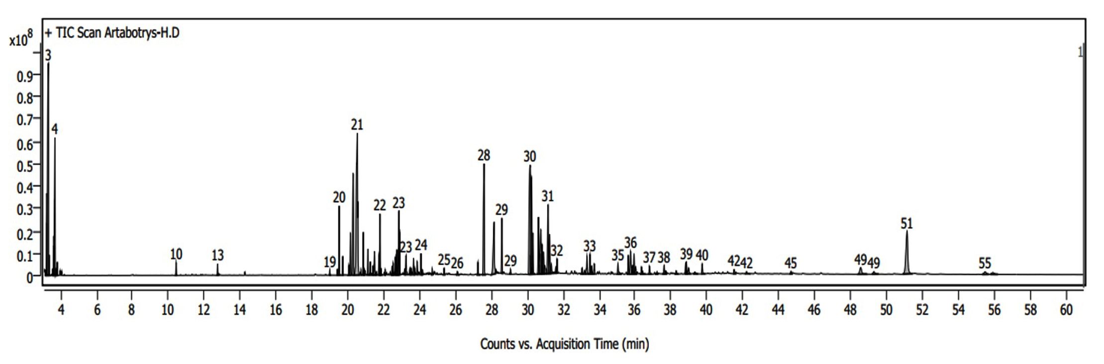
Figure. GC-MS chromatogram of the hexane extract of A. hexapetalus flowers showing the major chromatographic peaks corresponding to sesquiterpenes, fatty acid derivatives, and sterols identified in Table 3
Antibacterial activity of A. hexapetalus flower hexane extract
The antibacterial activity of the A. hexapetalus flower hexane extract against selected pathogenic bacteria is presented in Table 2. The extract exhibited inhibitory effects against all tested bacterial strains, with inhibition zones ranging from 6.33 ± 0.58 to 16.00 ± 1.73 mm. The largest inhibition zone was observed against S. enterica subsp. enterica serovar Typhi (16.00 ± 1.73 mm), followed by E. coli ATCC 25922 (14.33 ± 1.15 mm) and B. cereus (11.67 ± 0.58 mm). In comparison, streptomycin (3 mg/ml) exhibited significantly greater antibacterial activity against all tested bacteria, with inhibition zones ranging from 31.33 ± 2.31 to 41.67 ± 1.53 mm (P < 0.05). No inhibition was observed for the negative control (10% DMSO). The extract exhibited antibacterial activity against both Gram-positive and Gram-negative bacteria; however, its activity was significantly lower than that of streptomycin.
Table 2. Antibacterial activity of A. hexapetalus flower hexane extract against pathogenic bacteria
| Bacteria | Inhibition zone (mm) | |
|---|---|---|
| A. hexapetalus extract (500 mg/ml) | Streptomycin (3 mg/ml) | |
| B. cereus | 11.67 ± 0.58bAB | 31.33 ± 2.31aB |
| S. aureus | 7.33 ± 0.58bAB | 33.33 ± 1.53aAB |
| S. aureus DMST 20654 | 7.00 ± 0.00bAB | 41.33 ± 1.53aA |
| E. coli | 6.33 ± 0.58bB | 35.67 ± 2.89aAB |
| E. coli ATCC 25922 | 14.33 ± 1.15bAB | 33.67 ± 0.58aAB |
| P. aeruginosa | 7.00 ± 0.00bAB | 40.33 ± 0.58aAB |
| S. enterica subsp. enterica serovar Typhi | 16.00 ± 1.73bA | 41.67 ± 1.53aA |
Values are expressed as mean ± standard deviation (SD). Different lowercase letters (a,b) within the same row indicate significant differences between treatments, while different uppercase letters (A,B) within the same column indicate significant differences among bacterial strains, as determined by the Least Significant Difference (LSD) test at P < 0.05
Determination of minimum inhibitory concentration (MIC) and minimum bactericidal concentration (MBC)
The minimum inhibitory concentration (MIC) and minimum bactericidal concentration (MBC) values of the A. hexapetalus flower hexane extract against selected pathogenic bacteria are presented in Table 3. MIC values ranged from 0.488-500 mg/ml. The lowest MIC value was observed for E. coli ATCC 25922 (0.488 mg/ml), followed by B. cereus (7.812 mg/ml). S. aureus and P. aeruginosa exhibited MIC values of 250 mg/ml, whereas MIC values of 500 mg/ml were recorded for S. aureus DMST 20654, E. coli, and S. enterica subsp. enterica serovar Typhi. Regarding bactericidal activity, the extract exhibited MBC values of 500 mg/ml against B. cereus, S. aureus DMST 20654, P. aeruginosa, and S. enterica subsp. enterica serovar Typhi. However, no bactericidal effect was observed against S. aureus, E. coli, and E. coli ATCC 25922 at the tested concentrations (>500 mg/ml). The antibacterial activity of the extract varied among the tested bacterial strains. For several strains, MBC values were higher than the corresponding MIC values, indicating that higher extract concentrations were required to achieve bactericidal effects than to inhibit bacterial growth.
Table 3. Minimum inhibitory concentration (MIC) and minimum bactericidal concentration (MBC) of hexane extract from A. hexapetalus flowers against pathogenic bacteria
Bacteria |
MIC (mg/ml) |
MBC (mg/ml) |
|---|---|---|
Bacillus cereus |
7.812 |
500 |
Staphylococcus aureus |
250 |
>500 |
S. aureus DMST 20654 |
500 |
500 |
Escherichia coli |
500 |
>500 |
E. coli ATCC 25922 |
0.488 |
>500 |
Pseudomonas aeruginosa |
250 |
500 |
S. enterica subsp. enterica serovar Typhi |
500 |
500 |
>500 indicates that no bactericidal activity was observed within the tested concentration range (0.488-500 mg/mL)
The crude hexane extract of A. hexapetalus flowers exhibited a dark yellow color, viscous texture, and characteristic pungent odor. These physicochemical characteristics are typically associated with extracts enriched in lipophilic constituents such as terpenoids, fatty acids, and other non-polar metabolites efficiently extracted by hexane.11,12 The dark yellow coloration may be attributed to lipid-soluble pigments, including carotenoids and oxidized terpenoid derivatives, whereas the viscous consistency suggests the presence of fatty acids, fatty acid esters, and other hydrophobic compounds.13,14 The characteristic odor is likely associated with volatile terpenoids and sesquiterpenes. GC-MS analysis further supported these observations by confirming a chemically diverse profile composed mainly of sesquiterpenes, oxygenated sesquiterpenes, fatty acid derivatives, and phytosterols (Table 1). The predominance of hydrophobic constituents is also consistent with the insolubility of the extract in water and may contribute to antibacterial activity through interactions with bacterial cell membranes and disruption of membrane integrity.15,16
The extraction yield of the hexane extract was 20.78% (w/w), which may be attributed to the efficiency of hexane in solubilizing abundant lipophilic constituents in the flower matrix, including fatty acids, sterols, and other non-polar compounds identified by GC-MS analysis. Extraction yield is known to be influenced by solvent polarity, plant matrix composition, and extraction conditions.11,17
GC-MS profiling revealed 100 chromatographic peaks, of which 94 compounds were identified. The extract was predominantly composed of sesquiterpenes, fatty acid derivatives, and phytosterols, which is consistent with extraction using a non-polar solvent. Sesquiterpenes such as longifolene, cadinene derivatives, and murolene, as well as oxygenated derivatives including caryophyllene oxide and spathulenol, have been widely reported to exhibit antimicrobial activity, primarily through disruption of microbial membranes and interference with essential cellular functions.18,19 Fatty acid methyl esters, such as methyl palmitate and methyl linoleate, have also been associated with antimicrobial effects via membrane destabilization and metabolic disruption.20 In addition, phytosterols including gamma-sitosterol, campesterol, and stigmasterol may contribute to the overall biological activity, potentially through additive or synergistic interactions with other bioactive constituents.21
The antibacterial assay demonstrated that the extract exhibited inhibitory activity against all tested bacterial strains, although with lower potency compared to streptomycin. The extract showed broad-spectrum activity against both Gram-positive and Gram-negative bacteria. Notably, relatively stronger inhibition was observed against Gram-negative bacteria, including E. coli ATCC 25922 and S. enterica serovar Typhi, despite the presence of an outer membrane that typically limits antimicrobial penetration. This suggests that lipophilic constituents in the extract may interact with and destabilize bacterial membrane structures.22 The observed antibacterial activity is likely attributable to terpenoids and fatty acid derivatives identified by GC-MS, which are known to exert antimicrobial effects through membrane disruption, increased permeability, leakage of intracellular components, and interference with metabolic processes.6,18 However, the lower activity of the crude extract compared to standard antibiotics is expected due to the presence of complex mixtures in which active compounds occur at relatively low concentrations.23
MIC and MBC results further demonstrated variability in antibacterial susceptibility among tested bacterial strains. The lowest MIC observed for E. coli ATCC 25922 indicates higher susceptibility, whereas higher MIC values in other strains may be associated with intrinsic or acquired resistance mechanisms, including reduced membrane permeability, efflux pump activity, and enzymatic detoxification.24 Overall, the relatively high MIC and MBC values, together with the absence of bactericidal effects in some strains, indicate that the extract primarily exhibits bacteriostatic activity. This finding is consistent with previous reports showing that many plant-derived crude extracts tend to inhibit bacterial growth rather than induce cell death.25-27 The relatively high MIC and MBC values may also be attributed to the crude nature of the extract, where active compounds are diluted by inactive constituents. Fractionation and purification could enhance antibacterial potency by concentrating bioactive molecules and removing interfering components.23
Overall, the antibacterial activity observed in this study is likely the result of combined effects of multiple phytochemical classes, including fatty acid derivatives, terpenoids, and sterol-like compounds identified by GC-MS analysis. These compounds may act synergistically or additively to disrupt bacterial cell membranes and interfere with essential cellular processes. However, no experimental evaluation of synergistic interactions was conducted in this study. Therefore, further studies are required to isolate and characterize individual active compounds, evaluate their interaction effects, and elucidate detailed mechanisms of action against both Gram-positive and Gram-negative bacteria. Such investigations would support the potential application of A. hexapetalus flower extract as a natural antimicrobial agent.
The hexane extract of A. hexapetalus flowers exhibited antibacterial activity against both Gram-positive and Gram-negative bacteria. The extract showed more pronounced inhibitory effects against Salmonella enterica serovar Typhi and Escherichia coli ATCC 25922 compared with other tested strains, although its activity was lower than that of streptomycin. MIC and MBC analyses indicated that the extract primarily exerted bacteriostatic rather than bactericidal effects. GC-MS analysis indicated the presence of bioactive constituents, mainly sesquiterpenes, fatty acid derivatives, and phytosterols, which have been reported to possess antimicrobial properties. These findings are consistent with previous reports on A. hexapetalus and related Artocarpus species, which have demonstrated various biological activities including antimicrobial, antioxidant, and anti-inflammatory effects. Overall, this study highlights the potential of A. hexapetalus flower extract as a natural source of antimicrobial compounds. However, further studies are required to isolate active constituents, confirm their bioactivities, and elucidate their mechanisms of action for potential applications in pharmaceutical and food systems.
ACKNOWLEDGMENTS
The authors gratefully acknowledge Mahasarakham University for financial support and research facilities. The authors also thank Prof. Motoyuki Sumida for English language editing and Dr. Rungruedee Thiwthong for providing the Ylang-Ylang flower extract.
CONFLICT OF INTEREST
The authors declare that there is no conflict of interest.
AUTHORS’ CONTRIBUTION
All authors listed have made a substantial, direct and intellectual contribution to the work, and approved it for publication.
FUNDING
This research project was financially supported by Mahasarakham University.
DATA AVAILABILITY
All datasets generated or analyzed during this study are included in the manuscript.
ETHICS STATEMENT
This article does not contain any studies on human participants or animals performed by any of the authors.
- El-Saber Batiha G, Magdy Beshbishy A, Wasef LG, et al. Chemical constituents and pharmacological activities of garlic (Allium sativumL.): A review. Nutrients. 2020;12(3):872.
Crossref - Nakadate K, Ito N, Kawakami K, Yamazaki N. Anti-inflammatory actions of plant-derived compounds and prevention of chronic diseases: from molecular mechanisms to applications. Int J Mol Sci. 2025;26(11):5206.
Crossref - Newman DJ, Cragg GM. Natural products as sources of new drugs over the nearly four decades from 01/1981 to 09/2019. J Nat Prod. 2020;83(3):770-803.
Crossref - Atanasov AG, Waltenberger B, Pferschy-Wenzig EM, et al. Discovery and resupply of pharmacologically active plant-derived natural products: A review. Biotechnol Adv. 2015ַ 33(8):1582-1614.
Crossref - Krishnasamy V, Madhiyan S, Kathirvel P, Velliangiri P, Raman M, Anandhan A. chemical profile and biological activities of essential oil from Artabortrys hexapetalus (L.f.) Bhandari grown in Southern parts of Western Ghats. Orient J Chem. 2022;38(6).
Crossref - Pramila K, Mehta BK. GC-MS analysis of hexane extract of Artabotrys. Research Square. 2024.
Crossref - Ferri M, Ranucci E, Romagnoli P, Giaccone V. Antimicrobial resistance: A global emerging threat to public health systems. Crit Rev Food Sci Nutr. 2017;57(13):2857-2876.
Crossref - Dadgostar P. Antimicrobial resistance: implications and costs. Infect Drug Resist. 2019;12:3903-3910.
Crossref - Swamy MK, Akhtar MS, Sinniah UR. Antimicrobial properties of plant essential oils against human pathogens and their mode of action: An updated review. Evid Based Complement Alternat Med. 2016;2016:3012462.
Crossref - Sutthisa W, Sophachan H, Pimvichai P, Srisawad N. Antimicrobial efficiency and chemical composition of Jackfruit (Artocarpus heterophyllus Lam.) extracts from different plant parts. TJNPR, 2024;9(3):925-932.
Crossref - Chemat F, Abert-Vian M, Fabiano-Tixier AS, et al. Green extraction of natural products. Origins, current status, and future challenges. Trends Analyt Chem. 2019;118:248-263.
Crossref - Zhang QW, Lin LG, Ye WC. Techniques for extraction and isolation of natural products: a comprehensive review. Chin Med. 2018;13:20.
Crossref - Saini RK, Keum YS. Carotenoid extraction methods: A review of recent developments. Food Chemistry, 2018;240:90-103.
Crossref - Wahyuni, FD, Shalihah IM, Nurtiana W. Carotenoids as natural colorant: A review. Food ScienTech Journal. 2020;2(2):94.
Crossref - Iskandar K, Ahmed N, Paudyal N, et al. Essential oils as antimicrobial agents against WHO priority bacterial pathogens: A strategic review of in vitro clinical efficacy, innovations and research gaps. Antibiotics. 2025;14(12):1250.
Crossref - Mancianti F, Ebani VV. Biological activity of essential oils. Molecules. 2020;25(3):678.
Crossref - Zhang M, Zhao J, Dai X, Li X. Extraction and analysis of chemical compositions of natural products and plants. Separations. 2023;10(12):598.
Crossref - Sharifi-Rad J, Quispe C, Patra JK, eta l. Paclitaxel: application in modern oncology and nanomedicine-based cancer therapy. Oxid Med Cell Longev. 2021;2021: 3687700.
Crossref - Zhao DD, Jiang LL, Li HY, Yan PF, Zhang YL. Chemical components and pharmacological activities of terpene natural products from the genus Paeonia. Molecules. 2016;21(10):1362.
Crossref - Casillas-Vargas G, Ocasio-Malave C, Medina S, et al. Antibacterial fatty acids: An update of possible mechanisms of action and implications in the development of the next-generation of antibacterial agents. Prog Lipid Res, 2021;82:101093.
Crossref - Li X, Xin Y, Mo Y, Marozik P, He T, Guo H. The bioavailability and biological activities of phytosterols as modulators of cholesterol metabolism. Molecules. 2022;27(2):523.
Crossref - Breijyeh Z, Jubeh B, Karaman R. Resistance of gram-negative bacteria to current antibacterial agents and approaches to resolve it. Molecules. 2020;25(6):1340.
Crossref - Bakkali F, Averbeck S, Averbeck D, Idaomar M. Biological effects of essential oils- A review. Food Chem Toxicol. 2008;46(2):446-475.
Crossref - Blair JM, Webber MA, Baylay AJ, Ogbolu DO, Piddock LJV. Molecular mechanisms of antibiotic resistance. Nat Rev Microbiol. 2015;13(1):42-51.
Crossref - Cowan MM. Plant products as antimicrobial agents. Clinl Microbiol Rev. 1999;12(4):564–582.
Crossref - Cushnie TPT, Lamb AJ. Recent advances in understanding the antibacterial properties of flavonoids. Int J Antimicrob Agents. 2011;38(2):99-107.
Crossref - Borges A, Ferreira C, Saavedra MJ, Simoes M. Antibacterial activity and mode of action of ferulic and gallic acids against pathogenic bacteria. Microb Drug Resist. 2013;19(4):256–265.
Crossref
© The Author(s) 2026. Open Access. This article is distributed under the terms of the Creative Commons Attribution 4.0 International License which permits unrestricted use, sharing, distribution, and reproduction in any medium, provided you give appropriate credit to the original author(s) and the source, provide a link to the Creative Commons license, and indicate if changes were made.